Embed Size (px)
Citation preview

Split-TurboID enables contact-dependent proximitylabeling in cellsKelvin F. Choa, Tess C. Branonb,c,d,e, Sanjana Rajeevb, Tanya Svinkinaf, Namrata D. Udeshif, Themis Thoudamg
,Chulhwan Kwakh,i, Hyun-Woo Rheeh,j, In-Kyu Leeg,k,l, Steven A. Carrf, and Alice Y. Tingb,c,d,m,1
aCancer Biology Program, Stanford University, Stanford, CA 94305; bDepartment of Genetics, Stanford University, Stanford, CA 94305; cDepartment ofBiology, Stanford University, Stanford, CA 94305; dDepartment of Chemistry, Stanford University, Stanford, CA 94305; eDepartment of Chemistry,Massachusetts Institute of Technology, Cambridge, MA 02139; fBroad Institute of MIT and Harvard, Cambridge, MA 02142; gResearch Institute of Aging andMetabolism, Kyungpook National University, 37224 Daegu, South Korea; hDepartment of Chemistry, Seoul National University, 08826 Seoul, South Korea;iDepartment of Chemistry, Ulsan National Institute of Science and Technology, 44919 Ulsan, South Korea; jSchool of Biological Sciences, Seoul NationalUniversity, 08826 Seoul, South Korea; kDepartment of Internal Medicine, School of Medicine, Kyungpook National University, Kyungpook NationalUniversity Hospital, 41944 Daegu, South Korea; lLeading-edge Research Center for Drug Discovery and Development for Diabetes and Metabolic Disease,Kyungpook National University, 41944 Daegu, South Korea; and mChan Zuckerberg Biohub, San Francisco, CA 94158
Edited by Tony Hunter, The Salk Institute for Biological Studies, La Jolla, CA, and approved April 7, 2020 (received for review November 7, 2019)
Proximity labeling catalyzed by promiscuous enzymes, such asTurboID, have enabled the proteomic analysis of subcellular regionsdifficult or impossible to access by conventional fractionation-based ap-proaches. Yet some cellular regions, such as organelle contact sites, re-main out of reach for current PL methods. To address this limitation, wesplit the enzyme TurboID into two inactive fragments that recombinewhen driven together by a protein–protein interaction or membrane–membrane apposition. At endoplasmic reticulum–mitochondria contactsites, reconstituted TurboID catalyzed spatially restricted biotinyla-tion, enabling the enrichment and identification of >100 endoge-nous proteins, including many not previously linked to endoplasmicreticulum–mitochondria contacts. We validated eight candidates bybiochemical fractionation and overexpression imaging. Overall,split-TurboID is a versatile tool for conditional and spatially specificproximity labeling in cells.
proximity labeling | ER–mitochondria contacts | split-TurboID
Proximity labeling (PL) has been shown to be a valuable toolfor studying protein localization and interactions in living
cells (1–3). In PL, a promiscuous enzyme such as APEX (4, 5),BioID (6), or TurboID (7) is genetically targeted to an organelleor protein complex of interest. Addition of a biotin-derived small-molecule substrate then initiates biotinylation of endogenousproteins within a few nanometers of the promiscuous enzyme, viaa diffusible radical intermediate in the case of APEX, or an ac-tivated biotin adenylate intermediate in the case of BioID andTurboID. After cell lysis, biotinylated proteins are harvested usingstreptavidin beads and identified by mass spectrometry.PL has been applied in many cell types and species to map the
proteome composition of organelles, including mitochondria (5,8–10), synapses (11, 12), stress granules (13), and primary cilia(14). However, to increase the versatility of PL, new enzymevariants are needed. In particular, split enzymes could enablegreater spatial specificity in the targeting of biotinylation activity,as well as PL activity that is conditional on a specific input, suchas drug, calcium, or cell–cell contact. For example, contact sitesbetween mitochondria and the endoplasmic reticulum (ER) me-diate diverse biology, from lipid biosynthesis and Ca+2 signaling toregulation of mitochondrial fission (15). There is great interest inprobing the proteomic composition of ER–mitochondria contacts.However, direct fusion of a PL enzyme to one of the knownER–mitochondria contact resident proteins (e.g., Drp1 or Mff)would generate PL activity outside of ER–mitochondria con-tacts as well, because these proteins also reside in other sub-cellular locations (16, 17). On the other hand, use of a split PLenzyme, with one fragment targeted to the mitochondria andthe other targeted to the ER, would restrict biotinylation ac-tivity to ER–mitochondria contact sites specifically.
Split forms of APEX (18) and BioID (19–21) have previouslybeen reported. However, split-APEX (developed by us) has notbeen used for proteomics, and the requirement for exogenousH2O2 and heme addition limits its utility in vivo. Split-BioID wasfirst reported by De Munter et al. (19), followed by more activeversions from Schopp et al. (20) and Kwak et al. (21). All arederived from the parental enzyme BioID, which requires 18 to24 h of biotin labeling. We show below that the Schopp et al. (20)split-BioID does not produce detectable activity, while the Kwaket al. (21) split-BioID requires 16+ h of labeling to generatesufficient signal.Hence we sought to develop an improved, more active split PL
enzyme by starting from TurboID. In contrast to APEX, Tur-boID does not require any cofactors or cooxidants; just biotinaddition initiates labeling in cells or animals. TurboID is also>100-fold faster than BioID, requiring only 1 to 10 min of la-beling time (7). We performed a screen of 14 different TurboIDsplit sites to identify optimal fragments for high-affinity and low-affinity reconstitution. We converged upon TurboID split at L73/G74, which gave rapamycin-dependent reconstitution whenfused to FRB and FKBP in multiple subcellular organelles. We
Significance
Most of the thousands of proteins that comprise a human cellhave specific subcellular localization patterns essential for theirfunction. “Proximity labeling” (PL) is a method for mapping thelocalization of endogenous cellular proteins on a proteome-wide scale. To improve the specificity and versatility of PL,we developed split-TurboID, a promiscuous biotinylating en-zyme split into two inactive fragments. The fragments arecoexpressed in cells and brought together by a drug, protein–protein interaction, or organelle contact to reconstitute Tur-boID enzymatic activity. We used split-TurboID to map theprotein composition of endoplasmic reticulum–mitochondriacontact sites, which are essential for mitochondrial fission, lipidbiosynthesis, and calcium signaling. For conditional or higher-specificity PL, split-TurboID may be a valuable tool forbiological discovery.
Author contributions: K.F.C., S.A.C., and A.Y.T. designed research; K.F.C., T.C.B., S.R., T.S.,N.D.U., T.T., C.K., H.-W.R., and I.-K.L. performed research; K.F.C. and A.Y.T. analyzed data;and K.F.C. and A.Y.T. wrote the paper.
The authors declare no competing interest.
This article is a PNAS Direct Submission.
Published under the PNAS license.1To whom correspondence may be addressed. Email: [email protected].
This article contains supporting information online at https://www.pnas.org/lookup/suppl/doi:10.1073/pnas.1919528117/-/DCSupplemental.
First published May 18, 2020.
www.pnas.org/cgi/doi/10.1073/pnas.1919528117 PNAS | June 2, 2020 | vol. 117 | no. 22 | 12143–12154
CELL
BIOLO
GY
Dow
nloa
ded
by g
uest
on
Janu
ary
31, 2
022

then used this split-TurboID to perform proteomic mapping ofER–mitochondria contact sites in mammalian cells. The result-ing proteome of 101 proteins is highly specific and identifiesmany new ER–mitochondria contact site candidates, eight of whichwe validated by biochemical fractionation or overexpressionimaging.
ResultsDevelopment of a Split Promiscuous Biotin Ligase with High Activity.We started with TurboID, for the reasons given above, andsought to design split protein fragments with no detectable ac-tivity on their own, but high reconstituted activity. Given thediversity of ways in which split proteins are used, we envisionedengineering both a low-affinity fragment pair, whose reconstitutioncould be driven by a protein–protein or membrane–membrane as-sociation (Fig. 1A), and a high-affinity pair that would spontane-ously reconstitute upon cocompartmentalization of fragments.Previously, we developed split enzymes [split-APEX (18) and split-HRP (22)] by manually selecting cut sites in exposed loops, guidedby protein crystal structures. Here, we utilized a recently developedcomputational algorithm for predicting optimal protein split sites(23). SPELL (split protein reassembly by ligand or light) calculatesthe energy profile of each candidate fragment relative to that of thefull-length protein, and combines this information with solvent ac-cessibility, sequence conservation, and geometric constraints toevaluate potential split sites, aiming for fragment pairs that give highreconstitution efficiency and minimal spontaneous assembly (23).Because crystal structures for TurboID and BioID are not available,we applied the SPELL algorithm to wild-type Escherichia coli biotinligase [BirA; PDB ID 1HXD (24) and PDB ID 2EWN (25)], fromwhich both enzymes are derived.SPELL identified 10 potential split sites, all of which are in
exposed loops. We rejected some of them based on prior ex-perimental data: for example, cut site 62/63 was predicted bySPELL, but our previously developed miniTurbo is truncated atamino acid 64 and retains high activity (7). We selected five ofthe SPELL-predicted cut sites for experimental testing (Fig. 1B).In addition, we included in our screen five more cut sites used inprevious split-BioIDs (20, 21). Each fragment pair was cloned asfusions to FKBP and FRB, proteins whose association can beinduced by the small-molecule rapamycin (Fig. 1A). The constructswere expressed in the cytosol of HEK293T cells and incubated withbiotin for 24 h in the presence or absence of rapamycin. Cell lysateswere run on sodium dodecyl sulfate polyacrylamide gel electro-phoresis (SDS-PAGE) and blotted with streptavidin to evaluatethe extent of promiscuous biotinylation. Fig. 1 C and D and SIAppendix, Fig. S1A show that split-TurboIDs cut at 73/74, 78/79,and 98/99 give high reconstituted activity. Cut site 78/79 is themost active, in both the presence and absence of rapamycin,suggesting that the split fragments have high affinity for oneanother. The SPELL-predicted cut site 73/74 gave the greatestrapamycin-dependent activity, suggesting that it is a low-affinity,or conditional, split-TurboID.We performed a secondary screen around the cut site 73/74 to
further optimize low-affinity split-TurboID. Neighboring cut siteswere tested (SI Appendix, Fig. S1B), in addition to pairing of frag-ments with overlapping or gapped ends (SI Appendix, Fig. S1C).None of these were better than the original 73/74 pair, so we se-lected this as our optimal low-affinity split-TurboID (referred tosimply as “split-TurboID” henceforth).In a side-by-side comparison to previous split-BioIDs (Fig. 1 C
and D), both our high-affinity and low-affinity split-TurboIDswere far more active. The Kwak et al. (21) split-BioID (alsotermed Contact-ID) showed rapamycin-dependent reconstitutionwith activity ∼12-fold lower than that of split-TurboID. This isconsistent with the reported difference in catalytic activities of theparent enzymes TurboID and BioID (7). Interestingly, when theContact-ID cut site (78/79) is used to split TurboID, this yields our
best high-affinity pair (78/79). The discrepancy between the rapamycin-dependence of Contact-ID and the rapamycin-independence ofhigh-affinity split-TurboID is likely explained by their differentregimes of activity; Contact-ID labeling may not be detectable inthe omit-rapamycin condition because the intrinsic activity isso low.In our hands, the previously reported split-BioID from Schopp
et al. (20) did not give any detectable signal over backgroundafter 24 h of biotin incubation. Interestingly, TurboID split at thesame position (256/257) did show some labeling (Fig. 1C), butthis activity was also observed with the N-terminal fragmentalone (SI Appendix, Fig. S1A), suggesting that this cut site maynot yield a true protein complementation system. Notably, wefound that the activity of split-TurboID is even greater than thatof full-length BioID (Fig. 1D; side-by-side comparison using 24 hof biotin incubation), suggesting that split-TurboID’s activitylevel should be adequate for a wide range of applications.By referencing the protein structure of E. coli biotin ligase
(PDB ID 2EWN) (25), from which TurboID was evolved, we seethat the split-TurboID site (L73/G74) separates the protein intotwo globular domains (Fig. 1E). It is intriguing that just by moving thecut site five residues away (to 78/79), we produce a split-TurboIDsystem that is high-affinity/rapamycin-independent rather than low-affinity/rapamycin-dependent.
Further Characterization of Split-TurboID. To further characterizesplit-TurboID, we confirmed by confocal fluorescence imagingthat the constructs catalyze biotinylation in a biotin- and rapamycin-dependent manner (Fig. 2A). Reconstituted split-TurboID is not asactive as full-length TurboID, but gave detectable biotinylation afterjust 30 min of biotin incubation (Fig. 2B). To probe the kinetics ofreconstitution, we compared rapamycin preincubation to rapamycincoaddition with biotin. There was no difference in biotinylationactivity (SI Appendix, Fig. S2A), suggesting that split-TurboIDbecomes active and begins catalyzing biotinylation immediatelyupon rapamycin addition.We also generated constructs fusing split-TurboID with vari-
ous localization sequences to target the fragments to differentsubcellular compartments (cytosol, nucleus, mitochondrial matrix,and ER lumen) (Fig. 2C). Confocal fluorescence imaging of cellsexpressing these constructs labeled with rapamycin and biotin for1 h shows compartment-specific targeting and rapamycin-dependentbiotinylation in all compartments tested (Fig. 2D and SI Appendix,Fig. S2 B and C).
Using Split-TurboID for Proximity Labeling at ER–Mitochondria Contacts.ER–mitochondria contacts are important in a variety of biologicalprocesses, including Ca+2 signaling, lipid metabolism, nutrient sig-naling, and mitochondrial fission (15, 26, 27). There is tremendousinterest in understanding the molecular composition of these contacts.Biochemical purification of mitochondria-associated membranes(MAMs) has frequently been used to study ER–mitochondria con-tacts (15), but MAMs encompass much more than just mitochondria-associated ER microsomes; they also include contaminants from theplasma membrane, Golgi, peroxisomes, and nuclear membrane (28).To provide a more specific alternative, we recently applied APEX PLto produce separate proteomic maps of the ER membrane (ERM)and outer mitochondrial membrane (OMM), and then intersectedthe datasets to identify candidate ER–mitochondria contact residents(9). This resulted in the discovery of an ER–mitochondria tetheringprotein (SYNJ2BP), but many of the hits were merely dual-localizedER and mitochondria proteins.We sought to use split-TurboID reconstitution across ER–
mitochondria contacts in order to map this compartment di-rectly, with much greater specificity than both MAM purifica-tions and separate APEX tagging plus dataset intersection. Totarget split-TurboID to the ERM and OMM, we fused the frag-ments to the transmembrane domains of ERM-resident protein
12144 | www.pnas.org/cgi/doi/10.1073/pnas.1919528117 Cho et al.
Dow
nloa
ded
by g
uest
on
Janu
ary
31, 2
022

A B
C
E
80
58
46
32
25
Rapamycin:
Fragment: N C BB N C BB N C BB
high-affinitysplit-TurboID
24 hours(78/79)
split-BioID24 hours(256/257)
Contact-ID24 hours(78/79)
FL
Bio
ID (2
4h)
FL
Tur
boID
(30
min
)
TurboID sequence (red lines show split sites tested)
aa 1
aa 161
aa 160
aa 321
73* 98101112
212256258
270273
73* 747271 75
FRB
Tb(N)
Tb(C)
FKBP
RapamycinATP + B
AMP
reconstitutedsplit-TurboID
B
FKBP V5 Tb(N)
HA Tb(C)FRBHaloTag
D
L73/G74
Biotin-AMP
E256/G257
G78/G79
Streptavidin-HRP
100
* *
*
*
HA80
V5
46
32
25
58
78
0.0
0.5
1.0
1.5
2.0
No
rma
lize
d b
iotin
yla
tion
act
ivity
rela
tive
to V
5 e
xpre
ssio
n
Rapamycin:
spB
ioID
25
6/2
57
Co
nta
ct-ID
78
/79
73
/74
98
/99
11
2/1
13
10
1/1
02
21
2/2
13
25
8/2
59
27
0/2
71
27
3/2
74
FL
Tu
rbo
ID
FL
Bio
ID
Derived fromBioID Derived from TurboID
*
*
C-terminus
N-terminus
N C BB
low-affinitysplit-TurboID
24 hours(73/74)
Fig. 1. Engineering split-TurboID. (A) Schematic of split-TurboID reconstitution using the chemically inducible FRB-FKBP dimerization system. Upon rapa-mycin treatment, two inactive fragments of TurboID reconstitute to form an active enzyme capable of generating biotin-5′-AMP for promiscuous proximity-dependent labeling. N-terminal fragments [Tb(N)] were fused to FKBP and V5. C-terminal fragments [Tb(C)] were fused to HA, HaloTag, and FRB. The HaloTagwas used for initial screening as previous studies have shown that it can improve fragment stability (18). (B) Split sites tested. Ten split sites were tested in thefirst round. In the second round, four additional sites around 73/74 were tested. Split sites are indicated as red lines along the TurboID protein sequence. Theα-helices are shown in blue and the β-sheets are shown in purple. (C) Results of split site screen. Split-BioID (split at E256/G257) (20) and Contact-ID (split atG78/G79) (21) are shown for comparison. Each fragment pair was tested in HEK293T cells with 24 h biotin incubation in the presence or absence of rapamycin.At right, cells expressing full-length (FL) TurboID were incubated with biotin for 30 min. FL BioID was incubated with biotin for 24 h. Cell lysates were analyzedby streptavidin blotting as in D, and quantification was performed by dividing the streptavidin sum intensity by the anti-V5 intensity. Values were normalizedto that of FL TurboID. (D) Streptavidin blot comparing our best split-TurboIDs to FL TurboID and BioID, and the previously described split-BioID and Contact-ID(20, 21). Labeling conditions were the same as in C. For each construct pair, lanes are shown with both fragments present (B), N-terminal fragment only (N), orC-terminal fragment only (C). Anti-V5 and anti-HA blotting show expression levels of N-terminal fragments (V5-tagged), C-terminal fragments (HA-tagged),and full-length enzymes (V5-tagged). Dashed lines indicate separate blots performed at the same time and developed simultaneously. Asterisks indicateligase self-biotinylation. Full blots are shown in SI Appendix, Fig. S1A. (E) N- and C-terminal fragments (blue and purple, respectively) of split-TurboID (73/74),depicted on a structure of E. coli biotin ligase (PDB ID 2EWN), from which TurboID was evolved (7). Biotin-AMP in the active site is shown in yellow. The low-affinity split-TurboID cut site is shown in red, the high-affinity split-TurboID cut site is shown in blue, and the previous split-BioID cut site is shown in black (20).
Cho et al. PNAS | June 2, 2020 | vol. 117 | no. 22 | 12145
CELL
BIOLO
GY
Dow
nloa
ded
by g
uest
on
Janu
ary
31, 2
022

A-r
ap-b
iotin
+ra
p-b
iotin
DAPIFKBP-Tb(N)
(V5)FRB-Tb(C)
(HA) Neutravidin
-rap
+bi
otin
+ra
p+
biot
inB
D
80
58
46
32
80
58
46
32
Streptavidin-HRP
Anti-V5/Anti-HA
Rapamycin:
Unt
rans
.
FL
Bio
ID 1
8h
FL
Bio
ID 1
h
FL
Tur
boID
10m
split-TurboID (L73/G74)
10m 30m 1h 6h 18h
-rap
+ra
pcy
toso
l-r
ap+
rap
nucl
eus
-rap
+ra
pm
itoch
ondr
ial m
atrix
(zoom)
-rap
+ra
pER
lum
en
(zoom)
V5
HA
DAPIFKBP-Tb(N)
(V5)FRB-Tb(C)
(HA) Neutravidin DAPIFKBP-Tb(N)
(V5)FRB-Tb(C)
(HA) Neutravidin
C NESFKBP V5 Tb(N)Cytosol
Tb(C)HA FRB NES
NLSFKBP V5 Tb(N)Nucleus
Tb(C)HA FRB NLS
KDELFKBP V5 Tb(N)ER Lumen
Tb(C)HA FRB KDEL
MTS FKBP V5 Tb(N)Mitochondrial
Matrix Tb(C)HA FRBMTS
aa 1-24 of COX4
SS
SS
aa 1-21 of Ig kappa chain
Fig. 2. Characterization of low-affinity split-TurboID. (A) Confocal fluorescence imaging of low-affinity split-TurboID (split site L73/G74). HEK293T cells weretransiently transfected and incubated with 50 μM biotin and 100 nM rapamycin for 1 h, then fixed and stained with anti-V5 to detect the N-terminal fragment[Tb(N)], anti-HA to detect the C-terminal fragment [Tb(C)], and neutravidin-647 to detect biotinylated proteins. (Scale bars, 20 μm.) (B) Split-TurboID timecourse. HEK293T cells transiently transfected with split-TurboID constructs were treated with 50 μM biotin and 100 nM rapamycin for the indicated times, andwhole-cell lysates were analyzed by streptavidin blotting. FL TurboID (10 min) and FL BioID (1 h and 18 h) were included for comparison. (C) Design ofconstructs used to target split-TurboID fragments to various cellular compartments. Full descriptions of constructs are available in SI Appendix, Table S1 (MTS,mitochondrial targeting sequence; SS, signal sequence). (D) Confocal fluorescence imaging of split-TurboID targeted to various cellular compartments.HEK293T cells were labeled and imaged as in A. Fluorescence intensities are not normalized across cellular compartments. Zoomed images of the boxedregions are shown. (Scale bars, 10 μm.)
12146 | www.pnas.org/cgi/doi/10.1073/pnas.1919528117 Cho et al.
Dow
nloa
ded
by g
uest
on
Janu
ary
31, 2
022
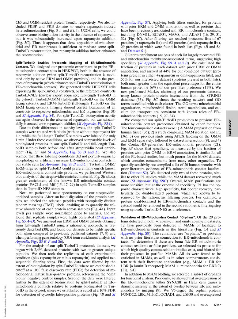
Cb5 and OMM-resident protein Tom20, respectively. We also in-cluded FKBP and FRB domains to enable rapamycin-inducedheterodimerization (Fig. 3 A and B). In U2OS cells, we couldobserve some biotinylation activity in the absence of rapamycin,but it was substantially increased upon rapamycin addition(Fig. 3C). Thus, it appears that close apposition of mitochon-drial and ER membranes is sufficient to mediate some split-TurboID reconstitution, but rapamycin addition further enhancesthe reconstitution.
Split-TurboID Enables Proteomic Mapping of ER–MitochondriaContacts. We designed our proteomic experiment to probe ER–
mitochondria contact sites in HEK293T, both in the absence ofrapamycin addition (when split-TurboID reconstitution is medi-ated only by native ERM and OMM proximity) and in the pres-ence of rapamycin (which enhances split-TurboID reconstitution atER–mitochondria contacts). We generated stable HEK293T cellsexpressing the split-TurboID constructs, or the reference constructsTurboID-NES (nuclear export sequence; full-length TurboID inthe cytosol), TurboID-OMM (full-length TurboID on the OMMfacing cytosol), and ERM-TurboID (full-length TurboID on theERM facing cytosol). Imaging showed correct localization of allconstructs to respective mitochondria and ER organelles (Fig. 3Dand SI Appendix, Fig. S4). For split-TurboID, biotinylation activitywas again observed in the absence of rapamycin, but was substan-tially increased upon rapamycin addition (SI Appendix, Fig. S3A).Due to their differences in activity levels, the split-TurboID
samples were treated with biotin (with or without rapamycin) for4 h, while the full-length TurboID samples were labeled for only1 min. Under these conditions, we observed comparable levels ofbiotinylated proteins in our split-TurboID and full-length Tur-boID samples both before and after streptavidin bead enrich-ment (Fig. 3F and SI Appendix, Fig. S3 D and E). We alsoverified that these labeling conditions did not perturb organellemorphology or artificially increase ER–mitochondria contacts inour stable cells (SI Appendix, Fig. S3 B and C). To test if OMM/ERM-targeted split-TurboID could preferentially enrich knownER–mitochondria contact site proteins, we performed Westernblot analysis of the streptavidin-enriched material. Fig. 3E showsgreater enrichment of the known ER–mitochondria contactproteins FACL4 and Mff (15, 17, 29) in split-TurboID samplesthan in TurboID-NES samples.Next, we performed mass spectrometry on our streptavidin-
enriched samples. After on-bead digestion of the protein sam-ples, we labeled the released peptides with isotopically distincttandem mass tag (TMT) labels, enabling us to quantify the rel-ative abundance of each protein across samples (Fig. 4A). Inputlevels per sample were normalized prior to analysis, and wefound that replicate samples were highly correlated (SI Appendix,Fig. S5 A–D). We analyzed our ERM and OMM datasets obtainedfrom full-length TurboID using a ratiometric approach, as pre-viously described (30), and found our datasets to be highly specificboth when compared to previously published datasets (7, 9) andwhen performing gene ontology (GO) term enrichment analysis (SIAppendix, Figs. S5 E–P and S6).For the analysis of our split-TurboID proteomic datasets, we
began with 2,496 detected proteins with two or greater uniquepeptides. We then took the replicates of each experimentalcondition (plus rapamycin or minus rapamycin) and applied twosequential filtering steps. First, the data were filtered by theextent of biotinylation by split-TurboID, where we established acutoff at a 10% false-discovery rate (FDR) for detection of mi-tochondrial matrix false-positive proteins, referencing the “omitbiotin” negative control samples. Second, the data were filteredfurther by the extent of biotinylation by split-TurboID at ER–
mitochondria contacts relative to proteins biotinylated by Tur-boID in the cytosol, where we established a cutoff at a 10% FDRfor detection of cytosolic false-positive proteins (Fig. 4B and SI
Appendix, Fig. S7). Applying both filters enriched for proteinswith prior ERM and OMM annotation, as well as proteins thathave been previously associated with ER–mitochondria contacts,including DNM1L, BCAP31, MAVS, and AKAP1 (16, 29, 31,32) (Fig. 4C). After filtering, we reached proteome lists of 67proteins (+rapamycin list) and 63 proteins (omit-rapamycin list),29 proteins of which were found in both lists (Figs. 4B and 5Aand Dataset S1).GO term enrichment analysis of each list largely recovered ER
and mitochondria membrane-associated terms, suggesting highspecificity (SI Appendix, Fig. S9 A and B). We calculated thefraction of proteins in each dataset with prior ERM or OMMannotation, and arrived at 44% for our combined dataset (pro-teins present in either +rapamycin or omit-rapamycin lists), and55% for our intersected dataset (proteins present in both lists),both much greater than the equivalent percentages for the entirehuman proteome (6%) or our pre-filter proteome (11%). Wenext performed Markov clustering of our proteomic datasets,using known protein–protein interactions from the STRINGdatabase (33). Fig. 5C shows the resulting network and GOterms associated with each cluster. The GO terms mitochondrialorganization, mitochondrial fission, sterol metabolism, and cal-cium ion transport are consistent with known roles of ER–
mitochondria contacts (15, 27, 34).We compared our split-TurboID proteomes to previous ER–
mitochondria contact proteomes obtained by other methods.The four comparison datasets were 1) A MAM preparation fromhuman tissue (35); 2) a study combining MAM isolation and PL(36); 3) our previous study using APEX labeling on the OMMand ERM separately, followed by dataset intersection (9); and 4)the Contact-ID–generated ER–mitochondria proteome (21).Fig. 5B shows that specificity, as measured by the fraction ofproteins with prior OMM or ERM annotation, is similar for allof the PL-based studies, but much poorer for the MAM dataset,which contains contaminants from many other organelles. Toquantify sensitivity, we compiled a list of 20 human proteins withprior literature evidence of ER–mitochondria contact localiza-tion (Dataset S2). We detected only two of these proteins, sim-ilar to other PL studies, while the MAM dataset recovered muchmore (SI Appendix, Fig. S9C). Overall, MAM purifications aremore sensitive, but at the expense of specificity. PL has the op-posite characteristics: high specificity, but poorer recovery, par-ticularly for dual-localized proteins, which are known to beremoved by the ratiometric filtering process. For example, aprotein dual-localized to ER–mitochondria contacts and thecytosol would be removed in the second ratiometric filtering stepusing cytosolic TurboID-NES as a reference.
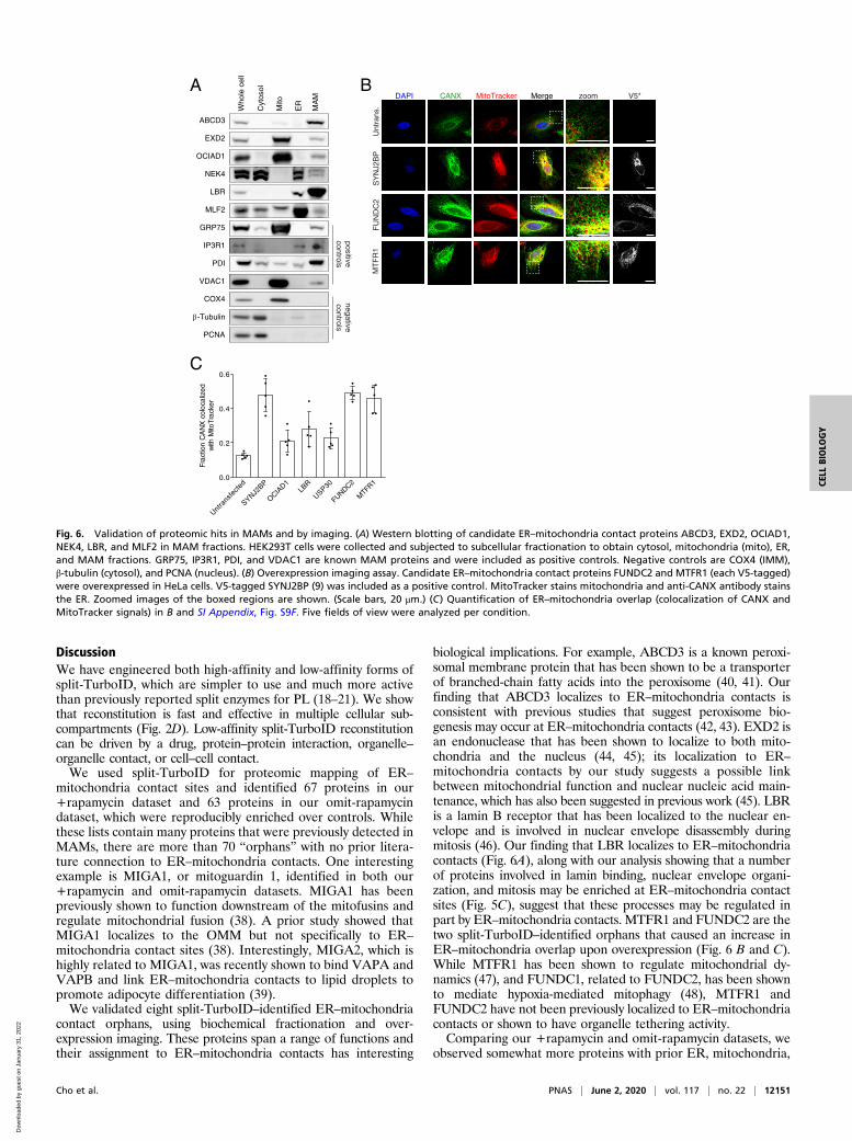
Validation of ER–Mitochondria Contact “Orphans”. Of the 29 pro-teins detected in both +rapamycin and omit-rapamycin datasets,12 have previously been detected in MAMs or localized toER–mitochondria contacts in the literature (Fig. 5A and SIAppendix, Fig. S8). The remainder are “orphans,” or proteinswith no prior literature connection to ER–mitochondria con-tacts. To determine if these are bona fide ER–mitochondriacontact residents or false positives, we selected six proteins forwhich high-quality commercial antibodies exist, and blotted fortheir presence in purified MAMs. All six were found to beenriched in MAMs, as well as in other compartments consis-tent with their literature annotation (e.g., MAM + ER forLBR [a lamin B receptor]; MAM + mitochondria for EXD2)(Fig. 6A).In addition to MAM blotting, we selected a subset of orphans
for functional analysis. Previously, we showed that overexpression ofthe ER–mitochondria tether SYNJ2BP in HeLa cells causes adramatic increase in the extent of overlap between ER and mito-chondria by imaging (9). We V5-tagged the orphan proteinsFUNDC2, LBR, MTFR1, OCIAD1, and USP30 and overexpressed
Cho et al. PNAS | June 2, 2020 | vol. 117 | no. 22 | 12147
CELL
BIOLO
GY
Dow
nloa
ded
by g
uest
on
Janu
ary
31, 2
022

1.0
0.0
0.2
0.4
0.6
0.8F
ract
ion
HA
co
loca
lize
d
with
mC
he
rry-K
DE
L
A
FRB
Tb(N)
Tb(C)
FKBP
reconstitutedsplit-TurboID
Mitochondria
ER
Mitochondria
ER
OMM anchor
ERM anchor
B
OMM FKBP V5 Tb(N)
Tb(C) HA FRB ERM
OMM-Tb(N)
Tb(C)-ERM
aa 1-34 of Tom20
aa 100-134 of Cb5
reconstitutedsplit-TurboID
Mitochondria
ER
+rapamycin-rapamycin
~30
-35
nm
~18
-20
nm
C
-rap
+ra
p
MergeOMM-FKBP-Tb(N) (V5)
Tb(C)-FRB-ERM (HA) NeutravidinDAPI
zoom
ATP + B
AMP B
ATP + B
AMP B
80
58
46
32
25
100
Silver stain after enrichment
NE
S
ER
M
FL TurboID
Rapamycin:
Biotin:
OM
M
NE
S
split-TurboID Unt
rans
.E F
58
46
32
25
5832
25
FACL4
Mff
SA-HRP
Rapamycin:
Biotin:
split-TurboID Unt
rans
.
NE
S
OM
M
ER
M
FL TurboID
80100
*
* *
split
-Tur
boID
stab
le
MergeDAPIOMM-FKBP-Tb(N) (V5) Tom20
split
-Tur
boID
stab
le
MergeDAPITb(C)-FRB-ERM (HA)
mCherry-KDEL
0.0
0.2
0.4
0.6
0.8
1.0
Fra
ctio
n V
5 c
olo
caliz
ed
w
ith T
om
20
D
Fig. 3. Reconstitution of split-TurboID at ER–mitochondria contact sites. (A) Schematic of split-TurboID reconstitution across ER–mitochondria contacts in thepresence or absence of rapamycin for inducing dimerization. (B) Design of constructs targeting split-TurboID fragments to the OMM and ERM. (C) Confocalfluorescence imaging of split-TurboID activity at ER–mitochondria contacts. Constructs were introduced into U2OS cells using lentivirus. Two days aftertransduction, cells were incubated with 50 μM biotin and 100 nM rapamycin for 1 h, then fixed and stained with anti-V5 to detect the N-terminal fragment[Tb(N)], anti-HA to detect the C-terminal fragment [Tb(C)], and neutravidin-647 to detect biotinylated proteins. Zoomed images of the boxed regions areshown. (Scale bars, 20 μm.) (D) Localization of split-TurboID in HEK293T cells stably expressing constructs from B. Cells were fixed and stained with anti-V5 todetect the OMM-targeted N-terminal fragment [Tb(N)] or with anti-HA to detect the ERM-targeted C-terminal fragment [Tb(C)]. Tom20 and mCherry-KDELwere used as mitochondrial and ER markers, respectively. (Scale bars, 10 μm.) Colocalization of V5 with Tom20 and HA with mCherry-KDEL are shown on theright. Quantitation from five fields of view per condition. (E) Enrichment of known ER–mitochondria contact proteins by split-TurboID-catalyzed PL.HEK293T cells stably expressing OMM/ERM-targeted split-TurboID constructs were treated with 50 μM biotin and 100 nM rapamycin for 4 h. HEK293T cellsstably expressing NES-, OMM-, or ERM-targeted FL-TurboID were treated with 50 μM biotin for 1 min. Biotinylated proteins were enriched from lysates usingstreptavidin beads, eluted, and analyzed by blotting with streptavidin and antibodies against FACL4 and Mff. Asterisks indicate ligase self-biotinylation. (F)Enrichment of biotinylated proteins for proteomics. Samples were generated as in E. Biotinylated proteins were enriched from lysates using streptavidinbeads, eluted, and analyzed by silver stain.
12148 | www.pnas.org/cgi/doi/10.1073/pnas.1919528117 Cho et al.
Dow
nloa
ded
by g
uest
on
Janu
ary
31, 2
022

them in HeLa cells, which were then stained for mitochondriaand ER markers. Confocal imaging shows that FUNDC2 andMTFR1, along with the positive control SYNJ2BP, cause asignificant increase in colocalization of ER and mitochondria,compared to untransfected control HeLa cells (Fig. 6 B and C).The other three proteins tested did not give as substantial aphenotype (SI Appendix, Fig. S9F). Our data suggest thatFUNDC2 and MTFR1 may have tethering functions at ER–
mitochondria contacts that are up-regulated in this gain-of-functionassay.
Cell–Cell Contact-Dependent Reconstitution of Split-TurboID. In addi-tion to permitting PL with greater spatial specificity, split-TurboIDcould potentially enable conditional PL dependent upon specific
inputs or signaling events. For example, in neuroscience, immu-nology, and cancer biology, there is great interest in characterizingthe transcriptomes and proteomes of cell subpopulations that havemade contact with specific “sender” cells (for example, neuronsdownstream of specific presynaptic inputs, or immune cells thatcome into contact with a tumor cell). If we could drive in-tracellular split-TurboID reconstitution, specifically in “receiver”cell populations that come into contact with defined “sender”cells, then this may enable PL-based proteomic analysis of func-tionally relevant cellular subpopulations.To test this, we designed a synthetic signaling network that
utilizes the transcellular interaction between glucagon peptideand its receptor (GCGR), employed in the transsynaptic tracingtool trans-Tango (37). In our design, “receiver” cells express the
A
TurboID-NES (Replicate 1)
+biotin
TurboID-NES(Replicate 2)
+biotin
ERM-TurboID+biotin
TurboID-OMM+biotin
spTurboID+rapamycin
+biotin(Replicate 1)
spTurboID+rapamycin
+biotin(Replicate 2)
spTurboID-rapamycin
-biotin
spTurboID+rapamycin
-biotin
spTurboID-rapamycin
+biotin(Replicate 2)
spTurboID-rapamycin
+biotin(Replicate 1)
Untransfected+rapamycin
+biotin
TMT Label:
TMT Label: 130N
127C
129C
129N128C128N126C 127N
130C 131N 131C
Combine samplesLC-MS/MS
Lyse cellsStreptavidin bead enrichment
On-bead trypsin digestionTMT labeling
B C
spTurboID(+R+B Rep. 1)
spTurboID(+R+B Rep. 2)
spTurboID(-R+B Rep. 2)
spTurboID(-R+B Rep. 1)
Proteins with 2 unique peptides detected2496 proteins
Ratio over omit biotin controlFilter 1 cutoff at FDR<0.1
607 proteins 619 proteins 466 proteins 606 proteins
Ratio over cytosolic controlFilter 2 cutoff at FDR<0.1
97 proteins 97 proteins 84 proteins 123 proteins
spTurboID +R+B Proteome67 proteins
spTurboID -R+B Proteome63 proteins
Overlap
log2(128C/130C) cutoff = 0.202
-4 -2 0 2 4-4.0
-2.0
0.0
2.0
4.0
log2(128C/130C)Extent of biotinylation
by split-TurboID (+R+B) (Rep. 1)
log 2
(128
C/1
26C
)R
atio
of b
iotin
ylat
ion
by
split
-Tur
boID
(+R
+B
) vs.
Tur
boID
-NE
S (R
ep. 1
)
RHOT2
MAVS
BCAP31
AKAP1
log2(128C/126C) cutoff = 0.805
DNM1L
ERM annotationOMM annotationERM + OMM annotationOthers
CISD2
(zoom)
Fig. 4. Proteomic mapping of ER–mitochondria contacts in HEK293T cells. (A) Experimental design and labeling conditions for MS-based proteomics. Cellsstably expressing the indicated constructs were labeled with 50 μM biotin and 100 nM rapamycin. Split-TurboID (ERM/OMM) samples were labeled for 4 h andFL-TurboID samples were labeled for 1 min. Cells were then lysed, and biotinylated proteins were enriched using streptavidin beads, digested to peptides, andconjugated to TMT labels. All samples were then combined and analyzed by LC-MS/MS. (B) Filtering scheme for mass spectrometric data. +R and −R refer torapamycin, and +B and −B refer to biotin. For each dataset, proteins were first ranked by the extent of biotinylation (ratiometric analysis referencing omitbiotin controls, filter 1). Next, proteins were ranked by relative proximity to ER–mitochondria contacts versus cytosol (ratiometric analysis referencingTurboID-NES, filter 2). (C) Scatterplot showing log2(128C/130C) (filter 1) versus log2(128C/126C) (filter 2) for each protein in replicate 1 of split-TurboID cellstreated with rapamycin and biotin. Known ERM and OMM proteins (as annotated by GOCC) are labeled blue and red, respectively; ERM and OMM dual-annotated proteins are colored purple, and all other proteins are shown in black. Cutoffs used to filter the mass spectrometric data and obtain the filteredproteome are shown by dashed lines. Zoom of boxed region is shown in the upper left.
Cho et al. PNAS | June 2, 2020 | vol. 117 | no. 22 | 12149
CELL
BIOLO
GY
Dow
nloa
ded
by g
uest
on
Janu
ary
31, 2
022

glucagon receptor (GCGR) and arrestin, each fused to a split-TurboID fragment. Upon cell–cell contact with “sender” cellsexpressing surface glucagon peptide, GCGR is activated, resulting inarrestin recruitment and reconstitution of split-TurboID (Fig. 7A).
Confocal fluorescence imaging of receiver and sender cell co-culturesafter biotin treatment for 1 h showed highly specific localization ofbiotinylated proteins, specifically at sender:receiver cell–cell contacts(red:green overlap sites in Fig. 7B).
B
Calcium iontransport
ER membrane
Outer mitochondrialmembrane
ERADSterol metabolism
Nitrate metabolism
ER-GolgiSNARE complexes
Lamin bindingNuclear env. org.Mitotic processes
Mito. org.Mito. fissionPerox. org.
log2 ratio:
0.5
2.5
+rapamycin -rapamycinintersection
AOMM and ERMOMM ERM
+rapamycinER-mito
proteome34
ABCA3, ABCD3, ACBD5, AKAP1, ANGEL1, ARMCX3,
BCAP31, C1QBP, CISD1, DNM1L, EMD, EXD2,
FKBP8, FUNDC2, LBR, MARC2, MAST4, MAVS, MIEF1, MIGA1, MLF2,
MTFR1, NEK4, OCIAD1,PKD2, RHOT2, TDRKH,
TMEM209, USP30
ANKLE2, ARID1B, ATP2C2, CDC23, CHEK1, CLCC1, FNDC3A, IFT57, KIF16B,
LEMD3, MAN1A1, MCM10,MIEF2, MTCH1, MTFR1L, MTX3, PEX13, POM121C,
PPP1R15B, PRR11, PTPN1, RTN3, SAMD1, SAMD11
SLC25A1, SLC35E1, STIM1, STIM2, SUCO, TMPO,
TRABD, TRIM61, VEZT, WFS1
Prior annotation for ERM, OMM, both, or neither(Prior enrichment in MAMs)
38 29-rapamycin
ER-mitoproteome
ALDH3A2, ATG2A, BAX, C1orf43, CAMLG, CHP1, CISD2, DGKE, DHCR7,
DIXDC1, EPHX1, ERLIN1,ERLIN2, GK5, ITGB1,
MARC1, MBOAT7, MLX, MMGT1, MYO19, NDC1,
PARP16, PEX14, PGRMC1,PKMYT1, RABL3, RAP2C,
REEP5, RFT1, RNF5, SCD, SOAT1, STX5,
TMEM201, TOR1AIP1, UBXN8,VKORC1L1, YKT6
C
Entire
hum
an p
rote
ome
Pre-fi
lter p
rote
ome
spTur
boID
+ra
p
spTur
boID
-rap
spTur
boID
inte
rsec
tion
spTur
boID
com
bined
Hung
et a
l. eL
ife, 2
017
Cho e
t al.
JBC, 2
017
Kwak e
t al.
PNAS, 202
0
Wan
g et
al.
Prote
omics
, 201
80
20
40
60
80
100
Pe
rce
nta
ge
of p
rote
om
e w
ithp
rior a
nn
ota
tion
2496
67
63
29
101 70
88115
214619694
Fig. 5. Analysis of ER–mitochondria contact proteome. (A) Venn diagram comparing proteome lists obtained using split-TurboID. Proteins that were pre-viously annotated ERM, OMM, both, or neither were labeled blue, red, purple, or black, respectively. Proteins previously enriched in MAMs are highlighted inyellow. (B) Specificity analysis for proteomic datasets generated using split-TurboID compared to the entire human proteome and to previously publisheddatasets. Bar graph shows the percentage of each proteome with prior OMM annotation only, ERM annotation only, or both OMM and ERM annotation.Each bar is labeled with the size of the proteome. Hung et al. used APEX (9), Cho et al. used APEX and MAM fractionation (36), Kwak et al. used Contact-ID(21), and Wang et al. used MAM purification (35). (C) Markov clustering of split-TurboID proteome using protein–protein interaction scores from the STRINGdatabase (33). Gray lines denote protein–protein interactions. Nodes are colored based on whether the corresponding protein was found in the +rapamycinproteome, omit-rapamycin proteome, or both proteomes. Node size is correlated with the relative enrichment of each protein at ER–mitochondria contactsversus cytosol [log2(128C/126C) or log2(129C/126C)]. Each cluster is labeled with associated GO terms (org., organization).
12150 | www.pnas.org/cgi/doi/10.1073/pnas.1919528117 Cho et al.
Dow
nloa
ded
by g
uest
on
Janu
ary
31, 2
022
DiscussionWe have engineered both high-affinity and low-affinity forms ofsplit-TurboID, which are simpler to use and much more activethan previously reported split enzymes for PL (18–21). We showthat reconstitution is fast and effective in multiple cellular sub-compartments (Fig. 2D). Low-affinity split-TurboID reconstitutioncan be driven by a drug, protein–protein interaction, organelle–organelle contact, or cell–cell contact.We used split-TurboID for proteomic mapping of ER–
mitochondria contact sites and identified 67 proteins in our+rapamycin dataset and 63 proteins in our omit-rapamycindataset, which were reproducibly enriched over controls. Whilethese lists contain many proteins that were previously detected inMAMs, there are more than 70 “orphans” with no prior litera-ture connection to ER–mitochondria contacts. One interestingexample is MIGA1, or mitoguardin 1, identified in both our+rapamycin and omit-rapamycin datasets. MIGA1 has beenpreviously shown to function downstream of the mitofusins andregulate mitochondrial fusion (38). A prior study showed thatMIGA1 localizes to the OMM but not specifically to ER–
mitochondria contact sites (38). Interestingly, MIGA2, which ishighly related to MIGA1, was recently shown to bind VAPA andVAPB and link ER–mitochondria contacts to lipid droplets topromote adipocyte differentiation (39).We validated eight split-TurboID–identified ER–mitochondria
contact orphans, using biochemical fractionation and over-expression imaging. These proteins span a range of functions andtheir assignment to ER–mitochondria contacts has interesting
biological implications. For example, ABCD3 is a known peroxi-somal membrane protein that has been shown to be a transporterof branched-chain fatty acids into the peroxisome (40, 41). Ourfinding that ABCD3 localizes to ER–mitochondria contacts isconsistent with previous studies that suggest peroxisome bio-genesis may occur at ER–mitochondria contacts (42, 43). EXD2 isan endonuclease that has been shown to localize to both mito-chondria and the nucleus (44, 45); its localization to ER–mitochondria contacts by our study suggests a possible linkbetween mitochondrial function and nuclear nucleic acid main-tenance, which has also been suggested in previous work (45). LBRis a lamin B receptor that has been localized to the nuclear en-velope and is involved in nuclear envelope disassembly duringmitosis (46). Our finding that LBR localizes to ER–mitochondriacontacts (Fig. 6A), along with our analysis showing that a numberof proteins involved in lamin binding, nuclear envelope organi-zation, and mitosis may be enriched at ER–mitochondria contactsites (Fig. 5C), suggest that these processes may be regulated inpart by ER–mitochondria contacts. MTFR1 and FUNDC2 are thetwo split-TurboID–identified orphans that caused an increase inER–mitochondria overlap upon overexpression (Fig. 6 B and C).While MTFR1 has been shown to regulate mitochondrial dy-namics (47), and FUNDC1, related to FUNDC2, has been shownto mediate hypoxia-mediated mitophagy (48), MTFR1 andFUNDC2 have not been previously localized to ER–mitochondriacontacts or shown to have organelle tethering activity.Comparing our +rapamycin and omit-rapamycin datasets, we
observed somewhat more proteins with prior ER, mitochondria,
A B
Who
le c
ell
Cyt
osol
Mito
ER
MA
M
ABCD3
EXD2
OCIAD1
NEK4
LBR
MLF2
GRP75
IP3R1
PDI
VDAC1
COX4
-Tubulin
PCNA
C
Untra
nsfe
cted
SYNJ2BP
OCIAD1
LBR
USP30
FUNDC2
MTFR1
0.0
0.2
0.4
0.6
Fra
ctio
n C
AN
X c
olo
caliz
ed
w
ith M
itoT
rack
er
Unt
rans
.S
YN
J2B
P
zoomDAPI CANX MitoTracker Merge
FU
ND
C2
MT
FR
1
positivecontrols
negativecontrols
V5*
Fig. 6. Validation of proteomic hits in MAMs and by imaging. (A) Western blotting of candidate ER–mitochondria contact proteins ABCD3, EXD2, OCIAD1,NEK4, LBR, and MLF2 in MAM fractions. HEK293T cells were collected and subjected to subcellular fractionation to obtain cytosol, mitochondria (mito), ER,and MAM fractions. GRP75, IP3R1, PDI, and VDAC1 are known MAM proteins and were included as positive controls. Negative controls are COX4 (IMM),β-tubulin (cytosol), and PCNA (nucleus). (B) Overexpression imaging assay. Candidate ER–mitochondria contact proteins FUNDC2 and MTFR1 (each V5-tagged)were overexpressed in HeLa cells. V5-tagged SYNJ2BP (9) was included as a positive control. MitoTracker stains mitochondria and anti-CANX antibody stainsthe ER. Zoomed images of the boxed regions are shown. (Scale bars, 20 μm.) (C) Quantification of ER–mitochondria overlap (colocalization of CANX andMitoTracker signals) in B and SI Appendix, Fig. S9F. Five fields of view were analyzed per condition.
Cho et al. PNAS | June 2, 2020 | vol. 117 | no. 22 | 12151
CELL
BIOLO
GY
Dow
nloa
ded
by g
uest
on
Janu
ary
31, 2
022

and MAM annotations in the +rapamycin condition. Because inour construct design, FRB and FKBP also function like linkersthat extend the reach of the split-TurboID fragments, we hy-pothesize that the +rapamycin proteome may be probing closerER–mitochondria contacts, while the omit-rapamycin proteomemay also be capturing proteins present at wider contacts, asshown in Fig. 3A. Perhaps MAM preparations in previous studiesare biased toward closer ER–mitochondria contacts if these aremore likely to survive the fractionation process, while widercontacts may be underrepresented. Thus, while there is less priorannotation information for proteins in the omit-rapamycin pro-teome, these proteins may still be bona fide ER–mitochondriacontact proteins.In addition to generating ER–mitochondria contact candi-
dates, our proteomic experiment also produced OMM and ERMproteomes, via the full-length TurboID constructs we targeted toOMM and ERM, respectively. Using these datasets, we couldcategorize our ER–mitochondria contact hits as additionally lo-calized to the OMM and/or ERM, or not (SI Appendix, Fig. S9D;each protein is classified as being a resident of “contacts only,”“contacts and OMM,” “contacts and ERM,” or “contacts, OMM,and ERM”). The categorizations based on our proteomic data arelargely consistent with prior literature, and with our fractionationblotting and imaging data as well (Fig. 6 and SI Appendix, Fig.S9 E and F). For several proteins, such as OCIAD1 and NEK4,our proteomic data newly assign them to mitochondrial and ERcompartments of the cell.When comparing our proteome with that obtained via Contact-
ID (21), we find that both proteomes detected a similar number ofproteins (101 proteins using split-TurboID and 115 proteins usingContact-ID). Of the eight proteins we validated, four (FUNDC2,EXD2, OCIAD1, and LBR) were also detected using Contact-ID(SI Appendix, Fig. S8). Conversely, FKBP8, which the authors ofContact-ID show facilitates ER–mitochondria contact formationand local calcium transport (21), was also present in our proteome.While both proteomes have high specificity, measured by the frac-tion of proteins with prior OMM or ERM annotation, we find thatthe proteome generated from Contact-ID is more biased towardERM proteins, whereas we observe more balance between OMMand ERM proteins in our split-TurboID proteome (Fig. 5B). Thisdifference may possibly arise from the longer labeling period usedfor Contact-ID (16 h vs. 4 h for split-TurboID).
Overall, we have developed a split-TurboID tool that can beconditionally reconstituted for spatially specific PL in cells.Especially when combined with functional assays and screens,split-TurboID–based PL can be a powerful tool for biologicaldiscovery around organelle contact sites or macromolecularcomplexes. Split-TurboID may also improve signal-to-noise forchallenging targeting applications, such as dCas9-directed PLof specific genomic loci (49, 50), or dCas13-directed PL ofspecific cellular RNAs (51).
MethodsMethods related to cloning, split-site pair selection, Western blots, confocalfluorescence imaging, proteomic sample preparation and analysis, additionaldata analysis, and subcellular fractionation are detailed in SI Appendix.
Mammalian Cell Culture, Transfection, and Stable Line Generation. HEK293T cellsfrom ATCC were cultured as a monolayer in DMEM with 4.5 g/L glucose andL-glutamine supplemented with 10% (vol/vol) fetal bovine serum, 50 U/mLpenicillin, and 50 μg/mL streptomycin at 37 °C under 5% CO2. For confocalimaging experiments, glass coverslips were coated with 50 μg/mL fibronectinin DPBS (Dulbecco’s phosphate-buffered saline) for at least 20 min at roomtemperature before plating; cells were grown on glass coverslips in 24-wellplates with 500 μL growth medium. For Western blot experiments, cells weregrown in six-well plates with 2 mL growth medium.
For transient expression, cells were transfected at ∼50% confluency using5 μL/mL Lipofectamine2000 and 250 ng/mL plasmid in serum-free media. Togenerate lentiviruses, HEK293T cells were cultured in T25 flasks and trans-fected at ∼60% confluency with 2,500 ng of the lentiviral vector containingthe gene of interest and lentiviral packaging plasmids pVSVG (250 ng) andΔ8.9 (2,250 ng) with 30 μL polyethyleneimine (PEI) (1 mg/mL in water, pH7.3) in serum-free media. After 48 h, the cell medium containing the lenti-virus was harvested and filtered using a 0.45-μm filter.
For generation of stable cell lines, HEK293T cells were infected with crudelentivirus at ∼50% confluence, followed by selection with 8 μg/mL blasticidin(at least 4 d) and 250 μg/mL hygromycin (at least 7 d) in growth mediumbefore use in experiments.
Biotin Labeling with TurboID and Split-TurboID. For biotin labeling of tran-siently transfected cells, biotin and rapamycin were added 18 h followingtransfection. Biotin (10 mM stock in DMSO) was diluted in complete mediaand added directly to cells to a final concentration of 50 μM. For full-lengthTurboID, cells were treated with 50 μM biotin and incubated at 37 °C for1–30 min; for split-TurboID, cells were treated with 50 μM biotin ± 100 nMrapamycin and incubated at 37 °C for 30 min–24 h (as indicated). In general,for split-TurboID labeling, 1 h is a good starting point, but optimization of
A B
-bio
tin+
biot
in
MergeGCGR-Tb(N)(Receiver, V5)
Glucagon(Sender, HA) NeutravidinDAPI
zoom
1
Receiver cell
Sender cell
GCGR
GlucagonPeptide
Arrestin
Tb(N)
Tb(C)
zoom
2*
1
2
cell-cellcontact GCGR
GlucagonPeptide
Arrestin ATP + B
AMP B
reconstitutedsplit-TurboID
Sender cell
Receiver cell
Fig. 7. Cell–cell contact dependent reconstitution of intracellular split-TurboID. (A) Schematic of cell–cell contact-dependent split-TurboID reconstitution. (B)Confocal fluorescence imaging of “sender” cells expressing cell surface targeted glucagon peptide co-cultured with “receiver” cells expressing split-TurboIDfragments. Zoomed images of the boxed regions are shown. (Scale bars, 20 μm.) *The contrast has been increased in the zoom 2 row.
12152 | www.pnas.org/cgi/doi/10.1073/pnas.1919528117 Cho et al.
Dow
nloa
ded
by g
uest
on
Janu
ary
31, 2
022

labeling times may be necessary depending on the application. For bothimaging and Western blot experiments, labeling was stopped after the in-dicated time periods by transferring cells on plates to ice and washing gentlywith cold DPBS.
Sample Preparation for ER–Mitochondria Proteomics. For each cell sampleshown in Fig. 4A, HEK293T cells were cultured in T150 flasks. All cells used inproteomics experiments were stably expressing the indicated TurboID orsplit-TurboID constructs. Split-TurboID samples were labeled with 50 μMbiotin and 100 nM rapamycin for 4 h, and full-length TurboID samples werelabeled with 50 μM biotin for 1 min. Labeling was stopped by placing cells onice and washing gently with cold DPBS twice. Cells were then detached fromthe well or flask by pipetting a stream of cold DPBS directly onto cells. Thecells were collected and pelleted by centrifugation at 2,500 rpm for 3 min at4 °C. The supernatant was removed, and the pellet was lysed by resus-pension in RIPA lysis buffer and incubation for 10 min at 4 °C. Lysates wereclarified by centrifugation at 12,000 rpm for 10 min at 4 °C.
For enrichment of biotinylated proteins, 300 μL streptavidin-coatedmagnetic beads (Pierce) were washed twice with RIPA lysis buffer and in-cubated with clarified lysates with rotation at 4 °C overnight. The beadswere then washed twice with 1 mL of RIPA lysis buffer, once with 1 mL 1MKCl, once with 1 mL 0.1M Na2CO3, once with 1 mL 2M urea in 10 mM Tris·HCl(pH 8.0), and twice with 1 mL RIPA lysis buffer. The beads were subsequentlywashed with 1 mL digestion buffer (75 mM NaCl, 1 mM EDTA, 50 mM Tris·HCl, pH 8.0) twice. The beads were then resuspended in 80 μL digestionbuffer, transferred to a new Eppendorf tube, frozen on dry ice, and shippedfor further processing and preparation for LC-MS/MS analysis.
For each proteomic sample, 0.2% of the lysate was removed prior toenrichment to verify construct expression and confirm successful bio-tinylation, as shown in SI Appendix, Fig. S3D. After enrichment, 5% of beadswere removed and biotinylated proteins were eluted by boiling the beads in80 μL 3× protein loading buffer supplemented with 20 mM DTT and 2 mMbiotin. The eluted proteins were separated by SDS-PAGE gel and analyzed byWestern blotting (1.25%) and silver stain (3.75%) to verify successful en-richment of biotinylated proteins (Fig. 3F and SI Appendix, Fig. S3E).
Generation of ER–Mitochondria Proteome Lists. Unprocessed mass spectrom-etry data for split-TurboID targeted to ERM/OMM (both +R+B and −R+B) areshown in Dataset S1, Tabs A–C. These data were filtered in three steps togenerate our proteome lists, as follows. First, to identify proteins bio-tinylated by TurboID over nonspecific binders, we generated a true positive(TP) list of literature-validated ER–mitochondria contact proteins (8, 16, 31,52–73; Dataset S2, Tab A) and a false positive list (FP1) of mitochondrial
matrix proteins, which should not be biotinylated by split-TurboID localizedto ER–mitochondria contacts, as determined by Gene Ontology CellularComponent (GOCC) (Dataset S2, Tab B; GO:0005759, but no additional an-notations for inner mitochondrial membrane [IMM], OMM, intermembranespace [IMS], or membrane). TMT log2 ratios for split-TurboID (+R+B) oversplit-TurboID (+R−B) and for split-TurboID (−R+B) over split-TurboID (−R−B)were ranked from highest to lowest. For each possible TMT log2 ratio cutoff,the false discovery rate (FDR), defined as the fraction of detected FP1 pro-teins detected above the cutoff, was calculated, and the cutoff was de-termined by setting the FDR < 0.1.
Second, we identified proteins preferentially biotinylated by ER–mitochondria contact-localized split-TurboID over cytosolic TurboID. To dothis, we generated another false positive list (FP2) of cytosol-resident pro-teins that should not be enriched at ER–mitochondria contacts, as de-termined by GOCC (Dataset S2, Tab C; GO:0005829, but no annotations formembrane). TMT log2 ratios for split-TurboID (+R+B) over TurboID-NES andfor split-TurboID (−R+B) over TurboID-NES were ranked from highest tolowest. For each possible TMT log2 ratio cutoff, the false discovery rate(FDR), defined as the fraction of detected FP2 proteins detected above eachcutoff, was calculated. The cutoff was determined by setting the FDR < 0.1.
Third, after applying both cutoff steps to each experimental replicate, weintersected the resulting lists to obtain our final +rapamycin and omit-rapamycin proteome lists, which are shown in Dataset S1, Tabs A and B.FKBP1 and mammalian target of rapamycin (mTOR) were removed from thelists as our split-TurboID fusions themselves generate these peptides (be-cause they incorporate FKBP and FRB domains). An additional list inter-secting split-TurboID (+R+B) and split-TurboID (−R+B) lists is also shown inDataset S1, Tab C.
Data Availability. Proteomics data and all log2 ratio values associated witheach protein detected (with two or greater unique peptides) are available inDataset S1.
ACKNOWLEDGMENTS. This work was supported by the NIH Grant R01-DK121409 (to A.Y.T. and S.A.C.); Stanford Wu Tsai Neurosciences InstituteBig Ideas Initiative (A.Y.T.); Korea Health Technology R&D Project GrantKHIDI HI16C1501 (to I.-K.L.); National Research Foundation of Korea (NRF)Grants 2017R1A2B3006406 (to I.-K.L.) and 2019R1A2C3008463 (to H.-W.R.);and Organelle Network Research Center Grant NRF-2017R1A5A1015366 (toH.-W.R.). K.F.C. was supported by NIH Training Grant 2T32CA009302-41 andthe Blavatnik Graduate Fellowship. T.C.B. was supported by Dow GraduateResearch and Lester Wolfe Fellowships. A.Y.T. is an investigator of the ChanZuckerberg Biohub.
1. D. I. Kim, K. J. Roux, Filling the void: Proximity-based labeling of proteins in living
cells. Trends Cell Biol. 26, 804–817 (2016).2. C. L. Chen, N. Perrimon, Proximity-dependent labeling methods for proteomic pro-
filing in living cells. Wiley Interdiscip. Rev. Dev. Biol. 6, e272 (2017).3. P. Li, J. Li, L. Wang, L. J. Di, Proximity labeling of interacting proteins: Application of
BioID as a discovery tool. Proteomics 17, 1700002 (2017).4. S. S. Lam et al., Directed evolution of APEX2 for electron microscopy and proximity
labeling. Nat. Methods 12, 51–54 (2015).5. H. W. Rhee et al., Proteomic mapping of mitochondria in living cells via spatially re-
stricted enzymatic tagging. Science 339, 1328–1331 (2013).6. K. J. Roux, D. I. Kim, M. Raida, B. Burke, A promiscuous biotin ligase fusion protein
identifies proximal and interacting proteins in mammalian cells. J. Cell Biol. 196,
801–810 (2012).7. T. C. Branon et al., Efficient proximity labeling in living cells and organisms with
TurboID. Nat. Biotechnol. 36, 880–887 (2018).8. V. Hung et al., Proteomic mapping of the human mitochondrial intermembrane space
in live cells via ratiometric APEX tagging. Mol. Cell 55, 332–341 (2014).9. V. Hung et al., Proteomic mapping of cytosol-facing outer mitochondrial and ER
membranes in living human cells by proximity biotinylation. eLife 6, e24463 (2017).10. S. Han et al., Proximity biotinylation as a method for mapping proteins associated
with mtDNA in living cells. Cell Chem. Biol. 24, 404–414 (2017).11. K. H. Loh et al., Proteomic analysis of unbounded cellular compartments: Synaptic
clefts. Cell 166, 1295–1307.e21 (2016).12. A. Uezu et al., Identification of an elaborate complex mediating postsynaptic in-
hibition. Science 353, 1123–1129 (2016).13. S. Markmiller et al., Context-dependent and disease-specific diversity in protein in-
teractions within stress granules. Cell 172, 590–604.e13 (2018).14. D. U. Mick et al., Proteomics of primary cilia by proximity labeling. Dev. Cell 35,
497–512 (2015).15. G. Csordás, D. Weaver, G. Hajnóczky, Endoplasmic reticulum-mitochondrial con-
tactology: Structure and signaling functions. Trends Cell Biol. 28, 523–540 (2018).16. J. R. Friedman et al., ER tubules mark sites of mitochondrial division. Science 334,
358–362 (2011).
17. S. Gandre-Babbe, A. M. van der Bliek, The novel tail-anchored membrane protein Mffcontrols mitochondrial and peroxisomal fission in mammalian cells. Mol. Biol. Cell 19,2402–2412 (2008).
18. Y. Han et al., Directed evolution of split APEX2 peroxidase. ACS Chem. Biol. 14,619–635 (2019).
19. S. De Munter et al., Split-BioID: A proximity biotinylation assay for dimerization-dependent protein interactions. FEBS Lett. 591, 415–424 (2017).
20. I. M. Schopp et al., Split-BioID a conditional proteomics approach to monitor thecomposition of spatiotemporally defined protein complexes. Nat. Commun. 8, 15690(2017).
21. C. Kwak et al., Contact-ID, a tool for profiling organelle contact sites, reveals regu-latory proteins of mitochondrial-associated membrane formation. Proc. Natl. Acad.Sci. U.S.A. 117, 12109–12120 (2020).
22. J. D. Martell et al., A split horseradish peroxidase for the detection of intercellularprotein-protein interactions and sensitive visualization of synapses. Nat. Biotechnol.34, 774–780 (2016).
23. O. Dagliyan et al., Computational design of chemogenetic and optogenetic splitproteins. Nat. Commun. 9, 4042 (2018).
24. L. H. Weaver, K. Kwon, D. Beckett, B. W. Matthews, Corepressor-induced organizationand assembly of the biotin repressor: A model for allosteric activation of a tran-scriptional regulator. Proc. Natl. Acad. Sci. U.S.A. 98, 6045–6050 (2001).
25. Z. A. Wood, L. H. Weaver, P. H. Brown, D. Beckett, B. W. Matthews, Co-repressorinduced order and biotin repressor dimerization: A case for divergent followed byconvergent evolution. J. Mol. Biol. 357, 509–523 (2006).
26. J. Rieusset, The role of endoplasmic reticulum-mitochondria contact sites in thecontrol of glucose homeostasis: An update. Cell Death Dis. 9, 388 (2018).
27. A. A. Rowland, G. K. Voeltz, Endoplasmic reticulum-mitochondria contacts: Functionof the junction. Nat. Rev. Mol. Cell Biol. 13, 607–625 (2012).
28. C. N. Poston, S. C. Krishnan, C. R. Bazemore-Walker, In-depth proteomic analysis ofmammalian mitochondria-associated membranes (MAM). J. Proteomics 79, 219–230(2013).
29. S. M. Horner, H. M. Liu, H. S. Park, J. Briley, M. Gale, Jr., Mitochondrial-associatedendoplasmic reticulum membranes (MAM) form innate immune synapses and aretargeted by hepatitis C virus. Proc. Natl. Acad. Sci. U.S.A. 108, 14590–14595 (2011).
Cho et al. PNAS | June 2, 2020 | vol. 117 | no. 22 | 12153
CELL
BIOLO
GY
Dow
nloa
ded
by g
uest
on
Janu
ary
31, 2
022

30. V. Hung et al., Spatially resolved proteomic mapping in living cells with the en-gineered peroxidase APEX2. Nat. Protoc. 11, 456–475 (2016).
31. R. Iwasawa, A. L. Mahul-Mellier, C. Datler, E. Pazarentzos, S. Grimm, Fis1 and Bap31bridge the mitochondria-ER interface to establish a platform for apoptosis induction.EMBO J. 30, 556–568 (2011).
32. R. A. Merrill, S. Strack, Mitochondria: A kinase anchoring protein 1, a signalingplatform for mitochondrial form and function. Int. J. Biochem. Cell Biol. 48, 92–96(2014).
33. D. Szklarczyk et al., STRING v10: Protein-protein interaction networks, integratedover the tree of life. Nucleic Acids Res. 43, D447–D452 (2015).
34. M. S. Herrera-Cruz, T. Simmen, Of yeast, mice and men: MAMs come in two flavors.Biol. Direct 12, 3 (2017).
35. X. Wang, Y. Wen, J. Dong, C. Cao, S. Yuan, Systematic in-depth proteomic analysis ofmitochondria-associated endoplasmic reticulum membranes in mouse and humantestes. Proteomics 18, e1700478 (2018).
36. I. T. Cho et al., Ascorbate peroxidase proximity labeling coupled with biochemicalfractionation identifies promoters of endoplasmic reticulum-mitochondrial contacts.J. Biol. Chem. 292, 16382–16392 (2017).
37. M. Talay et al., Transsynaptic mapping of second-order taste neurons in flies by trans-Tango. Neuron 96, 783–795.e4 (2017).
38. Y. Zhang et al., Mitoguardin regulates mitochondrial fusion through MitoPLD and isrequired for neuronal homeostasis. Mol. Cell 61, 111–124 (2016).
39. C. A. C. Freyre, P. C. Rauher, C. S. Ejsing, R. W. Klemm, MIGA2 links mitochondria, theER, and lipid droplets and promotes de novo lipogenesis in adipocytes. Mol. Cell 76,811–825.e14 (2019).
40. T. Imanaka, K. Aihara, Y. Suzuki, S. Yokota, T. Osumi, The 70-kDa peroxisomalmembrane protein (PMP70), an ATP-binding cassette transporter. Cell Biochem. Bio-phys. 32, 131–138 (2000).
41. Y. Kashiwayama et al., Role of Pex19p in the targeting of PMP70 to peroxisome.Biochim. Biophys. Acta 1746, 116–128 (2005).
42. A. Sugiura, G. L. McLelland, E. A. Fon, H. M. McBride, A new pathway for mito-chondrial quality control: Mitochondrial-derived vesicles. EMBO J. 33, 2142–2156(2014).
43. A. Sugiura, S. Mattie, J. Prudent, H. M. McBride, Newly born peroxisomes are a hybridof mitochondrial and ER-derived pre-peroxisomes. Nature 542, 251–254 (2017).
44. R. Broderick et al., EXD2 promotes homologous recombination by facilitating DNAend resection. Nat. Cell Biol. 18, 271–280 (2016).
45. F. Hensen, A. Moretton, S. van Esveld, G. Farge, J. N. Spelbrink, The mitochondrialouter-membrane location of the EXD2 exonuclease contradicts its direct role in nu-clear DNA repair. Sci. Rep. 8, 5368 (2018).
46. A. R. English, G. K. Voeltz, Endoplasmic reticulum structure and interconnections withother organelles. Cold Spring Harb. Perspect. Biol. 5, a013227 (2013).
47. M. Monticone et al., The nuclear genes Mtfr1 and Dufd1 regulate mitochondrialdynamic and cellular respiration. J. Cell. Physiol. 225, 767–776 (2010).
48. W. Wu et al., FUNDC1 regulates mitochondrial dynamics at the ER-mitochondrialcontact site under hypoxic conditions. EMBO J. 35, 1368–1384 (2016).
49. S. A. Myers et al., Discovery of proteins associated with a predefined genomic locusvia dCas9-APEX-mediated proximity labeling. Nat. Methods 15, 437–439 (2018).
50. X. D. Gao et al., C-BERST: Defining subnuclear proteomic landscapes at genomic el-ements with dCas9-APEX2. Nat. Methods 15, 433–436 (2018).
51. S. Han et al., RNA-protein interaction mapping via MS2 or Cas13-based APEX tar-geting. bioRxiv:10.1101/2020.02.27.968297 (28 February 2020).
52. K. S. Lee et al., Altered ER-mitochondria contact impacts mitochondria calcium ho-
meostasis and contributes to neurodegeneration in vivo in disease models. Proc. Natl.Acad. Sci. U.S.A. 115, E8844–E8853 (2018).
53. T. Simmen et al., PACS-2 controls endoplasmic reticulum-mitochondria communica-
tion and Bid-mediated apoptosis. EMBO J. 24, 717–729 (2005).54. Y. Hirabayashi et al., ER-mitochondria tethering by PDZD8 regulates Ca2+ dynamics in
mammalian neurons. Science 358, 623–630 (2017).55. X. Wang, T. L. Schwarz, The mechanism of Ca2+ -dependent regulation of kinesin-
mediated mitochondrial motility. Cell 136, 163–174 (2009).56. K. J. De vos et al., VAPB interacts with the mitochondrial protein PTPIP51 to regulate
calcium homeostasis. Hum. Mol. Genet. 21, 1299–1311 (2012).57. R. Stoica et al., ER-mitochondria associations are regulated by the VAPB-PTPIP51 in-
teraction and are disrupted by ALS/FTD-associated TDP-43. Nat. Commun. 5, 3996
(2014).58. S. B. Wortmann et al., Mutations in the phospholipid remodeling gene SERAC1 impair
mitochondrial function and intracellular cholesterol trafficking and cause dystonia
and deafness. Nat. Genet. 44, 797–802 (2012).59. T. Hayashi, T. P. Su, Sigma-1 receptor chaperones at the ER-mitochondrion interface
regulate Ca(2+) signaling and cell survival. Cell 131, 596–610 (2007).60. M. Sugo et al., Syntaxin 17 regulates the localization and function of PGAM5 in mi-
tochondrial division and mitophagy. EMBO J. 37, e98899 (2018).61. A. Vecchione et al., MITOSTATIN, a putative tumor suppressor on chromosome
12q24.1, is downregulated in human bladder and breast cancer. Oncogene 28,
257–269 (2009).62. C. Cerqua et al., Trichoplein/mitostatin regulates endoplasmic reticulum-
mitochondria juxtaposition. EMBO Rep. 11, 854–860 (2010).63. P. Gomez-Suaga et al., The ER-mitochondria tethering complex VAPB-PTPIP51 regu-
lates autophagy. Curr. Biol. 27, 371–385 (2017).64. W. H. Yu, W. Wolfgang, M. Forte, Subcellular localization of human voltage-
dependent anion channel isoforms. J. Biol. Chem. 270, 13998–14006 (1995).65. M. Hamasaki et al., Autophagosomes form at ER-mitochondria contact sites. Nature
495, 389–393 (2013).66. A. Raturi, T. Simmen, Where the endoplasmic reticulum and the mitochondrion tie
the knot: The mitochondria-associated membrane (MAM). Biochim. Biophys. Acta
1833, 213–224 (2013).67. O. M. de Brito, L. Scorrano, An intimate liaison: Spatial organization of the endo-
plasmic reticulum-mitochondria relationship. EMBO J. 29, 2715–2723 (2010).68. E. Area-Gomez et al., Upregulated function of mitochondria-associated ER mem-
branes in Alzheimer disease. EMBO J. 31, 4106–4123 (2012).69. S. Missiroli et al., Mitochondria-associated membranes (MAMs) and inflammation.
Cell Death Dis. 9, 329 (2018).70. G. Szabadkai et al., Chaperone-mediated coupling of endoplasmic reticulum and
mitochondrial Ca2+ channels. J. Cell Biol. 175, 901–911 (2006).71. F. Korobova, V. Ramabhadran, H. N. Higgs, An actin-dependent step in mitochondrial
fission mediated by the ER-associated formin INF2. Science 339, 464–467 (2013).72. C. N. Poston, E. Duong, Y. Cao, C. R. Bazemore-Walker, Proteomic analysis of lipid
raft-enriched membranes isolated from internal organelles. Biochem. Biophys. Res.
Commun. 415, 355–360 (2011).73. E. A. Schon, E. Area-Gomez, Mitochondria-associated ER membranes in Alzheimer
disease. Mol. Cell. Neurosci. 55, 26–36 (2013).
12154 | www.pnas.org/cgi/doi/10.1073/pnas.1919528117 Cho et al.
Dow
nloa
ded
by g
uest
on
Janu
ary
31, 2
022