Embed Size (px)
DESCRIPTION
amplatzer
Citation preview
St. Jude Medical
AMPLATZER® Atrial Septal Occluder
Executive Summary Advisory Committee Briefing Materials: Available for Public Release
St. Jude Medical 24 May 2012
1 | P a g e

St. Jude Medical – AMPLATZER® ASO Executive Summary
1 INTRODUCTION................................................................................................................ 3
1.1 SURGICAL CLOSURE VS. TRANSCATHETER CLOSURE ..........................................................................3 1.2 ATRIAL SEPTAL DEFECT OVERVIEW ...............................................................................................4
2 DEVICE SUMMARY ........................................................................................................... 7
2.1 BENEFITS OF TRANSCATHETER CLOSURE.........................................................................................7 2.2 INTENDED USE AND PATIENT POPULATION.....................................................................................7 2.3 DEVICE DESCRIPTION .................................................................................................................8 2.4 REGULATORY PATHWAY TO US APPROVAL ...................................................................................12 2.5 MARKET EXPERIENCE ...............................................................................................................12
3 CLINICAL TRIALS SUMMARY ............................................................................................15
3.1 IDE PIVOTAL TRIAL..................................................................................................................15 3.1.1 ASO US Pivotal Trial Overview ..............................................................................15 3.1.2 ASO US Pivotal Trial Results ..................................................................................16
3.2 POST‐APPROVAL STUDY ...........................................................................................................17 3.2.1 Post‐Approval Study Overview..............................................................................17 3.2.2 Post‐Approval Study Data (54 month interim results) ..........................................18
4 POST COMMERCIALIZATION EXPERIENCE ........................................................................19
4.1 ADVERSE EVENT REPORTING .....................................................................................................19 4.1.1 Arrhythmia...........................................................................................................20 4.1.2 Device Embolization.............................................................................................21 4.1.3 Erosion .................................................................................................................22 4.1.4 Explants................................................................................................................23 4.1.5 Fracture................................................................................................................23 4.1.6 Malfunction..........................................................................................................23 4.1.7 Malposition ..........................................................................................................23 4.1.8 Mortality (all‐cause).............................................................................................24 4.1.9 Stroke ...................................................................................................................24 4.1.10 Thrombus on Device.............................................................................................24 4.1.11 Post Commercialization Experience Conclusion ...................................................24
4.2 CORRECTIVE ACTIONS ..............................................................................................................25 4.2.1 Embolization .........................................................................................................25 4.2.2 Erosion ..................................................................................................................25
5 CONCLUSION ..................................................................................................................33
6 SUMMARY OF SAFETY AND EFFECTIVENESS DATA (SSED) ................................................34
7 INSTRUCTIONS FOR USE ..................................................................................................35
2 | P a g e
3 | P a g e
1 INTRODUCTION
1.1 Surgical Closure vs. Transcatheter Closure
Atrial Septal Defect (ASD) is one of the most common congenital heart defects requiring procedural intervention. An untreated symptomatic ASD leads to significant morbidity and reduces life expectancy. Complications of an untreated ASD can include right ventricular failure, atrial arrhythmias, paradoxical embolism, pulmonary hypertension and cyanosis secondary to reversal of shunt from pulmonary vascular disease. Mortality rates for untreated ASDs are as high as 25%1. American Heart Association (AHA) guidelines recommend closure of secundum ASDs, either surgically or percutaneously, in patients with right atrial heart volume overload.
Surgical closure of the secundum ASD has been standard‐of‐care for nearly five decades. Despite good clinical outcomes following surgical repair, right ventricular enlargement and abnormal ventricular septal wall motion may persist. Over the last 25 years, minimally‐invasive transcatheter closure has evolved, becoming the preferred treatment option in the majority of secundum ASDs. Transcatheter closure of secundum ASDs has been demonstrated to be safe and effective in both children and adults, with similar success and complication rates to surgery, and the potential for decreased hospital stay2.
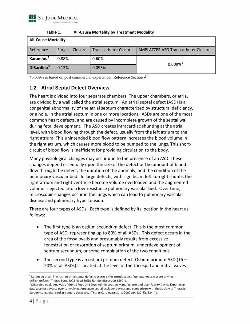
In February 2012, Kutty et al reported the results of a retrospective observational outcomes study published in the American Journal of Cardiology, comparing long‐term results of transcatheter and surgical ASD closure. This study represents the longest reported duration of follow‐up after transcatheter closure with a mean follow‐up of 10 years. Overall complication rates observed in both groups were low and differed by less than 2%3. All‐cause mortality rates following transcatheter ASD closure compare favorably to surgical closure. Table 1 summarizes recently reported all‐cause mortality rates segmented by treatment modality compared to the AMPLATZER Atrial Septal Occlusion (ASO) device.
These contemporary results validate transcatheter secundum ASD closure as a safe and effective therapeutic option for patients in whom the need for defect closure is clearly indicated.
1 Kirklin/Barratt‐Boyes Cardiac Surgery vol.1 (Kochoukas, Kirklin/Barratt‐Boyes Cardiac Surgery (2 vol. Set)) 2 Du ZD, Hijazi ZM, Kleinman CS, et al. for the AMPLATZER Investigators. Comparison between transcatheter and surgical closure of secundum atrial septal defect in children and adult: results of multicenter nonrandomized trial. J AM Coll Cardiology. 2002; 8:CR 787‐791. 3 Kutty et al., Long‐Term (5‐ to 20‐Year) Outcomes After Transcatheter or Surgical Treatment of Hemodynamically Significant Isolated Secundum Atrial Septal Defect, American Journal of Cardiology, Feb 13 2012 Ahead of Print
4 | P a g e
Table 1. All‐Cause Mortality by Treatment Modality
All‐Cause Mortality
Reference Surgical Closure Transcatheter Closure AMPLATZER ASO Transcatheter Closure
Karamlou4 0.88% 0.60%
DiBardino5 0.13% 0.093% 0.009%*
*0.009% is based on post commercial experience. Reference Section 4.
1.2 Atrial Septal Defect Overview
The heart is divided into four separate chambers. The upper chambers, or atria, are divided by a wall called the atrial septum. An atrial septal defect (ASD) is a congenital abnormality of the atrial septum characterized by structural deficiency, or a hole, in the atrial septum in one or more locations. ASDs are one of the most common heart defects, and are caused by incomplete growth of the septal wall during fetal development. The ASD creates intracardiac shunting at the atrial level, with blood flowing through the defect, usually from the left atrium to the right atrium. This unintended blood flow pattern increases the blood volume in the right atrium, which causes more blood to be pumped to the lungs. This short‐circuit of blood flow is inefficient for providing circulation to the body.
Many physiological changes may occur due to the presence of an ASD. These changes depend essentially upon the size of the defect or the amount of blood flow through the defect, the duration of the anomaly, and the condition of the pulmonary vascular bed. In large defects, with significant left‐to‐right shunts, the right atrium and right ventricle become volume overloaded and the augmented volume is ejected into a low‐resistance pulmonary vascular bed. Over time, microscopic changes occur in the lungs which can lead to pulmonary vascular disease and pulmonary hypertension.
There are four types of ASDs. Each type is defined by its location in the heart as follows:
The first type is an ostium secundum defect. This is the most common type of ASD, representing up to 80% of all ASDs. This defect occurs in the area of the fossa ovalis and presumably results from excessive fenestration or resorption of septum primum, underdevelopment of septum secundum, or some combination of the two conditions.
The second type is an ostium primum defect. Ostium primum ASD (15 – 20% of all ASDs) is located at the level of the tricuspid and mitral valves
4 Karamlou et al., The rush to atrial septal defect closure: is the introduction of percutaneous closure driving utilization?,Ann Thorac Surg. 2008 Nov;86(5):1584‐90; discussion 1590‐1. 5 DiBardino et al., Analysis of the US Food and Drug Administration Manufacturer and User Facility Device Experience database for adverse events involving Amplatzer septal occluder devices and comparison with the Society of Thoracic Surgery congenital cardiac surgery database, J Thorac Cardiovasc Surg. 2009 Jun;137(6):1334‐41.
5 | P a g e
and presumably results from failure of the endocardial cushions to close the ostium primum. Because endocardial cushions also form the mitral and tricuspid valves, ostium primum defects are typically associated with a cleft in the anterior mitral valve leaflet.
The third type is a sinus venosus defect. This ASD is found in the posterior aspect of the septum near the superior vena cava (where it may coexist with partial anomalous pulmonary venous connection of the right upper pulmonary vein) or the inferior vena cava (where it may coexist with partial anomalous pulmonary venous defect of the right lower pulmonary vein). Sinus venosus ASDs represent 5 – 10% of all ASDs.
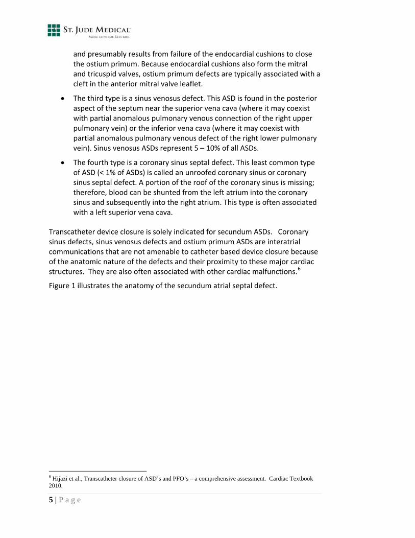
The fourth type is a coronary sinus septal defect. This least common type of ASD (< 1% of ASDs) is called an unroofed coronary sinus or coronary sinus septal defect. A portion of the roof of the coronary sinus is missing; therefore, blood can be shunted from the left atrium into the coronary sinus and subsequently into the right atrium. This type is often associated with a left superior vena cava.
Transcatheter device closure is solely indicated for secundum ASDs. Coronary sinus defects, sinus venosus defects and ostium primum ASDs are interatrial communications that are not amenable to catheter based device closure because of the anatomic nature of the defects and their proximity to these major cardiac structures. They are also often associated with other cardiac malfunctions.6
Figure 1 illustrates the anatomy of the secundum atrial septal defect.
6 Hijazi et al., Transcatheter closure of ASD’s and PFO’s – a comprehensive assessment. Cardiac Textbook 2010.
6 | P a g e
Figure 1. Heart with Atrial Septal Defect and Cross Sectional View of Atrial Septum
The anatomic margins (also referred to as “rims”) surrounding the atrial septum are defined below and detailed in Figure 1:
Aortic rim: rim related to the aorta that abuts the anterior‐superior septum of the defect. It may also be called the anterior‐superior rim, retro‐aortic rim, or retro‐aortic mound.
Superior rim: the rim that abuts the superior wall of the atrium.
Atrioventricular Valve (AV) rim: rim that abuts the atrioventricular valves or crux of the heart, also called the inferior‐anterior rim.
Inferior Vena Cava (IVC) rim: rim that abuts the inferior vena cava, also called the inferior‐posterior rim.
Posterior rim: most rightward and posterior rim opposite the aortic rim, and anatomically related to the right upper pulmonary vein.
Superior Vena Cava (SVC) rim: posterior‐superior rim which is bordered by the superior vena cava and is near the upper pulmonary vein.
The secundum ASD can vary greatly in size and shape, but does not directly involve the major cardiac structures (vena cava, right pulmonary veins, coronary sinus, or atrioventricular valves). Relationships to these structures, however, are important considerations for transcatheter closure. The defect tissue rims must be present and substantial enough to anchor the device.
Atrial Septal Defect
2 DEVICE SUMMARY
2.1 Benefits of Transcatheter Closure
The AMPLATZER ASO device was developed with the intent to treat ostium secundum atrial septal defects using minimally invasive techniques as a reasonable alternative to surgical closure (suture or patch). Results with an AMPLATZER ASO device have demonstrated to be comparable to surgical closure, with the added benefits of:
No exposure to cardiopulmonary bypass
No chest incision required
Decreased need for blood or blood product transfusion
Reduction in hospital stay
Significantly reduced convalescence time
Rapid return to normal activities
Potential health care economic benefits
The AMPLATZER ASO device was designed for ease of use with simple mechanics and nitinol construction, which allows the device to be placed and easily retrieved and/or repositioned within a 6 – 8 Fr. delivery system. Because device position relative to the defect anatomy is an important factor in transcatheter closure, the ability to reposition and retrieve the device allows the implanting physician greater flexibility for optimal device placement. The device’s design is self‐centering and durable, and is able to effectively close most ASDs in patients who meet the indications for use (Figure 2).
2.2 Intended Use and Patient Population
The AMPLATZER ASO is a percutaneous, transcatheter, ASD closure device intended for the occlusion of ASDs in the secundum position and for patients who have undergone a fenestrated Fontan procedure and require closure of the fenestration.
Patients indicated for ASD closure have echocardiographic evidence of ostium secundum atrial septal defect and clinical evidence of right ventricular volume overload (i.e., 1.5:1 degree of left‐to‐right shunt or right ventricle enlargement).
7 | P a g e
2.3 Device Description
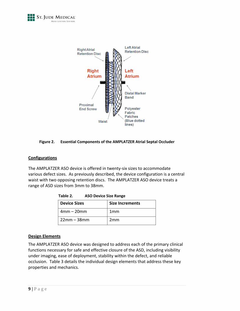
The AMPLATZER ASO is a self‐expandable device constructed from 0.004” – 0.008” braided nitinol wires. These braided nitinol wires are manufactured in a tubular form and are set to a designated length by placing and laser welding platinum marker bands on both the proximal and distal ends. Additionally, a female threaded component, or end screw, is laser welded over the proximal platinum marker band for attachment to the delivery system. The braided nitinol tube with welded ends is formed into the ASO geometry using a mold and heat treatment process. Following heat treatment and cooling, polyester fabric patches are sewn securely into the device to increase occlusion properties. The final ASO device consists of a continuous braided structure comprised of two retention disks: a right and left atrial retention disk connected by a central waist that is either 3 or 4mm in length.
Because of the importance of device sizing relative to the defect, AMPLATZER ASO devices are constructed in a variety of sizes ranging from 4mm to 38mm.
The required size of the AMPLATZER ASO is determined by the diameter of the waist, which should be equal to or slightly larger than the measured size of the ASD. When implanted, the connecting waist fills or occludes the defect, and the two opposing retention discs hold the device securely in place.
The radial span of the retention discs was minimized as much as possible in the device design in order to lower the risk of disc encroachment on adjacent cardiac structures. The left atrial retention disc is slightly larger than the right atrial retention disc, to account for the typical left‐to‐right trans‐atrial pressure gradient and to facilitate deployment.
Key features of the AMPLATZER ASO device are summarized as follows:
ASD treatment size range of 3mm to 38mm
Central positioning of the device within the ASD
Stable positioning of the device within the ASD
Device waist fills the ASD for reliable occlusion
Polyester material sewn into each device increases the occlusive properties and provides a substrate for tissue ingrowth
Retrievability; the AMPLATZER ASO remains attached to the delivery cable until the physician releases the device by unscrewing the device from the delivery cable. Correct device placement is first confirmed, and the device can be retrieved if placement is not satisfactory
Figure 2 provides an illustration of the ASO device design elements.
8 | P a g e
9 | P a g e
Figure 2. Essential Components of the AMPLATZER Atrial Septal Occluder
Configurations
The AMPLATZER ASO device is offered in twenty‐six sizes to accommodate various defect sizes. As previously described, the device configuration is a central waist with two opposing retention discs. The AMPLATZER ASO device treats a range of ASD sizes from 3mm to 38mm.
Table 2. ASO Device Size Range
Device Sizes Size Increments
4mm – 20mm 1mm
22mm – 38mm 2mm
Design Elements
The AMPLATZER ASO device was designed to address each of the primary clinical functions necessary for safe and effective closure of the ASD, including visibility under imaging, ease of deployment, stability within the defect, and reliable occlusion. Table 3 details the individual design elements that address these key properties and mechanics.
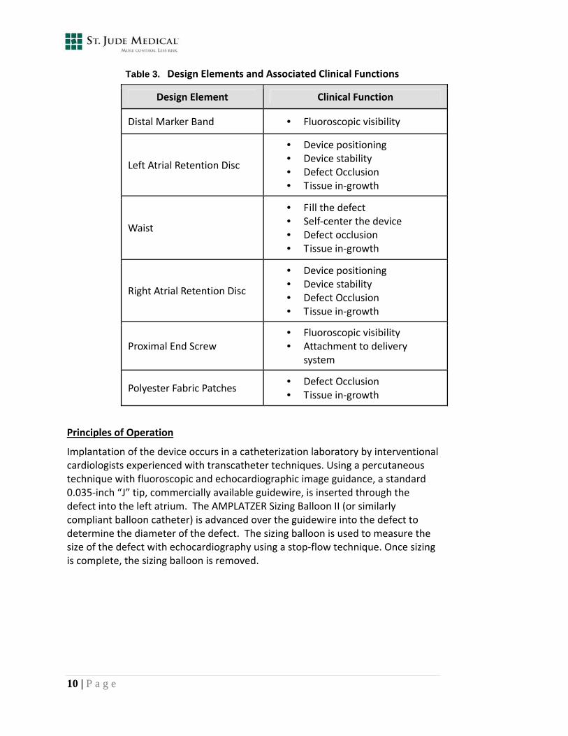
Table 3. Design Elements and Associated Clinical Functions
Design Element Clinical Function
Distal Marker Band • Fluoroscopic visibility
Left Atrial Retention Disc
• Device positioning • Device stability • Defect Occlusion • Tissue in‐growth
Waist
• Fill the defect • Self‐center the device • Defect occlusion • Tissue in‐growth
Right Atrial Retention Disc
• Device positioning • Device stability • Defect Occlusion • Tissue in‐growth
Proximal End Screw • Fluoroscopic visibility • Attachment to delivery
system
Polyester Fabric Patches • Defect Occlusion • Tissue in‐growth
Principles of Operation
Implantation of the device occurs in a catheterization laboratory by interventional cardiologists experienced with transcatheter techniques. Using a percutaneous technique with fluoroscopic and echocardiographic image guidance, a standard 0.035‐inch “J” tip, commercially available guidewire, is inserted through the defect into the left atrium. The AMPLATZER Sizing Balloon II (or similarly compliant balloon catheter) is advanced over the guidewire into the defect to determine the diameter of the defect. The sizing balloon is used to measure the size of the defect with echocardiography using a stop‐flow technique. Once sizing is complete, the sizing balloon is removed.
10 | P a g e
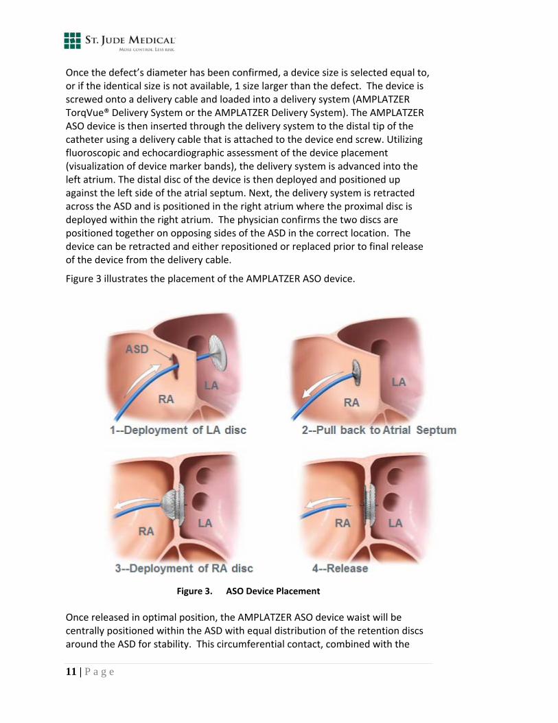
Once the defect’s diameter has been confirmed, a device size is selected equal to, or if the identical size is not available, 1 size larger than the defect. The device is screwed onto a delivery cable and loaded into a delivery system (AMPLATZER TorqVue® Delivery System or the AMPLATZER Delivery System). The AMPLATZER ASO device is then inserted through the delivery system to the distal tip of the catheter using a delivery cable that is attached to the device end screw. Utilizing fluoroscopic and echocardiographic assessment of the device placement (visualization of device marker bands), the delivery system is advanced into the left atrium. The distal disc of the device is then deployed and positioned up against the left side of the atrial septum. Next, the delivery system is retracted across the ASD and is positioned in the right atrium where the proximal disc is deployed within the right atrium. The physician confirms the two discs are positioned together on opposing sides of the ASD in the correct location. The device can be retracted and either repositioned or replaced prior to final release of the device from the delivery cable.
Figure 3 illustrates the placement of the AMPLATZER ASO device.
Figure 3. ASO Device Placement
Once released in optimal position, the AMPLATZER ASO device waist will be centrally positioned within the ASD with equal distribution of the retention discs around the ASD for stability. This circumferential contact, combined with the
11 | P a g e
polyester patch (sewn securely into each disc) is intended to prevent the flow of blood through the defect and the device, thereby promoting tissue in‐growth. The device becomes fully endothelialized in a matter of months.
2.4 Regulatory Pathway to US Approval
The AMPLATZER ASO device was originally evaluated in a US clinical trial under an Investigational Device Exemption (IDE). This US clinical trial (pivotal study) was a multi‐center, non‐randomized, controlled study with a total of 442 patients enrolled in the device arm and 154 patients enrolled in the surgical arm. Additionally, a registry group was studied to evaluate the device in 48 patients with a Fenestrated Fontan (FF).
Upon successful completion of the pivotal study and the registry, a premarket approval (PMA) application was filed with the FDA. An advisory meeting was held on September 10, 2001, and the Circulatory System Devices Panel recommended approval with conditions that related to labeling, training, and a 5‐year follow‐up of a subset of the pivotal trial cohort. An additional post‐approval study, with a 2‐year follow‐up of patients implanted outside the pivotal trial cohort, was initiated in 2007 to evaluate the long‐term safety and efficacy of the ASO device.
All approval conditions were agreed upon and the AMPLATZER ASO device was approved in the US on December 5, 2001.
2.5 Market Experience
The AMPLATZER ASO device received CE mark in the European Union on February 24, 1998. The device also received approval in several other major foreign markets including Canada (May 17, 2000), Japan (March 25, 2005), and Australia (December 20, 2006).
As of January 2012, total sales of the AMPLATZER ASO device were 223,965 devices. Of these, 72,566 devices were shipped to centers in the US and the remaining 151,399 devices were shipped internationally.
12 | P a g e

In addition to these markets, the AMPLATZER ASO is approved for marketing in the following countries as of March 2012:
Iraq Romania Argentina
Ireland Russia Armenia
Israel Saudi Arabia Austria
Italy Serbia Belarus
Kazakhstan Singapore Belgium
Korea Slovakia Brazil
Kyrgyzstan Slovenia Bulgaria
Latvia Spain China
Libya Sri Lanka Colombia
Liechtenstein Sweden Costa Rica
Lithuania Switzerland Croatia
Luxembourg Taiwan Cyprus
Macedonia Tajikistan Czech Republic
Malta Thailand Denmark
Mexico Tunisia Ecuador
Moldova Turkey Estonia
Monaco Turkmenistan Finland
Netherlands Ukraine France
New Zealand United Kingdom Georgia
Norway Uruguay Germany
Panama Uzbekistan Greece
Peru Venezuela Hungary
Vietnam Philippines Iceland
Poland India
Indonesia Portugal
13 | P a g e

14 | P a g e
3 CLINICAL TRIALS SUMMARY
3.1 IDE Pivotal Trial
The data from the US IDE pivotal trial provided evidence supporting the safety and efficacy of the AMPLATZER ASO in secundum ASDs. Transcatheter closure results were comparable to surgical closure, with added benefits of a shorter hospital stay. Adverse event rates were within the limits defined by the protocol, and significantly lower than those observed in the surgical arm.
3.1.1 ASO US Pivotal Trial Overview
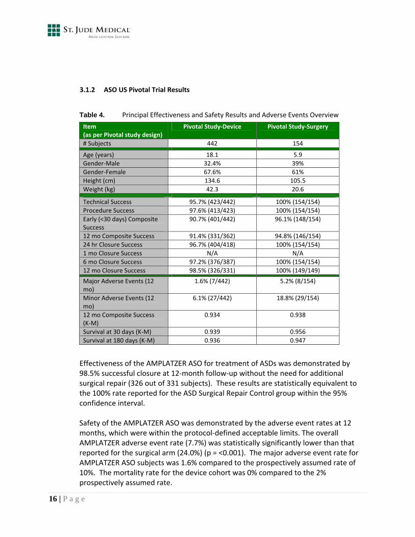
The pivotal trial was designed to evaluate the clinical performance of the AMPLATZER ASO device for ASD closure compared to the ASD surgical repair procedure. The study was a multi‐center, non‐randomized, controlled study with a total of 442 subjects enrolled in the device arm and 154 subjects enrolled in the ASD surgical arm. Enrolled subjects had echocardiographic evidence of ostium secundum ASD with a minimum of 1.5:1 left‐to‐right shunt, right ventricle enlargement, or had clinical symptoms such as paradoxical embolism or atrial dysrhythmia in the presence of a minimal shunt. Additional demographic information as well as a results summary can be found in Table 4 below.
Details of the pivotal trial results can be found in the Summary of Safety and Effectiveness (SSED) submitted as part of the original PMA (Section 5) and the Instruction for Use (IFU) (Section 6).
PMA Approval was granted on December 5, 2001.
15 | P a g e
3.1.2 ASO US Pivotal Trial Results
Table 4. Principal Effectiveness and Safety Results and Adverse Events Overview
Item (as per Pivotal study design)
Pivotal Study‐Device Pivotal Study‐Surgery
# Subjects 442 154
Age (years) 18.1 5.9
Gender‐Male 32.4% 39%
Gender‐Female 67.6% 61%
Height (cm) 134.6 105.5
Weight (kg) 42.3 20.6
Technical Success 95.7% (423/442) 100% (154/154)
Procedure Success 97.6% (413/423) 100% (154/154)
Early (<30 days) Composite Success
90.7% (401/442) 96.1% (148/154)
12 mo Composite Success 91.4% (331/362) 94.8% (146/154)
24 hr Closure Success 96.7% (404/418) 100% (154/154)
1 mo Closure Success N/A N/A
6 mo Closure Success 97.2% (376/387) 100% (154/154)
12 mo Closure Success 98.5% (326/331) 100% (149/149)
Major Adverse Events (12 mo)
1.6% (7/442) 5.2% (8/154)
Minor Adverse Events (12 mo)
6.1% (27/442) 18.8% (29/154)
12 mo Composite Success (K‐M)
0.934 0.938
Survival at 30 days (K‐M) 0.939 0.956
Survival at 180 days (K‐M) 0.936 0.947
Effectiveness of the AMPLATZER ASO for treatment of ASDs was demonstrated by 98.5% successful closure at 12‐month follow‐up without the need for additional surgical repair (326 out of 331 subjects). These results are statistically equivalent to the 100% rate reported for the ASD Surgical Repair Control group within the 95% confidence interval.
Safety of the AMPLATZER ASO was demonstrated by the adverse event rates at 12 months, which were within the protocol‐defined acceptable limits. The overall AMPLATZER adverse event rate (7.7%) was statistically significantly lower than that reported for the surgical arm (24.0%) (p = <0.001). The major adverse event rate for AMPLATZER ASO subjects was 1.6% compared to the prospectively assumed rate of 10%. The mortality rate for the device cohort was 0% compared to the 2% prospectively assumed rate.
16 | P a g e
In addition to strong safety and effectiveness results, the mean length of hospital stay for AMPLATZER subjects (1 day) was statistically‐significantly lower than the mean of 3.4 days documented for the ASD Surgical Repair control group (p <0.001).
3.2 Post‐Approval Study
3.2.1 Post‐Approval Study Overview
The comprehensive post‐approval study was approved by FDA on August 16, 2007 and initiated in March 2008. The post‐approval study is a prospective, non‐randomized, multi‐center evaluation of 1000 subjects. A minimum of 800 subjects will be followed to two years. The subject population includes both pediatric and adult subjects. St. Jude Medical submits interim study reports on a six month basis. The company recently submitted the 54‐month interim report to the FDA, dated February 15, 2012. Data presented herein is reflective of this interim report (876 patients). However, as of March 30, 2012, greater than 970 subjects have been enrolled.
17 | P a g e

3.2.2 Post‐Approval Study Data (54 month interim results)
This study was developed to reflect real world use. Rates as seen in Table 5 are consistent with IDE findings but are more reflective of real world experience.
Table 5. Summary of Safety and Effectiveness Data
Item (as per Post‐Approval study design)
Post‐Approval Study 54 mo Annual Report
# Subjects 876
Age (years) 20.9
Gender‐Male 35%
Gender‐Female 65%
Height (cm) 135.1
Weight (kg) 45.4
Rate N
Technical Success 98.1% 859/876
1 mo Closure Success 97% 789/813
Rate N
Major Adverse Events 3.8% 33
Minor Adverse Events 28.2% 247
Deaths
Due to Erosion 0% 0
Due to Embolization 0% 0
Due to Fracture 0% 0
Due to Stroke 0% 0
Due to Thrombus 0% 0
Due to Arrhythmia 0% 0
Due to Other 0.003%
3 (2‐cardiac arrest, 1‐pneumonia)
Rate N
Erosion 0% 0
Embolization‐Serious 0.3% 3
Embolization‐Minor 0.1% 1
Fracture 0% 0
Stroke 0.1% 1
Thrombus‐Serious 0% 0
Thrombus‐Minor 0% 0
Arrhythmias‐Serious 1.6% 14
Arrhythmias‐Minor 11.9% 105
Note: All potential erosion events observed in the post‐approval study under the definition of hemodynamic compromise are evaluated by an independent “Erosion Board” according to the uniform methods described in Section 4.2.2. Since the 54‐month follow‐up report was submitted to FDA, two hemodynamic compromise events have been adjudicated as confirmed erosion events. These events are captured in the total adverse events reported in Section 4.
18 | P a g e
Interim results for the post‐approval study demonstrate the continued safety and efficacy of the AMPLATZER ASO for percutaneous, transcatheter closure of atrial septal defects in secundum position.
4 POST COMMERCIALIZATION EXPERIENCE
4.1 Adverse Event Reporting
St. Jude Medical continuously monitors product in commerce to determine if the device is performing as expected, and takes action in the event that there are unexpected occurrences. St. Jude Medical meets post‐market regulatory requirements through the use of standard operating procedures designed to ensure post‐market product performance, safety, and distribution channel traceability. St. Jude Medical becomes aware of field events, associated with the AMPLATZER ASO, in a variety of ways including physician or patient concerns or comments, published literature, or customer‐reported adverse clinical events. Adverse event reporting is a key component of monitoring field events. Per regulatory requirements, all field events are reviewed and, as appropriate, are reported to the FDA under the Medical Device Reporting regulation (21 CFR §803). With adverse events reported outside of a controlled clinical investigation, device manufacturers are reliant on information from reporting health care providers and published literature, which may be incomplete or anecdotal when compared to the type of data obtained through a clinical trial. St. Jude Medical conducts routine trending of events reported in all commercially‐released products and attempts are made to gain as much information as possible on these field events.
Table 6 represents the major adverse event categories; other less frequent and/or less severe adverse events are not discussed in this document. Event numbers (see Table 6) are as reported through January 2012, with the exception of erosion events, where the event numbers are reflective of the latest Erosion Board adjudication; encompassing events through 15 March 2012. All events described herein include MDR‐reported major adverse events for the on‐label use of the AMPLATZER ASO device, in the specified categories, with associated rates based on world‐wide reported events and world‐wide sales of 223,965 devices.
19 | P a g e

20 | P a g e
Table 6. World‐Wide Serious Adverse Event Overview – Through January 2012*
Adverse Event Total Reported Events (on‐label, ASO, world‐wide)
Rate (based on sales, world‐wide)**
Arrhythmia 54 0.024%
Embolization 347 0.155%
Erosion 97 0.043%
Fracture 1 0.0004%
Malfunction 0 0.000%
Malposition 9 0.004%
Stroke 6 0.003%
Thrombus on Device 11 0.005%
*Erosion events are as of 15 March 2012
** All adverse event rates are calculated using total world‐wide sales of 223,965 units.
NOTE: The same patient may be counted in more than one of the above reported adverse events.
4.1.1 Arrhythmia
Arrhythmias, occurring both during the procedure and within several months after treatment, are anticipated potential adverse events for any cardiac intervention and are usually transient in nature7. Clinically significant arrhythmias include those that result in heart block and / or require the placement of a permanent pacemaker, those requiring long‐term medication and those requiring intervention during the catheterization procedure. The reported incidence of serious arrhythmia following surgical repair of ASDs is approximately 8%.8,9,10 This surgical rate is significantly higher than that observed with transcatheter closure using the AMPLATZER ASO as evidenced in the pivotal trial, post‐approval study and the post commercialization ASO experience The observed rate of serious arrhythmias in the pivotal study was 0.5%, and in the post‐approval study (PAS) was 1.6%. The world‐wide event rate of 0.024% remains lower than the serious arrhythmia event rates seen in the pivotal study and the PAS. The arrhythmia events reported in Table 6 include atrial fibrillation, atrial flutter, bradycardia, palpitations, premature ventricular contractions, ST wave abnormality, tachycardia, ventricular fibrillation, and heart block.
7 Hijazi et al., Transcatheter closure of ASD’s and PFO’s – a comprehensive assessment. Cardiac Textbook 2010. 8 Roos‐Hesselink et al., Excellent survival and low incidence of arrhythmias, stroke and heart failure long‐term after surgical ASD closure at young age. A prospective follow‐up study of 21–33 years. European Heart Journal (2003) 24, 190–197 9 Mascio et al., Outcomes in adult congenital heart surgery: Analysis of the Society of Thoracic Surgeons Database. The Journal of Thoracic and Cardiovascular Surgery, Nov 2011: 1090‐1097. 10 Berger et al., Arrhythmias in patients with surgically treated atrial septal defects. Swiss Medical Weekly, 2005; 135:175–178

21 | P a g e
Figure 4. Arrhythmia Adverse Event Comparison Chart
4.1.2 Device Embolization
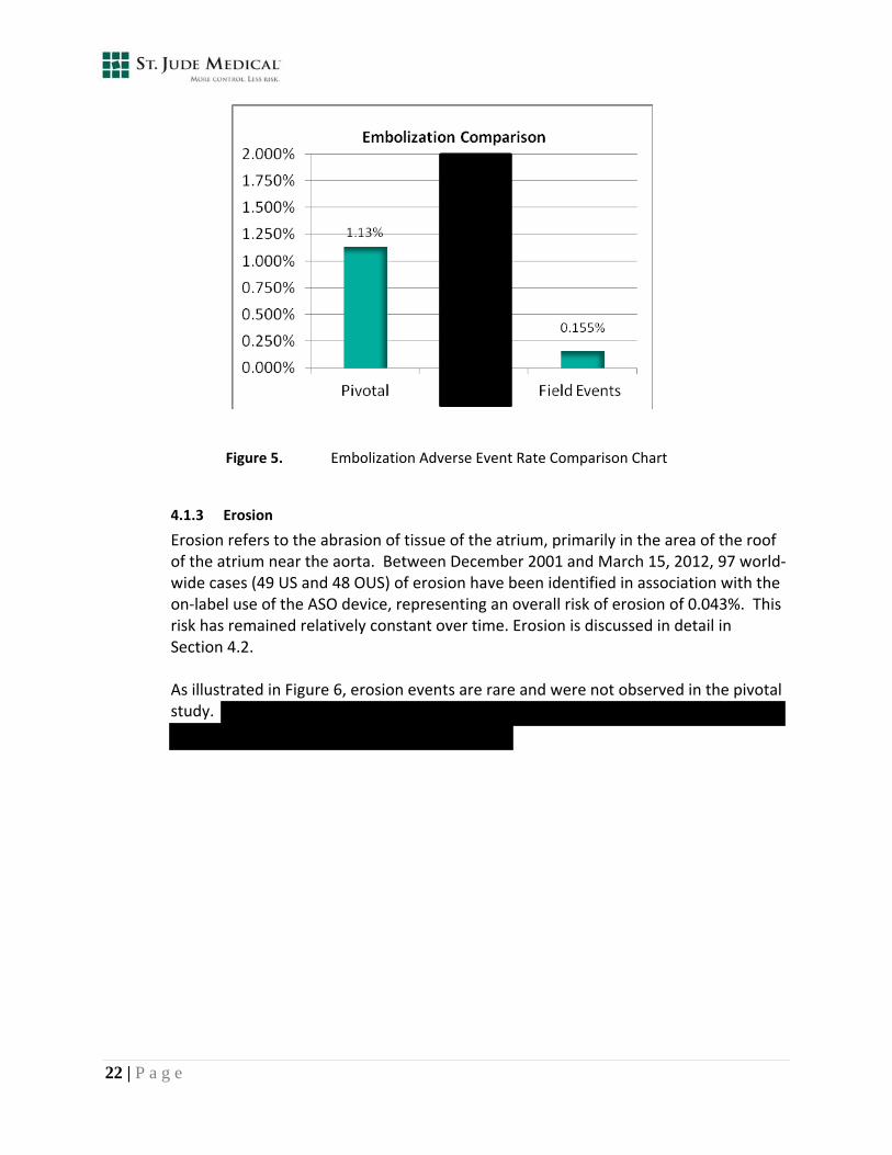
Device embolization is movement or migration of the device outside of the atrial septal defect after device release from the delivery cable. Movement into the right ventricle and pulmonary artery is more common compared to the left ventricle, which is rare. Following proper device implant technique and sizing guidance, as defined within the IFU, is critical to preventing device embolizations. In the AMPLATZER ASO US pivotal trial experience, embolization was reported in two categories, serious and minor; the event rates observed were 0.9% and 0.2%, respectively (overall rate of 1.13% for the device arm). Within the post‐approval clinical study, an adverse event rate of 0.3% for serious and 0.1% for minor embolization was observed; with an overall rate of 0.46%. As shown in Figure 5, the world‐wide event rates (0.155%) remain lower than the event rates observed in the pivotal study and the PAS.
22 | P a g e
Figure 5. Embolization Adverse Event Rate Comparison Chart
4.1.3 Erosion
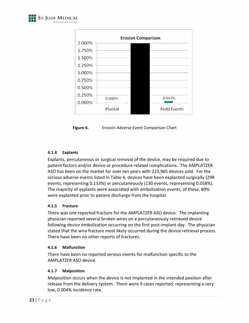
Erosion refers to the abrasion of tissue of the atrium, primarily in the area of the roof of the atrium near the aorta. Between December 2001 and March 15, 2012, 97 world‐wide cases (49 US and 48 OUS) of erosion have been identified in association with the on‐label use of the ASO device, representing an overall risk of erosion of 0.043%. This risk has remained relatively constant over time. Erosion is discussed in detail in Section 4.2. As illustrated in Figure 6, erosion events are rare and were not observed in the pivotal study. However, as in post commercialization experience, erosion events have been observed in the post‐approval study (2 of 970).
23 | P a g e
Figure 6. Erosion Adverse Event Comparison Chart
4.1.4 Explants
Explants, percutaneous or surgical removal of the device, may be required due to patient factors and/or device or procedure related complications. The AMPLATZER ASO has been on the market for over ten years with 223,965 devices sold. For the serious adverse events listed in Table 6, devices have been explanted surgically (298 events, representing 0.133%) or percutaneously (130 events, representing 0.058%). The majority of explants were associated with embolization events; of these, 89% were explanted prior to patient discharge from the hospital.
4.1.5 Fracture
There was one reported fracture for the AMPLATZER ASO device. The implanting physician reported several broken wires on a percutaneously retrieved device following device embolization occurring on the first post‐implant day. The physician stated that the wire fracture most likely occurred during the device retrieval process. There have been no other reports of fractures.
4.1.6 Malfunction
There have been no reported serious events for malfunction specific to the AMPLATZER ASO device.
4.1.7 Malposition
Malposition occurs when the device is not implanted in the intended position after release from the delivery system. There were 9 cases reported; representing a very low, 0.004% incidence rate.
24 | P a g e
4.1.8 Mortality (all‐cause)
Since market introduction of the AMPLATZER ASO device, there have been 20 reported deaths through January 2012 for an all‐cause mortality rate of 0.009%. Deaths occurred secondary to the following events: embolization (2), erosion (8), stroke and thrombus (1) cardiac arrest (1), perforation (2), tamponade (1), effusion (2), CNS bleed (1), CVA (1) and seizure (1). The all‐cause mortality rate associated with ASO implantation is significantly lower than the rate for surgical closure of ASDs cited in the most recent published literature for all‐cause mortality in both adult and pediatric patients (0.13% to 0.88%, as described in Section 1). The mortality rate associated with erosion has been carefully assessed and is detailed in Section 4.3.2.
4.1.9 Stroke
Stroke is an anticipated adverse event with repair of ASDs (surgical or percutaneous). The reported incidence of stroke following surgical repair of ASD ranges from 2% to 6.5%.11,12,13 This surgical rate is higher than that observed with transcatheter closure using the AMPLATZER ASO as evidenced through the pivotal trial (0.0%), post‐approval study (0.1%) and the post commercialization stroke adverse event rate (0.003%).
4.1.10 Thrombus on Device
Thrombus has been demonstrated to be a rare adverse event for transcatheter closure using the AMPLATZER ASO. This is evidenced by the observed post commercialization adverse event rate of 0.005%. In the pivotal study, only minor thrombus events were noted; however, thrombus remains a potential adverse event but at an extremely low incidence rate.
4.1.11 Post Commercialization Experience Conclusion
The adverse events presented in this section represent post commercialization experience across the range of patient demographics and implanter experience. These data confirm that the reported adverse event rates for the AMPLATZER ASO device are consistent with, and in some cases, lower than, those reported in the pivotal and post‐approval studies. St. Jude Medical maintains a global complaint handling process to ensure that potential nonconformances and adverse events are analyzed, investigated and acted
11 Kutty S, Hazeem AA, Brown K, Danford CJ, Worley SE, Delaney JW, Danford DA, Latson LA. Long‐Term (5‐ to 20‐Year) Outcomes After
Transcatheter or Surgical Treatment of Hemodynamically Significant Isolated Secundum Atrial Septal Defect. Am J Cardiol. 2012 Feb 13. [Epub ahead of print] PubMed PMID: 22335856. 12 Shibata Y, Abe T, Kuribayashi R, Sekine S, Seki K, Yamagishi I, Chanda J. Surgical treatment of isolated secundum atrial septal defect in patients
morethan 50 years old. Ann Thorac Surg. 1996 Oct;62(4):1096‐9. PubMed PMID: 8823095. 13 Murphy JG, Gersh BJ, McGoon MD, Mair DD, Porter CJ, Ilstrup DM, McGoon DC,Puga FJ, Kirklin JW, Danielson GK. Long‐term outcome after
surgical repair of isolated atrial septal defect. Follow‐up at 27 to 32 years. N Engl J Med. 1990 Dec 13;323(24):1645‐50. PubMed PMID: 2233961.
upon appropriately. Any corrective actions are verified, validated, and monitored for effectiveness. All formal Corrective and Preventative Action (CAPA) steps are documented through the St. Jude Medical quality system.
4.2 Corrective Actions
In monitoring the safety and effectiveness of the AMPLATZER ASO, there have been two
adverse event types upon which St. Jude Medical has provided additional focus: embolization
and erosion. These events were analyzed for both world‐wide and US experience.
4.2.1 Embolization
Following proper device sizing guidance and implant technique as defined within
the IFU is critical to preventing device embolization. St. Jude Medical recently
implemented retraining of all implanting physicians on implant technique and
device‐sizing to ensure the ASO device is accurately sized to the defect being
treated. St. Jude Medical continues to actively monitor these events in an effort to
ensure rates remain at the lowest possible level.
4.2.2 Erosion
Background
Erosion is defined as abrasion of tissue of the atrium, primarily in the area of the
roof of the atrium near the aorta. Although rare in occurrence, erosion can
potentially result in serious health consequences for the patient. St. Jude Medical
recognizes the potential severity of this event, and has thus ensured (1)
adjudication of all potential erosion events by the Erosion Board, (2) completion of
a rigorous root cause analysis, and (3) implementation of substantial corrective
actions.
Following the first report of an erosion field event in 2002, AGA Medical, now a St.
Jude Medical company, established an independent panel of expert physicians (the
Erosion Board) to assist in the monitoring and analysis of these events. The Erosion
Board is actively involved in the adjudication of all potential erosion events,
monitoring of incidence trends, and providing recommendations to the company
on potential contributing factors to erosion. The Erosion Board also assists in the
development of appropriate clinical guidance through updates of the IFU, technical
notes, and publications in order to minimize the potential for any future erosion
events. In addition to the internal analysis conducted by St. Jude Medical, the
Erosion Board also conducts an independent review of all potential erosion events
on behalf of St. Jude Medical.
25 | P a g e
26 | P a g e
The methods of identifying and investigating events are uniformly applied to each
potential event identified through field / complaint event reporting, post‐approval
clinical study and monitoring of MAUDE and published literature. Potential erosion
events reported under a pre‐specified set of event terms are reviewed and
adjudicated by the Erosion Board. The Erosion Board confirms evidence of erosion
and differentiates, when possible, the causality between the event, defect
anatomy, implant procedure and /or implant device. This process serves to
continually monitor the incidence of erosion and guide the company towards
necessary modifications to labeling, implant technique and physician materials for
the purpose of reducing the incidence of erosion and further mitigating patient risk.
A summary of the actions taken by AGA Medical / St. Jude Medical since the first
reported erosion event are summarized below:
1999
2001
AMPLATZER ASO CE Mark Approval
AMPLATZER ASO PMA Approval
2002 First reported erosion case (US)
Independent Erosion Board formed by AGA Medical with
established adjudication process for all reported potential
erosions to determine clinical contributors and root‐cause.
The Erosion Board determined that device oversizing was
a significant contributing factor
2004 IFU updated with device sizing recommendation to
mitigate the risk of device oversizing
Erosion Board published results and recommendations to
mitigate risk of device oversizing
2005 AGA Medical / FDA discussion to review incidence of
erosion events and mitigations implemented
2006 Physician Tech Note published, patient ID card and patient
guide verbiage updated for guidance
2009 IFU updated with additional warning statements (balloon
sizing and stop flow technique)
2011 St. Jude Medical / FDA discussion regarding incidence and

27 | P a g e
mitigating actions
FDA approves additional IFU modifications to further
mitigate against potential erosion events; St. Jude Medical
publishes modifications to IFU and Patient Guide in
January 2012; St. Jude Medical proposes physician letter
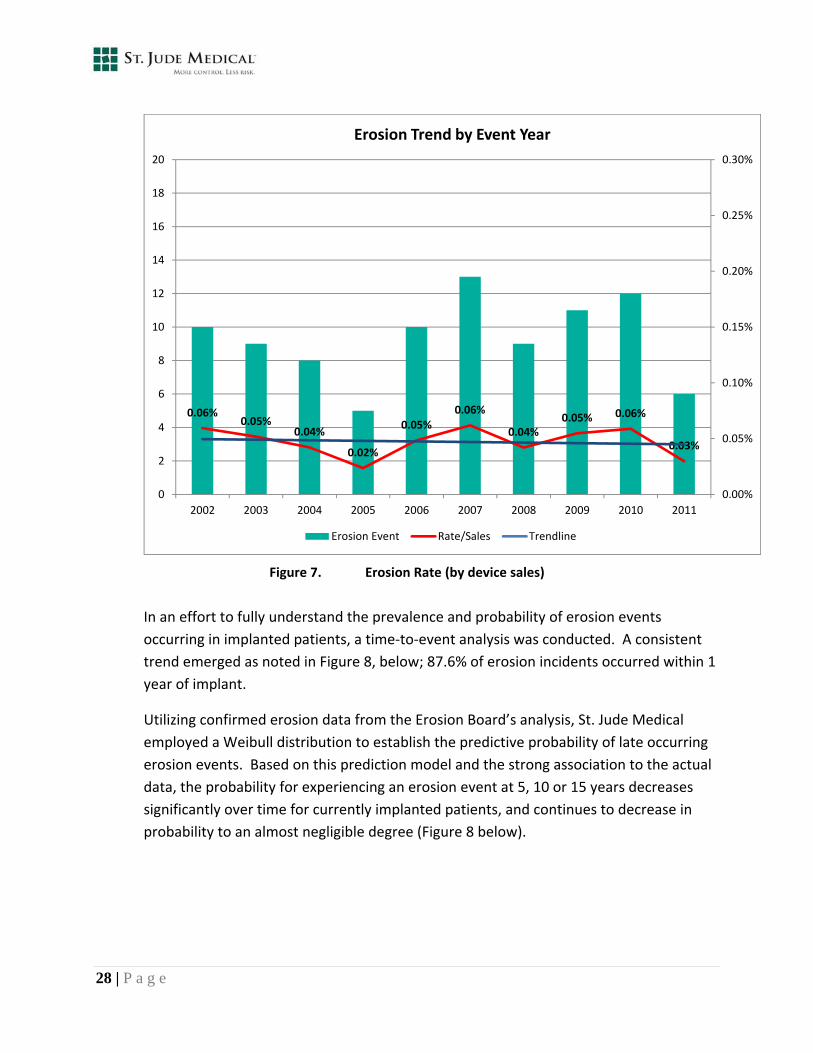
As evidenced by the actions summarized above, the company has consistently and progressively implemented corrective actions to help mitigate and decrease the potential incidence of erosion when additional knowledge is learned. The actions previously taken were based upon internal analysis, recommendations from the Erosion Board, and recommendations from the FDA. In November 2010, St. Jude Medical acquired AGA Medical. In early 2011, St. Jude Medical completed a thorough root cause analysis supplementing the previous analysis conducted by AGA Medical. This comprehensive analysis resulted in a recent update to the IFU and the Patient Guide, and focused training and re‐training for all physicians specific to atrial septal anatomy and device implant technique. Additionally, the company developed a proposed physician letter with a summary of investigation findings and specific mitigation guidance. These most recent actions represent the company’s ongoing diligence and commitment towards monitoring and mitigation of this rare but serious event. Prevalence and Probability Erosion events recorded annually since 2002 (Figure 7) indicate a stable trend over time.
0.06%0.05%
0.04%
0.02%
0.05%0.06%
0.04%0.05% 0.06%
0.03%
0.00%
0.05%
0.10%
0.15%
0.20%
0.25%
0.30%
0
2
4
6
8
10
12
14
16
18
20
2002 2003 2004 2005 2006 2007 2008 2009 2010 2011
Erosion Trend by Event Year
28 | P a g e
Erosion Event Rate/Sales Trendline
Figure 7. Erosion Rate (by device sales)
In an effort to fully understand the prevalence and probability of erosion events
occurring in implanted patients, a time‐to‐event analysis was conducted. A consistent
trend emerged as noted in Figure 8, below; 87.6% of erosion incidents occurred within 1
year of implant.
Utilizing confirmed erosion data from the Erosion Board’s analysis, St. Jude Medical
employed a Weibull distribution to establish the predictive probability of late occurring
erosion events. Based on this prediction model and the strong association to the actual
data, the probability for experiencing an erosion event at 5, 10 or 15 years decreases
significantly over time for currently implanted patients, and continues to decrease in
probability to an almost negligible degree (Figure 8 below).

Figure 8. Predictive Probability of Late Event
Mortality
The mortality rate associated with this rare event has been carefully assessed. The
overall incidence of mortality is 0.004% (8 events out of 223,965 devices). Not all
erosions have resulted in death, as there are recognized clinical symptoms for erosion
events that facilitate identification and intervention. Importantly, no deaths associated
with erosion occurred in patients younger than 15 years of age, and all reported deaths
occurred within 16 months of implant. Each event was adjudicated with the confirmed
presence of device oversizing, deficient rim, or both.
The mortality rate associated with erosion following ASO implantation (0.004%)
compares favorably to the all‐cause mortality rate for surgical closure of ASDs cited in
the most recent published literature for all‐cause mortality in both adult and pediatric
patients as previously described in Section 1.1, Table 1 (0.13% ‐ 0.88%) as well as the
0.07% mortality rate derived from the STS pediatric database (January 2006 to
December 2009).
29 | P a g e
Root Cause Analysis
As concluded by the Erosion Board from 2002 to 2010, the most frequently observed
causal relationship to erosion was device over‐sizing; noted in 40% of cases (31%
pediatric and 46% of adult cases). In 2011, St. Jude Medical, in coordination with the
Erosion Board, completed an additional root cause analysis for all erosion events
reported between December 1998 and November 2011 (97 world‐wide erosion cases;
49 US and 48 OUS). All available echocardiographic images were reviewed, coupled
with source medical records. Erosion events were examined across multiple factors,
including but not limited to: age (pediatric vs. adult), sufficiency of atrial septal defect
rims (specifically anterior‐superior rim), gender, device size (over‐sizing), event outcome
and time‐to‐event.
From this analysis, St. Jude Medical concluded that erosion is caused by (1) a device
implanted within a defect with insufficient rim distance (anterior‐superior rim), or (2) by
an over‐sized device, thus resulting in device edge contact with adjacent structures. In
point of fact, review of all available echocardiographic images confirmed the presence
of deficient rim and/or over‐sizing of the device in each erosion event.
More specifically, anterior‐superior rim deficiency was noted in 90% of all cases. From
the data available, 100% of pediatric erosion cases and 84% of adult erosion cases
demonstrated deficient anterior‐superior rims. A sufficient rim was defined as the
presence of at least 5mm rim in multiple AND sequential short‐axis views confirmed by
ICE or TEE.
Corrective Actions
Prior to 2011, corrective actions were focused on device over‐sizing since this was
determined to be the leading causal relationship to erosion. Incidence of device over‐
sizing has decreased over time due to mitigating actions taken (prior IFU updates,
physician technical notes, etc.) and overall increased experience and awareness.
The comprehensive analysis, conducted in 2011, revealed that although device over‐
sizing remains an important contributor to erosion, a second causal relationship is
evident, i.e., a device implanted within a defect with insufficient rim distance (anterior‐
superior rim). Based upon these incremental findings, St. Jude Medical implemented
the following mitigating actions in 2012:
30 | P a g e

31 | P a g e
Updated labeling (IFU) including an additional contraindications, modified
warning, and specific echocardiographic imaging guidance
Addition of new physician training modules addressing erosion mitigation and
retraining of physicians on implant technique; specific training of the IFU has
been initiated at both implanting physician and institutional levels
Updated Patient Guide to provide additional awareness and guidance to patients
These mitigating actions provide implanting physicians with additional clear and
actionable guidance for avoidance of erosion events.
The new contraindications added to the IFU were an intentionally conservative measure
aimed at eliminating the potential for erosions by avoiding device edge contact between
the device and adjacent cardiac structures. The addition of this language aligned
contraindications across the industry (i.e., all currently marketed ASO transcatheter
closure devices are contraindicated for contact with adjacent non‐septal intra‐cardiac
structures).
The modification to an existing warning and the new echocardiographic imaging
guidance, within the IFU, was specifically added to address rim sufficiency; advising the
physician to conduct a full assessment of defect rim anatomy prior to device
implantation. Particularly, rims must be observed in multiple and sequential views to
thoroughly evaluate device contact with any adjacent cardiac structure.
The specific IFU updates, implemented in January 2012, are detailed as follows:
Additional contraindications:
Any patient in whom the device would interfere with or contact other
intracardiac or intravascular structures (e.g. atrial roof, cardiac valves,
pulmonary veins, coronary sinus, or aorta).
Any patient with echocardiographic evidence of absent or deficient
anterior – superior rim (sufficient rim is defined as the presence of at
least 5mm of rim in multiple AND sequential short axis views confirmed
by ICE or TEE).
Updated warning:
Do not release the AMPLATZER Septal Occluder from the delivery cable if
the device does not conform to its original configuration, or if the device
position is unstable and/or in contact with any adjacent cardiac structure.
Recapture the device and redeploy. If still unsatisfactory, recapture the
device and replace with a new device or refer the patient for alternative
treatment.
Additional echocardiography guidance:
To allow for comprehensive assessment of anterior‐superior rim
adequacy, the rim must be evaluated in multiple and sequential short‐
axis views (e.g. 20, 30, 40, 50, 60 or 70 degrees) confirmed by ICE or TEE.
Rim adequacy is defined as sufficient tissue (at least 5mm) surrounding
the defect to ensure the device does not come in contact with adjacent
cardiac structures.
Updated Patient Guide:
The Patient Guide is provided to patients at time of the procedure and is
also available on the St. Jude Medical website. The Patient Guide was
updated to ensure patients are aware of erosion related symptoms and
the need to seek immediate medical assistance in the event of these
symptoms.
Lastly, St. Jude Medical proposed a physician communication (technical note) to include
a summary of the root cause analysis findings and to provide specific mitigation
guidance to avoid the potential of erosion events and to ultimately mitigate risk to
patients. St. Jude Medical has proposed a technical note as well; this communication
was previously provided to the FDA with the intent of finalizing subsequent to the panel
discussion.
32 | P a g e
5 CONCLUSION
Minimally invasive transcatheter closure has become the preferred treatment for symptomatic ostium secundum ASDs over surgery. The AMPLATZER ASO device is designed to achieve stability within the defect and reliable occlusion, addressing the primary mechanisms necessary for safe and effective transcatheter closure of the ASD. The AMPLATZER ASO device has a strong record of reliable and effective performance. The device has been studied in more than 1,200 subjects between the original US IDE pivotal and post‐approval studies, and has been on the market for more than 10 years with 223,965 devices sold worldwide.
While the AMPLATZER ASO device continues to be a safe and effective means for transcatheter ASD closure, St. Jude Medical recognizes the potential severity associated with the rare adverse event of erosion. The company’s diligent post‐market surveillance process employs a rigorous and uniform method to investigate all potential erosion events. Through this vigilant post‐market surveillance process, and the subsequent analysis described in previous sections, two causal relationships have been clearly identified with the occurrence of erosion events: device over‐sizing, and a device implanted within a defect with insufficient rim distance. The substantial and intentionally conservative corrective actions implemented by St. Jude Medical are intended to significantly decrease the potential for erosion events, thus mitigating and reducing patient risk.
Results from the pivotal and post‐approval studies, paired with the device’s post‐commercialization data, confirm that the AMPLATZER ASO is a safe and effective treatment for closure of ASDs and continues to demonstrate overall complication rates comparable or favorable to surgical closure. Furthermore, the safety profile of the AMPLATZER ASO remains within the anticipated ranges predicted at time of PMA approval (2001). In summation, the totality of the benefits to the patient continue to far outweigh the risks associated with on‐label use of the ASO in light of the context of the disease and existing treatment options.
33 | P a g e

6 SUMMARY OF SAFETY AND EFFECTIVENESS DATA (SSED)
34 | P a g e

7 INSTRUCTIONS FOR USE
35 | P a g e