Embed Size (px)
Citation preview

CASE REPORT Open Access
Spontaneous community-acquired PVL-producing Staphylococcus aureusmediastinitis in an immunocompetentadult – a case reportJosselin Brisset1,2, Thomas Daix1,3* , Jérémy Tricard4, Bruno Evrard1, Philippe Vignon1,3, Olivier Barraud3,5
and Bruno François1,3
Abstract
Background: Mediastinitis caused by hematogenous spread of an infection is rare. We report the first known caseof community-acquired mediastinitis from hematogenous origin in an immunocompetent adult. This rare invasiveinfection was due to Panton-Valentine Leucocidin-producing (PVL+) methicillin-susceptible Staphylococcus aureus (MSSA).
Case presentation: A 22-year-old obese man without other medical history was hospitalized for febrile precordial chestpain. He reported a cutaneous back abscess 3 weeks before. CT-scan was consistent with mediastinitis and blood culturesgrew for a PVL+ MSSA. Intravenous clindamycin (600mg t.i.d) and cloxacillin (2 g q.i.d.), secondary changed for fosfomycin(4 g q.i.d.) because of a related toxidermia, was administered. Surgical drainage was performed and confirmed thepresence of a mediastinal abscess associated with a fistula between the mediastinum and right pleural space. All localbacteriological samples also grew for PVL+ MSSA. In addition to clindamycin, intravenous fosfomycin was switched totrimethoprim-sulfamethoxazole after 4 weeks for a total of 10 weeks of antibiotics.
Conclusions: We present the first community-acquired mediastinitis of hematogenous origin with PVL+ MSSA. Clinicalevolution was favorable after surgical drainage and 10weeks of antibiotics. The specific virulence of MSSA PVL+ strainsplayed presumably a key role in this rare invasive clinical presentation.
Keywords: Community-acquired mediastinitis, MSSA, Panton-valentine Leucocidin
BackgroundMediastinitis is mainly due to deep sternal wound infec-tion, esophageal perforation, or descending necrotizingmediastinitis originating from ear-nose-throat (ENT) in-fections and exceptionally to hematogenous spread [1].In case of hematogenous spread, mediastinitis is usuallyhealthcare-acquired [2, 3].
Panton Valentin Leucocidin-producing (PVL+)methicillin-susceptible Staphylococcus aureus (MSSA)was mostly described in community-acquired necro-tizing pneumonia, bone and joint infections and skinand soft tissue infections such as furunculosis [4]. Asfar as we know, we present the first case of acommunity-acquired mediastinitis caused by MSSA.The strain was PVL+ and seemed to belong toUSA300 strains [5] which are increasingly associatedwith invasive infections.
© The Author(s). 2020 Open Access This article is licensed under a Creative Commons Attribution 4.0 International License,which permits use, sharing, adaptation, distribution and reproduction in any medium or format, as long as you giveappropriate credit to the original author(s) and the source, provide a link to the Creative Commons licence, and indicate ifchanges were made. The images or other third party material in this article are included in the article's Creative Commonslicence, unless indicated otherwise in a credit line to the material. If material is not included in the article's Creative Commonslicence and your intended use is not permitted by statutory regulation or exceeds the permitted use, you will need to obtainpermission directly from the copyright holder. To view a copy of this licence, visit http://creativecommons.org/licenses/by/4.0/.The Creative Commons Public Domain Dedication waiver (http://creativecommons.org/publicdomain/zero/1.0/) applies to thedata made available in this article, unless otherwise stated in a credit line to the data.
* Correspondence: [email protected]éanimation polyvalente, CHU Dupuytren, 2 avenue Martin Luther King,F-87000 Limoges, France3Inserm CIC 1435 & UMR 1092, CHU Dupuytren, F-87000 Limoges, FranceFull list of author information is available at the end of the article
Brisset et al. BMC Infectious Diseases (2020) 20:354 https://doi.org/10.1186/s12879-020-05076-6
Case presentationA 22-year-old obese (BMI = 38 kg/m2) man withoutother medical history was admitted to the emergency de-partment (ED) for precordial chest pain worsening for 5days and radiating to the back and shoulders. The pa-tient had low dysphagia, progressive-onset dyspnea andunproductive cough for 2 days but without fever orshiver. This patient, usually living in Illinois, had workedas a teacher in France for the last 8 months and did nottravel outside Western Europe and the USA.On admission, the patient presented with fever
(38.5 °C), tachypnea (RR: 30/min) and required 3 l/minof oxygen (SpO2: 97%) but had no signs of respiratorydistress. Lung auscultation revealed decreased breathsounds in the right lower lobe. Blood pressure and heartrate were normal. There was no evidence for a dental,oro-pharyngeal infection, or cervical cellulitis. Secondquestioning of the patient highlighted a skin lesion de-scribed as an abscess in the back 3 weeks before admis-sion which was successfully treated by Povidone-Iodinealcohol but it was absent on the current clinical examin-ation. None of his relatives or colleagues described anysigns of skin infection.Blood tests were consistent with a marked inflammatory
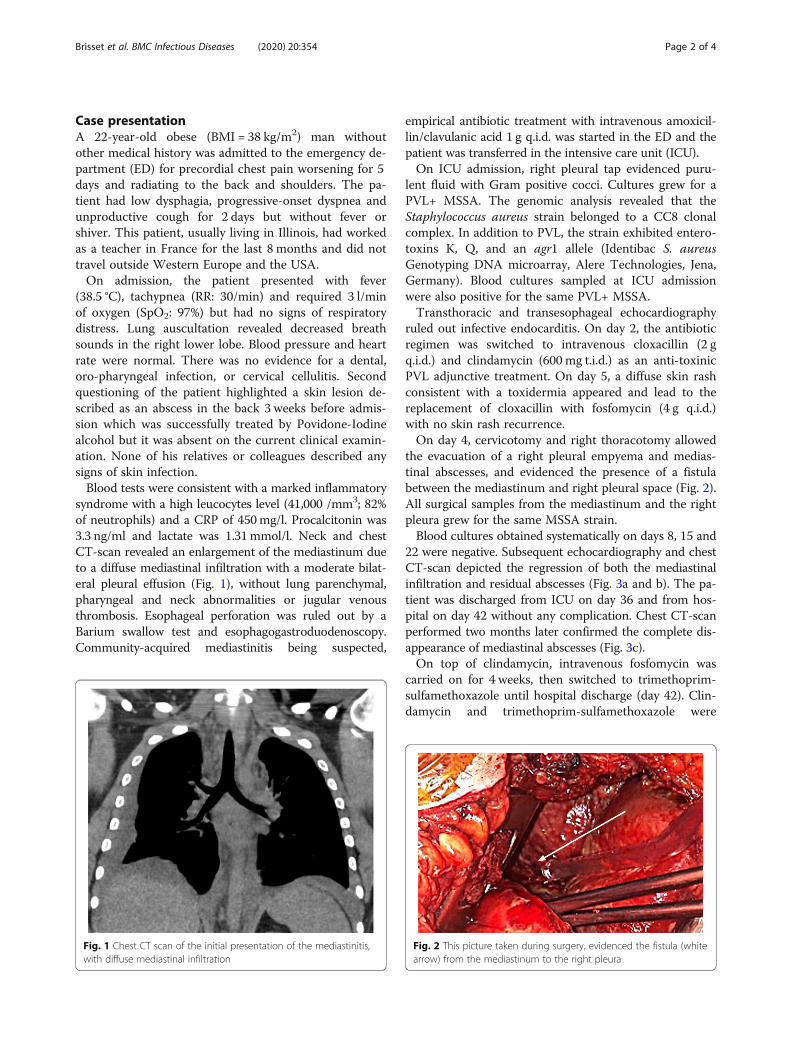
syndrome with a high leucocytes level (41,000 /mm3; 82%of neutrophils) and a CRP of 450mg/l. Procalcitonin was3.3 ng/ml and lactate was 1.31mmol/l. Neck and chestCT-scan revealed an enlargement of the mediastinum dueto a diffuse mediastinal infiltration with a moderate bilat-eral pleural effusion (Fig. 1), without lung parenchymal,pharyngeal and neck abnormalities or jugular venousthrombosis. Esophageal perforation was ruled out by aBarium swallow test and esophagogastroduodenoscopy.Community-acquired mediastinitis being suspected,
empirical antibiotic treatment with intravenous amoxicil-lin/clavulanic acid 1 g q.i.d. was started in the ED and thepatient was transferred in the intensive care unit (ICU).On ICU admission, right pleural tap evidenced puru-
lent fluid with Gram positive cocci. Cultures grew for aPVL+ MSSA. The genomic analysis revealed that theStaphylococcus aureus strain belonged to a CC8 clonalcomplex. In addition to PVL, the strain exhibited entero-toxins K, Q, and an agr1 allele (Identibac S. aureusGenotyping DNA microarray, Alere Technologies, Jena,Germany). Blood cultures sampled at ICU admissionwere also positive for the same PVL+ MSSA.Transthoracic and transesophageal echocardiography
ruled out infective endocarditis. On day 2, the antibioticregimen was switched to intravenous cloxacillin (2 gq.i.d.) and clindamycin (600 mg t.i.d.) as an anti-toxinicPVL adjunctive treatment. On day 5, a diffuse skin rashconsistent with a toxidermia appeared and lead to thereplacement of cloxacillin with fosfomycin (4 g q.i.d.)with no skin rash recurrence.On day 4, cervicotomy and right thoracotomy allowed
the evacuation of a right pleural empyema and medias-tinal abscesses, and evidenced the presence of a fistulabetween the mediastinum and right pleural space (Fig. 2).All surgical samples from the mediastinum and the rightpleura grew for the same MSSA strain.Blood cultures obtained systematically on days 8, 15 and
22 were negative. Subsequent echocardiography and chestCT-scan depicted the regression of both the mediastinalinfiltration and residual abscesses (Fig. 3a and b). The pa-tient was discharged from ICU on day 36 and from hos-pital on day 42 without any complication. Chest CT-scanperformed two months later confirmed the complete dis-appearance of mediastinal abscesses (Fig. 3c).On top of clindamycin, intravenous fosfomycin was
carried on for 4 weeks, then switched to trimethoprim-sulfamethoxazole until hospital discharge (day 42). Clin-damycin and trimethoprim-sulfamethoxazole were
Fig. 1 Chest CT scan of the initial presentation of the mediastinitis,with diffuse mediastinal infiltration
Fig. 2 This picture taken during surgery, evidenced the fistula (whitearrow) from the mediastinum to the right pleura
Brisset et al. BMC Infectious Diseases (2020) 20:354 Page 2 of 4

continued for 6 more weeks for a total of 10 weeks oftreatment.No immunodepression was evidenced with normal im-
munoglobulin levels, no complement pathway defect,negative HIV testing and antibody titers of previouslyadministered vaccines (diphtheria, tetanus, HBV) with-out any abnormal response.Long-term antibiotics and early surgery benefited our
patient which is now doing well and was able to go backto the United States of America (USA) on June 6th,2019.
Discussion and conclusionsThe MSSA strain was PVL+ and a member of CC8clonal complex. Regarding the genetic marker interfer-ence methodology, this strain belongs to USA300 strains[5]. The prevalence of this strain seems to increase andto be associated with invasive infection [6].Undoubtedly, the PVL toxin may have significantly
contributed to the severity of the disease. PVL isexpressed by major methicillin-resistant Staphylococcusaureus (MRSA) clones, which have now spread through-out the world [7] even if PVL+ SA remains especiallypresent in the USA, with 48.1% of MRSA strains and11.5% of MSSA strains being identified in this region(8.2% in northeast, region of origin of our patient) [8]. Incontrast in France, the majority of strains expressingPVL are MSSA strains. As in our patient, PVL+ SAstrains affect mainly young adults without underlyingdisorder [9] and are more often found in community-acquired infections than in hospital-acquired infections[10]. In community-acquired infections, PVL is an inde-pendent marker of severity regardless the resistance ofthe Staphylococcus aureus strain to methicillin [11]. PVLis known to lyse neutrophils and boost the inflammatorycascade which can explain the progressive tissue necrosisin case of invasive infection associated with necrotic skinand soft tissue infection such as furuncles [12] andcommunity-acquired pneumonia [13]. In our patient,
these properties presumably contributed to the develop-ment of a fistula from which facilitated the spread of thelocal infection from the mediastinum to the right pleuralspace. PVL+ SA toxins have been most frequently asso-ciated with skin and soft tissue infections (folliculitis andcutaneous abscesses), and with necrotizing pneumonia[4]. Since the medical history, clinical examination andCT-scan did not show any signs of dental, pharyngeal orcervical infections, we can assume that the skin abscesswhich developed 3 weeks before the severe infectioncould be the source of the initial bacteremia and second-ary location in the chest.Three main etiologies of acute infective mediatinitis
are traditionally distinguished: deep sternal wound infec-tion following cardiovascular and thoracic surgery,esophageal perforation and descending necrotizing med-iastinitis originating from dental, pharyngeal or cervicalinfections [14]. Overall mortality of acute mediastinitisranges between 20% (esophageal perforation) to 30%(descending necrotizing mediastinitis) [1]. Exceptionally,primary mediastinitis may result from a bacteremia. Toour knowledge, only two cases have been reported sofar. Both of them were healthcare-associated mediastini-tis, one in a dialysis patient with an arteriovenous shuntdue to MRSA which was not identified as PVL+ [2] andone associated with a central venous catheter and due toCandida species [3].Treatment of community-acquired acute mediastinitis
is based on antimicrobial therapy and control of thesource of infection using surgical debridement. With theexception of mediastinitis developing in the postopera-tive setting which requires rapid surgical debridementwithin 24 h [15], there are currently no guidelines forthe timing of the surgery. Multiple surgical managementis required in approximatively 30% of patients [16].There are currently no guidelines for empirical antibiotictherapy which should be adapted on the expected originof mediastinitis. With the exception of deep sternalwound infections which are commonly related to SA,
Fig. 3 (a) Day 7 CT scan- acme of the mediastinal infiltration, with abscesses formation (3 days after surgery); (b) CT scan at M1 after intensivecare unit admission; (c) CT scan at M2 after intensive care unit admission
Brisset et al. BMC Infectious Diseases (2020) 20:354 Page 3 of 4

polymicrobial infection with flora from the oral cavityand/or the upper respiratory tract is common in othercauses of mediastinitis [1]. In our patient, the empiricaltherapy with amoxicillin-clavulanic acid targeted aerobicand anaerobic oral and upper gastro-intestinal bacteria.Antibiotic regimen was adapted secondary to MSSAidentification using high-dose of cloxacillin since beta-lactam antibiotics can increase PVL secretion in case ofinsufficient concentration in vitro [17]. In addition, clin-damycin was associated since it strongly inhibits PVL se-cretion in vitro, as linezolid [17].PVL-producing MSSA can result in community-
acquired infective mediastinitis secondary to bacteremiaafter a cutaneous abscess in young immunocompetentadult.
AbbreviationsBMI: Body mass index; CRP: C-reactive protein; ED: Emergency department;HBV: Hepatitis B virus; HIV: Human immunodeficiency virus; ICU: Intensivecare unit; MRSA: Methicillin-resistant Staphylococcus aureus; MSSA: Methicillin-susceptible Staphylococcus aureus; PCT: Procalcitonin; PVL: Panton-ValentineLeucocidin; RR: Respiratory rate; TTE: Transthoracic echocardiography;USA: United States of America; WBC: White blood cells
AcknowledgementsNA
Authors’ contributionsJB, TD, BE, JT, PV, and BF took care of the patient. OB analyzed the samples.JB and TD gathered the data and drafted the manuscript. PV, BE, OB and BFcritically reviewed the manuscript. All authors approved the final version.
FundingNone.
Availability of data and materialsData sharing is not applicable to this article as no datasets were generatedor analyzed during the current case.
Ethics approval and consent to participateNA
Consent for publicationWritten informed consent was obtained from the patient for publication ofthis case report and any accompanying images. A copy of the writtenconsent is available for review by the Editor-in-Chief of this journal.
Competing interestsThe authors declare that they have no competing interests.
Author details1Réanimation polyvalente, CHU Dupuytren, 2 avenue Martin Luther King,F-87000 Limoges, France. 2Maladies infectieuses, CHU Dupuytren, F-87000Limoges, France. 3Inserm CIC 1435 & UMR 1092, CHU Dupuytren, F-87000Limoges, France. 4Chirurgie cardiaque, CHU Dupuytren, F-87000 Limoges,France. 5Laboratoire de Bactériologie - Virologie – Hygiène, CHU Dupuytren,F-87000 Limoges, France.
Received: 2 April 2020 Accepted: 7 May 2020
References1. Pastene B, Cassir N, Tankel J, Einav S, Fournier PE, Thomas P, Leone M.
Mediastinitis in the intensive care unit patient: a narrative review. ClinMicrobiol Infect. 2020;26(1):26–34.
2. Chang CH, Huang JY, Lai PC, Yang CW. Posterior mediastinal abscess in ahemodialysis patient – a rare but life-threatening complication ofStaphylococcus bacteremia. Clin Nephrol. 2009;71:92–5.
3. Er F, Nia AM, Caglayan E, Gassanov N. Mediastinitis as a complication ofcentral venous catheterization. Infection. 2010;38:509.
4. Lina G, Piémont Y, Godail-Gamot F, Bes M, Peter MO, Gauduchon V,Vandenesch F, Etienne J. Involvement of Panton-valentine leukocidin-producing Staphylococcus aureus in primary skin infections and pneumonia.Clin Infect Dis. 1999;29:1128–32.
5. Bowers JR, Driebe EM, Albrecht V, McDougal LK, Granade M, Roe CC,Lemmer D, Rasheed JK, Engelthaler DM, Keim P, Limbago BM. ImprovedSubtyping of Staphylococcus aureus Clonal Complex 8 Strains Based onWhole-Genome Phylogenetic Analysis. mSphere. 2018;3(3):pii:e00464–17.
6. McCaskill ML, Mason EO Jr, Kaplan SL, Hammerman W, Lamberth LB, HulténKG. Increase of the USA300 clone among community-acquired methicillin-susceptible Staphylococcus aureus causing invasive infections. Pediatr InfectDis J. 2007;26(12):1122–7.
7. Tristan A, Bes M, Meugnier H, Lina G, Bozdogan B, Courvalin P, Reverdy ME,Enright MC, Vandenesch F, Etienne J. Global distribution of Panton-valentineLeukocidin–positive methicillin-resistant Staphylococcus aureus, 2006.Emerg Infect Dis. 2007;13:594–600.
8. Brown ML, O’Hara FP, Close NM, Mera RM, Miller LA, Suaya JA, Amrine-Madsen H. Prevalence and sequence variation of panton-valentineleukocidin in methicillin-resistant and methicillin-susceptible Staphylococcusaureus strains in the United States. J Clin Microbiol. 2012;50:86–90.
9. Gillet Y, Issartel B, Vanhems P, Fournet JC, Lina G, Bes M, Vandenesch F,Piémont Y, Brousse N, Floret D, Etienne J. Association betweenStaphylococcus aureus strains carrying gene for Panton-valentine leukocidinand highly lethal necrotising pneumonia in young immunocompetentpatients. Lancet. 2002;359(9308):753–9.
10. Bádue Pereira MF, Bando SY, Sasagawa SM, da Silva CB, Mimica MJ, BerezinEN. Panton-valentine positive Staphylococcus aureus in community-acquired and hospital-acquired pediatric infections. Pediatr Infect Dis J.2019;38(10):1068–70.
11. Gijón M, Bellusci M, Petraitiene B, Noguera-Julian A, Zilinskaite V, SanchezMoreno P, Saavedra-Lozano J, Glikman D, Daskalaki M, Kaiser-Labusch P,Falup-Pecurariu O, Montagnani C, Prieto L, Gené A, Trumpulyte G,Kulecnikova I, Lepe JA, Cercenado E, Kudinsky R, Makri A, Huppertz HI,Bleotu L, Cocchi P, García-Hierro P, Vitkauskiene A, Fortuny C, Zukovskaja V,Neth O, Santos M, Rokney A, Petra M, Lixandru R, Galli L, Guillén S, Chaves F,Rojo Conejo P. Factors associated with severity in invasive community-acquired Staphylococcus aureus infections in children: a prospectiveEuropean multicentre study. Clin Microbiol Infect. 2016;22(7):643.e1–6.
12. Lina G, Piémont Y, Godail-Gamot F, Bes M, Peter MO, Gauduchon V,Vandenesch F, Etienne J. Involvement of Panton-valentine leukocidin-producing Staphylococcus aureus in primary skin infections andpneumonia. Clin Infect Dis. 1999;29(5):1128–32.
13. Labandeira-Rey M, Couzon F, Boisset S, Brown EL, Bes M, Benito Y, BarbuEM, Vazquez V, Höök M, Etienne J, Vandenesch F, Bowden MG.Staphylococcus aureus Panton-Valentine leukocidin causes necrotizingpneumonia. Science. 2007;315(5815):1130–3.
14. Chen K-C, Chen J-S, Kuo S-W, Huang PM, Hsu HH, Lee JM, Lee YC.Descending necrotizing mediastinitis: a 10-year surgical experience in asingle institution. J Thorac Cardiovasc Surg. 2008;136:191–8.
15. Abu-Omar Y, Kocher GJ, Bosco P, Barbero C, Waller D, Gudbjartsson T,Sousa-Uva M, Licht PB, Dunning J, Schmid RA, Cardillo G. EuropeanAssociation for Cardio-Thoracic Surgery expert consensus statement on theprevention and management of mediastinitis. Eur J Cardiothorac Surg. 2017;51(1):10–29.
16. Prado-Calleros HM, Jiménez-Fuentes E, Jiménez-Escobar I. Descendingnecrotizing mediastinitis: systematic review on its treatment in the last 6years, 75 years after its description. Head Neck. 2016;38(Suppl 1):E2275–83.
17. Dumitrescu O, Boisset S, Badiou C, Bes M, Benito Y, Reverdy ME,Vandenesch F, Etienne J, Lina G. Effect of antibiotics on Staphylococcusaureus producing Panton-valentine leukocidin. Antimicrob AgentsChemother. 2007;51:1515–9.
Publisher’s NoteSpringer Nature remains neutral with regard to jurisdictional claims inpublished maps and institutional affiliations.
Brisset et al. BMC Infectious Diseases (2020) 20:354 Page 4 of 4