Embed Size (px)
Citation preview
Sports Concussion: Basic Science
VSCC ImPACT WorkshopNovember 15, 2013
Gary Solomon, Ph.D., FACPNCo-Director, Vanderbilt Sports Concussion CenterAssociate Professor, Departments of Neurological Surgery, Orthopaedic Surgery & Rehabilitation, and PsychiatryVanderbilt University School of MedicineTeam Neuropsychologist, Nashville PredatorsConsulting Neuropsychologist, Tennessee Titans

Please leave your pagers, cell phones, iPads, laptops, and
other electronic devices ON.
The more distracted you are, the easier this will be for me.
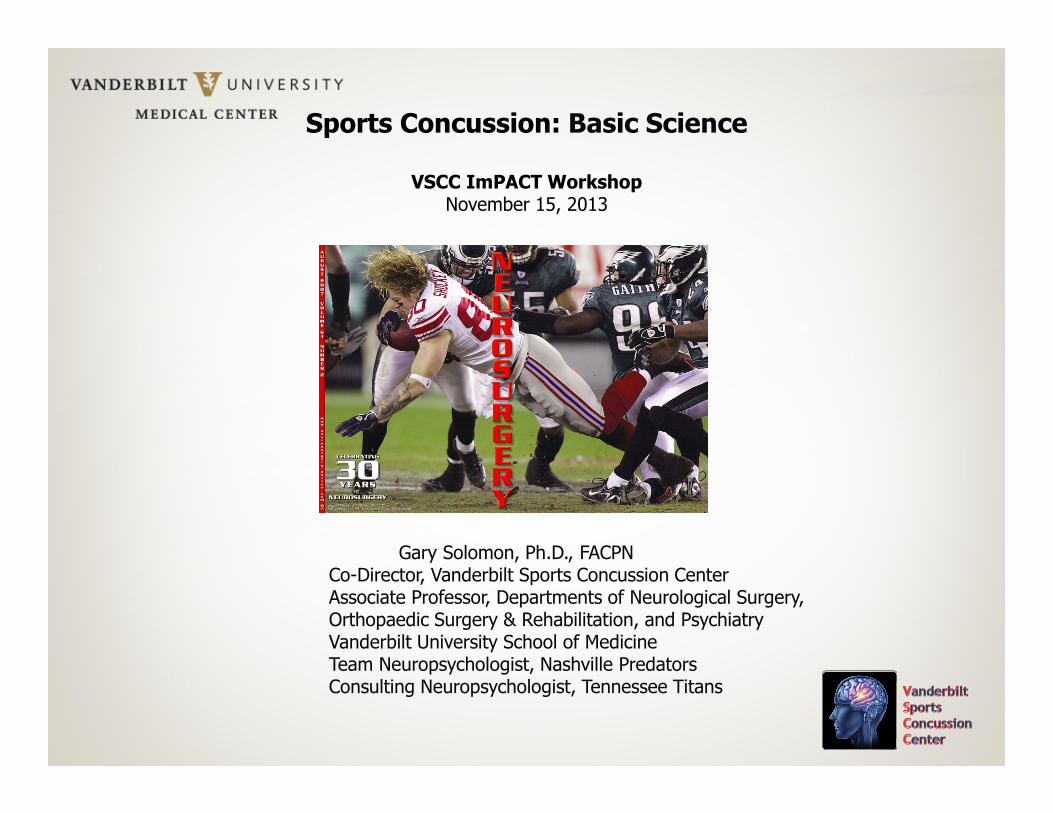
• Consulting fees from the Nashville Predators and Tennessee Titans
• Involved in beta testing new versions of ImPACT and receive free use during the testing
• Member of the ImPACT Professional Advisory Board; reimbursed for expenses associated with board meetings
• This presentation is not endorsed by any organization with which I am affiliated
• “Bottom Line Conclusions” are my opinions and are notevidence-based
Disclosures/Competing Interests
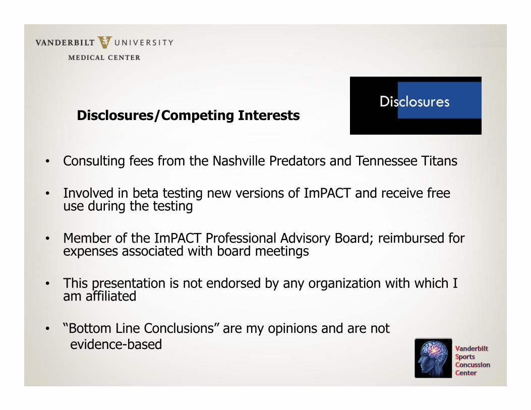
Overview
• Biomechanics• Biochemistry• Structural Neuroimaging• Functional Neuroimaging• Neurophysiology• TBI Biomarkers• Animal studies
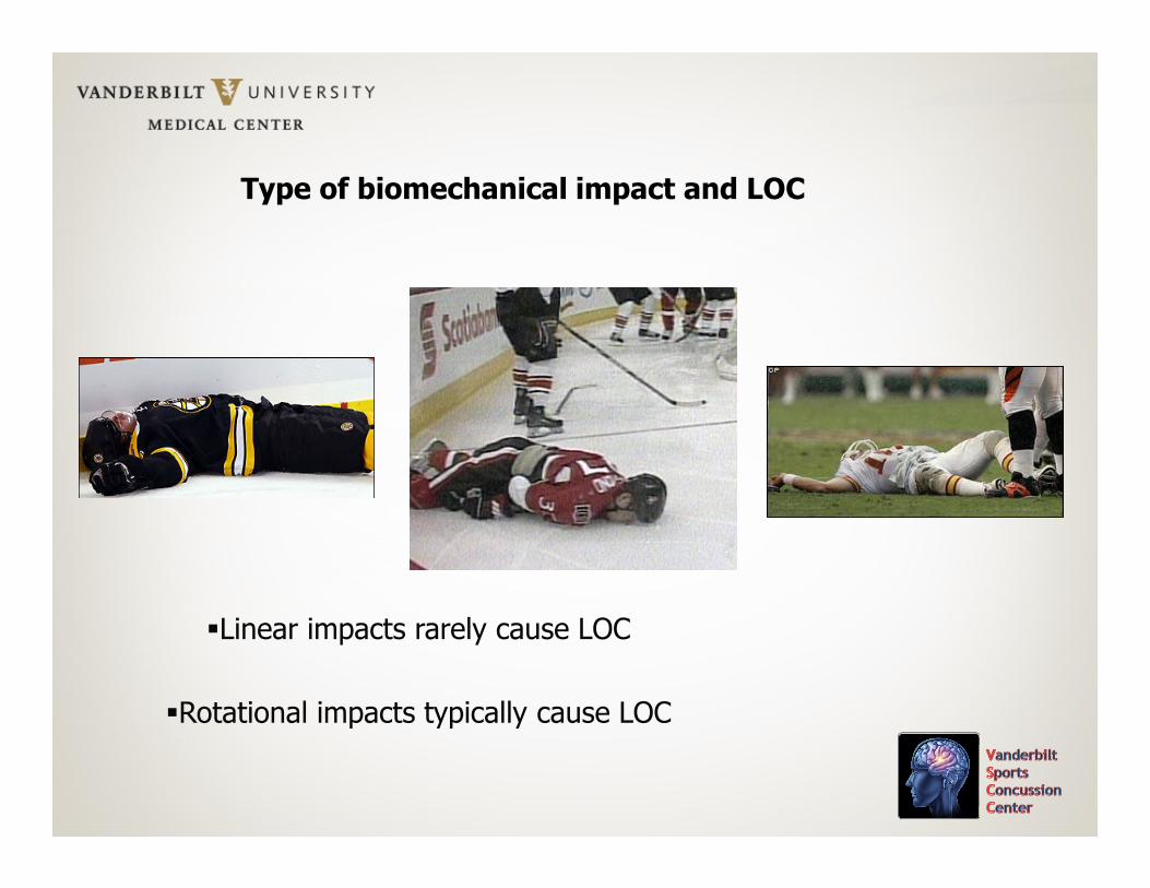
Type of biomechanical impact and LOC
�Linear impacts rarely cause LOC
�Rotational impacts typically cause LOC


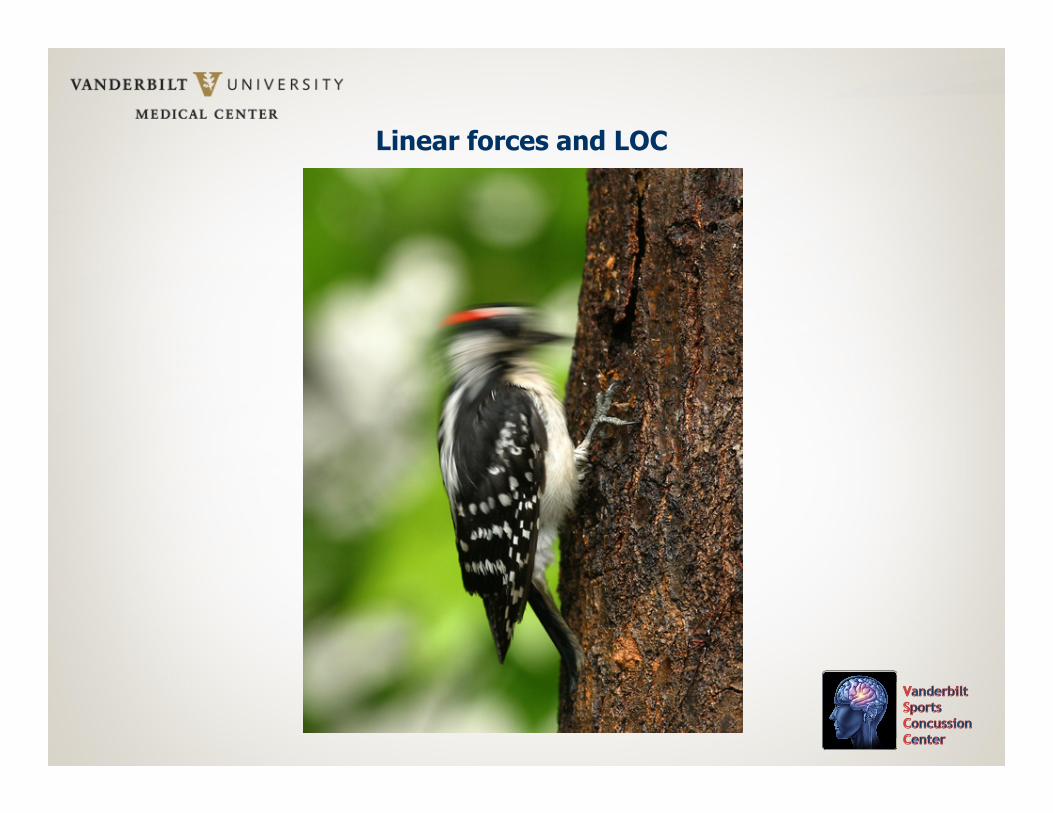
Linear forces and LOC
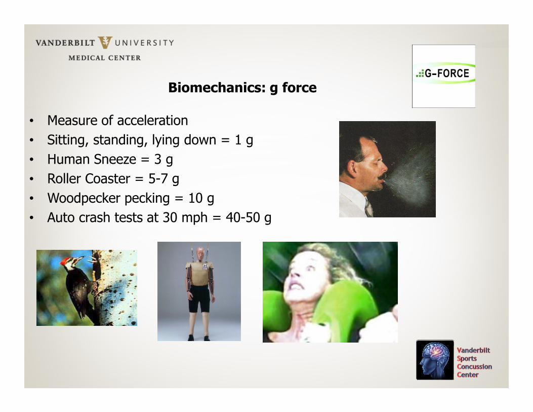
Biomechanics: g force
• Measure of acceleration
• Sitting, standing, lying down = 1 g
• Human Sneeze = 3 g
• Roller Coaster = 5-7 g
• Woodpecker pecking = 10 g
• Auto crash tests at 30 mph = 40-50 g
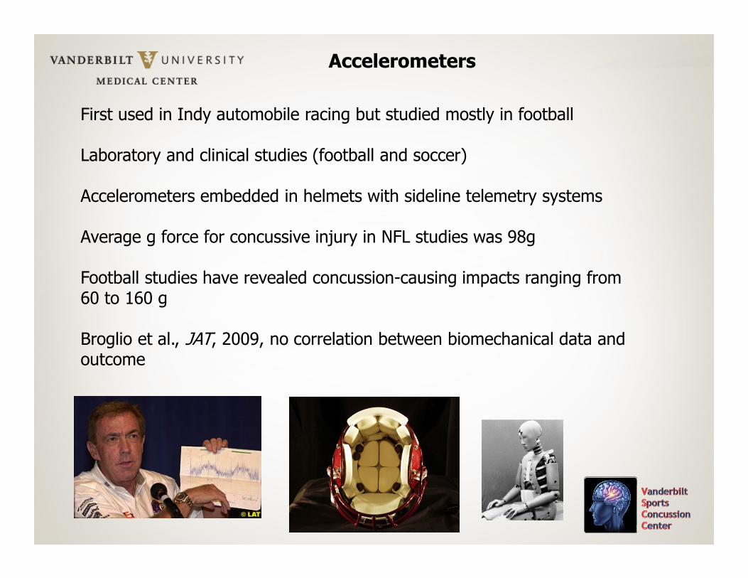
Accelerometers
First used in Indy automobile racing but studied mostly in football
Laboratory and clinical studies (football and soccer)
Accelerometers embedded in helmets with sideline telemetry systems
Average g force for concussive injury in NFL studies was 98g
Football studies have revealed concussion-causing impacts ranging from 60 to 160 g
Broglio et al., JAT, 2009, no correlation between biomechanical data and outcome
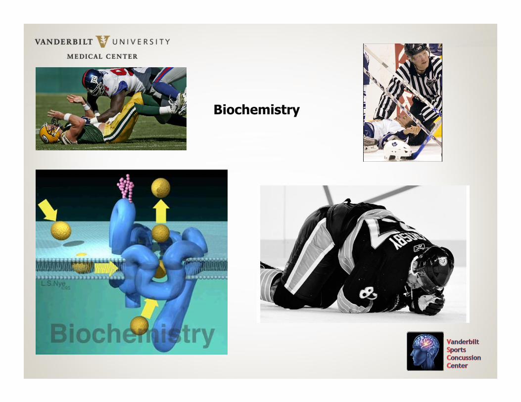
Biochemistry
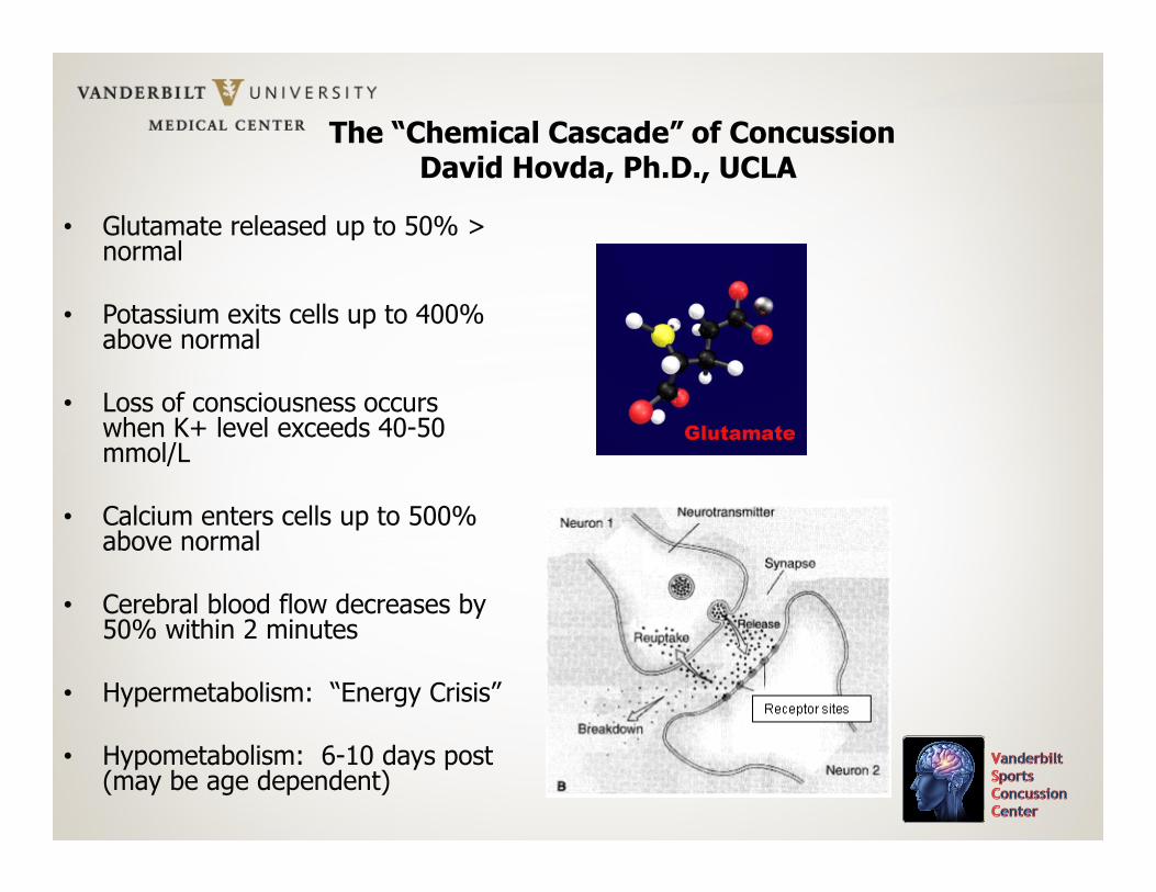
The “Chemical Cascade” of ConcussionDavid Hovda, Ph.D., UCLA
• Glutamate released up to 50% > normal
• Potassium exits cells up to 400% above normal
• Loss of consciousness occurs when K+ level exceeds 40-50 mmol/L
• Calcium enters cells up to 500% above normal
• Cerebral blood flow decreases by 50% within 2 minutes
• Hypermetabolism: “Energy Crisis”
• Hypometabolism: 6-10 days post (may be age dependent)
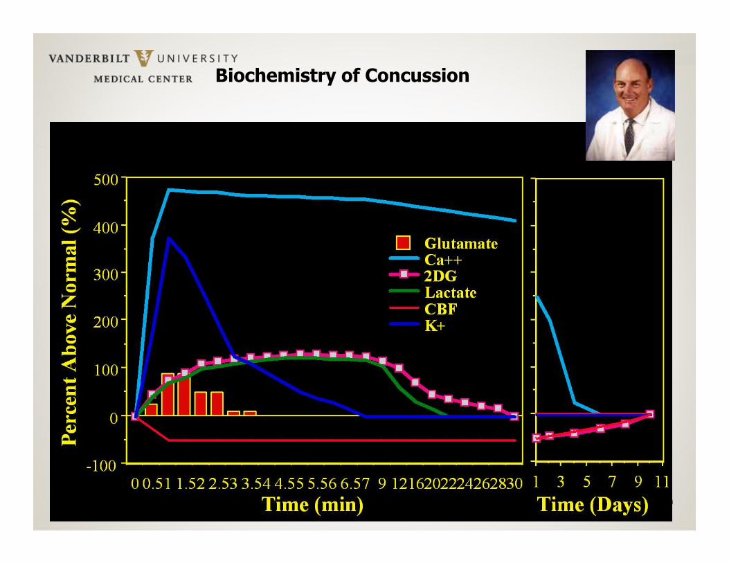
Biochemistry of Concussion
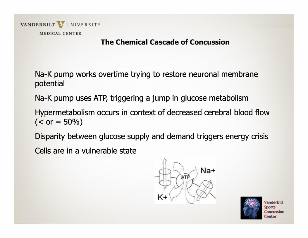
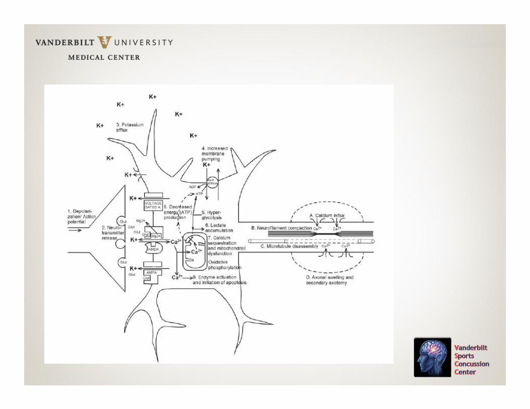
Na-K pump works overtime trying to restore neuronal membrane potential
Na-K pump uses ATP, triggering a jump in glucose metabolism
Hypermetabolism occurs in context of decreased cerebral blood flow (< or = 50%)
Disparity between glucose supply and demand triggers energy crisis
Cells are in a vulnerable state
The Chemical Cascade of Concussion
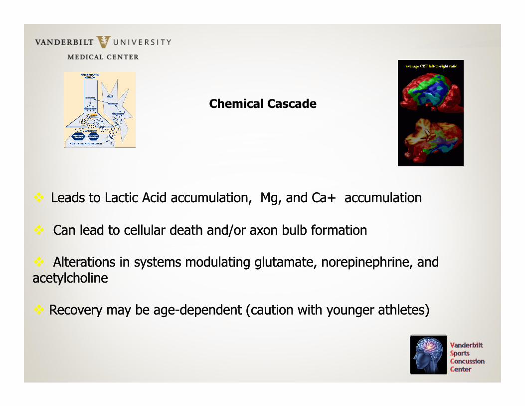
� Leads to Lactic Acid accumulation, Mg, and Ca+ accumulation
� Can lead to cellular death and/or axon bulb formation
� Alterations in systems modulating glutamate, norepinephrine, and acetylcholine
� Recovery may be age-dependent (caution with younger athletes)
� Leads to Lactic Acid accumulation, Mg, and Ca+ accumulation
� Can lead to cellular death and/or axon bulb formation
� Alterations in systems modulating glutamate, norepinephrine, and acetylcholine
� Recovery may be age-dependent (caution with younger athletes)
Chemical Cascade
Bottom Line Conclusions: Biochemistry
Animal models (and to a lesser extent, data from humans with severe TBI) support the hypothesis of biochemical changes in the brain post-concussion. These changes persist, on average, for 7-10 days post-trauma. Current evidence suggests that age, gender, genetic and host factors, activity level during the recovery period, and prior concussion history are some of the major moderating variables.
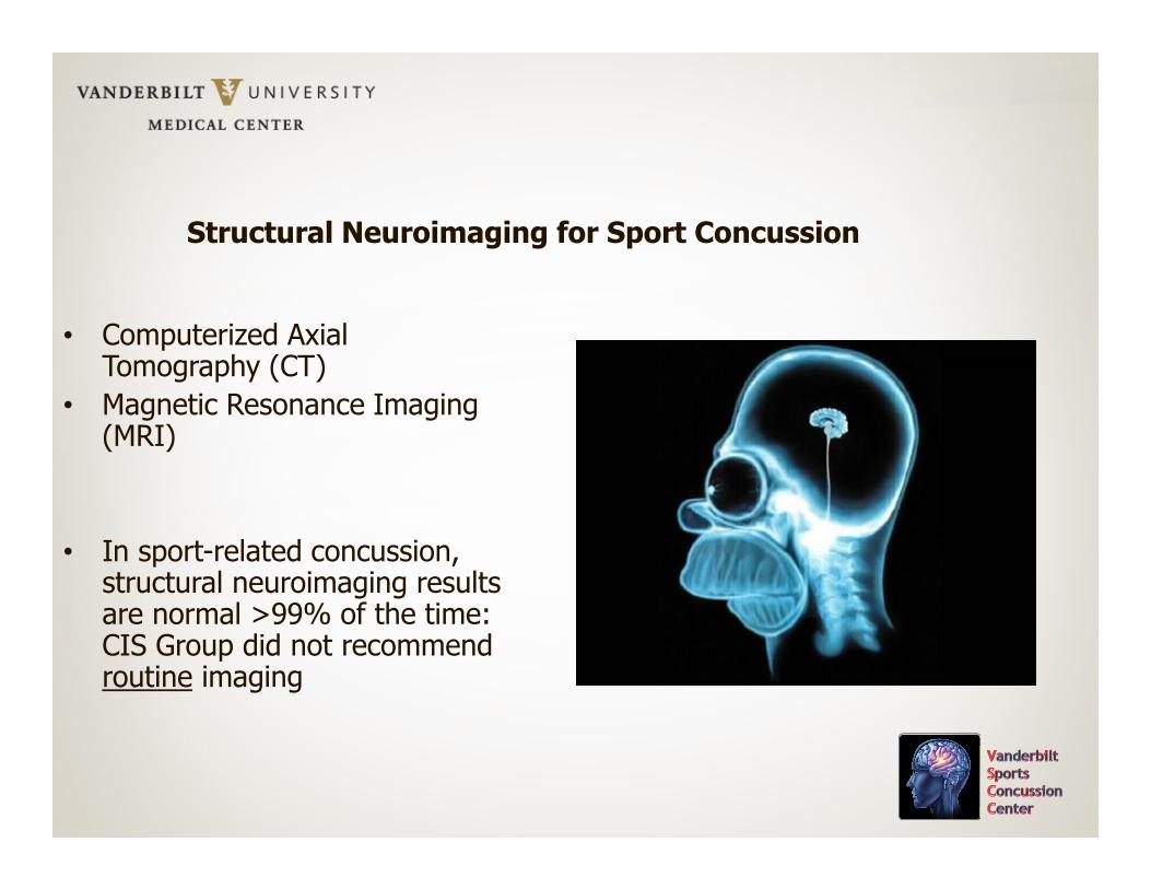
Structural Neuroimaging for Sport Concussion
• Computerized Axial Tomography (CT)
• Magnetic Resonance Imaging (MRI)
• In sport-related concussion, structural neuroimaging results are normal >99% of the time: CIS Group did not recommend routine imaging
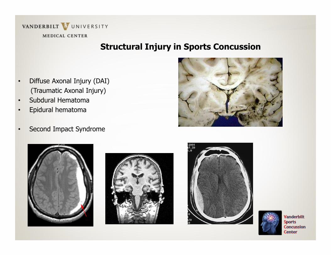
Structural Injury in Sports Concussion
• Diffuse Axonal Injury (DAI)
(Traumatic Axonal Injury)
• Subdural Hematoma
• Epidural hematoma
• Second Impact Syndrome
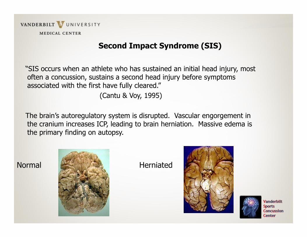
Second Impact Syndrome (SIS)
“SIS occurs when an athlete who has sustained an initial head injury, most often a concussion, sustains a second head injury before symptoms associated with the first have fully cleared.”
(Cantu & Voy, 1995)
The brain’s autoregulatory system is disrupted. Vascular engorgement in the cranium increases ICP, leading to brain herniation. Massive edema is the primary finding on autopsy.
Normal Herniated
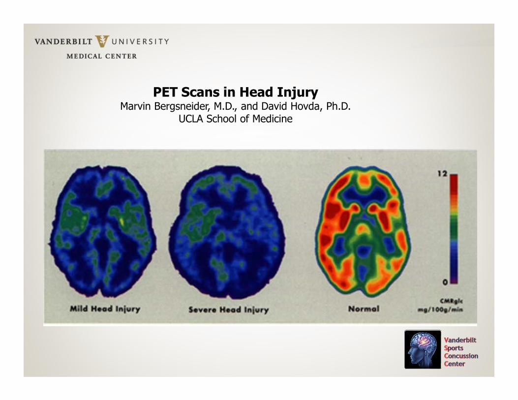
PET Scans in Head InjuryMarvin Bergsneider, M.D., and David Hovda, Ph.D.
UCLA School of Medicine
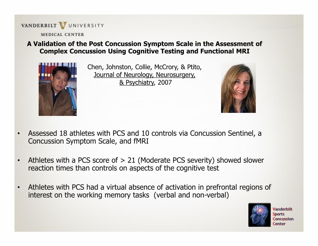
A Validation of the Post Concussion Symptom Scale in the Assessment of Complex Concussion Using Cognitive Testing and Functional MRI
Chen, Johnston, Collie, McCrory, & Ptito, Journal of Neurology, Neurosurgery,
& Psychiatry, 2007
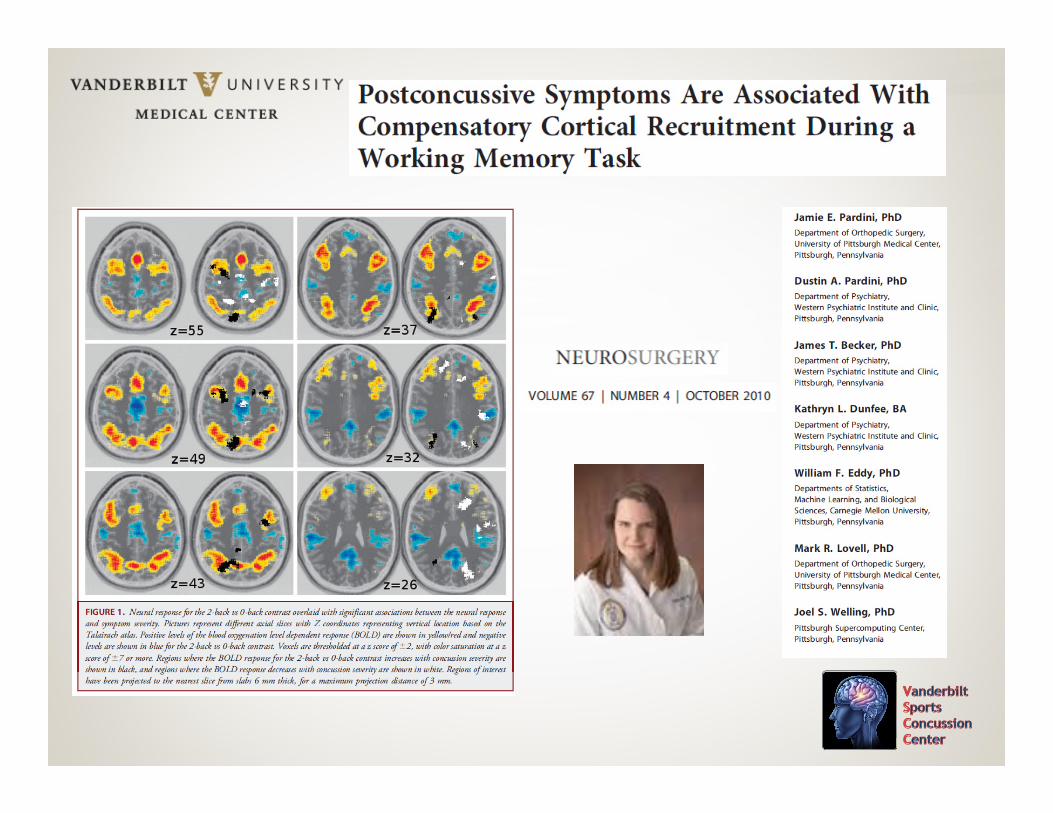
• Assessed 18 athletes with PCS and 10 controls via Concussion Sentinel, a Concussion Symptom Scale, and fMRI
• Athletes with a PCS score of > 21 (Moderate PCS severity) showed slower reaction times than controls on aspects of the cognitive test
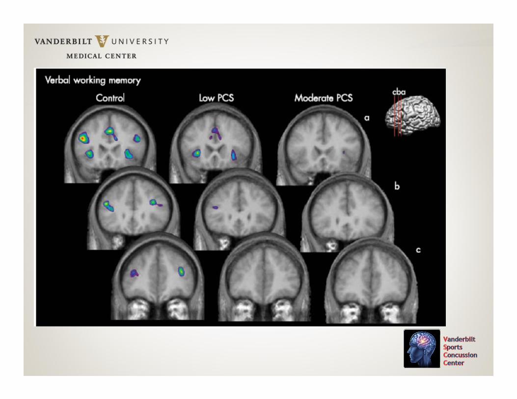
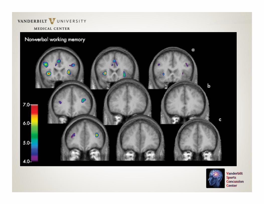
• Athletes with PCS had a virtual absence of activation in prefrontal regions of interest on the working memory tasks (verbal and non-verbal)
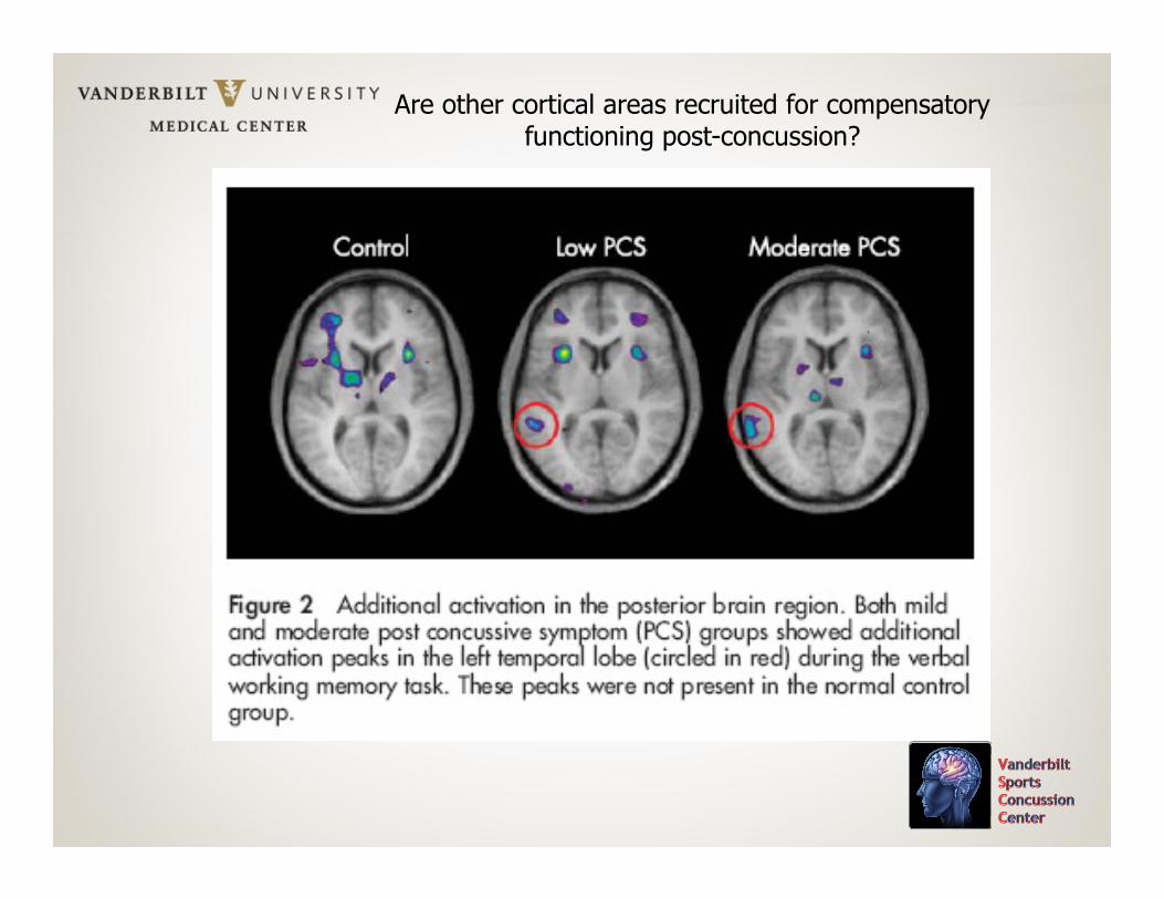
Are other cortical areas recruited for compensatory functioning post-concussion?
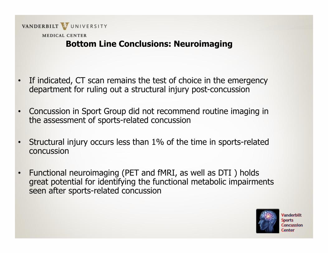
Bottom Line Conclusions: Neuroimaging
• If indicated, CT scan remains the test of choice in the emergency department for ruling out a structural injury post-concussion
• Concussion in Sport Group did not recommend routine imaging in the assessment of sports-related concussion
• Structural injury occurs less than 1% of the time in sports-related concussion
• Functional neuroimaging (PET and fMRI, as well as DTI ) holds great potential for identifying the functional metabolic impairments seen after sports-related concussion
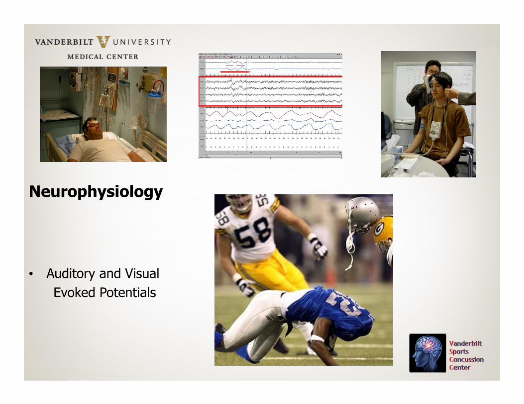
Neurophysiology
• Auditory and Visual
Evoked Potentials
Long Term and Cumulative Effects of SportsConcussion on Motor Cortex Inhibition
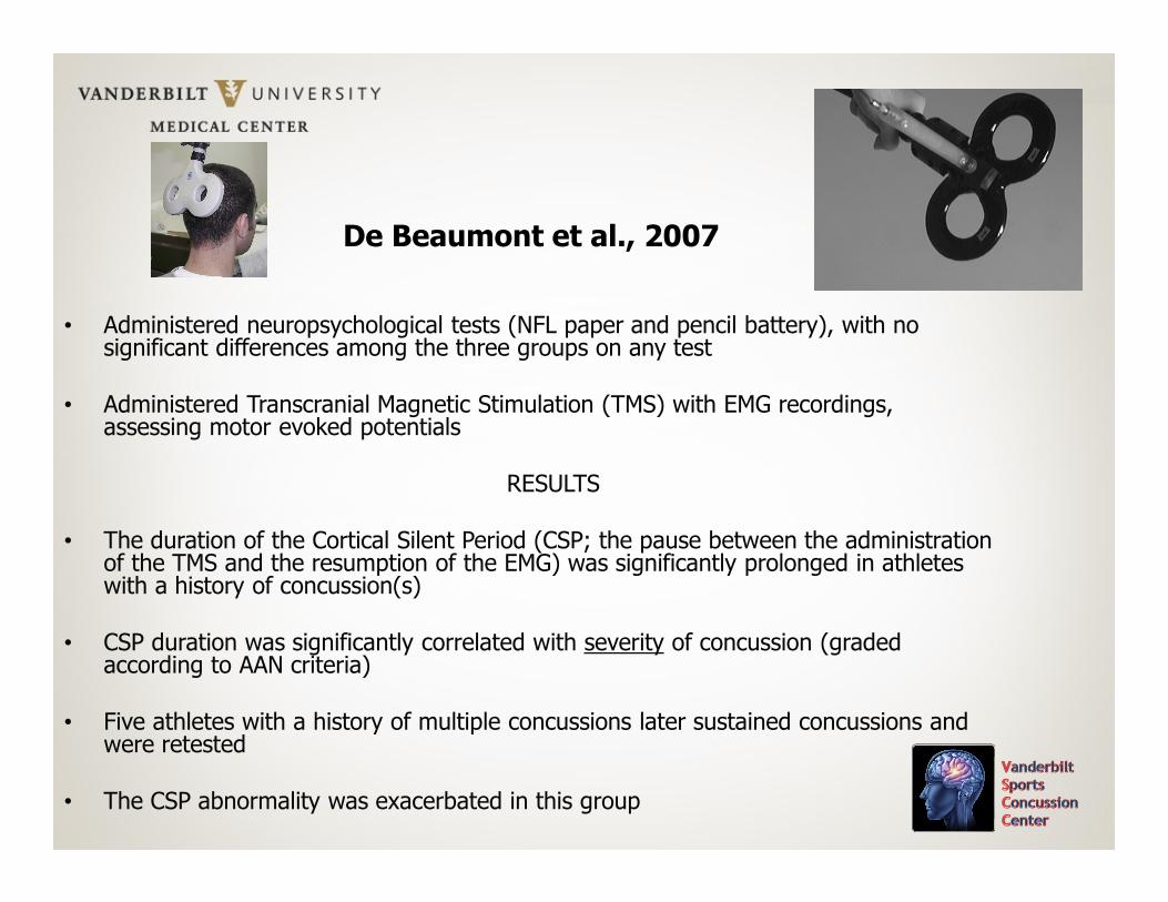
De Beaumont et al., Neurosurgery, 2007, 61, 329-337
• 45 University of Montreal Football players
• No history of psychiatric illness, LD, substance abuse, or non-sports related TBI
• Group 1: 15 asymptomatic athletes with 2 or more (<6) concussions (at least 9 months post), with all concussions diagnosed by the Team Physician and graded according to AAN criteria
• Group 2: 15 athletes with one sports concussion
• Group 3: 15 athletes with no history of concussion
De Beaumont et al., 2007
• Administered neuropsychological tests (NFL paper and pencil battery), with no significant differences among the three groups on any test
• Administered Transcranial Magnetic Stimulation (TMS) with EMG recordings, assessing motor evoked potentials
RESULTS
• The duration of the Cortical Silent Period (CSP; the pause between the administration of the TMS and the resumption of the EMG) was significantly prolonged in athletes with a history of concussion(s)
• CSP duration was significantly correlated with severity of concussion (graded according to AAN criteria)
• Five athletes with a history of multiple concussions later sustained concussions and were retested
• The CSP abnormality was exacerbated in this group

De Beaumont et al., 2007: Conclusion
“Findings from this study show that sports concussions result in long-term motor system dysfunctions that seem to be attributable to subclinical intracortical inhibitory system abnormalities. This study also shows that sustaining subsequent concussions exacerbates this deficit, thus providing support for the contention that the adverse effects of concussions on intracortical inhibitory systems are cumulative” (p. 7).
Bottom Line Conclusions: Electrophysiology
• There is accumulating evidence that electrophysiological changes (especially a decrement in visual and auditory P300 amplitude) persist well beyond symptom resolution and return to baseline cognitive functioning in concussed athletes. qEEG changes may also be evident in the acute recovery phases, but resolve.
• The electrophysiological changes may be a function of concussion severity and frequency.
• The duration of the P300 changes could be months or years.
• The clinical significance of these changes is unknown.

TBI Biomarkers
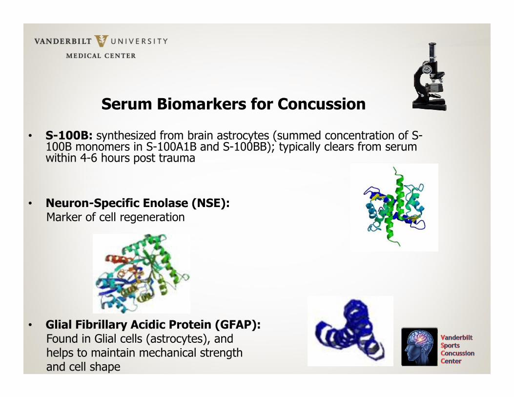
Serum Biomarkers for Concussion
• S-100B: synthesized from brain astrocytes (summed concentration of S-100B monomers in S-100A1B and S-100BB); typically clears from serum within 4-6 hours post trauma
• Neuron-Specific Enolase (NSE):Marker of cell regeneration
• Glial Fibrillary Acidic Protein (GFAP):Found in Glial cells (astrocytes), andhelps to maintain mechanical strengthand cell shape
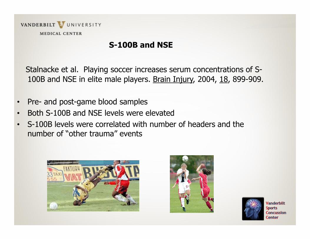
S-100B and NSE
Stalnacke et al. Playing soccer increases serum concentrations of S-
100B and NSE in elite male players. Brain Injury, 2004, 18, 899-909.
• Pre- and post-game blood samples
• Both S-100B and NSE levels were elevated
• S-100B levels were correlated with number of headers and the number of “other trauma” events
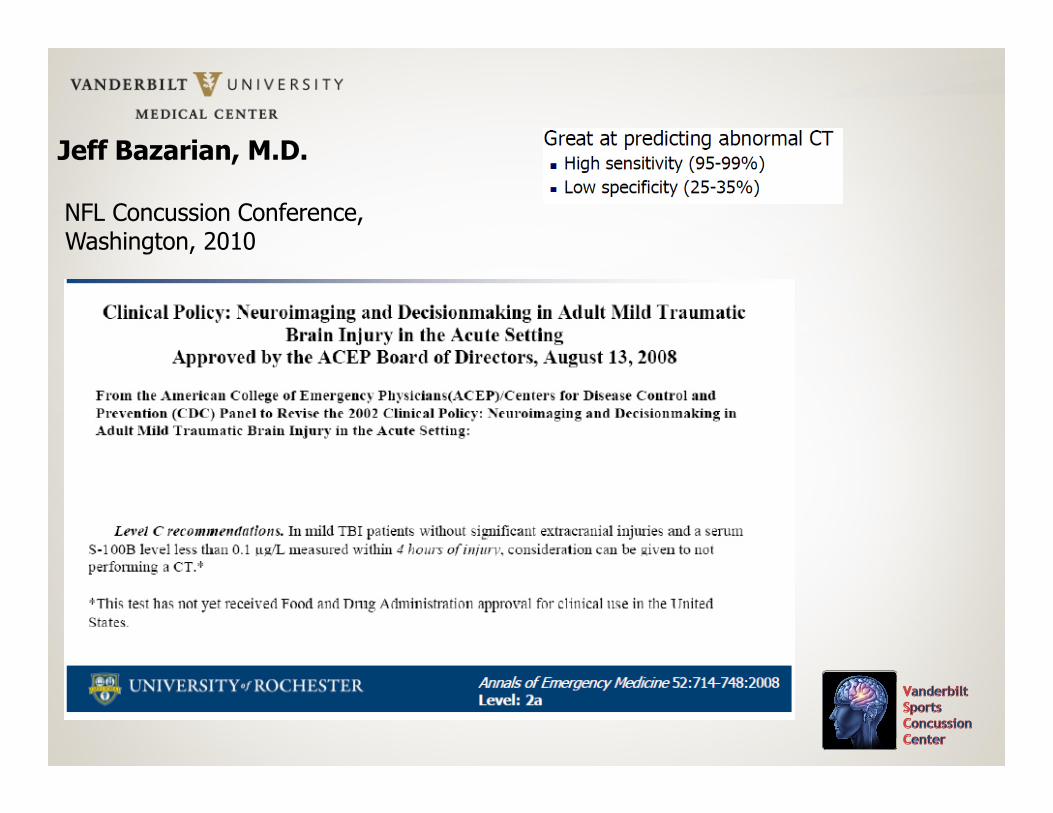
Jeff Bazarian, M.D.
NFL Concussion Conference, Washington, 2010
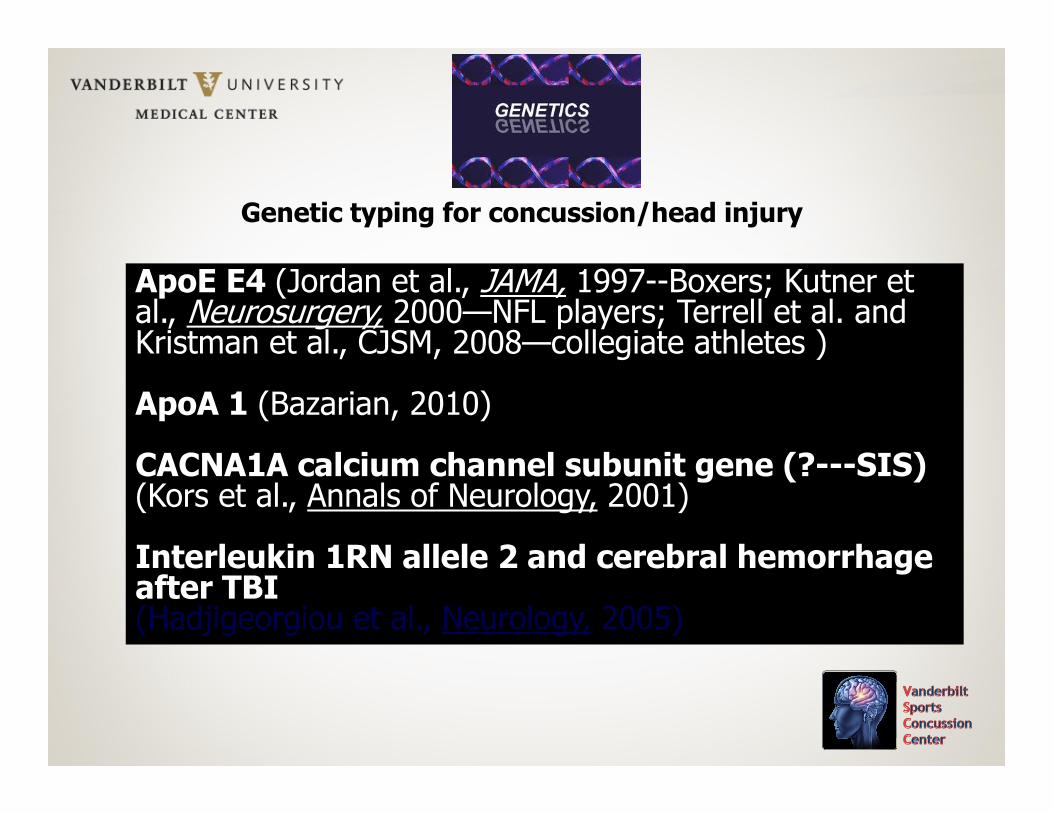
Genetic typing for concussion/head injury
ApoE E4 (Jordan et al., JAMA, 1997--Boxers; Kutner et al., Neurosurgery, 2000—NFL players; Terrell et al. and Kristman et al., CJSM, 2008—collegiate athletes )
ApoA 1 (Bazarian, 2010)
CACNA1A calcium channel subunit gene (?---SIS)(Kors et al., Annals of Neurology, 2001)
Interleukin 1RN allele 2 and cerebral hemorrhage after TBI(Hadjigeorgiou et al., Neurology, 2005)
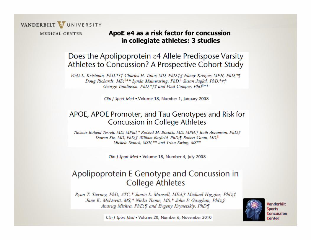
ApoE e4 as a risk factor for concussionin collegiate athletes: 3 studies
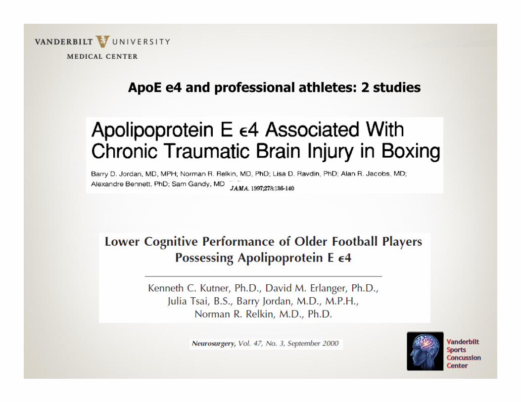
ApoE e4 and professional athletes: 2 studies
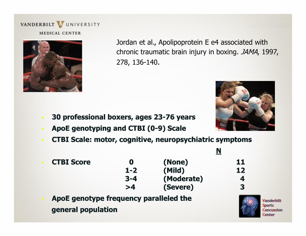
� 30 professional boxers, ages 23-76 years
� ApoE genotyping and CTBI (0-9) Scale
� CTBI Scale: motor, cognitive, neuropsychiatric symptoms
N
� CTBI Score 0 (None) 111-2 (Mild) 123-4 (Moderate) 4>4 (Severe) 3
� ApoE genotype frequency paralleled the
general population
� 30 professional boxers, ages 23-76 years
� ApoE genotyping and CTBI (0-9) Scale
� CTBI Scale: motor, cognitive, neuropsychiatric symptoms
N
� CTBI Score 0 (None) 111-2 (Mild) 123-4 (Moderate) 4>4 (Severe) 3
� ApoE genotype frequency paralleled the
general population
Jordan et al., Apolipoprotein E e4 associated with
chronic traumatic brain injury in boxing. JAMA, 1997,
278, 136-140.
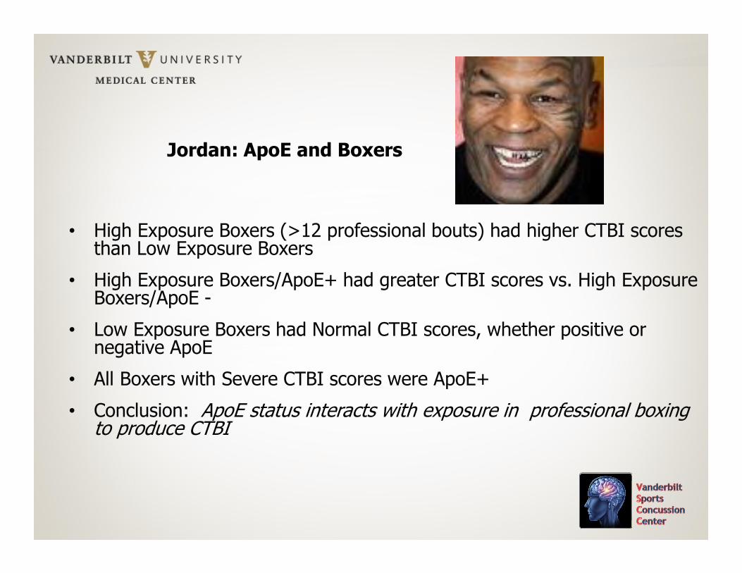
Jordan: ApoE and Boxers
• High Exposure Boxers (>12 professional bouts) had higher CTBI scores than Low Exposure Boxers
• High Exposure Boxers/ApoE+ had greater CTBI scores vs. High Exposure Boxers/ApoE -
• Low Exposure Boxers had Normal CTBI scores, whether positive or negative ApoE
• All Boxers with Severe CTBI scores were ApoE+
• Conclusion: ApoE status interacts with exposure in professional boxing to produce CTBI
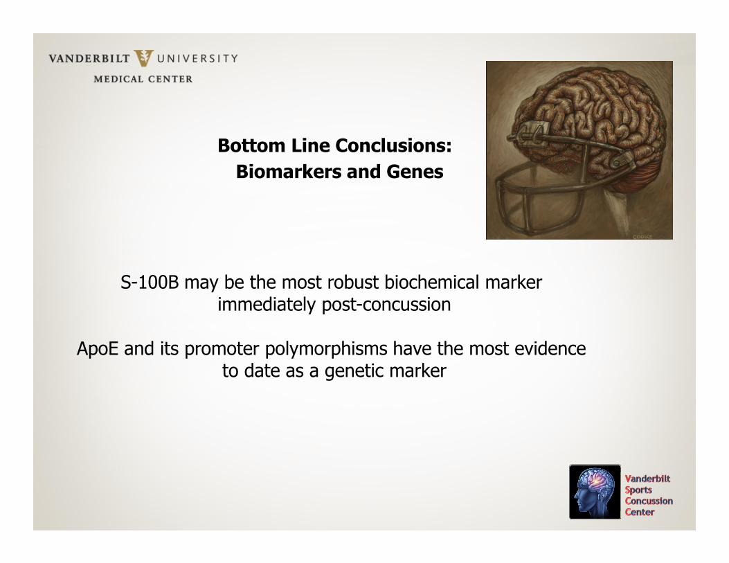
Bottom Line Conclusions:
Biomarkers and Genes
S-100B may be the most robust biochemical markerimmediately post-concussion
ApoE and its promoter polymorphisms have the most evidenceto date as a genetic marker
Animal Studies
Neuropathology and Biochemistry
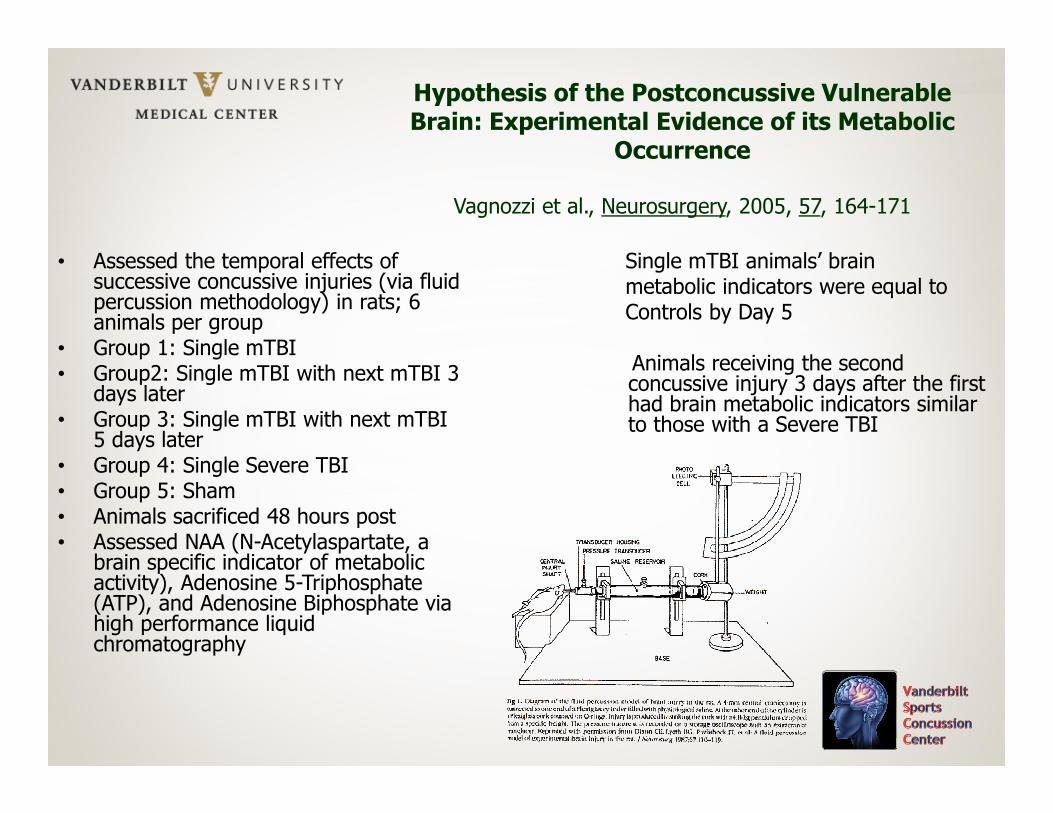
Hypothesis of the Postconcussive Vulnerable Brain: Experimental Evidence of its Metabolic
Occurrence
Vagnozzi et al., Neurosurgery, 2005, 57, 164-171
• Assessed the temporal effects of successive concussive injuries (via fluid percussion methodology) in rats; 6 animals per group
• Group 1: Single mTBI• Group2: Single mTBI with next mTBI 3
days later• Group 3: Single mTBI with next mTBI
5 days later• Group 4: Single Severe TBI• Group 5: Sham• Animals sacrificed 48 hours post• Assessed NAA (N-Acetylaspartate, a
brain specific indicator of metabolic activity), Adenosine 5-Triphosphate (ATP), and Adenosine Biphosphate via high performance liquid chromatography
Single mTBI animals’ brainmetabolic indicators were equal toControls by Day 5
Animals receiving the second concussive injury 3 days after the first had brain metabolic indicators similar to those with a Severe TBI
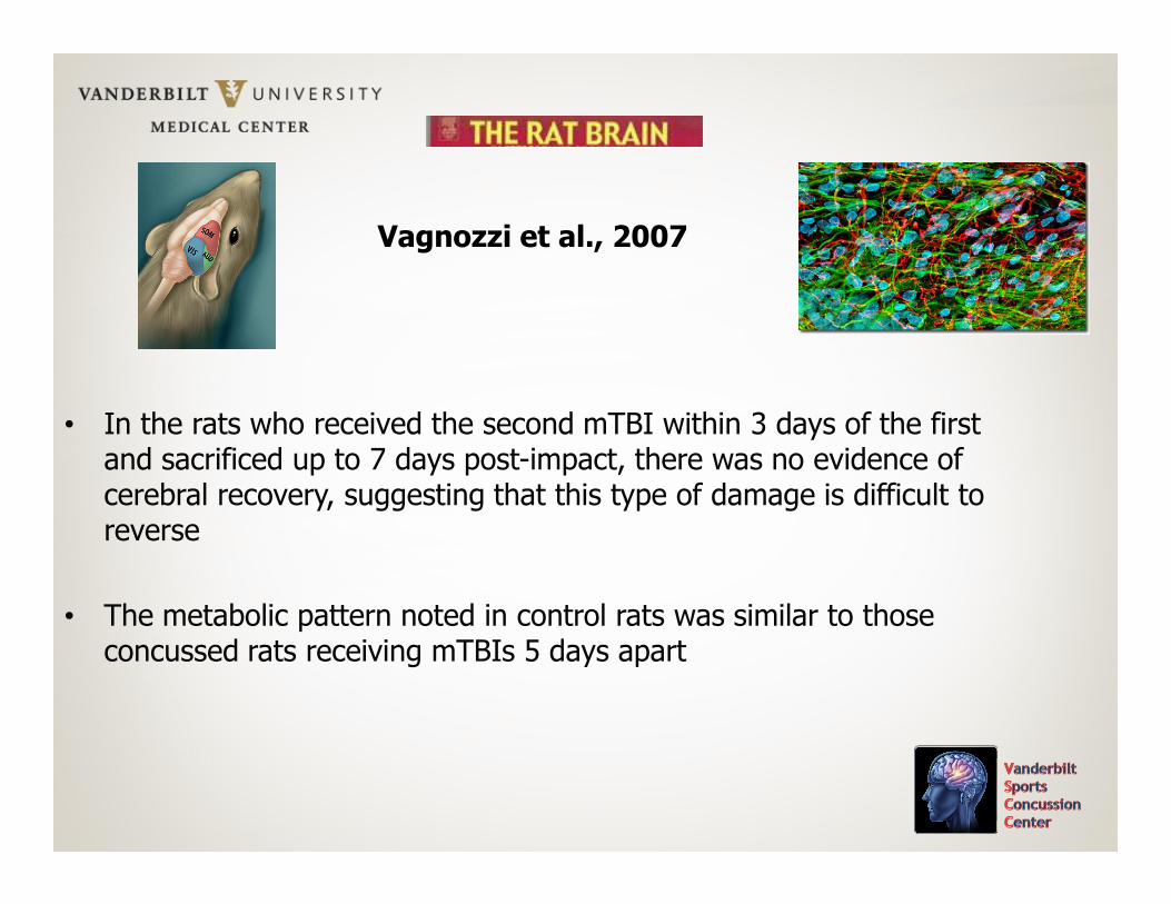
Vagnozzi et al., 2007
• In the rats who received the second mTBI within 3 days of the first and sacrificed up to 7 days post-impact, there was no evidence of cerebral recovery, suggesting that this type of damage is difficult to reverse
• The metabolic pattern noted in control rats was similar to those concussed rats receiving mTBIs 5 days apart
Vagnozzi et al., 2007
Conclusion: “This study shows that the existence of a temporal window of brain vulnerability after mTBI. A second concussive event falling within this time range had profound consequences on mitochondrial-related metabolism. Furthermore, because NAA recovery coincided with normalization of all other metabolites, it is conceivable to hypothesize that NAA measurement by 1-H-NMR spectroscopy might be a valid tool in assessing full cerebral metabolic activity in the clinical setting and with particular reference to sports medicine in establishing when to return mTBI-affected athletes to play”.
Thanks!