Embed Size (px)
Citation preview

The Plant Cell, Vol. 8, 429-445, March 1996 O 1996 American Society of Plant Physiologists
SRK, the Stigma-Specific S Brassica, 1s Targeted to the Transgenic Tobacco
Locus Receptor Kinase of Plasma Membrane in
Joshua C. Stein,’ Ram Dixit, Mikhail E. Nasrallah, and June B. Nasrallah2 Section of Plant Biology, Division of Biological Sciences, Cornell University, Ithaca, New York 14853
The S locus receptor kinase (SRK) gene is one of two S locus genes required for the self-incompatibility response in Brassica. We have identified the product of the SRKs gene in 6. oleracea stigmas and have shown that it has character- istics of an integral membrane protein. When expressed in transgenic tobacco, SRK6 is glycosylated and targeted to the plasma membrane. These results provide definitive biochemical evidence for the existence in plants of a plasma membrane-localized transmembrane protein kinase with a known cell-cell recognition function. The timing of SRK ex- pression in stigmas follows a time course similar to that previously described for another S locus-linked gene, the S locus glycoprotein (SLG) gene, and correlates with the ability of stigmas to mount a self-incompatibility response. Based on SRK6 promoter studies, the site of gene expression overlaps with that of SLG and exhibits predominant expression in the stigmatic papillar cells. Although reporter gene studies indicated that the SRK promoter was active in pollen, SRK protein was not detected in pollen, suggesting that SRK functions as a cell surface receptor exclusively in the papillar cells of the stigma.
INTRODUCTION
Many plant species with hermaphroditic flowers are able to limit self-fertilization by a mechanism called self-incompatibility (SI). In Brassica, the SI response represents the culmination of cell-to-cell recognition and signal transduction events that occur soon after pollen makes contact with the papillar cells of the stigma surface. Specificity of this response is controlled by the S locus complex, a highly polymorphic cluster of genes that are organized into distinct haplotypes (Boyes and Nasrallah, 1993, 1995). In general, SI is elicited when parents of a cross carry an identical S locus haplotype, resulting in the disrup- tion of pollen germination and tube growth at the stigma surface. Depending on their combination in heterozygotes, S haplotypes may be codominant or show dominantlrecessive interactions. For example, class I haplotypes, such as S,, con- fer a strong SI reaction and are dominant to the weaker class II haplotypes, such as S2 (Nasrallah et al., 1991).
At least some of the genes within the S locus complex are thought to encode recognition molecules that act at the pol- len-papillar cell interface. Among these are the S locus glycoprotein (SLG) gene and the S receptor kinase (SRK) gene, two stigma-expressed genes that are required for phenotypic expression of SI (Toriyama et al., 1991a; Nasrallah et al., 1992,
Current address: CIBA-GEIGY Corporation, Research Triangle Park,
To whom correspondence should be addressed. NC 27709-2257.
1994a; Goring et al., 1993). SLG encodes an abundant glyco- protein that accumulates in the cell wall of papillar cells (Nasrallah et al., 1985b; Kandasamy et al., 1989). Based on its sequence, we had predicted SRK to encode a plasma mem- brane receptor protein kinase having an extracellular domain that is highly similar to SLG, a single pass transmembrane domain, and a cytoplasmic kinase domain (Stein, et al., 1991). When expressed in Escherichia coli, the kinase domain is capa- ble of autophosphorylating on serine and threonine residues (Goring and Rothstein, 1992; Stein and Nasrallah, 1993).
Both SLG and SRK have the potential to function as recog- nition molecules by virtue of their extensive polymorphism (Nasrallah et al., 1987; Stein et al., 1991). Greatest amino acid divergence, up to 33%, is observed between alleles derived from class I and class II haplotypes (Chen and Nasrallah, 1990; Stein et al., 1991), and this divergence has allowed the devel- opment of class I-specific antibodies to SLG (Kandasamy et al., 1989). Of great interest is the finding that despite their ex- tensive divergence between haplotypes, SLG and the SLG-like domain of SRK share a high degree of sequence identity within a given haplotype, showing as little as 10% divergence (Stein et al., 1991; Goring and Rothstein, 1992; Delorme et al., 1995). Because of this apparent concerted evolution of the SLGISRK gene pair within each haplotype, we had suggested that the protein products of these genes interact functionally, perhaps as components of a receptor system involving binding of a com- mon ligand (Nasrallah et al., 1994b). The validity of such a

430 The Plant Cell
hypothesis rests in part on whether SRK is in fact localizedto the plasma membrane and is expressed within the samecells as SLG.
In this study, we focus primarily on B. oleracea SRKB, theSRK allele contained within the S6 haplotype (a class I haplo-type) that was previously cloned and sequenced (Stein et al.,1991). We show that stigmatic SRK6 is an integral membraneprotein, consistent with sequence-based predictions. Moreover,when expressed in transgenic tobacco leaves, SRK6 is tar-geted to the plasma membrane. We also show by RNA andprotein blot analyses that SRK and SLG are coordinately regu-lated genes, displaying similar temporal patterns of expression.The SRK promoter contains functional elements similar to thatof SLG and drives reporter gene expression in papillar cells,where SLG is also known to be expressed. Finally, despite evi-dence that both of these genes are transcribed at low levelswithin male reproductive organs (Sato et al., 1991; Stein et al.,1991; Goring and Rothstein, 1992), we found no evidence ofSLG or SRK proteins in pollen grains. These findings supportthe hypothesis that SRK functions as a plasma membrane-localized receptor and, along with SLG, acts principally as afemale determinant of SI recognition.
RESULTS
Identification of SRK in Stigmas of Brassica
Previously, we raised monoclonal antibody MAb/H8 againstpurified SLG6 (Kandasamy et al., 1989). Because the SLG-like domain of SRK6 has 89% amino acid sequence identitywith SLG6 (Stein et al., 1991), we hypothesized that MAb/H8would also recognize SRK6 and possibly other class I SRKproteins as well. To test this hypothesis, we expressed the SLG-like domain of SRK6 as a glutathione S-transferase (GST) fu-sion protein in £ co//and subjected the affinity-purified proteinto immunoblotting. In Figure 1A, the protein blot stained withCoomassie blue shows that a GST-SRK6 fusion protein of theexpected size was produced and that this band was absentin £ co// harboring the parental pGEX vector. In the accom-panying blot subjected to immunostaining, MAb/H8 detectedthe GST-SRK6 fusion protein but did not detect GST in thenegative control lane (Figure 1A). In addition to the full-lengthfusion protein, several smaller immunoreactive proteins werealso detected with MAb/H8 (Figure 1A). These bands, whichare absent from control extracts that lack GST-SRK6 andtherefore do not represent endogenous bacterial proteins, areproteolytic derivatives of the GST-SRK6 fusion protein, result-ing from the commonly observed instability of recombinantproteins in £ co//. The cross-reactivity of the GST-SRK6 fu-sion protein with MAb/H8 indicates that MAb/H8 does indeedbind to SRK6 and establishes the utility of this antibody instudies of SRK.
To study the native SRK protein in Brassica stigmas, we usedF2 populations segregating for the S6 haplotype. One such
population was derived from a cross between an SgSg ho-mozygote and an S2S2 homozygote. Among 32 plants in thispopulation, eight were S6S6 homozygotes, 18 were S2S6 het-erozygotes, and six were S2S2 homozygotes, as determinedby pollination tests and DNA gel blot analysis (see Methods).
Coomassie MAb/H8
1 2 1 297 -66 -43 -
31 -I
22 -
Anti-FLAG
•GST-SRK6
GST
MAb/H8
26-
Figure 1. Immunodetection of the SLG-like Domain of SRKe andSRK2.(A) GST (lanes 1) and a GST-SRKe fusion protein (lanes 2) expressedin bacteria were affinity purified with glutathione-agarose beads. Pro-teins were fractionated by SDS-PAGE, blotted onto PVDF membrane,either stained with Coomassie blue or probed with MAb/H8, and de-veloped using chromogenic substrates.(B) Total extracts of bacteria expressing GST-FLAG-SRKg (lanes 1),GST-FLAG-SRK2 (lanes 2), or GST-FLAG (lanes 3) were subjectedto SDS-PAGE, blotted onto PVDF membrane, probed with anti-FLAGantibody (see Methods) or MAb/H8, and developed using chemilumi-nescent substrates. The bands corresponding to the full-length fusionproteins are indicated by an arrowhead for SRK6 and an asterisk forSRK2. The GST-FLAG protein encoded by the modified pGEX vec-tor is indicated by an open circle.The immunoreactive bands that migrate faster than the full-length SRKfusion proteins are breakdown products. Molecular mass standardsshown at left are given in kilodaltons.

Cellular and Subcellular Localization of SRK 431
- 200
SRK
SLG
MAb/H8
B
ctfctf
- 200
-1 1 6
- 97
- 66
R1-254Figure 2. Detection of Stigmatic SRKe in Brassica with Two Antibod-ies and Demonstration of Linkage to the S Locus Complex.Stigma microsomal membrane proteins (20 ng per lane) were preparedfrom individuals in an F2 population segregating for the S6 and S2haplotypes. The S locus genotype of each plant is indicated aboveeach lane.(A) Immunoblot probed with MAb/H8.(B) The same blot was stripped and reprobed with affinity-purified R1-254, a polyclonal antiserum raised against a peptide from the C-terminaldomain of SRK6.The blots in (A) and (B) were developed with chemiluminescent sub-strates. SRK6 is detected by both antibodies as a band of ~108 kD.The band of ~94 kD evident in the blot probed with the R1-254 rabbitantiserum was detected in all lanes, regardless of genotype, and isattributed to nonspecific reactivity. Numbers at right indicate molecu-lar mass markers in kilodaltons.
S2 is a class II haplotype whose SRK and SLG alleles arehighly diverged from SRK6 and SLGS (Stein et al., 1991), andMAb/H8 cross-reacts with neither SLG2, as previously shown(Kandasamy et al., 1989), nor the S domain of SRK2, asdemonstrated by immunological analysis of the GST-SRK2 fu-
sion protein (Figure 1B). The S2 haplotype therefore servedas a negative control in these studies.
Figure 2A shows an immunoblot of stigma proteins extractedfrom six representative F2 individuals. SRK6 was identifiedtentatively as an immunoreactive band of 108 kD, a size con-sistent with a predicted SRK6 polypeptide of 92 kD havingmultiple N-linked glycosylation sites (Stein et al., 1991). In ad-dition to this 108-kD band, MAb/H8 detected multiple bandsof 55 to 65 kD (Figure 2A) previously identified as SLGe glyco-forms(Umbachetal., 1990). Because SLG is highly abundantin stigmas, representing up to 5% of the total synthesizedprotein (Nasrallah et al., 1985a), its detection by the chem-iluminescence method used here resulted in a highly intenseand diffuse signal. The additional minor immunoreactive bandsthat migrated more slowly than did the 116-kD molecular massmarker (Figure 2A) appear to be artifacts and probably resultedfrom oxidative cross-linking of SLG during tissue homogeni-zation. Their detection was increased when antioxidants andpolyvinylpyrrolidone were excluded from the homogenizationbuffer (data not shown).
By testing the entire F2 population, the eight S6S6 and 18S6S2 plants were found to exhibit identical immunoblot pat-terns in which both the 108-kD protein and the 55- to 65-kDSLG6 glycoforms were evident. In contrast, all six S2S2 indi-viduals lacked the 108-kD and 55- to 65-kD immunoreactiveproteins. This perfect cosegregation of the 108-kD protein withthe S6 haplotype and with SLG6 demonstrates linkage to theS locus complex.
To confirm the identity of the 108-kD band as SRK6, wetested whether this protein binds to R1-254, an antiserum raisedagainst a peptide sequence within the C-terminal domain ofSRK6 and previously shown to recognize a bacterially ex-pressed SRK6 fusion protein containing only the kinase andC-terminal domains (Stein and Nasrallah, 1993). The peptidesequence used to raise antiserum R1-254 intentionally waschosen to be specific to SRK6, having <36% sequence iden-tity with SRK2 (Stein et al., 1991). The blot in Figure 2A wasstripped and reacted with affinity-purified R1-254 antibodies.The results, shown in Figure 2B, demonstrate that R1-254recognized a 108-kD protein identical to the one detectedwith MAb/H8. This protein was detected in all plants bearingthe Ss haplotype but was absent in the S2S2 individuals (Fig-ure 2B).
Cosegregation of the 108-kD protein with the S6 haplotypewas also demonstrated in a second F2 population segregat-ing for the S6 and S» haplotypes. The S» haplotype, whichspecifies a self-fertile phenotype in S/, homozygotes, is a non-functional haplotype that carries a null allele of SRK in whichthe promoter and a sizable portion of the coding sequenceare deleted (Nasrallah et al., 1994a). St1 does, however, pos-sess an intact and expressed allele of SLG, which is classifiedas a class I allele (Nasrallah et al., 1994a). We examined 18individuals in an F2 population derived from crossing an S6S6parent with an Sf,Sf1 parent. In this population, 14 individu-als (six S6S6 homozygotes and eight S6S« heterozygotes)were self-incompatible, whereas four individuals exhibited a
432 The Plant Cell
F2 progeny
A
SRK § - -_
alternative product of the SLG gene. Based on the above find-ings showing genetic linkage of the 108-kD protein to the Slocus complex, its cross-reactivity with antibodies directedagainst both the SLG-like domain and C-terminal domain, andthe absence of a similar protein in a strain carrying a null SRKallele, we conclude that the 108-kD protein is the product ofthe SRK6 gene.
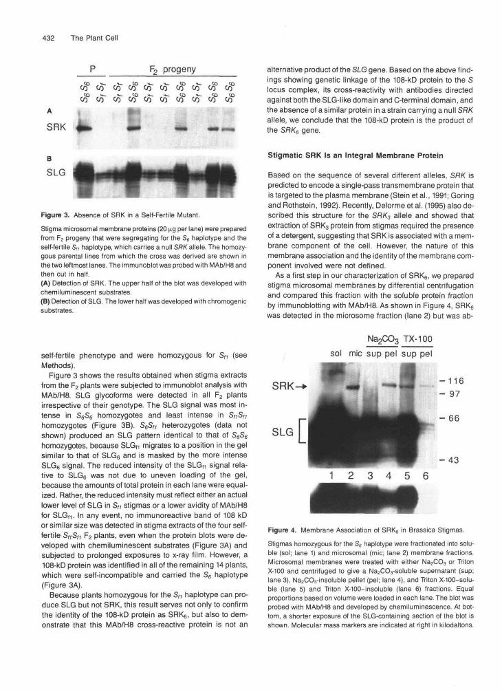
Figure 3. Absence of SRK in a Self-Fertile Mutant.
Stigma microsomal membrane proteins (20 ng per lane) were preparedfrom F2 progeny that were segregating for the S6 haplotype and theself-fertile S,, haplotype, which carries a null SRK allele. The homozy-gous parental lines from which the cross was derived are shown inthe two leftmost lanes. The immunoblot was probed with MAb/H8 andthen cut in half.(A) Detection of SRK. The upper half of the blot was developed withchemiluminescent substrates.(B) Detection of SLG. The lower half was developed with chromogenicsubstrates.
Stigmatic SRK Is an Integral Membrane Protein
Based on the sequence of several different alleles, SRK ispredicted to encode a single-pass transmembrane protein thatis targeted to the plasma membrane (Stein etal., 1991; Goringand Rothstein, 1992). Recently, Delorme et al. (1995) also de-scribed this structure for the SRK3 allele and showed thatextraction of SRK3 protein from stigmas required the presenceof a detergent, suggesting that SRK is associated with a mem-brane component of the cell. However, the nature of thismembrane association and the identity of the membrane com-ponent involved were not defined.
As a first step in our characterization of SRK6, we preparedstigma microsomal membranes by differential centrifugationand compared this fraction with the soluble protein fractionby immunoblotting with MAb/H8. As shown in Figure 4, SRK6
was detected in the microsome fraction (lane 2) but was ab-
self-fertile phenotype and were homozygous for Sf1 (seeMethods).
Figure 3 shows the results obtained when stigma extractsfrom the F2 plants were subjected to immunoblot analysis withMAb/H8. SLG glycoforms were detected in all F2 plantsirrespective of their genotype. The SLG signal was most in-tense in S6S6 homozygotes and least intense in Sf,SH
homozygotes (Figure 3B). S6SM heterozygotes (data notshown) produced an SLG pattern identical to that of S6S6homozygotes, because SLGf1 migrates to a position in the gelsimilar to that of SLG6 and is masked by the more intenseSLG6 signal. The reduced intensity of the SLGn signal rela-tive to SLG6 was not due to uneven loading of the gel,because the amounts of total protein in each lane were equal-ized. Rather, the reduced intensity must reflect either an actuallower level of SLG in S/» stigmas or a lower avidity of MAb/H8for SLG(1. In any event, no immunoreactive band of 108 kDor similar size was detected in stigma extracts of the four self-fertile SnSti F2 plants, even when the protein blots were de-veloped with chemiluminescent substrates (Figure 3A) andsubjected to prolonged exposures to x-ray film. However, a108-kD protein was identified in all of the remaining 14 plants,which were self-incompatible and carried the S6 haplotype(Figure 3A).
Because plants homozygous for the Sft haplotype can pro-duce SLG but not SRK, this result serves not only to confirmthe identity of the 108-kD protein as SRK6, but also to dem-onstrate that this MAb/H8 cross-reactive protein is not an
Na2CO3 TX-100sol mic sup pel sup pel
SRK
SLG
1 2 3 4 5 6-43
Figure 4. Membrane Association of SRK6 in Brassica Stigmas.Stigmas homozygous for the S6 haplotype were fractionated into solu-ble (sol; lane 1) and microsomal (mic; lane 2) membrane fractions.Microsomal membranes were treated with either Na2CO3 or TritonX-100 and centrifuged to give a Na2CO3-soluble supernatant (sup;lane 3), Na2CO3-insoluble pellet (pel; lane 4), and Triton X-100-solu-ble (lane 5) and Triton X-100-insoluble (lane 6) fractions. Equalproportions based on volume were loaded in each lane. The blot wasprobed with MAb/H8 and developed by chemiluminescence. At bot-tom, a shorter exposure of the SLG-containing section of the blot isshown. Molecular mass markers are indicated at right in kilodaltons.

Cellular and Subcellular Localization of SRK 433
sent from the soluble protein fraction (lane l), consistent with the prediction that SRK is bound to the membrane.
To test the nature of this interaction, we compared the abil- ity of sodium carbonate and detergent treatments to solubilize membrane-bound SRK,. Sodium carbonate has been shown to convert closed membrane vesicles into open membrane sheets, thereby releasing proteins held in the lumen (Fujiki et al., 1982). Sodium carbonate also strips away peripheral mem- brane proteins while leaving integral membrane proteins embedded in the membrane. On the other hand, detergents generally are required to solubilize integral membrane proteins but also will solubilize peripheral membrane proteins indirectly. In this study, proteins were deemed soluble if they remained in the supernatant after treated membranes were centrifuged at 100,000gor greater for 1 hr (Penefsky and Tzagoloff, 1971).
When microsomal membranes were washed with sodium carbonate, virtually all of the SRK6 protein remained as- sociated with the pelleted membranes (Figure 4, lanes 3 and 4). However, almost complete solubilization of SRK6 was achieved by treating the microsomal membranes with Triton X-100 (Figure 4, lanes 5 and 6). These solubility characteris- tics are consistent with SRK being an integral rather than peripheral membrane protein and contrast markedly with the solubility characteristics of SLG. Most of SLG6 was detected in the soluble fraction (Figure 4, lane l), as expected for this cell wall-localized secreted glycoprotein, but a significant amount of SLG6 was detected also in the microsomal mem- brane fraction (Figure 4, lane 2). Unlike SRK6, most of this membrane-associated SLG6 was released into the superna- tant after treatment with sodium carbonate (Figure 4, lanes 3 and 4).
SRK Encodes an Integral Plasma Membrane-Bound Glycoprotein
Further biochemical localization of SRK at the subcellular leve1 was restricted by the limited quantities of stigmatic tissue avail- able. To provide an abundant Source of SRK protein, the SRK6 gene was expressed in transgenic tobacco leaves by fusing its coding region to the cauliflower mosaic virus (CaMV) 35s promoter. Tobacco has been used frequently as a heterolo- gous plant system for the study of protein targeting, production of correctly targeted proteins, and determination of the sub- cellular localization of proteins (reviewed in Frommer and Ninnemann, 1995). In addition, we had shown previously that Brassica SLG is properly glycosylated and targeted to the ex- tracellular matrix in tobacco either by cells of the transmitting tract when expressed under the control of the SLG promoter (Kandasamy et al., 1990; Moore and Nasrallah, 1990) or by tobacco cell cultures when expressed under the control of the CaMV 35s promoter (Perl-Treves et al., 1993).
5A shows six independent transgenic lines, each of which ex- pressed a protein that reacted with the antibody. This protein was not detected in untransformed leaf material or in material that was transformed with a binaryvector lacking SRKcoding sequences and was thus encoded by the SRK6 transgene. As in Brassica stigmas, tobacco-expressed SRK6 has an appar- ent molecular mass of 108 kD.
To determine the subcellular location of SRK6 in transgenic tobacco, we purified plasma membranes away from intracel- lular membranes, using the aqueous two-phase partitioning method (Larsson, 1985). Membrane purity was confirmed by assaying for marker enzymes immunologically, as shown in Figure 5B, and biochemically, as shown in Table 1. The 100- kD plasma membrane H+-ATPase was enriched in the plasma membrane fraction (Figure 5B), and this was reflected in a >4.5- fold enrichment of vanadate-sensitive ATPase activity (Table 1). Purity of this plasma membrane fraction was confirmed by the absence of markers for the tonoplast (Figure 5B), endoplas- mic reticulum, Golgi apparatus, and mitochondria (Table 1). SRK, was assayed in soluble, crude microsome, intracellular membrane, and plasma membrane fractions by immuno- blotting. As shown in Figure 5C, SRK6 was associated with microsomal membranes and copurified with the plasma mem- brane; it was largely depleted from intracellular membranes and could not be detected in the lane containing soluble proteins.
Tobacco-expressed SRK, has solubility characteristics simi- lar to the native stigmatic protein. As shown in Figure 50, treatment of microsomal membranes containing S R G (lane 1) with sodium carbonate resulted in little or no solubilization of SRK6 (lane 2): all of the detectable SRK6 remained bound to the pelleted membranes (lane 3). In contrast, treatment of membranes with RIPA, a buffer that contains a mixture of ionic and nonionic detergents (see Methods), resulted in the release of most of the bound SRK6 (lane 4), although trace amounts of the protein still were detected in the pelleted insoluble ma- terial (lane 5). Thus, tobacco-expressed appears to be embedded in the membrane.
The SRK6 sequence contains seven potential N-linked glycosylation sites within its SLG-like domain, severa1 of which are conserved in the SLG6 sequence (Stein et al., 1991). We tested whether tobacco-expressed SRK6 is glycosylated by assessing its ability to bind the lectin concanavalin A (ConA). ConA-Sepharose beads were incubated with detergent-solu- bilized microsomal membranes containing SRK6, and bound proteins were analyzed by immunoblotting. As shown in Fig- ure 5D, SRK6 was detected in the ConA-bound fraction (lane 6), consistent with the presence of glycan chains covalently linked to the SRK6 polypeptide. These results agree with those of Delorme et al. (1995), who showed by different methods that SRKB is glycosylated.
Forty-six independent transgenic lines were screened for the presence of SRK RNA, and those showing high relative levels of transcript were selected for further study. To test for the presence of the SRK protein, whole extracts of leaf pro- teins were analyzed by immunoblotting with MAb/H8. Figure
Coordinate Regulation of SRK and SLG Expression
We and others have shown that transcription of SRK and SLG is confined to reproductive organs, predominantly the stigma

434 The Plant Cell
C1 C2 35S::Sf?K6 tobacco
1 2 3 4 5 6 7 8200-
116-97-
66-
Table 1. Marker Enzyme Analysis of Microsomes, and Upperand Lower Phases of Phase-Partitioned Membranes from CaMV35S::SftK(5-Transgenic Tobacco Leaves
Marker (Organelle)3
VO4-sensitive ATPase (PM)NADH cyt-c reductase (ER)UDPase (Golgi)Cyt-c oxidase (Mt)
Specific Activity"(nmol mg~1 min-')
M L U
0.074 0.046 0.2140.063 0.054 00.028 0.043 0.0060.068 0.070 0.005
Enrichment(U/L)
4.6500.1400.071
a PM, plasma membrane; cyt, cytochrome; ER, endoplasmic reticu-lum; Mt, mitochondria.b M, total microsomes; L, lower phase containing intracellular mem-branes; U, upper phase enriched in plasma membrane.
B M IM PM
P-ATPase t
V-ATPase
S M IM PM
SRK6
and, to a lesser extent, the anther (Nasrallah et al., 1985b; Satoet al., 1991; Stein et al., 1991; Goring and Rothstein, 1992;Kandasamy et al., 1993). We investigated whether these twogenes exhibit qualitatively similar spatial and temporal ex-pression patterns. Evidence that SRK and SLG are subject tocommon transcriptional control mechanisms was found in acomparison of their promoter sequences. As shown in Figure6, the alignment of two SRK alleles, SRKe and SftK2, with thepromoter sequence of Si.G,3 revealed a conserved region of140 bp (-86 to -227 of SLG,3), in which 74% of the nucleo-tides were identical among the three sequences. Within thisshared domain were located five sequence motifs, designatedboxes I through V, which are highly conserved among SLGpromoters and have been shown to function as c/s-actingregulatory elements that direct pistil- and anther-specific ex-pression (Dzelzkalns et al., 1993). These motifs also wereidentified recently in the SRK3 allele (Delorme et al., 1995).
Na2CO3 RIP A
SRK6-
1 2 3 4 5 6Figure 5. Tobacco-Expressed SRK6 Is Bound to the PlasmaMembrane.
(A) Tobacco proteins (100 ng/lane) from leaf whole-cell extracts weresubjected to immunoblot analysis with MAb/H8 and chemilumines-cence. Lanes 1 and 2 are negative controls that contain, respectively,protein extracts of untransformed plant material and transgenic plantmaterial transformed with a binary vector that lacks SRK coding se-quences. Lanes 3 to 8 contain extracts of six independent transgenictobacco lines, each harboring the SRK6 coding region under controlof the CaMV 35S promoter. Molecular mass markers are indicated atleft in kilodaltons.
(B) and (C) Transgenic leaf material harboring the CaMV 35S::SflK6
construct was fractionated into soluble (lane S in JC]) and microsomalmembrane (lane M) fractions. Microsomal membranes were fraction-ated further by the aqueous two-phase partitioning method to givefractions enriched in intracellular membranes (lane IM) and plasmamembranes (lane PM). Proteins (12 |ig per lane) were immunoblottedand probed with antibodies raised against the indicated marker pro-teins (B) or with MAb/H8 (C) and then developed with chemiluminescentsubstrates. P-ATPase, plasma membrane H+-ATPase; V-ATPase,vacuolar H+-ATPase.(D) Microsomal membranes prepared from transgenic tobacco wereuntreated (lane 1), washed with Na2CO3 (lanes 2 and 3), or washedwith RIPA buffer (lanes 4 and 5). Membrane washes were centrifuged,giving soluble fractions in the supernatant (lanes 2 and 4) and insolu-ble fractions in the pellet (lanes 3 and 5). Equal proportions basedon volume were loaded in each lane. Proteins in RIPA-solubilizedmicrosomal membranes were bound to ConA-Sepharose beads (conA)and analyzed in lane 6. The blots were probed with MAb/H8 and de-veloped by chemiluminescence. mic, microsomal fraction; sup,supernatant; pel, pellet.

Cellular and Subcellular Localization of SRK 435
SRU-6 -391 F&lTl!XCISA ...............wvIcpEuTlCIG.......GCz.uLpc.. AWCA SRU-2 -300 'Tr- SLG-13 -287 P m.. .... .GCXWC.. .PCCR ***** **** **
SRK-6 -355 TAlmTAmm- TAGAT SRK-2 -263 TAn;cAArr;T ................... n ; G C P G T G l W G A A . C T A G A T SLG-13 -231 TAlmTAl" ...................- TAGAT
tt*t **** *** t **.**** **** +* *****e*
SRK-6 -295 ARA GGAAA GAPGGA SRK-2 -223 " G T A O l T CAT GCAAG ATGGGA
ATGGCA SLG-13 -196 ATAGRpGTIy;TA TAG GGGAR .** ........................ K X V
SRK-6 -242 ATAn;CAATATIyI: PGG" SRK-2 -169 CTA- PGG" SLG-13 -142 FC2A-w PM;" ............................................
** *** ** ** *
SRK-6 -126 A T A V G W X W & Y A W P I C K ~ ~ ~ ~ T SRK-2 -54 aa"x- TG SLG-13 -32 P ** +*
SRK-6 -66 T X T P TAAPGTAWYX;TGAPGT-T
SW-6 -6 zy;AGpGATG
Figure 6. SRK Promoter Analysis.
The SLGIJ, SRK2, and SRKs promoter sequences were aligned using the program PILEUP Final adjustments were made by eye. Dots represent gaps introduced to optimize the alignment. Asterisks indicate nucleotides that are conserved in ali three sequences. Boxes I through V delineate highly conserved sequence motifs that were identified previously in a comparison of SLG and SLR7 promoter sequences (Dzelzkalns et al., 1993). The sequences are numbered from the translation initiation codon of each gene.
To investigate the hypothesis that the expression patterns of SRK and SLG overlap spatially and temporally, we exam- ined the expression of SRK in detail. First, we monitored SRK RNA in pistils collected from successive stages of maturation and compared this developmental profile with that observed for SLG. The maturation of Brassica and other cruciferous flowers may be defined in terms of the number of days until flower opening or anthesis (stage O). Stigmas from young buds at 5,4,3, and 2 days before anthesis (stages -5, -4, -3, and -2) are self-fertile but become self-incompatible at 1 day be- fore anthesis (stage -1). In the RNA blot hybridization shown in Figure 7A, SRK transcripts were detected first at the -3 stage, increased during stigma maturation and reached a maximum at the -1 stage, and declined in open flowers. This course of developmental regulation paralleled that observed for SLG (Figure 78; Nasrallah et al., 1985b). In contrast to the dynamic patterns of expression displayed by SRK and SLG, actin tran- scripts varied little during stigma maturation (Figure 7C).
To investigate the temporal regulation of SRK protein, we prepared stigma microsomal membranes from successive stages of floral development and subjected them to immuno- blot analysis with MAWH8. Two strains from two different Brassica species were used for this study: a B. oleracea strain homozygous for the S6 haplotype and a 6. campestris strain homozygous for the S, haplotype. Figure 8A shows that SR& protein levels are regulated tightly during flower maturation
in 6. oleracea. Consistent with the results of RNA gel blot anal- ysis, S R k protein was detected first at 3 days before flowering (stage -3), increased during maturation to maximal levels at stage -1, and declined in open flowers. The levels of SRK were regulated also during stigma maturation in 6. campes- tris, as shown in Figure 8B. However, in contrast to SRK6, the B. campestris SRKa protein was not detected until the -1 stage when it reached maximal levels, and relatively high lev- els of SRK8 were maintained in open flowers.
These differences in the details of SRK regulation in the two strains likely are due to the fact that flower maturation in B. campestris is compressed into a shorter time frame relative to 6. oleracea, in keeping with the overall smaller stature, more rapid development, and shorter life cycle of members of this species. An additional observation made in the course of this developmental analysis concerns the minor immunoreactive bands that migrate slower than SRK. These bands, which were noted earlier as being due to oxidative cross-linking of SLG, appeared to increase in intensity with stigma maturation and were evident particularly in mature 6. campestris stigmas (stage O; Figure 88). This increase may be related to an in- creased leve1 of SLG and of other factors that promote cross- linking upon flower maturation.
To identify the cells in which SRK is expressed, we moni- tored SRK promoter activity by using a reporter gene in transgenic plants. The construct, SRK6::uidA, consisted of

436 The Plant Cell
B. oleracea S6
stage: 0 -1 -2 -3 -4 -5A
SRK
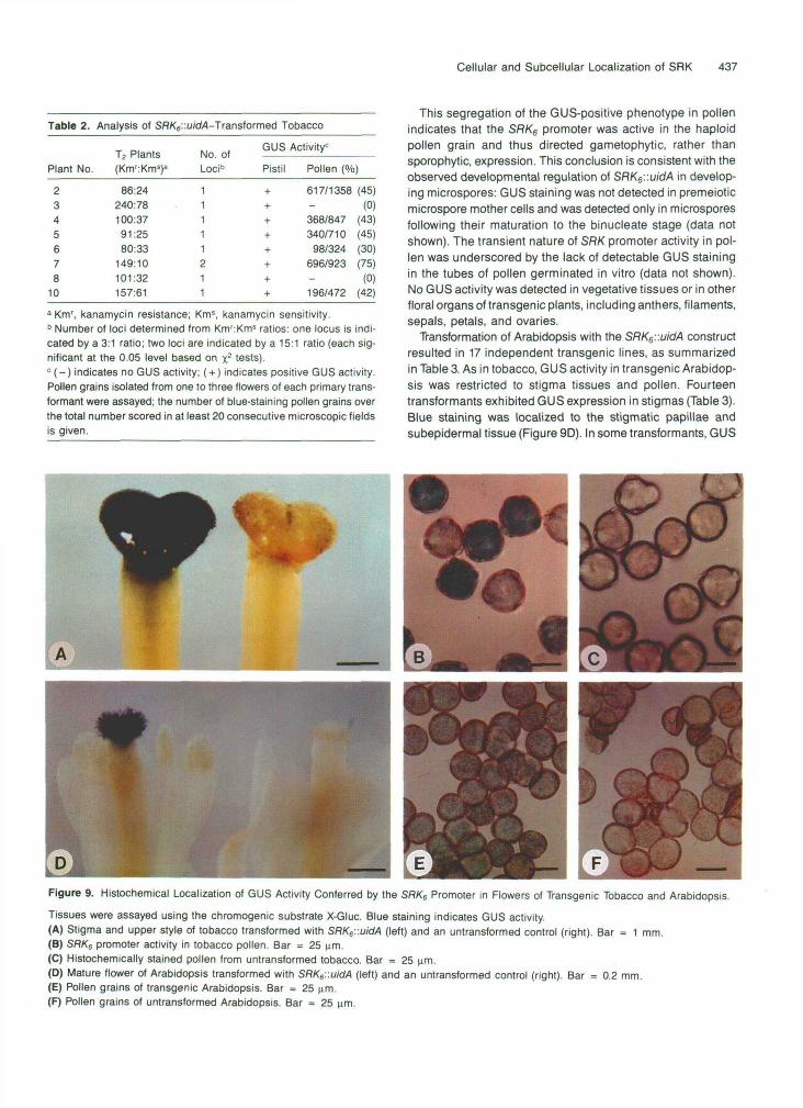
tobacco transformants expressed GUS activity in the stigma(Table 2). Blue staining was observed throughout the stigma,including the papillar cells and subepidermal secretory cells(Figure 9A). In six out of eight transgenic lines, GUS activitywas also detected in pollen (Figure 9B). In transgenic linescarrying a single integration of the transgene, the proportionof blue-staining pollen grains approached 50% (Table 2). Intransformant 7, which was predicted to carry T-DNA integra-tions at two loci, 75% of the pollen grains stained blue.
SLG
B. oleracea SQI I
stage: 0 - 1 - 2 - 3 - 4
SRK-
-200
-116- 97
actin
Figure 7. Temporal Regulation of SLG and SRK RNA in Maturing Bras-sica Stigmas.Poly(A)+ RNA (2 ng per lane) was isolated from developing stigmasof B. oleracea S6 homozygotes. Flower stages ranging from -5 (5days before flowering) to 0 (open flowers) are indicated above eachlane. The blot was sequentially hybridized with 32P-labeled probes thatdetect SRK (top panel; exposed 22 hr), SLG (middle panel; exposed5 hr), and actin (bottom panel; exposed 20 hr) transcripts.
SLG-66
B. campestris S8I I
stage: 0 - 1 - 2
SRK- -116
-97
452 bp of the SRK6 promoter (shown in Figure 6) fused to theE. coliuidA gene, which encodes p-glucuronidase (GUS). Thechimeric gene was introduced into tobacco and Arabidopsisby Agrobacterium-mediated transformation. These heterolo-gous hosts were chosen because they have previously servedas model systems for studying the SLG promoter in similarstudies (Thorsness et al., 1991;Toriyamaetal., 1991b; Dzelzkalnset al., 1993).
Using the SRK6::uidA construct, eight independent tobaccotransformants were obtained, and their characterization is sum-marized in Table 2. Histochemical GUS assays of controluntransformed plants and transgenic plants were performed,as described in Methods, on vegetative tissues and on floraltissues isolated from a variety of developmental stages. Asshown in Figure 9, no GUS staining was observed in any tis-sues of nontransformed control plants, including stigmas(Figure 9A) and pollen (Figure 9C). All eight of the SRK6::uidA
SLG-66
Figure 8. Temporal Regulation of SRK Protein in Maturing Stigmas.(A) Stigma microsomal membranes (20 ng per lane) were preparedfrom 8. oleracea S6 homozygotes. Flower stages ranging from -4 (4days before flowering) to 0 (open flowers) are indicated above each lane.(B) Stigma microsomal membranes (20 ng per lane) were preparedfrom 8. campestris S8 homozygotes. Flower stages ranging from -2(2 days before flowering) to 0 (open flowers) are indicated above eachlane.The blots were probed with MAb/H8 and developed with chemilumi-nescent substrates. Numbers at right indicate molecular mass markersin kilodaltons.
Cellular and Subcellular Localization of SRK 437
Table 2. Analysis of SflK6::u/d4-Transformed Tobacco
Plant No.
2345678
10
T2 Plants(Km':Kms)a
86:24240:78100:3791:2580:33
149:10101:32157:61
No. ofLoci"
11111211
GUS Activity0
Pistil Pollen (%)
+ 617/1358 (45)+ - (0)+ 368/847 (43)+ 340/710 (45)+ 98/324 (30)+ 696/923 (75)+ - (0)+ 196/472 (42)
a Km', kanamycin resistance; Km5, kanamycin sensitivity.b Number of loci determined from Km':Kms ratios: one locus is indi-cated by a 3:1 ratio; two loci are indicated by a 15:1 ratio (each sig-nificant at the 0.05 level based on x2 tests).c (-) indicates no GUS activity; ( + ) indicates positive GUS activity.Pollen grains isolated from one to three flowers of each primary trans-formant were assayed; the number of blue-staining pollen grains overthe total number scored in at least 20 consecutive microscopic fieldsis given.
This segregation of the GUS-positive phenotype in pollenindicates that the SRK6 promoter was active in the haploidpollen grain and thus directed gametophytic, rather thansporophytic, expression. This conclusion is consistent with theobserved developmental regulation of SRK6'.'.uidA in develop-ing microspores: GUS staining was not detected in premeioticmicrospore mother cells and was detected only in microsporesfollowing their maturation to the binucleate stage (data notshown). The transient nature of SRK promoter activity in pol-len was underscored by the lack of detectable GUS stainingin the tubes of pollen germinated in vitro (data not shown).No GUS activity was detected in vegetative tissues or in otherfloral organs of transgenic plants, including anthers, filaments,sepals, petals, and ovaries.
Transformation of Arabidopsis with the SRK6::uidA constructresulted in 17 independent transgenic lines, as summarizedin Table 3. As in tobacco, GUS activity in transgenic Arabidop-sis was restricted to stigma tissues and pollen. Fourteentransformants exhibited GUS expression in stigmas (Table 3).Blue staining was localized to the stigmatic papillae andsubepidermal tissue (Figure 9D). In some transformants, GUS
Figure 9. Histochemical Localization of GUS Activity Conferred by the SflK6 Promoter in Flowers of Transgenic Tobacco and Arabidopsis.
Tissues were assayed using the chromogenic substrate X-Gluc. Blue staining indicates GUS activity.(A) Stigma and upper style of tobacco transformed with SRKe::uidA (left) and an untransformed control (right). Bar = 1 mm.(B) SRKe promoter activity in tobacco pollen. Bar = 25 urn.(C) Histochemically stained pollen from untransformed tobacco. Bar = 25 urn.(D) Mature flower of Arabidopsis transformed with SRK6::uidA (left) and an untransformed control (right). Bar = 0.2 mm.(E) Pollen grains of transgenic Arabidopsis. Bar = 25 urn.(F) Pollen grains of untransformed Arabidopsis. Bar = 25 urn.

438 The Plant Cell
Table 3. Analysis of SflK6::u/cM-Transformed Arabidopsis
LineNo.
1235689
10111314151618192324
Km':Kmsa
18:523:1424:1519:939:027:198:22
159:119:0
104:2613:472:0
100:3685:1955:155:22:69
No. ofLoci"
1111
>2212211
>21111
ND
T-j GUS Activity0
Stigma Pollen (%)
ND+
ND++++ + (100%)+ + (100%)++-++ + (100%)++ + (100%)+ 51/179 (32%)+ ND
Generation
T2
T2
T2
T2T3,T4T3
T2
T3T2
T3
T3
T3
T3
T2
a Km', kanamycin resistance; Kms, kanamycin sensitivity.b Number of loci determined from Km':Km5 ratios: one locus is indi-cated by a 3:1 ratio; two loci are indicated by a 15:1 ratio (eachsignificant at the 0.05 level based on x2 tests). Ratios that are statisti-cally greater than 15:1 were assigned >2 loci. ND, number of locicould not be determined based on the data.c (-) indicates no GUS activity; (+) indicates GUS activity. Stigmaactivity was evaluated in the T2 generation and in subsequentgenerations as indicated. Pollen activity was evaluated only in thegenerations indicated. Pollen GUS staining was scored in at least10 consecutive microscopic fields. The number of blue-staining pollengrains over the total number scored is given only when <100% stainingwas observed. ND, not determined.
activity was detected also within the transmitting tissue of thestyle, with the intensity of blue staining decreasing toward theovary. In the maturing inflorescence, GUS activity in the stigmawas detected first in buds at 1 day before flower opening,reached a maximum in open flowers, and persisted until flowersenescence. Five of the transgenic Arabidopsis lines alsoexhibited GUS activity in pollen (Table 3 and Figure 9E).Segregation of the GUS-positive pollen phenotype was ob-served in transformant 23, which was tested in the T2generation, indicating gametophytic expression of the trans-gene (Table 3). Plants tested in the T3 and T4 generationswere true breeding for kanamycin resistance, and as expectedfor plants homozygous for the transgene, 100% of the pollengrains produced by these plants were positive for GUS activ-ity. No GUS activity was detected in the pistil (Figure 9D) orpollen (Figure 9F) of untransformed control plants.
SRK and SLG Proteins Are Absent in Pollen
In light of the above results showing SRK promoter activity inpollen and similar findings obtained with the SLG promoter
(Sato et al., 1991; Thorsness et al., 1991, 1993; Toriyama etal., 1991b; Dzelzkalns et al., 1993; Kandasamy et al., 1993),it was of interest to determine whether SRK and SLG proteinscould be detected in pollen by using the highly sensitive che-miluminescent detection system. We therefore subjected pollenextracts, as well as leaf extracts that were not expected to con-tain SRK and SLG, to immunoblot analysis with MAb/H8. Figure10 shows that, although both SRK and SLG were detected eas-ily in stigmas after a short exposure to film, no bands weredetected in either pollen or leaf extracts, despite the fact thatthe pollen and leaf lanes contained nearly eight times moretotal protein than the stigma lanes. Upon longer exposure tofilm, some bands were detected in the pollen lanes. However,these bands did not comigrate with SRK and SLG and appar-ently were due to nonspecific reactivity with the secondaryantibody used in the assay: they were observed in the absenceof primary antibody and were present in pollen from S2 as wellas S6 homozygotes. Microsomal membrane preparations andextraction with RIPA, SDS, and Triton X-100 similarly have failedto reveal SLG or SRK proteins in pollen (data not shown).
DISCUSSION
As a putative receptor protein kinase, SRK has a postulatedrole in both the recognition and signal transduction phasesof SI. Genetic evidence for this role includes linkage to theS locus and the identification of mutant alleles that are as-sociated with self-fertility; structural and biochemical evidenceinclude the high degree of allelic polymorphism in the SLG-like domain and the demonstrated catalytic activity of the ki-nase domain. The present study adds to this body of evidenceby showing that SRK is expressed in the papillar cell, the siteat which the SI reaction is manifested, and that SRK is an in-
&
S<3<o
%<b %<b CJQ
SRK
SLG
Figure 10. Distribution of SRK and SLG Proteins in Brassica Tissues.Protein extracts of stigma (11 ng), leaf (85 ng), and pollen (85 ng) fromthe indicated S genotypes were immunoblotted, probed with theMAb/H8 antibody, and developed with chemiluminescent substrates.

Cellular and Subcellular Localization of SRK 439
tegral plasma membrane glycoprotein, placing it at the cell surface, where it would be available for mediating cell-to-cell signaling events.
SRK6 1s a 108-kD Integral Plasma Membrane Glycoprotein
lmmunological and genetic criteria were used to identify SRK6 as a 108-kD protein in B. oleracea stigmas. The polypep- tide was recognized by two different antibodies, one that binds the N-terminal domain of SRK6 and a second that binds the C-terminal domain (Figure 2). Genetic evidence that the 108- kD stigma protein is encoded by SRK6 derives from the find- ing that, among F2 plants segregating for the s6 haplotype and the highly diverged class II S2 haplotype, this protein was detected only in plants carrying the SRK6 gene (Figure 2). Ad- ditionally, a protein of similar size was detected in self-incom- patible plants carrying other class I haplotypes, such as B. campesfris s8s8 plants (Figure 86), but not in S&, plants (Figure 3), which have a null allele of SRK but do express im- munoreactive SLG proteins. Corroborating this evidence, SRK6 sequences placed under the control of the CaMV 35s promoter direct the synthesis of a 108-kD protein in leaves of transgenic tobacco (Figure 5A).
The 108-kD SRK6 protein cofractionates with stigma mi- crosomal membranes, indicating its association with stigma membranes. Our procedure for isolating microsomes included a centrifugation step to remove cell debris and large organelles and a washing step to remove residual soluble proteins. Al- though the resulting cell fraction would include the plasma membrane, the bulk of membranes would be derived from the tonoplast, endoplasmic reticulum, and Golgi apparatus, thus preventing a precise determination of the subcellular location of SRK6. We therefore subjected SRK6-expressing tobacco leaves to aqueous two-phase partitioning, which yielded well- purified plasma membranes (Table 1 and Figure 56).
Localization of SRK6 to plasma membranes was indicated clearly by its enrichment in the plasma membrane fraction rel- ative to the fraction containing intracellular membranes (Figure 5C). In Brassica stigmas (Figure 4) and transgenic tobacco (Figure 5D), solubilization studies demonstrated that mem- brane-bound SRK6 exhibited properties of an integral membrane protein, in agreement with sequence data show- ing that SRK6 possesses a hydrophobic region located between the SLG-like and kinase domains that conforms to the requirements of a single-pass transmembrane domain (Stein et al., 1991).
Tobacco-expressed SRK6 also was shown to bind ConA, a lectin that recognizes the a-D-mannose moiety of glycopro- teins. The S R k sequence contains seven potential N-linked glycosylation sites within its SLG-like domain, four of which are conserved with at least four different alleles of SLG (Stein et al., 1991). Moreover, at least some of these sites are known to be glycosylated in SLG (Takayama et al., 1986, 1987). The observed glycosylation of SRK6 (see also Delorme et al.,
1995) provides experimental evidence that the SLG-like do- main was translocated into the lumen of the endoplasmic reticulum during its synthesis, as was originally proposed based on the presence of a putative signal sequence at the N termi- nus of the polypeptide (Stein et al., 1991). In light of our results demonstrating that SRK is embedded in the plasma mem- brane, the SLG-like domain would be exposed to the exterior face of the cell, whereas the kinase domain would be located cytoplasmically. However, this topology remains to be verified by direct experimental methods.
In contrast to SRK, SLG is a soluble glycoprotein that ac- cumulates in the papillar cell wall (Kandasamy et al., 1989). Accordingly, in fractionated stigma extracts, most of the SLG6 glycoprotein was present in the soluble fraction, but we also identified a membrane-associated pool of SLG6 (Figure 4). Al- though at least one SLG allele has been shown to produce an alternative transcript that encodes a membrane-bound iso- form (Tantikanjana et al., 1993), the SLG6 allele is not predicted to encode such a form. Therefore, we hypothesize that the microsomal membrane pool of SLG6 represents pro- tein in transit through the secretory pathway. In agreement with this hypothesis, we found that the microsome-associated SLG6 was solubilized in the presence of sodium carbonate (Figure 4), consistent with a location of this SLG pool within the lumen of membrane vesicles or a peripheral interaction with the membrane. Additional support for this hypothesis can be found in immunocytochemical localization studies of SLG in the papillar cell (Kandasamy et al., 1989). In these studies, a fraction of antigens recognized by MAblH8 and likely to be SLG because of its high abundance relative to SRKiyas local- ized to the rough endoplasmic reticulum and Golgi apparatus.
Coordinate Regulation of SRK and SLG in the Stigma
The conservation of cis-acting regulatory elements in SLG and SRK suggests that these genes (Figure 6) are subject to com- mon transcriptional control mechanisms that, in effect, would coordinately regulate their expression. In support of this hy- pothesis, we showed that SRK and SLG have similar spatial and temporal patterns of expression by analyzing levels of RNA and protein and by monitoring SRK6 promoter activity in trans- genic plants. In a previous study, we had estimated that in the stigma, SLG transcripts are 140 to 180 times more abundant than SRK transcripts (Stein et al., 1991), and an abundance of SLG far in excess of SRK is obvious in the immunoblots displayed in Figures 4 and 10. Yet, despite these quantitative differences, the steady state transcript levels of the two genes increased coordinately during maturation and reached a max- imum at 1 day before flower opening, a timing that coincides with the ability of stigmas to distinguish between self- and non-self-pollen (Figure 7). The levels of SLG and SRK protein followed a similar time course. However, whereas maximal SLG levels are attained in open flowers (Nasrallah et al., 1985a), the leve1 of SRK was observed to decline at this stage in at least one genotype (Figure 8). This decline, which may be

440 The Plant Cell
genotype specific, is not understood but may reflect differences in the relative strengths of the SI response in different genotypes.
The SRK and SLG genes overlap in their expression pat- terns not only temporally but also spatially. The reporter gene studies described in this article demonstrate conclusively that the sRK6 promoter is active primarily in the papillar cells of the stigma, as shown previously for the SLG13 promoter in transgenic tobacco (Thorsness et al., 1991; Dzelzkalns et al., 1993), Arabidopsis (Toriyama et al., 1991b), and Brassica (Sato et al., 1991). In addition to this primary site of activity, the sRK6 promoter also was active in the subepidermal cellsof thestigma in Arabidopsis and tobacco and the transmitting tissue of the style in Arabidopsis but not in tobacco. In this respect, the SRK6 promoter differs from the SLG13 promoter, which was found to be active in the subepidermal cells and transmitting tissue of tobacco but not Arabidopsis. However, the activity of the SRK, promoter in subepidermal cells and transmitting tissue of the Arabidopsis pistil does coincide with a similar activity observed for the SLG13 promoter in B. oleracea (Sato et al., 1991).
Because different transgenic hosts impose somewhat differ- ent patterns of activity on the sRK6 and SLG13 promoters at secondary sites of activity, it is not clear to what extent pro- moter activity at these sites reflects promoter activity in the native plant. However, no such variability has been observed in the primary site of activity for either the SRK or SLG pro- moter. Indeed, reporter studies have been predictive of the primary site of expression of the SLG gene in Brassica papil- lar cells, as determined by in situ hybridization (Nasrallah et al., 1988) and immunolocalization studies (Kandasamy et al., 1989). We therefom conclude that SLG and SRKare transcribed simultaneously in the papillar cells of the stigma.
SLG and SRK Expression in Anthers and Pollen
Because genetic models of SI based on singla-locus control historically have predicted the expression of a common S gene in both male and female tissues, much attention has focused on demonstrating the expression of SLG and SRK in anthers and pollen. In Brassica, expression is expected to occur ei- ther in the sporophytic tissue of the anther tapetum or in premeiotic microspores, because the recognition phenotype of pollen is determined by the diploid genome of the parent plant. Conventional RNA blot hybridization techniques have revealed the presence of SLG and SRK transcripts in micro- spores andlor anthers (Sato et al., 1991; Stein et al., 1991). However, these transcripts are rare, and several studies have resorted to amplification by the polymerase chain reaction (PCR) to detect expression in anthers and pollen (Guilluy et al., 1991; Goring and Rothstein, 1992; Delorme et al., 1995).
Consistent with the presence of SRKtranscripts in male tis- sues, we found that the sRK6 promoter directed expression of the GUS reporter in pollen grains of tobacco and Arabidop- sis (Figure 9). Similarly, the SLG13 promoter had been shown
to direct expression of the GUS reporter as well as the diph- theria subunit A toxin in cells of the anther and/or developing pollen grains in transgenic tobacco (Thorsness et al., 1991), Arabidopsis (Toriyama et al., 1991b; Thorsness et al., 1993), and Brassica(Sat0 et al., 1991; Kandasamyet al., 1993). How- ever, we found no evidence of SRK6 or SLG6 proteins in pollen or anther extracts even when we used several different extrac- tion protocols and the highly sensitive chemiluminescent immunodetection method (Figure 10). It thus appears that, although the SRK6 and SLG6 promoters are active transcrip- tionally in cells of the anther, additional regulatory mechanisms that affect either transcript stability, protein translation, or pro- tein stability must function to prevent the accumulation of the SRK6 or SLG6 proteins to detectable levels in these cells.
lmplications for the Mechanism of SI in Brassica and Receptor-Mediated Signaling in Plants
The lack of SLG and SRK proteins in male reproductive tis- sues refutes models of SI based on the homophilic interaction of S locus products and the notion of control by a single S gene. However, control of SI by a single genetic locus is still valid, given the potential for additional recognition genes within the physical and genetic limits of the S locus complex (Boyes and Nasrallah, 1993). In particular, we anticipate the presence of a pollen-expressed determinant of recognition that would act as a haplotype-specific ligand of SRK and SLG. A recently de- scribed gene, the S locus anther (SLA) gene, possesses several features expected of a recognition determinant, including link- age to the S locus, haplotype-specific polymorphism, and anther-specific expression (Boyes and Nasrallah, 1995). It re- mains to be determined whether the polypeptides of 7.5 and 10 kD potentially encoded by SLA transcripts are expressed on the pollen surface and are capable of interacting with SRK and SLG.
The simultaneous expression of SRK and SLG in the papil- lar cells of the stigma supports the hypothesis that they aCt together as determinants of the SI response in the stigma, per- haps by binding to a common ligand via their highly related S domains. Models of SI based on competition between SRK and SLG for ligand binding are inconsistent with genetic evi- dente that expression of both proteins is required for pollen recognition. We propose therefore that SLG functions cooper- atively with SRK. The presence of a thick cell wall in plants may represent an obstacle for plasma membrane-based sig- naling. A soluble molecule like SLG theoretically could solve this problem by shuttling ligand from the outer cell wall and presenting it to the plasma membrane-localized SRK. Such a mechanism may be based on a higher affinity of the ligand for SRK than for SLG. These possibilities remain open questions.
Also to be resolved are exactly how cooperativity in SLG and SRK function is achieved and whether there is a direct inter- action between these two proteins. The different levels of SLG and SRK that accumulate in papillar cells might imply that the

Cellular and Subcellular Localization of SRK 441
mechanism of action of SLG and SRK as female determinants of SI requires a high local molar excess of SLG relative to SRK rather than stoichiometric amounts of the two proteins. How- ever, it is important to note that SLG is distributed throughout the volume of the cell wall, whereas SRK is restricted to the area of the plasma membrane. Based on a value of 615 nm for the thickness of the papillar cell wall (Kandasamy et al., 1989), we calculated that an excess of SLG over SRK of two orders of magnitude would be required to give equal local molar concentrations of protein.
SRK belongs to a family of related genes that includes in maize the Zea mays protein kinase gene ZmPKl (Walker and Zhang, 1990) and in Arabidopsis the Arabidopsis receptor ki- nase genesARK7,ARK2, andARK3(Tobias et al., 1992; Dwyer et al., 1994) and the receptor-like kinase genes RLK7 and RLK4 (Walker, 1993). These genes are expressed in a variety of vegetative tissues, but they all share structural features in com- mon with SRK. Based on our results, which provide conclusive experimental evidence that the product of a member of this family is an integral plasma membrane protein, it may be inferred that proteins similar to SRK exist in the plasma mem- branes of vegetative plant cells, where they presumably fulfill cell surface signaling functions unrelated to pollination. SRK would have been recruited from such proteins for the special- ized function of cell-cell signaling and the recognition of self during reproduction.
METHODS
Plant Material
Brassica oleracea inbred lines bearing s& S2, and St, haplotypes and the B. campestris SB inbred line have been described previously (Nasrallah et al., 1987, 1994a; Chen and Nasrallah, 1990; Toriyama et al., 1991a). Homozygous parenta1 lines were crossed, and the result- ing F, hybrids were selfed by bud pollination to generate F2 progeny. S locus genotypes within segregating populations were determined by pollination to testei lines. Genotype assignments also were made on the basis of restriction fragment length polymorphism analysis using allele-specific S locus receptor kinase (SRK) and S locus glycopro- tein (SLG) probes (Boyes and Nasrallah, 1993) and on the basis of allele-specific charge polymorphisms displayed by the SLG protein when subjected to isoelectric focusing and immunoblotting (Nasrallah and Nasrallah, 1984).
DNA Cloning, Sequence Analysis, and RNA Gel Blot Hybridization
lsolation of genomic clones for SRKs and SRK2 was described by Stein et al. (1991). Dideoxy sequencing was accomplished using the Sequenase Kit (United States Biochemical). Sequence alignment was performed with the GCG version 6.0 software package (University of Wisconsin Biotechnology Center, Madison).
lsolation of poly(A)+ RNA and gel blot hybridization with 3*P-labeled probes was performed as described by Stein et al. (1991).
Construction ot a uidA Reporter Gene Fusion, Transformation of Arabidopsis and Tobacco, and Histochemical Analysis of p-Glucuronidase Activity
To construct an SRKs promoter::uidA chimeric gene, a 452-bp frag- ment upstream of the protein coding region was amplified by the polymerase chain reaction (PCR) and cloned into vector pCR1000 (In- vitrogen, San Diego, CA). The integrity of the amplified sequence was confirmed by dideoxy sequencing and subsequently inserted into the theTi binaryvector pBllOl (Jefferson et al., 1987) upstream of the uidA coding region. The resulting construct was introduced into Agrobac- terium tumefaciens pCIB524/A136 (derived from helper plasmid pEJAlO1; Hood e! al., 1986).
Nicotiana tabacum cv Petit Havana was transformed by the method of Horsch et al. (1988). Arabidopsis thaliana strain C24 was transformed by the method of Valvekens et al. (1988). Kanamycin-resistant tobacco and Arabidopsis plants resulting from independent transformation events were transferred to soil and grown to maturity in a greenhouse. DNA gel blot analysis confirmed the presence of the transgene in all primary regenerants. Subsequent generations derived by selfing were germinated from seed on Murashige and Skoog medium (Murashige and Skoog, 1962) containing either 300 pg/mL kanamycin (tobacco) or 25 pg/mL kanamycin (Arabidopsis) and grown in a 25OC growth cham- ber with a 16-hr lightl8-hr dark cycle. To asses antibiotic resistance, the number of green kanamycin-resistant seedlings and bleached kanamycin-sensitive seedlings were scored 2 to 4 weeks after sowing.
Histochemical localization of P-glucuronidase (GUS) activity in vegeta- tive and floral organs was performed by using 5-bromo-4-chloro-3- indolyl-P-D-glucuronide (X-Gluc), as described previously (Thorsness et al., 1991; Toriyama et al., 1991b). Pollen and isolated microspores were assayed with 2 mM X-Gluc, 0.1 M NaP04, pH 7.0, O.lO/o (vh) Triton X-100, without vacuum infiltration, and were observed without ethanol destaining. Constant mixing of the pollen was required during incu- bation with X-Gluc to avoid false-positive staining. The nuclear stages of the grains were assessed by staining with 4: 6-diamidino-2-phenyl- indole and fluorescence microscopy, as detailed by Thorsness et al. (1991). Pollen was isolated from Arabidopsis by rinsing whole mature flowers with buffer and pipetting off the suspended pollen grains. To avoid possible contaminating GUS activity from stigmas, the pollen was washed four times with 1 mL of buffer. As an additional control, the pollen from nontransformed plants was assayed in buffer that was used previously in the first rime of transgenic flowers.
Expression of SRK in Escherichia coli
A 1.2-kb fragment consisting of the SLG homologous domain of SRK, exclusive of the signal peptide was amplified by PCR, ligated into the pCRlOOO vector, and subsequently inserted into the bacterial expres- sion vector pGEX-3X (Pharmacia, Piscataway, NJ). The resulting plasmid encoded a 73-kD glutathione S-transferase (GST) fusion pro- tein, which could be affinity purified from JM109 cells using glutathione-agarose beads (Smith and Johnson, 1988). Purified fu- sion proteins were visualized following transfer to lmmobilon PVDF membranes (Millipore, Bedford, MA) by staining with Coomassie Brilliant Blue R 250 or by probing with MAblH8.
The SLG homologous domain of SRK, also was cloned into a modi- fied pGEX vector containing a FLAG epitope at the 3’end of the GST coding region. A similar construct was generated by using the SLG homologous domain of SRK2. The resulting fusion proteins were transferred to PVDF membranes and probed with the commercially

442 The Plant Cell
available monoclonal M2 anti-FLAG antibody (International Biotech- nologies, New Haven, CT) or with MAb/H8.
Because immunostaining repeatedly revealed the presence of pro- teolytic derivatives of the full-length fusion proteins for all three GST-SRK constructs, we prepared bacterial extracts in the presence of a spectrum of protease inhibitors. This treatment did not prevent degradation of the fusion protein, indicating that the proteolytic prod- ucts occurred not as a consequence of cell disruption and protein extraction but rather as a result of in vivo instability, a commonly ob- served feature of recombinant proteins produced in E. coli.
Construction of an S R K s Plant Expression Vector and Selection of Transgenic Tobacco Lines Expressing SRK Protein
To construct a cauliflower mosaic virus (CaMV) 355 prOmOter::SRK6 chimeric gene, we used the pCT37 vector (Tobias, 1995), a Ti binary vector derived from pBIN19 (Bevan, 1984) and containing a duplicated CaMV 35s promoter (Kay et al., 1987) and a nos terminator between the T-DNA borders. Because only partia1 SRK6 cDNA clones were available (Stein et al., 1991), we had to reconstitute the full-length coding sequence. This was accomplished in a series of cloning and PCR steps involving the ligation of sequences from different cDNA clones and extension of the cDNA sequence to include the ATG initiation codon. The SRK6 genomic clone was used as template for PCR. After verify- ing the fidelity of PCRs by sequence analysis, the reconstituted SRK6 full-length clone, which included three nucleotides upstream of the SRK6 start codon, was inserted between the CaMV 35s promoter and the nos terminator within pCT37.
Leaves of greenhouse-grown primary transformants were screened for SRK transcript 3 weeks after transfer to soil by using the rapid screen- ing method described by Verwoerd et al. (1989). Those individuals expressing relatively high levels of SRK, RNA were screened further for the expression of SRK6 protein in primary transformants and in their kanamycin-resistant T2 progeny. Leaf segments were ground to a powder in liquid nitrogen, extracted with hot SDS-PAGE sample buffer (2% Iwhr] SDS, 100 mM DTT, 80 mM Tris-HCI, pH 6.8, 10% [vlv] glycerol, 5 mg/mL bromphenol blue), and analyzed by immunoblotting. Because highest SR& protein levels were observed in leaves of 1- to 2-month- old, growth chamber-grown seedlings (Murshige and Skoog medium; 50 pglmL kanamycin), all subsequent experiments used plant mate- rial grown in this manner.
Preparation of Tobacco Membrane Fractions
Unless otherwise specified, all steps were carried out at 4OC and all buffers contained the following protease inhibitors: phenylmethylsul- fonyl fluoride (1 mM), aprotinin (10 pglml), leupeptin (10 pglml), and pepstatin A (1 pglmL). Tobacco leaves (15 g fresh weight) were homogenized with a polytron in buffer (2 mLlg fresh weight) contain- ing 30 mM Tris-HCI, pH 8.0, 150 mM NaCI, 1 mM EDTA, 1 mM DTT, 5 mM ascorbate, 20% (v/v) glycerol. The homogenate was filtered through one layer of Miracloth (Calbiochem, LaJolla, CA), and the result- ing filtrate was centrifuged at IO,OOOg(9OOO rpm, SM 24 rotor; Beckman, Fullerton, CA) for 15 min. The supernatant was centrifuged at 100,OOOg (33,200 rpm, 50.2 Ti rotor; Beckman) for 1 hr. Soluble protein retained in the supernatant was stored in liquid nitrogen.
The microsomal membrane pellet was fractionated by the aqueous two-phase partitioning method described by Larsson (1985). Phase
separations were carried out in a series of 5-9 phase systems that con- tained 6.2% (wlw) dextran T500 and 6.2% (wlw) polyethylene glycol 3350 (omitting phenylmethylsulfonyl fluoride). Three successive rounds of partitioning yielded a colorless upper phase enriched in plasma membranes and a green lower phase containing intracellular mem- branes. After washing, pelleting, and resuspending in 250 mM sucrose, 5 mM Bis-Tris-propane (BTP)/Mes, pH 7.2, the partitioned membranes were assayed for marker enzyme activity and stored in liquid nitrogen.
Marker Enzyme Assays
Vanadate-sensitive ATPase was assayed by a protocol modified from Hodges and Leonard (1972). Membranes were added to a reaction mix- ture containing 3 mM ATP-2Na, 3 mM MgS04, 50 mM KCI, 25 mM Hepes, 0.03% (v/v) Triton X-100 (pH adjusted to 6.5 with BTP), with or without 100 pM sodium orthovanadate. The reaction proceeded at 30% for 15 min and was stopped by the addition of 0.25% (wlv) ammonium molybdate, 1.3% (wlv) SDS, and 1.4% (v/v) sulfuric acid. Activity was represented by the difference in amount of phosphate released (Fiske and Subbarow, 1952) in the presence and absence of vanadate.
Triton-stimulated UDPase was assayed by a protocol modified from Nagahashi and Kane (1982). Membranes were added to a reaction mixture containing 3 mM UDP-Na, 3 mM MnS04, 50 mM KCI, 25 mM Hepes (pH adjusted to 7.4 with BTP), with or without 0.03% (vlv) Triton X-100. Reactions proceeded as described above for 10 min. Activity was represented by the difference in activity observed in the presence and absence of Triton X-100.
Spectrophotometric assays'for NADH cytochrome c reductase and cytochrome c oxidase were performed essentially as described by Yoshida (1979)%
Preparation of Brassica Stigma Microsomes
All steps were performed at 4OC or less, andall buffers contained the protease inhibitors listed above. For routine preparation of microsomes, 30 stigmas ( ~ 6 mg fresh weight) were ground with a Teflon (Kontes, Vineland, NJ) pestle in a homogenization buffer (2.5 pL per stigma) containing 30 mM Tris-HCI, pH 7.5, 150 mM NaCI, 10 mM EDTA, 10% (vlv) glycerol. For most experiments, it was necessary to include DTT (5 mM), potassium metabisulfite (2.5 mM), ascorbate (5 mM), and poly- vinylpyrrolidone (3 to 5% [wlw]) to prevent the artifactual formation of high molecular mass SLG protein. Centrifugation was carried out at 40009 for 5 min (7000 rpm; Eppendorf centrifuge), and the recov- ered supernatant was recentrifuged to remove welb debrisand large organelles. The supernatant was fractionated into soluble and microsomal membrane fractions by centrifugation at 100,OOOg (49,000 rpm, TLA 100.4 rotor; Beckman) for 1 hr. After washing with homogeni- zation buffer, the pelleted membranes were solubilized in RIPA buffer (50 mM TriS-HCI, pH 8.0,150 mM NaCI, 1% [whr] Nonidet P-40 (Pierce), 0.5% [w/v] deoxycholate, 0.1% [w/v] SDS) for 30 min and then cen- trifuged at 1~0,IlOOg k r 1' hr. The supernatant, containing solubilized membrane profeins, was stored at -8OOC.
Membrane Protein Solubility Studies and Concanavalin A Binding
Tobacco microsomes were divided into three equal pellets. One pellet was extracted directly with SDS-PAGE sample buffer to give a total

Cellular and Subcellular Localization of SRK 443
microsome fraction. The second pellet was resuspended with 100 mM sodium carbonate, pH 11.5, and incubated on ice for 30 min (Fujiki et al., 1982). The third pellet was resuspended with RIPA buffer and incubated on ice for 30 min. Tubes were centrifuged at 150,OOOg at 4OC (60,000 rpm, TLA 100.4 rotor; Beckman) for 1 hr. Solubilized pro- teins were recovered in the supernatant, and the insoluble pellets were dissolved in SDS-PAGE sample buffer. Equal volume propoations of ‘each fraction were analyzed by immunoblotting. Solubilitystudies with Brassica stigma microsomes were carried out as above;except that 50 mM Tris-HCI, pH 8.0,150 mM NaCI, 1.5% (vlv) Triton X-100 was used instead of RIPA buffer.
RIPA-solubilized tobacco membranes were incubated wlth con- canavalin A (ConA)-Sepharose beads (Sigma) at 4OC:for 915 hr. Beads were washed fourtimes with 1 mL of RIPA. Bound,material\was,rleased by boiling with SDSPAGE sample buffer and,analyzed byimmunoblotting.
Other Protein Extraction Procedures
For the experiment depicted in Figure 10, stigmas and leaves were ground in 30 mM Tris-HCI, pH 7.5, 75 mM NaCI, 10 mM EDTA, 10% (vlv) glycerol, and protease inhibitors. Pollen was ground in 20 mM Tris-HCI, pH 8.0, 1% (vlv) P-mercaptoethanol, 10 mM EDTA, 2.5 mM potassium metabisulfite, and protease inhibitors. Homogenates were centrifuged at 40009 for 5 min (7000 rpm; Eppendorf centrifuge). The supernatants were boiled with SDS-PAGE sample buffer and analyzed by immunoblotting.
Protein lmmunoblots and Antibodies
Proteins were quantified with the Bio-Rad (Hercules, CA) protein as- say dye reagent (Bradford, 1976) with BSA as a standard; for protein solutions containing detergent, a modified Lowry assay was employed (Bensadoun and Weinstein, 1976). Proteins were resolved by electropho- resis on 7% SDS-polyacrylamide gels (unless otherwise specified) and electroblotted onto PVDF membranes. Membrane blocking for 30 min and antibody binding for I hr were carried out in TBST (10 mM Tris-HCI, pH 7.5, 150 mM NaCI, 0.05% [vlv] Tween 20) containing 5% (wlv) nonfat dry milk at room temperature. After primary and second- ary antibody treatments, membranes were washed three times for 10 min each with TBST. As specified in the text, blots were either probed with alkaline phosphatase-conjugated second antibodies and devel- oped with chromogenic substrates 5-bromo-4-chloro-3-indoyl phosphate and nitro blue tetrazolium (Bio-Rad) or probed with horseradish per- oxidase-conjugated secondary antibody and developed with the BM Chemiluminescence Western Blotting Kit (Boehringer Mannheim, In- dianapolis, IN). Blots were stripped and reprobed using the method described by Kain et al. (1994).
MAb/H8, raised against SLG (Kandasamy et al., 1989), was used ata 1:50 dilution. MAb 2E7, raised against the 60-kD subunit of vacuolar H+-ATPase (Ward et al., 1992), was used at a 1:200 dilution. Rabbit antiserum raised against the plasma membrane H+-ATPase (Harper et al., 1990) was used at a 1:10,000 dilution. The use of rabbit antise- rum RI-254, raised against residues 790 to 803 within the C-terminal region of SR&, was described by Stein and Nasrallah (1993). R1-254 was affinity purified over a column containing the synthetic peptide antigen covalently coupled to an agarose matrix. The column was pre- pared by using SulfoLink Coupling Gel (Pierce) and resulted in 3.3 mg of peptide immobilized on 2 mL of gel. The column was loaded with antiserum and washed with PBS, pH 7.4. Fractions were eluted
with 0.2 M glycine, pH 2.8,0.5 M NaCI, and neutralized with 1.0 M Tris- HCI, pH 9.0. Fractions containing protein were pooled and concen- trated with a Centricon 10 (Amicon) device. The affinity-purified antibody was used at a concentration of 5 pg/mL.
ACKNOWLEDGMENTS
We thank Mike Sussman for the antibodies to plasma membrane H+- ATPase, Heven Sze for the antibodies to vacuolar H+-ATPase, and Michael Blanar for the pGEX-FLAG expression vector. We also thank Debbie Letham for generating the pGEX-FLAG-SRK6 and SRK2 fu- sions, Warren Huang for help in preparing RNAfrom transgenic plants, and Matsuo Uemura for advice relating to protocols for plasma mem- brane preparation. This work was supported by Grant No. DE-FG02-88ER13909 from the U.S. Department of Energy and by Grant No. IBN-9220401 from the National Science Foundation.
Received October 23, 1995; accepted January 12, 1996.
REFERENCES
Bensadoun, A., and Weinstein, D. (1976). Assay of proteins in the presence of interfering materials. Anal. Biochem. 70, 241-250.
Bevan, M.W. (1984). Binary Agrobacterium vectors for plant transfor- mation. Nucleic Acids Res. 12, 8711-8721.
Boyes, D.C., and Nasrallah, J.B. (1993). Physical linkage of the SLG and SRK genes at the self-incompatibility locus of Brassica oler- acea. MOI. Gen. Genet. 236, 369-373.
Boyes, D.C., and Nasrallah, J.B. (1995). An anther-specific gene en- coded by an S locus haplotype of Brassica produces complementary and differentially regulated transcripts. Plant Cell 7, 1283-1294.
Bradford, M.M. (1976). A rapid and sensitive method for the quantifi- cation of microgram quantities of protein utilizing the principle of protein-dye binding. Anal. Biochem. 72, 248-254.
Chen, C.-H., and Nasrallah, J.B. (1990). A new class of S sequences defined by a pollen recessive self-incompatibility allele of Brassica oleracea. MOI. Gen. Genet. 222, 241-248.
Delorme, V., Giranton, J.-L., Hatzfeld, Y., Friry, A,, Heizmann, P., Ariza, M.J., Dumas, C., Gaude, T., and Cock, J.M. (1995). Char- acterization of the S locus genes, SLG and SRK, of the Brassica S3 haplotype: ldentification of a membrane-localized protein en- coded by the S locus receptor kinase gene. Plant J. 7, 429-440.
Dwyer, K.G., Kandasamy, M.K., Mahosky, D.I., Acciai, J., Kudish, B.I., Miller, J.E., Nasrallah, M.E., and Nasrallah, J.B. (1994). A superfamily of S locus-related sequences in Arabidopsis: Diverse structures and expression patterns. Plant Cell 6, 1829-1843.
Dzelzkalns, V.A., Thorsness, M.K., Dwyer, K.G., Baxter, J.S., Balent, M.A., Nasrallah, M.E., and Nasrallah, J.B. (1993). Distinct cis-acting elements direct pistilspecific and pollen-specific activity of the Bras- sica S locus glycoprotein gene promoter. Plant Cell 5, 855-863.
Fiske, C.H., and Subbarow, Y. (1952). The colorimetric determina- tion of phosphorus. J. Biol. Chem. 66, 375-400.

444 The Plant Cell
Frommer, W.B., and Ninnemann, O. (1995). Heterologous expres- sion of genes in bacterial, fungal, animal, and plant cells. Annu. Rev. Plant Physiol. Plant MOI. Biol. 46, 419-444.
Fujiki, Y., Hubbard, A.L., Fowler, S., and Lazarow, P.B. (1982). Iso- lation of intracellular membranes by means of sodium carbonate treatment: Application to endoplasmic reticulum. J. Cell Biol. 93,
Goring, D.R., and Rothstein, S.J. (1992). The S-locus receptor ki- nase gene in a self-incompatible Brassica napus line encodes a functional serinelthreonine kinase. Plant Cell 4, 1273-1281.
Goring, D.R., Glavin, T.L., Schafer, U., and Rothstein, S.J. (1993). An S receptor kinase gene in self-compatible Brassica napus has a 1-bp deletion. Plant Cell 5, 531-539.
Guilluy, C.-M., Trick, M., Heizmann, P., and Dumas, C. (1991). PCR detection of transcripts homologous to the self-incompatibility gene in anthers of Brassica. Theor. Appl. Genet. 82, 466-472.
Harper, J.F., Manney, L., DeWitt, N.D., Yoo, M.H., and Sussman, M.R. (1990). The Arabidopsis thaliana plasma membrane H+- ATPase multigene family: Genomic sequence and expression of a third isoform. J. Biol. Chem. 265, 13601-13608.
Hodges, T.K., and Leonard, R.T. (1972). Purification of a plasma mem- brane-bound adenosine triphosphatase from plant roots. Methods Enzymol. 32, 392-406.
Hood, E.E., Helmer, G.L., Fraley, R.T., and Chilton, M.-D. (1986). The hypervirulence of Agrobacterium tumefaciens A281 is encoded in a region of pTiBo542 outside of T-DNA. J. Bacteriol. 168,1291-1301.
Horsch, R.B., Fry, J., Hoffmann, N., Neidermeyer, J., Rogers, S.G., and Fraley, R.T. (1988). Leaf disc transformation. In Plant Molecu- lar Biology Manual, S.B. Gelvin and R.A. Schilperoort, eds (Dordrecht, The Netherlands: Kluwer Academic Publishers), pp.
Jefferson, R.A., Kavanagh, T.A., and Bevan, M.W. (1987). GUS fu- sions: R-Glucuronidase as a sensitive and versatile gene fusion marker in higher plants. EMBO J. 6, 3901-39m.
Kain, S.R., Mai, K., and Sinai, P. (1994). Human multiple tissue western blots: A new immunological tool for the analysis of tissue-specific protein expression. BioTechniques 17, 982-987.
Kandasamy, M.K., Paolillo, D. J., Faraday, C.D., Nasrallah, J.B., and Nasrallah, M.E. (1989). The S-locus specific glycoproteins of Bras- sica accumulate in the cell wall of developing stigma papillae. Dev. Biol. 134, 462-472.
Kandasamy, M.K., Dwyer, K.G., Paolillo, D.J., Doney, R.C., Nasrallah, J.B., and Nasrallah, ME. (1990). Brassica S-proteins accumulate in the intercellular matrix along the path of pollen tubes in trans- genic tobacco pistils. Plant Cell 2, 39-49.
Kandasmy, M.K., Thorsness, M.K., Rundle, S.J., Goldberg, M.L., Nasrallah, J.B., and Nasrallah, M.E. (1993). Ablation of papillar cell function in Brassica flowers results in the loss of stigma recep- tivity to pollination. Plant Cell 5, 263-275.
Kay, R., Chan, A., Daly, M., and McPherson, J. (1987). Duplication of CaMV 35s promoter sequences creates a strong enhancer for plant genes. Science 236, 1299-1302.
Lanson, C.H. (1985). Plasma membranes. In Modern Methods in Plant Analysis, New Series, Volume 1, Cell Components, H.F. Linskens and J.F. Jackson, eds (Berlin: Springer-Verlag), pp. 85-104.
Moore, H., and Nasrallah, J.B. (1990). A Brassica self-incompatibility gene is expressed in the stylar transmitting tissue of transgenic tobacco. Plant Cell 2, 29-38.
97-1 02.
A5: 1-9.
Murashige, T., and Skoog, F. (1962). A revised medium for rapid growth and bioassays with tobacco tissue cultures. Physiol. Plant. 15, 473-497.
Nagahashi, J., and Kane, A.P. (1982). Triton-stimulated nucleoside diphosphatase activity: Subcellular localization in corn root homogen- ates. Protoplasma 112, 167-173.
Nasrallah, J.B., and Nasrallah, M.E. (1984). Electrophoretic hetero- geneity exhibited by the S-allele specific glycoproteins of Brassica. Experimentia 40, 279-281.
Nasrallah, J.B., Doney, R.C., and Nasrallah, M.E. (1985a). Biosyn- thesis of glycoproteins involved in the pollen-stigma interaction of incompatibility in developing flowers of Brassica oleracea L. Planta 165, 100-107.
Nasrallah, J.B., Kao, T.-H., Goldberg, M.L., and Nasrallah, M.E. (1985b). A cDNA clone encoding an S-locus-specific glycoprotein from Brassica oleracea. Nature 318, 263-267.
Nasrallah, J.B., Kao, T.-H., Chen, C.-H., Goldberg, M.L., and Nasrallah, M.E. (1987). Amino-acid sequence of glycoproteins en- coded by three alleles of the S-locus of Brassica oleracea. Nature 326, 617-619.
Nasrallah, J.B., Yu, S.-M., and Nasrallah, M.E. (1988). Self-incompat- ibility genes of Brassica olearacea: Expression, isolation, and structure. Proc. Natl. Acad. Sci. USA 85, 5551-5555.
Nasrallah, J.B., Nishio, T., and Nasrallah, M.E. (1991). The self- incompatibility genes of Brassica: Expression and use in genetic ablation of floral tissues. Annu. Rev. Plant Physiol. Plant MOI. Biol.
Nasrallah, J.B., Rundle, S.B., and Nasrallah, M.E. (1994a). Genetic evidence for the requirement of the Brassica S-locus receptor ki- nase gene in the self-incompatibility response. Plant J. 5,373-384.
Nasrallah, J.B., Stein, J.C., Kandasamy, M.K., and Nasrallah, M.E. (1994b). Signaling the arrest of pollen tube development in self- incompatible plants. Science 266, 1505-1508.
Nasrallah, M E , Kandasamy, M.K., and Nasrallah, J.B. (1992). A genetically defined trans-acting locus regulates S-locus function in Brassica. Plant J. 2, 497-506.
Penefsky, H.S., and Tzagoloff, A. (1971). Extraction of water-soluble enzymes and proteins from membranes. Methods Enzymol. 22, 204-219.
Perl-Treves, R., Howlett, B., and Nasrallah, M.E. (1993). Self- incompatibility related glycoproteins of Brassica are produced and secreted by transgenic tobacco cell cultures. Plant Sci. 92,99-110.
Sato, T., Thorsness, M.K., Kandasamy, M.K., Nishio, T., Hirai, M., Nasrallah, J.B., and Nasrallah, M.E. (1991). Activity of an S locus gene promoter in pistils and anthers of transgenic Brassica. Plant Cell 3, 867-876.
Smith, D.B., and Johnson, K.S. (1988). Single-step purification of polypeptides expressed in Escherichia colias fusions with glutathione S-transferase. Gene 67, 31-40.
Stein, J.C., and Nasrallah, J.B. (1993). A plant receptor-like gene, the S-locus receptor kinase of Brassica oleracea L., encodes a func- tional serinelthreonine kinase. Plant Physiol. 101, 1103-1106.
Stein, J.C., Howlett, B., Boyes, D.C., Nasrallah, M.E., and Nasrallah, J.B. (1991). Molecular cloning of a putative receptor protein kinase gene encoded at the self-incompatibility locus of Brassica oleracea. Proc. Natl. Acad. Sci. USA 88, 8816-8820.
Takayama, S., Isogai, A., Tsukamoto, C., Shiozawa, H., Ueda, Y., Hinata, K., Okazaki, K., and Suzuki, A. (1986). Structures of
42, 393-422.

Cellular and Subcellular Localization of SRK 445
N-glycosidic saccharide chains in S-glycoproteins, products of S-genes associated with self-incompatibility in Brassica campes- Vis. Agric. Biol. Chem. 53, 713-722.
Takayama, S., Isogai, A., Tsukamoto, C., Ueda, Y., Hinata, K., Okazaki, K., and Suzuki, A. (1987). Sequences of S-glycoproteins, products of the Brassica campestris setf-incompatibility locus. Na- ture 326, 102-104.
Tantikanjana, T., Nasrallah, M.E., Stein, J.C., Chen, C.-H., and Nasrallah, J.B. (1993). An alternative transcript of the S locus gly- coprotein gene in a class II pollen-recessive self-incompatibility haplotype of Brassica oleracea encodes a membrane-anchored pro- I
tein. Plant Cell 5, 657-666.
Thorsness, M.K., Kandasamy, M.K., Nasrallah, M.E., and Nasrallah, J.B. (1991). A Brassica S-locus gene promoter targets toxic gene expression and cell death to the pistil and pollen of transgenic Nico- tiana. Dev. Biol. 143, 173-184.
Thorsness, M.K., Kandasamy, M.K., Nasrallah, M.E., and Nasrallah, J.B. (1993). Genetic ablation of floral cells in Arabidopsis. Plant Cell 5, 253-261.
Tobias, C.M. (1995). Characterization of an Arabidopsis thaliana Gene That Encodes a Receptor Protein Kinase Potentially lnvolved in Sig- naling Events at the Plasma Membrane. PhD Dissertation (Ithaca, NY: Cornell University).
Tobias, C.M., Howlett, B., and Nasrallah, J.B. (1992). An Arabidop- sis thaliana gene with sequence similarity to the S-locus receptor kinase of Brassica oleracea. Plant Physiol. 99, 284-290.
Toriyama, K., Stein, J.C., Nasrallah, M.E., and Nasrallah, J.B. (1991a). Transformation of Brassica oleracea with an S-locus gene from 6.
campestris changes the self-incompatibility phenotype. Theor. Appl. Genet. 81, 769-776.
Toriyama, K., Thorsness, M.K., Nasrallah, J.B., and Nasrallah, M.E. (1991b). A Brassica S-locus gene promoter directs sporophytic ex- pression in the anther tapetum of transgenic Arabidopsis. Dev. Biol.
Umbach, A.L., Lalonde, B.A., Kandasamy, M.K., Nasrallah, J.B., and Nasrallah, M.E. (1990). lmmunodetection of protein glycoforms encoded by two independent genes of the self-incompatibility mul- tigene family of Brassica. Plant Physiol. 93, 739-747.
Valvekens, D., Van Montagu, M., and Van Lijsebettens, M. (1988). Agrobacterium tumefaciens-mediated transformation of Arabidop- sis thaliana root explants by using kanamycin selection. Proc. Natl. Acad. Sci. USA 85, 5536-5540.
Verwoerd, T.C., Dekker, B.M.M., and Hoekema, A. (1989). A small- scale procedure for the rapid isolation of plant RNAs. Nucleic Acids Res. 17, 2362.
Walker, J.C. (1993). Receptor-like protein kinase genes of Arabidop- sis thaliana. Plant J. 3, 451-456.
Walker, J.C., and Zhang, R. (1990). Relationship of a putative recep- tor protein kinase from maize to the S-locus glycoproteins of Brassica. Nature 345, 743-746.
Ward, J.M., Reinderr, A., Hsu, H-.T., and Sze, H. (1992). Dissocia- tion and reassembly of the vacuolar H+-ATPase complex from oat roots. Plant Physiol. 99, 161-169.
Yoshida, S. (1979). Freezing injury and phospholipid degradation in vivo in woody plant cells. Plant Physiol. 64, 241-246.
143, 427-431.

DOI 10.1105/tpc.8.3.429 1996;8;429-445Plant Cell
J C Stein, R Dixit, M E Nasrallah and J B Nasrallahin transgenic tobacco.
SRK, the stigma-specific S locus receptor kinase of Brassica, is targeted to the plasma membrane
This information is current as of February 3, 2019
Permissions X
https://www.copyright.com/ccc/openurl.do?sid=pd_hw1532298X&issn=1532298X&WT.mc_id=pd_hw1532298
eTOCs http://www.plantcell.org/cgi/alerts/ctmain
Sign up for eTOCs at:
CiteTrack Alerts http://www.plantcell.org/cgi/alerts/ctmain
Sign up for CiteTrack Alerts at:
Subscription Information http://www.aspb.org/publications/subscriptions.cfm
is available at:Plant Physiology and The Plant CellSubscription Information for
ADVANCING THE SCIENCE OF PLANT BIOLOGY © American Society of Plant Biologists