Embed Size (px)
Citation preview
JOURNAL OF VIROLOGY, Sept. 2011, p. 9176–9187 Vol. 85, No. 170022-538X/11/$12.00 doi:10.1128/JVI.02173-10Copyright © 2011, American Society for Microbiology. All Rights Reserved.
Effective Antiviral Treatment of Systemic Orthopoxvirus Disease:ST-246 Treatment of Prairie Dogs Infected with Monkeypox Virus�
Scott K. Smith,1* Josh Self,1 Sonja Weiss,1 Darin Carroll,1 Zach Braden,1 Russell L. Regnery,1Whitni Davidson,1 Robert Jordan,2 Dennis E. Hruby,2 and Inger K. Damon1
Poxvirus Team, Poxvirus and Rabies Branch, Division of High-Consequence Pathogens and Pathology, National Center forEmerging and Zoonotic Infectious Diseases, Centers for Disease Control and Prevention, Atlanta,
Georgia,1 and SIGA Technologies, Inc., Corvallis, Oregon2
Received 15 October 2010/Accepted 9 June 2011
Smallpox preparedness research has led to development of antiviral therapies for treatment of seriousorthopoxvirus infections. Monkeypox virus is an emerging, zoonotic orthopoxvirus which can cause severe andtransmissible disease in humans, generating concerns for public health. Monkeypox virus infection results ina systemic, febrile-rash illness closely resembling smallpox. Currently, there are no small-molecule antiviraltherapeutics approved to treat orthopoxvirus infections of humans. The prairie dog, using monkeypox virus asa challenge virus, has provided a valuable nonhuman animal model in which monkeypox virus infection closelyresembles human systemic orthopoxvirus illness. Here, we assess the efficacy of the antiorthopoxvirus com-pound ST-246 in prairie dogs against a monkeypox virus challenge of 65 times the 50% lethal dose (LD50).Animals were infected intranasally and administered ST-246 for 14 days, beginning on days 0, 3, or after rashonset. Swab and blood samples were collected every 2 days and analyzed for presence of viral DNA by real-timePCR and for viable virus by tissue culture. Seventy-five percent of infected animals that received vehicle alonesuccumbed to infection. One hundred percent of animals that received ST-246 survived challenge, and animalsthat received treatment before symptom onset remained largely asymptomatic. Viable virus and viral DNA wereundetected or at greatly reduced levels in animals that began treatment on 0 or 3 days postinfection, comparedto control animals or animals treated post-rash onset. Animals treated after rash onset manifested illness, butall recovered. Our results indicate that ST-246 can be used therapeutically, following onset of rash illness, totreat systemic orthopoxvirus infections.
Despite the eradication of smallpox 30 years ago, orthopox-viruses (OPXVs) continue to cause human illness. Variolavirus (VARV), the causative agent of smallpox, remains abioterror agent of concern. Monkeypox virus (MPXV) is en-demic to regions of Africa and caused epidemic human diseasein the United States following its introduction to North Amer-ican prairie dogs after importation of, and cohousing with,species from a consignment of infected African animals. Evenwithin the United States, where endemic human OPXVs arenot thought to exist, recent phylogenetic analysis describingNorth American OPXVs suggests that unidentified emergingand/or zoonotic OPXVs may be circulating in wild animals (2,4, 7, 23). Although severe disease can be associated with hu-man OPXV infection, supportive care is the current mainstayof most treatment approaches. Postexposure administration ofsmallpox vaccine for prevention of an OPXV infection must begiven within 3 days of exposure to provide maximal benefit andwithin 4 to 7 days to ameliorate disease (17, 18). However,vaccination is restricted due to numerous contraindications forthe vaccine, which predispose some recipients to vaccine-re-lated adverse events. People who should not receive the vac-cine are those who have weakened immune systems (e.g., HIV-infected subjects, transplant recipients, or those receiving
cancer treatment), are pregnant, or suffer from heart or certainskin conditions (e.g., eczema or atopic dermatitis). While mostpeople experience normal, mild inflammatory reactions to thevaccine, some may experience serious to life-threatening reac-tions such as eczema vaccinatum, progressive vaccinia, or post-vaccinial encephalitis. Although vaccinia immune globulin islicensed to treat severe smallpox vaccine adverse events, inorder to treat OPXV infections and adverse reactions to small-pox vaccine, it is critical to additionally develop an antiviralcompound against OPXVs, especially VARV and MPXV.
Antiviral agents with activity against OPXVs have been ex-tensively studied to provide additional strategies for postexpo-sure and therapeutic benefit. In the United States, recommen-dations call for the development of two antiviral drugs havingdifferent mechanisms of action against poxviruses. ST-246 isone anti-OPXV drug that was identified by a high-throughputscreening assay designed to evaluate compounds for their abil-ity to inhibit cytopathic effects (CPE) induced in cell culture byvaccinia virus (VACV) and cowpox virus (CPXV) (27). It tar-gets the F13L gene product of VACV, a 37-kDa palmitoylatedperipheral membrane protein required for formation of extra-cellular virus (EV). In vitro, ST-246 selectively inhibits severalmembers of the OPXV genus, including camelpox virus,CPXV, ectromelia virus (ECTV), MPXV, VACV, and VARV(6, 25, 27). The effective concentration of compound requiredto inhibit virus-induced cytopathic effect by 50% is less than0.07 �M and 0.04 �M against six VARV strains and fiveMPXV strains, respectively (25). The potent, prophylactic orpostexposure protective antiviral activity of ST-246 in vivo has
* Corresponding author. Mailing address: Centers for Disease Con-trol and Prevention, Poxvirus and Rabies Branch, Mail Stop G-06,1600 Clifton Road NE, Atlanta, GA 30333. Phone: (404) 639-1681.Fax: (404) 639-1060. E-mail: [email protected].
� Published ahead of print on 22 June 2011.
9176
on April 11, 2019 by guest
http://jvi.asm.org/
Dow
nloaded from
been described by numerous investigators using multiple ani-mal models of virulent OPXV illness (1, 8, 10, 19, 20, 24, 27).Although these studies document ST-246 as an effective anti-viral drug for prophylactic or postexposure treatment ofOPXV infections, they do not address potential human ther-apeutic use of ST-246, i.e., after definitive rash illness onset.
The current study utilizes wild-caught prairie dogs, Cynomysludovicianus, inoculated intranasally with MPXV in order tomimic systemic monkeypox or smallpox infection in humans.Human infection with VARV or MPXV has similar clinicaldisease progression consisting of an asymptomatic period fol-lowed by a febrile prodrome prior to development of a gener-alized rash (2, 3, 13). Lesions initially erupt on surfaces of theoral mucosa such as the mouth, tongue, and oropharynx, fol-lowed by systemic spread to the skin of the face and other partsof the body. Lesions evolve through stages of macule, papule,vesicle, pustule, and scab before desquamating, often leavingscars. Prairie dogs were first discovered to be susceptible toinfection with MPXV during an outbreak in 2003 in the mid-western United States (21). Prairie dogs that were cohousedwith imported African rodents became infected and subse-quently transmitted the virus to humans, resulting in 37 labo-ratory-confirmed and 10 probable cases of monkeypox (22).
Since this outbreak, several studies have been conducted todetermine the potential of employing the prairie dog as asurrogate animal model for systemic OPXV infection in hu-mans. Prairie dogs have been infected with MPXV by multipleroutes, including intranasal (i.n.), intradermal (i.d.), and intra-peritoneal (i.p.), thus confirming their high susceptibility to thevirus (12, 9, 26). Animals that were infected i.p. succumbed tooquickly for a complete description of disease presentation, butanimals infected by other infection routes exhibited similarsymptoms. The most common symptoms included lethargy,nasal discharge, inappetence, weight loss, and lesion develop-ment between 9 to 12 days postinoculation. More importantly,the illness in prairie dogs followed a course similar to that seenwith human monkeypox virus infection, consisting of an asymp-tomatic period followed by a generalized rash that progressedfrom macules to pustules. These observations provide clearevidence that MPXV infection in prairie dogs is a valuablesurrogate system for monkeypox virus or smallpox virus infec-tion in humans.
In this report, we evaluate the efficacy of ST-246 to protectprairie dogs infected i.n. with 65 times the 50% lethal dose(LD50) of virions from a Central African strain of MPXV fromdisease and death. ST-246 was administered by oral gavage fora period of 14 days starting on days 0 and 3 or on the day ofrash onset to assess its prophylactic, postexposure, and thera-peutic potential. Animals were monitored daily for symptompresentation, and blood and swabs were collected every 2 daysfor viral analysis by tissue culture and real-time PCR.
MATERIALS AND METHODS
Many of the materials and methods have been previously described (12).Animals. Black-tailed, juvenile prairie dogs (Cynomys ludovicianus) were
caught from the wild in western Kansas. At the time this study began, animalswere 3 years old. During the course of study, prairie dogs were housed in largerat cages covered with aerosol filter tops. Cages were maintained inside a Duo-Flow biosafety cabinet (Biochem Lab Products, Seaford, DE) in an animalbiosafety level 3 (ABSL-3) room. Animals were cared for in accordance with theCDC Institutional Animal Care and Use Committee (IACUC) under an ap-
proved protocol (1563DAMPRAC). Animal handling was performed using bio-safety level 3� personal protective equipment (PPE). In addition to prairie dogchow and hay, animals were provided with monkey biscuits for added dietaryenrichment. The average starting weight of animals was 952 g (range, 752 to1,210 g).
Virus. MPXV strain ROC-2003-358, was collected from a 2003 outbreak ofMPXV in the Republic of Congo (ROC) and has been fully sequenced (16). Ithad been passaged twice in African green monkey kidney cells (BSC-40) prior toseed pool production. Viral stocks were titrated prior to storage at �80°C. TheVACV mutant virus VACV-N267D was provided by SIGA Technologies.
Virus isolates obtained from selected swab samples collected from prairie dogsbefore (prairie dog 14 [PD14] oral sample, day 8) or posttreatment (PD7 oral,day 22; PD10 oral, day 22; PD11 oral, day 20; PD12 ocular, day 24; PD16 oral,day 18; PD17 oral, day 18) were propagated in tissue culture. Briefly, 100 �l ofeach original swab eluate was combined with 900 �l of RPMI medium containing2% fetal bovine serum (FBS) and inoculated onto a T25 flask confluent withBSC-40 cells. Cultures were incubated at 35.5°C until cytopathic effects reached90%. Monolayers were collected by scraping, and samples were frozen at �80°C.Passage 1 (P1) lysates were thawed, and 1 ml from each isolate was diluted inRPMI medium containing 2% FBS and inoculated onto eight T162 flasks con-fluent with BSC-40 cells. Cultures were incubated at 35.5°C until CPE reached90%, at which time P2 lysates were collected in 250-ml Nalgene bottles aftermonolayers were dislodged with a cell scraper. Lysates were centrifuged at 2,500rpm for 30 min in a Beckman J14 rotor. Supernatants were removed, and pelletswere resuspended in RPMI medium containing 2% FBS. Crude virus prepara-tions were freeze-thawed two times in a dry ice-ethanol bath and 35.5°C incu-bator, sonicated three times in a cup horn sonicator at 40% output with 1-minintervals, and frozen at �80°C until drug resistance testing. A small aliquot ofeach P2 crude preparation was removed prior to freezing, and titers were de-termined on BSC-40 cells.
Animal challenge. Virus stock was serially diluted 10-fold to obtain an infec-tious dose of 105 PFU. Inoculum titer was retitrated by plaque counts postin-fection (p.i.) and was found to be slightly higher at 3.8 � 105 PFU. All animalswere infected by an i.n. route of inoculation while under anesthesia (5% isoflu-rane). Sixteen prairie dogs were inoculated by use of a micropipettor with a totalvolume of 10 �l (5 �l in each nostril) of virus suspended in phosphate-bufferedsaline (PBS). Additionally, two animals were mock infected with the same vol-ume of PBS.
ST-246 treatment. ST-246, provided by SIGA Technologies (Corvalis, OR),was formulated at 30 mg/ml in a suspension vehicle containing 1% methylcellu-lose and 0.5% Tween 80. Prairie dogs were administered ST-246 or vehicle aloneat 1 ml/kg of body weight (30 mg/kg) daily by oral gavage for a period of 14 days.Prairie dogs were divided into four weight-adjusted groups (range of averagegroup weights, 876 to 976 g) of 4 animals, each containing 2 males and 2 females.Two additional female animals served as uninfected control animals. Threegroups were treated with ST-246 either prophylactically (day 0), postexposure(day 3), or therapeutically (post-rash onset). The fourth group received vehiclealone beginning on day 3, and the uninfected control animals received ST-246also beginning on day 3.
Observations and sampling. Animals were monitored daily and observations(food consumption, mobility, general symptoms, and disease progression) wererecorded for a period of 30 days, with exceptions for uninfected control animalsand animals in the therapeutic treatment group, which were extended to day 50due to one animal having a delayed onset of rash illness. Every 2 days the animalswere anesthetized with 5% isoflurane, and samples were collected. Prairie dogswere weighed, and sterile Dacron swabs were used to collect ocular, anal, andoropharyngeal (oral) samples which were stored at �70°C without diluent. Bloodwas collected from the femoral vein in EDTA-coated tubes (Sarstedt) prior tostudy onset, on sampling days, and when the animal expired or was euthanized.Swabs and blood were processed and prepared for DNA analysis and virustitration (see below). Lesion counts were conducted on areas of the face, innerhind legs, and genitalia, which have relatively less fur. Additionally, an areaapproximately 5 cm2 on the back of the animal was shaved prior to the start ofthe study in order to better visualize the development and progression of lesionson the trunk.
Necropsy and tissue specimen collection. Animals that survived the challengewere humanely euthanized, and necropsies were performed at 36 days p.i. withexceptions for uninfected control animals and animals in the therapeutic treat-ment group, which were extended to day 57 due to one animal having a delayedonset of rash illness. Carcasses from animals that did not survive the challengewere frozen at �70°C, and necropsies were performed on day 57. Necropsies onall animals were performed according to IACUC standards in an ABSL-3 lab-oratory and utilizing full ABSL-3 PPE. Tissues harvested during necropsy in-
VOL. 85, 2011 ST-246 TREATMENT OF SYSTEMIC ORTHOPOXVIRUS DISEASE 9177
on April 11, 2019 by guest
http://jvi.asm.org/
Dow
nloaded from
cluded heart, liver, kidney, lung, skin (lesion material or inner leg), spleen,mesenteric lymph node, tongue, and eyelid. Instruments were cleaned and de-contaminated with 3% Amphyl and 10% bleach between collections of eachtissue. Tissues were frozen at �70°C prior to further processing. Oral, ocular,and anal swabs were collected with sterile Dacron swabs and stored withoutdiluent at �70°C. Serum was separated from whole blood by centrifugation andprocessed for serology (see below). Tissues and swabs were subsequently pro-cessed and further prepared for DNA analysis and virus titration (see below).
Sample preparation. Sample processing was performed under BSL-2 condi-tions with BSL-3 work practices. For DNA analysis of blood, 100 �l of sterilewater was added to 100 �l of blood in order to achieve a total volume of 200 �land to prevent coagulation. Samples were incubated at 55°C for 1 h, and genomicDNA was extracted using a BioRobot EZ-1 Workstation (Qiagen). The swabextraction tube system (SETS; Roche) protocol was used to recover sample fromswabs. Swabs were hydrated with 400 �l of phosphate-buffered saline (PBS), andeluates were collected by centrifugation. A 100-�l aliquot of the swab eluate wasmixed with 90 �l of buffer G2 (Qiagen) and 10 �l of proteinase K and incubatedat 55°C for 1 h. DNA was extracted using the BioRobot EZ-1 Workstation.Subsequently, remaining swab eluate was used for virus titration (see below).Tissue samples were suspended in 1 ml of PBS in a 2-ml Sarstedt tube containinga single grinding ball. Tissues were ground using a GenoGrinder 2000 (OPSDiagnostics) at 600 strokes per min for 2 min. Tissue sample aliquots of 100 �lwere mixed with 90 �l of buffer G2 (Qiagen) and 10 �l of proteinase K andincubated at 55°C for 1 h. Genomic DNA was extracted using the BioRobotEZ-1 Workstation. The remaining homogenized tissue samples were used forvirus titration (see below).
Real-time PCR analysis. Samples were tested in duplicate by real-time PCRusing forward and reverse primers and probe complementary to the conservedOPXV E9L (DNA polymerase) gene (14). Representative samples from eachanimal were confirmed for MPXV DNA using forward and reverse primers andprobe specific to the MPXV cytokine response modifier B gene (15). A standardset of MPXV DNAs in 10-fold dilutions (5 fg to 5 ng) was used as positivecontrols for both tests. The limit of detection for a positive sample was 5 fg.
Virus titration. Previous analyses demonstrated that real-time PCR detectionof MPXV DNA was significantly more sensitive than detection of plaque-form-ing virus as an indicator of virus infection (11). Therefore, specimens were firsttested for the presence of OPXV DNA by real-time PCR and, if positive, weresubsequently evaluated for viable virus by tissue culture propagation. Each swabeluate or tissue sample homogenate was sonicated in a cup horn sonicator set atan output level of 40%. Titrations were made by preparing 10-fold dilutions ofswab eluate or tissue homogenate and inoculating confluent monolayers ofBSC-40 cells. Monolayers were incubated at 35.5°C and 6% CO2 for 72 h.Plaques were visualized by staining cell monolayers with crystal violet containingformalin. The limit of detection was 10 PFU.
Serologic analysis. As previously described, a modified enzyme-linked immu-nosorbent assay (ELISA) was used for analysis of anti-OPXV immunoglobulintypes A and G (12). Ninety-six-well plates were divided into two halves, onecontaining killed VACV and the other containing BSC-40 cell lysate. Serumsamples were added identically to both halves of the plates at a dilution of 1:100in assay diluent. Optical densities (OD) were measured on a spectrophotometer(Victor 3 Multilabel Counter; PerkinElmer, Waltham, MA) at 450 nm. Reportedvalues represent the average of duplicate wells for each sample. Both positive
and negative human anti-VACV sera were used as assay controls. The BSC-40cell lysate half of each plate was used to generate a cutoff value (COV) for eachplate by averaging lysate OD values and adding 2 standard deviations. Specimenswere considered positive if the test sample’s value was above the COV.
Evaluation of drug resistance: plaque assessment and virus comet reductionIHC. Confluent monolayers of BSC-40 cells were infected with approximately 50PFU of virus suspended in RPMI medium containing 2% FBS. After a 1-hincubation at 35.5°C, inocula were removed and replaced with 2 ml of RPMImedium containing 2% FBS–0.1% dimethyl sulfoxide (DMSO) with or without0.005 to 5 �M ST-246. Cultures were incubated at 35.5°C on a fixed incline ofapproximately 5° for 2 (VACV) or 3 (prairie dog isolates) days. Medium wasremoved, and monolayers were fixed for 20 min with 2 ml of PBS containing 10%formalin. Immunohistochemistry (IHC) was performed as previously described(5, 25).
Virus isolates obtained from selected swab samples collected from prairiedogs, previously quantified to have greater than 50 PFU/ml, before (PD3 oralswab, day 22), during (PD14 anal swab, day 22), or after (PD11 oral swab, day 18;PD11 oral swab, day 20) treatment were plaqued in the presence or absence of0, 0.03, 0.3, or 5.0 �M. Control viruses were wild-type MPXV-ROC-358 and ahighly ST-246-resistant virus, VACV-N267D.
RESULTS
Prophylactic or 3-day-postexposure treatment with ST-246protects animals from death and most signs of illness. Begin-ning on day 8, infected, sham-treated animals administeredvehicle alone displayed general symptoms such as inappetance,facial swelling, nasal discharge, and congestion (Table 1). In-dividual animal symptoms also included mouth breathing andred, crusty, and/or bloody nose. After initially gaining weightdue to daily supplementation of their diet with monkey bis-cuits, animals began losing weight on days 6 to 8. On day 10 avesicular rash was observed on PD8 over much of the bodyincluding areas of the mouth, tongue, chest, extremities, andgroin. Three of the four animals died between days 10 and 12.PD5 and PD15 succumbed to infection by day 10, and PD8expired during sample collection on day 12. They lost 2 to 10%of their starting weights at the time of death (Fig. 1). Althoughno papular or pustular lesions were observed at the time ofdeath on PD5 or PD15, blood was present around the nose andmouth, and a petechial rash was present on the chest of PD5.MPXV DNA was initially detected in blood (Fig. 2) and oralswabs on days 4 to 6 for these 3 animals, increased 3 to 5 logs(with the exception of PD15) in oral swabs, and persisted untildeath (Fig. 3). MPXV DNA was initially detected in ocular andanal swabs on days 6 to 8 for 2 (PD5 and PD8) of the 3 animals
TABLE 1. Comparison of disease presentation and molecular findings between prairie dog treatment groupsa
ST-246 treatment Symptoms(no. of PDs affected)
Day ofsymptom onsetGroup (n)b Day(s) of administration
Prophylaxis (4) 0–13 NA NAPostexposure (4) 3–16 NA NATherapeutic (4) 10–23, 24–37 (PD3) Facial swelling, nasal discharge, nasal crust, bloody nose,
inappetance, weight loss, pustular lesions (4)8 (PD2, PD12, PD14),
24 (PD3)Vehicle control (4) None Facial swelling, nasal discharge, nasal crust, bloody nose,
inappetance, weight loss, pustular lesions, petechialrash, mouth breathing (3)
8 (PD5, PD8, PD15)
Uninfected control (2) 3–16 NA NA
a Clinical and molecular observations in prairie dogs (PDs) infected with MPXV and treated with ST-246. Prairie dogs were inoculated intranasally with 3.8 � 105
PFU of MPXV or mock infected with PBS and treated orally with ST-246 or vehicle for a period of 14 days. Observations were recorded for 30 days p.i., with swaband blood sample collections every 2 days for analysis of viral DNA and viable virus. Serum was collected from blood and monitored for antibody production. SpecificPDs are indicated in parentheses where appropriate.
b Therapeutic, post-rash onset; n, number of animals.
9178 SMITH ET AL. J. VIROL.
on April 11, 2019 by guest
http://jvi.asm.org/
Dow
nloaded from
and persisted through death. Infectious virus was recoveredfrom oral swabs initially on day 6 (1.7 � 101 PFU/ml), peakedat days 10 to 12 (4.0 � 105 to 4.3 � 107 PFU/ml), and persistedthrough death; infectious virus from ocular swabs was bothinitially recovered and peaked on day 8 (4.3 � 102 to 2.8 � 104
PFU/ml), and persisted through death; infectious virus from
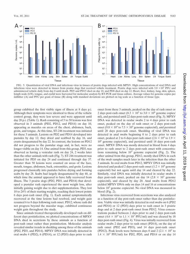
anal swabs was initially recovered on day 8 (8.8 � 104 PFU/ml),peaked at days 10 to 12 (3.6 � 105 to 1.7 � 106 PFU/ml), andpersisted through death (Fig. 4). Viral DNA and viable viruswere detected in all tissues collected during necropsy (Fig. 5).Concentrations of virus were highest in different tissues foreach animal: for PD5, liver; PD8, tongue; and PD15, lymph
TABLE 1—Continued
Day ofrash onset Lesion count No. of
surviving PDs
Day(s) of MPXV DNA detection in: Days of viablevirus detection
Detection of Ig antibody(no. of positive PDs)Swabs Blood
NA NA 4 8–26 24 (PD10) 12–24 4NA NA 4 6–28 None 8–26 410 (PD2, PD12,
PD14), 24 (PD3)�10 (PD2, PD12,
PD14), �50 (PD3)4 4–30 6–26 (PD2,
PD3, PD14)4–30 3 (PD2, PD3, PD14)
10 (PD8) 10–25 (PD8) 1 (PD6) 4–12 (PD5,PD8, PD15)
4–12 (PD5,PD8, PD15)
6–12 (PD5,PD8, PD15)
1 (PD8)
NA NA 2 None None None 0
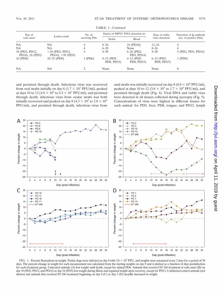
FIG. 1. Percent fluctuations in weight. Prairie dogs were infected on day 0 with 3.8 � 105 PFU, and weights were measured every 2 days for a period of 30days. The percent change in weight for each measurement was calculated from the starting weights on day 0 and is plotted as a function of days postinfectionfor each treatment group. Untreated animals (A) lost weight until death, except for animal PD6. Animals that received ST-246 treatment at rash onset (B) onday 10 (PD2, PD12, and PD14) or day 24 (PD3) lost weight during illness and regained weight upon recovery, except for PD12. Uninfected control animals (notshown) and animals that received ST-246 treatment beginning on day 0 (C) or day 3 (D) steadily increased in weight.
VOL. 85, 2011 ST-246 TREATMENT OF SYSTEMIC ORTHOPOXVIRUS DISEASE 9179
on April 11, 2019 by guest
http://jvi.asm.org/
Dow
nloaded from
node. Anti-OPXV antibodies were detected in one animal,PD8, from this group on day 12 (Fig. 6).
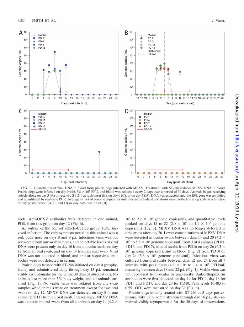
An outlier of the control vehicle-treated group, PD6, sur-vived infection. The only symptom noted in this animal was ared, puffy nose on days 8 and 9 p.i. Infectious virus was notrecovered from any swab samples, and detectable levels of viralDNA were present only on day 10 from an ocular swab, on day12 from an oral swab, and on day 14 from an anal swab. ViralDNA was not detected in blood, and anti-orthopoxvirus anti-bodies were not detected in serum.
Prairie dogs treated with ST-246 initiated on day 0 (prophy-lactic) and administered daily through day 13 p.i. remainedvisibly asymptomatic for the entire 30 days of observations. Noanimals lost more than 5% body weight, and all animals sur-vived (Fig. 1). No viable virus was isolated from any swabsamples while animals were on treatment except for two oralswabs on day 12. MPXV DNA was detected on day 8 in oneanimal (PD11) from an oral swab. Interestingly, MPXV DNAwas detected in oral swabs from all 4 animals on day 14 (4.2 �
101 to 2.2 � 102 genome copies/ml), and quantitative levelspeaked on days 18 to 22 (2.0 � 104 to 4.1 � 105 genomecopies/ml) (Fig. 3). MPXV DNA was no longer detected inoral swabs after day 26. Lower concentrations of MPXV DNAwere detected in ocular swabs between days 14 and 20 (4.2 �101 to 5.5 � 102 genome copies/ml) from 3 of 4 animals (PD11,PD16, and PD17), in anal swabs from PD16 on day 26 (6.3 �101 genome copies/ml), and in blood (Fig. 2) from PD10 onday 24 (5.6 � 101 genome copies/ml). Infectious virus wascultured from oral swabs between days 12 and 24 from all 4animals, with peak titers (4.6 � 102 to 1.4 � 105 PFU/ml)occurring between days 18 and 22 p.i. (Fig. 4). Viable virus wasnot recovered from ocular or anal swabs. Antiorthopoxvirusantibodies were first detected on day 14 for PD11, day 16 forPD16 and PD17, and day 20 for PD10. Peak levels (0.403 to0.551 OD) were measured on day 30 (Fig. 6).
Prairie dogs initially treated with ST-246 at 3 days postex-posure, with daily administration through day 16 p.i., also re-mained visibly asymptomatic for the 30 days of observations.
FIG. 2. Quantitation of viral DNA in blood from prairie dogs infected with MPXV. Treatment with ST-246 reduces MPXV DNA in blood.Prairie dogs were infected on day 0 with 3.8 � 105 PFU, and blood was collected every 2 days over a period of 30 days. Animals began receivingvehicle alone on day 3 (A) or received ST-246 at rash onset (B), on day 0 (C), or on day 3 (D). DNA was extracted, and the E9L gene was amplifiedand quantitated by real-time PCR. Average values of genome copies per milliliter and standard deviations were plotted on a log scale as a functionof day postinfection (A, C, and D) or day post-rash onset (B).
9180 SMITH ET AL. J. VIROL.
on April 11, 2019 by guest
http://jvi.asm.org/
Dow
nloaded from
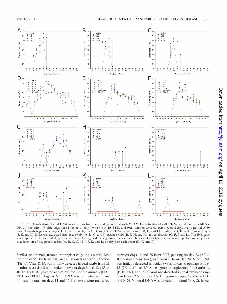
Similar to animals treated prophylactically, no animals lostmore than 5% body weight, and all animals survived infection(Fig. 1). Viral DNA was initially detected in oral swabs from all4 animals on day 8 and peaked between days 8 and 12 (2.3 �103 to 3.6 � 103 genome copies/ml) for 3 of the animals (PD1,PD4, and PD13) (Fig. 3). Viral DNA was not detected in anyof these animals on days 14 and 16, but levels were measured
between days 18 and 26 from PD7, peaking on day 24 (2.7 �104 genome copies/ml), and from PD4 on day 24. Viral DNAwas initially detected in ocular swabs on day 8, peaking on day12 (7.9 � 101 to 2.5 � 102 genome copies/ml) for 3 animals(PD1, PD4, and PD7), and was detected in anal swabs on days6 and 12 (6.2 � 101 to 2.7 � 102 genome copies/ml) from PD1and PD4. No viral DNA was detected in blood (Fig. 2). Infec-
FIG. 3. Quantitation of viral DNA in secretions from prairie dogs infected with MPXV. Early treatment with ST-246 greatly reduces MPXVDNA in secretions. Prairie dogs were infected on day 0 with 3.8 � 105 PFU, and swab samples were collected every 2 days over a period of 30days. Animals began receiving vehicle alone on day 3 (A, B, and C) or ST-246 at rash onset (D, E, and F), on day 0 (G, H, and I), or on day 3(J, K, and L). DNA was extracted from oral swabs (A, D, G, and J), ocular swabs (B, E, H, and K), and anal swabs (C, F, I, and L). The E9L genewas amplified and quantitated by real-time PCR. Average values of genome copies per milliliter and standard deviations were plotted on a log scaleas a function of day postinfection (A, B, C, G, H, I, J, K, and L) or day post-rash onset (D, E, and F).
VOL. 85, 2011 ST-246 TREATMENT OF SYSTEMIC ORTHOPOXVIRUS DISEASE 9181
on April 11, 2019 by guest
http://jvi.asm.org/
Dow
nloaded from
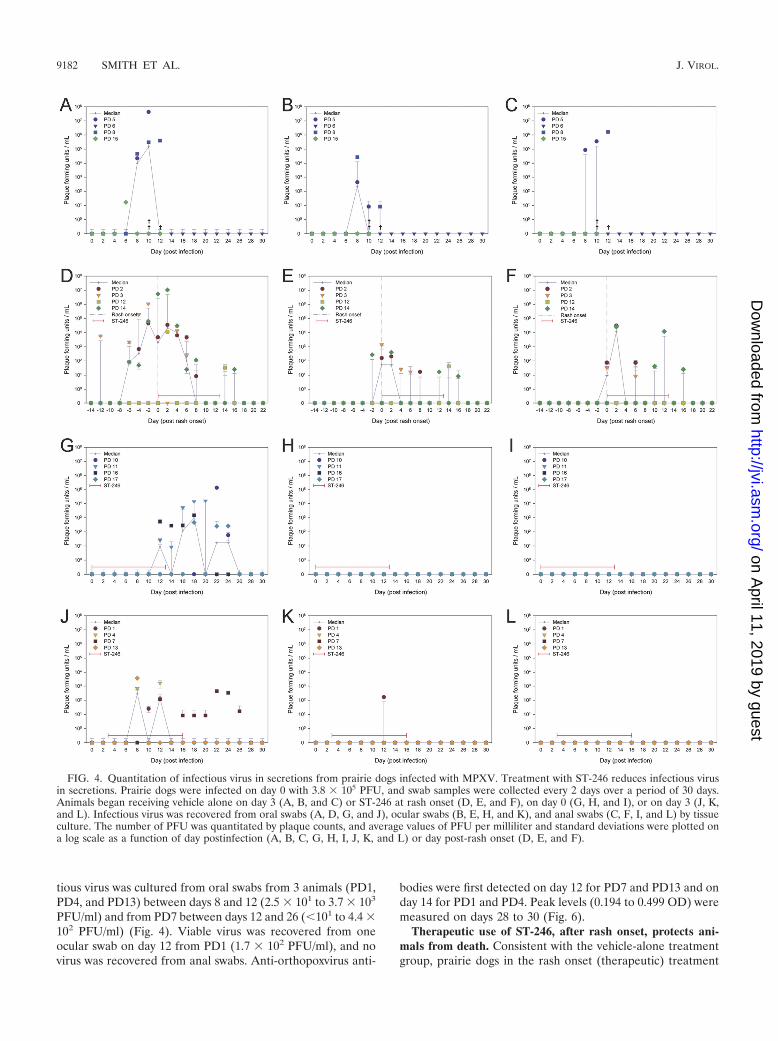
tious virus was cultured from oral swabs from 3 animals (PD1,PD4, and PD13) between days 8 and 12 (2.5 � 101 to 3.7 � 103
PFU/ml) and from PD7 between days 12 and 26 (�101 to 4.4 �102 PFU/ml) (Fig. 4). Viable virus was recovered from oneocular swab on day 12 from PD1 (1.7 � 102 PFU/ml), and novirus was recovered from anal swabs. Anti-orthopoxvirus anti-
bodies were first detected on day 12 for PD7 and PD13 and onday 14 for PD1 and PD4. Peak levels (0.194 to 0.499 OD) weremeasured on days 28 to 30 (Fig. 6).
Therapeutic use of ST-246, after rash onset, protects ani-mals from death. Consistent with the vehicle-alone treatmentgroup, prairie dogs in the rash onset (therapeutic) treatment
FIG. 4. Quantitation of infectious virus in secretions from prairie dogs infected with MPXV. Treatment with ST-246 reduces infectious virusin secretions. Prairie dogs were infected on day 0 with 3.8 � 105 PFU, and swab samples were collected every 2 days over a period of 30 days.Animals began receiving vehicle alone on day 3 (A, B, and C) or ST-246 at rash onset (D, E, and F), on day 0 (G, H, and I), or on day 3 (J, K,and L). Infectious virus was recovered from oral swabs (A, D, G, and J), ocular swabs (B, E, H, and K), and anal swabs (C, F, I, and L) by tissueculture. The number of PFU was quantitated by plaque counts, and average values of PFU per milliliter and standard deviations were plotted ona log scale as a function of day postinfection (A, B, C, G, H, I, J, K, and L) or day post-rash onset (D, E, and F).
9182 SMITH ET AL. J. VIROL.
on April 11, 2019 by guest
http://jvi.asm.org/
Dow
nloaded from
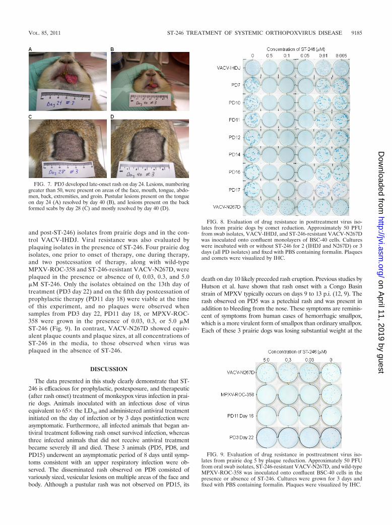
group exhibited the first visible signs of illness at 8 days p.i.Although their symptoms were identical to those of the vehiclecontrol group, they were less severe and were apparent untilday 20 p.i. (Table 1). Rash consisting of 5 to 50 lesions was firstobserved in 3 animals (PD2, PD12, and PD14) on day 10,appearing as macules on areas of the chest, abdomen, back,groin, and tongue. At this time, ST-246 treatment was initiatedfor these 3 animals. Lesions on PD2 and PD14 developed intopustules by day 12; they dried and scabbed by day 16, andcrusts desquamated by day 22. In contrast, the lesions on PD12did not progress to the pustular stage and, in fact, were nolonger visible on day 14. One animal from this group, PD3, wasobserved as having a vesicular rash on day 24, 2 weeks laterthan the other animals with rash (Fig. 7). ST-246 treatment wasinitiated for PD3 on day 24 and continued through day 37.Greater than 50 lesions were counted on areas of the face,mouth, tongue, abdomen, back, extremities, and groin. Lesionsprogressed classically into pustules before drying and formingscabs by day 28. Scabs had largely desquamated by day 40, atwhich time the animal appeared to have fully recovered fromillness. The 3 prairie dogs (PD2, PD3, and PD14) that devel-oped a pustular rash experienced the most weight loss, afterinitially gaining weight due to diet supplementation. They lost10 to 24% of their starting weights, reaching their lowest points6 days post-rash onset (Fig. 1). Each animal symptomaticallyrecovered at the time lesions had resolved, and weight gainresumed 6 to 8 days following rash onset. PD12, whose rash didnot progress beyond the macular stage, exhibited continuousweight gain throughout the study.
Since animals treated therapeutically developed rash on dif-ferent days postinfection, we plotted concentrations of MPXVDNA shed in secretions by these animals as a function ofpost-rash onset rather than postinfection. Molecular analysisrevealed similar trends in shedding among three of the animals(PD2, PD3, and PD14). MPXV DNA was initially detected inoral swabs 4 (PD2), 6 (PD14), or 12 (PD3) days prior to rash
onset from these 3 animals, peaked on the day of rash onset or2 days post-rash onset (8.3 � 105 to 3.0 � 106 genome copies/ml), and persisted until 22 days post-rash onset (Fig. 3). MPXVDNA was detected in ocular swabs 2 to 4 days prior to rashonset, peaked on the day of rash onset or 2 days post-rashonset (9.8 � 104 to 7.5 � 105 genome copies/ml), and persisteduntil 20 days post-rash onset. Shedding of viral DNA wasdetected in anal swabs beginning 0 to 2 days prior to rashonset, peaked at 2 to 6 days post rash onset (2.6 � 104 to 1.9 �106 genome copies/ml), and persisted until 18 days post-rashonset. MPXV DNA was mostly detected in blood from 4 daysprior to rash onset to 2 days post-rash onset with concentra-tions remaining below 103 genome copies/ml (Fig. 2). Theother animal from this group, PD12, mostly shed DNA in eachof the swab samples much later in the infection than the other3 animals. In oral swabs from PD12, MPXV DNA was initiallydetected and peaked 2 days post-rash onset (1.2 � 104 genomecopies/ml) but not again until day 14 and cleared by day 22.Similarly, viral DNA was initially detected in ocular swabs 4days post-rash onset, peaked on day 14 (2.8 � 103 genomecopies/ml), and cleared by day 20. Anal swabs from PD12yielded MPXV DNA only on days 14 and 16 at concentrationsbelow 103 genome copies/ml. No viral DNA was measured inblood (Fig. 2).
Infectious virus recovered from secretions was also analyzedas a function of day post-rash onset rather than day postinfec-tion. Viable virus was initially detected in oral swabs on 6 (PD2and PD14) or 12 (PD3) days prior to rash onset in 3 prairiedogs and at 2 days post-rash onset from PD12. Virus concen-trations peaked between 2 days prior to and 2 days post-rashonset (4.6 � 104 to 1.1 � 107 PFU/ml) and was cleared by 18days post-rash onset (Fig. 4). Virus was initially recovered fromocular swabs 2 days prior to rash onset (PD14), on the day ofrash onset (PD2 and PD3), and 14 days post-rash onset(PD12). Peak levels were between days 0 and 2 (2.1 � 102 to1.4 � 103 PFU/ml) for PD2, PD3, and PD14 or on day 14
FIG. 5. Quantitation of viral DNA and infectious virus in tissues of prairie dogs infected with MPXV. High concentrations of viral DNA andinfectious virus were detected in tissues from prairie dogs that received vehicle treatment. Prairie dogs were infected with 3.8 �105 PFU andadministered vehicle daily from day 0 until death. PD5 and PD15 died on day 10, and PD8 died on day 12. Heart, liver, kidney, lung, skin, spleen,lymph node (LN), tongue, and eyelid were harvested for molecular analysis by RT-PCR and tissue culture. Average values for genome copies permilliliter (A) and PFU per gram of tissue (B) along with standard deviations are plotted on log scale as a function of tissue type.
VOL. 85, 2011 ST-246 TREATMENT OF SYSTEMIC ORTHOPOXVIRUS DISEASE 9183
on April 11, 2019 by guest
http://jvi.asm.org/
Dow
nloaded from
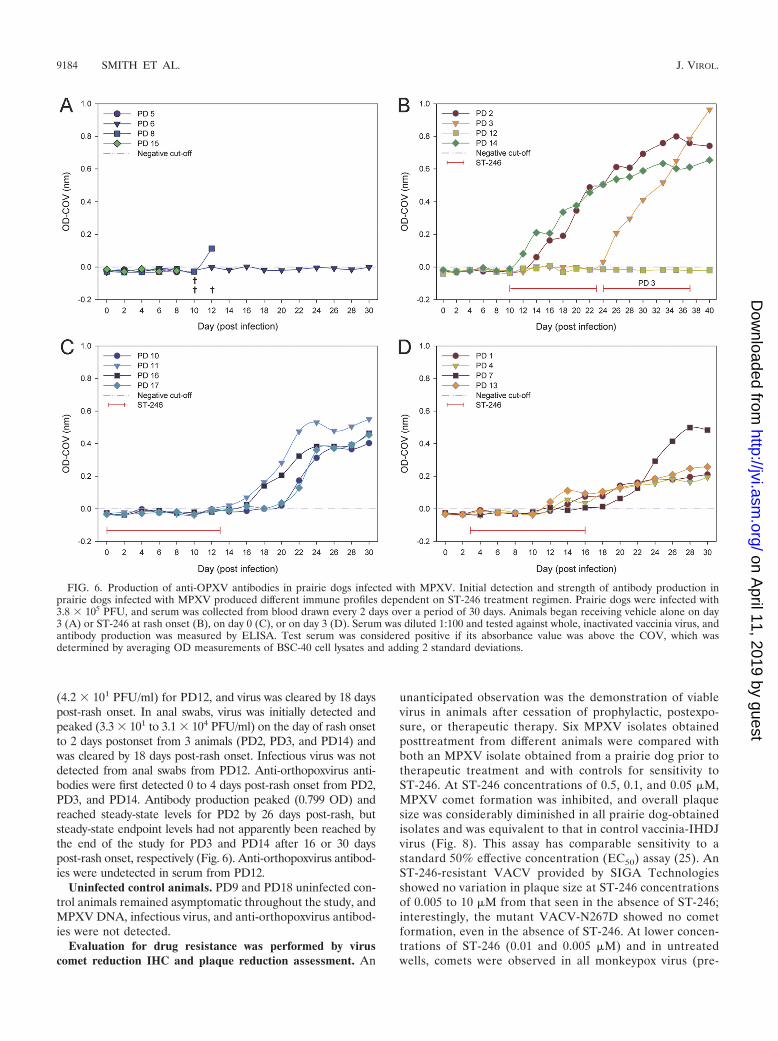
(4.2 � 101 PFU/ml) for PD12, and virus was cleared by 18 dayspost-rash onset. In anal swabs, virus was initially detected andpeaked (3.3 � 101 to 3.1 � 104 PFU/ml) on the day of rash onsetto 2 days postonset from 3 animals (PD2, PD3, and PD14) andwas cleared by 18 days post-rash onset. Infectious virus was notdetected from anal swabs from PD12. Anti-orthopoxvirus anti-bodies were first detected 0 to 4 days post-rash onset from PD2,PD3, and PD14. Antibody production peaked (0.799 OD) andreached steady-state levels for PD2 by 26 days post-rash, butsteady-state endpoint levels had not apparently been reached bythe end of the study for PD3 and PD14 after 16 or 30 dayspost-rash onset, respectively (Fig. 6). Anti-orthopoxvirus antibod-ies were undetected in serum from PD12.
Uninfected control animals. PD9 and PD18 uninfected con-trol animals remained asymptomatic throughout the study, andMPXV DNA, infectious virus, and anti-orthopoxvirus antibod-ies were not detected.
Evaluation for drug resistance was performed by viruscomet reduction IHC and plaque reduction assessment. An
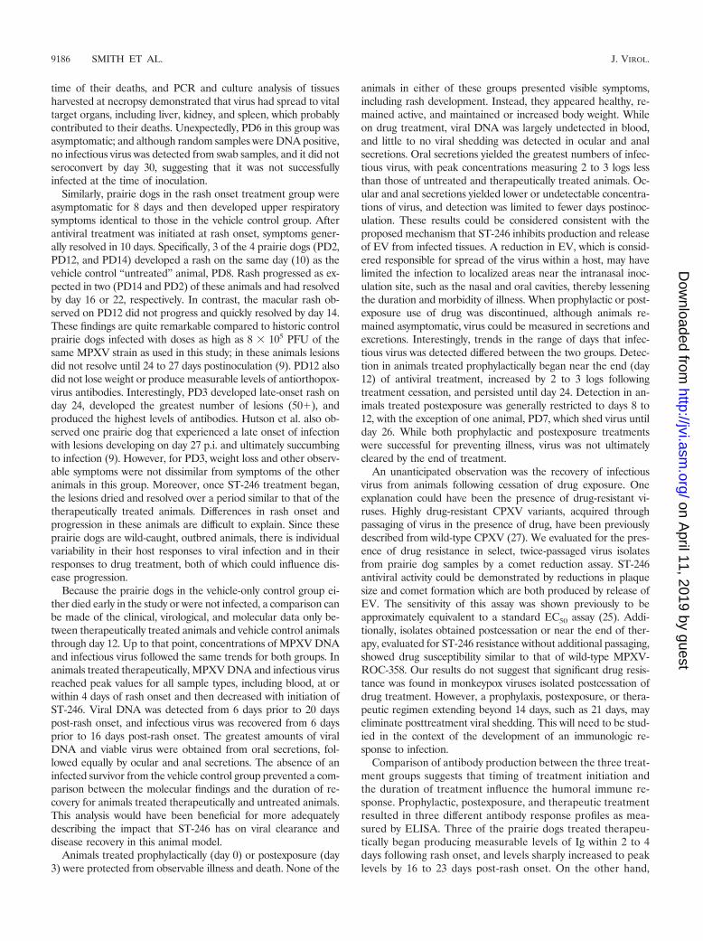
unanticipated observation was the demonstration of viablevirus in animals after cessation of prophylactic, postexpo-sure, or therapeutic therapy. Six MPXV isolates obtainedposttreatment from different animals were compared withboth an MPXV isolate obtained from a prairie dog prior totherapeutic treatment and with controls for sensitivity toST-246. At ST-246 concentrations of 0.5, 0.1, and 0.05 �M,MPXV comet formation was inhibited, and overall plaquesize was considerably diminished in all prairie dog-obtainedisolates and was equivalent to that in control vaccinia-IHDJvirus (Fig. 8). This assay has comparable sensitivity to astandard 50% effective concentration (EC50) assay (25). AnST-246-resistant VACV provided by SIGA Technologiesshowed no variation in plaque size at ST-246 concentrationsof 0.005 to 10 �M from that seen in the absence of ST-246;interestingly, the mutant VACV-N267D showed no cometformation, even in the absence of ST-246. At lower concen-trations of ST-246 (0.01 and 0.005 �M) and in untreatedwells, comets were observed in all monkeypox virus (pre-
FIG. 6. Production of anti-OPXV antibodies in prairie dogs infected with MPXV. Initial detection and strength of antibody production inprairie dogs infected with MPXV produced different immune profiles dependent on ST-246 treatment regimen. Prairie dogs were infected with3.8 � 105 PFU, and serum was collected from blood drawn every 2 days over a period of 30 days. Animals began receiving vehicle alone on day3 (A) or ST-246 at rash onset (B), on day 0 (C), or on day 3 (D). Serum was diluted 1:100 and tested against whole, inactivated vaccinia virus, andantibody production was measured by ELISA. Test serum was considered positive if its absorbance value was above the COV, which wasdetermined by averaging OD measurements of BSC-40 cell lysates and adding 2 standard deviations.
9184 SMITH ET AL. J. VIROL.
on April 11, 2019 by guest
http://jvi.asm.org/
Dow
nloaded from
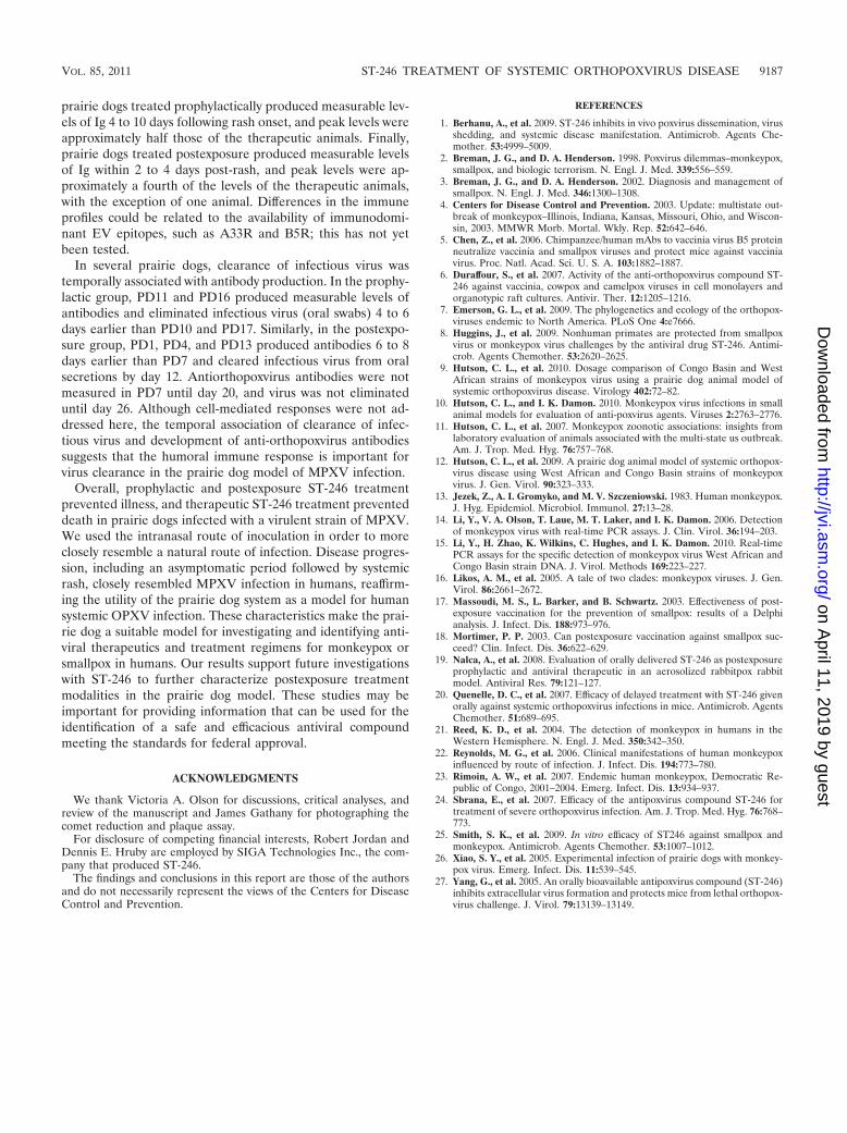
and post-ST-246) isolates from prairie dogs and in the con-trol VACV-IHDJ. Viral resistance was also evaluated byplaquing isolates in the presence of ST-246. Four prairie dogisolates, one prior to onset of therapy, one during therapy,and two postcessation of therapy, along with wild-typeMPXV-ROC-358 and ST-246-resistant VACV-N267D, wereplaqued in the presence or absence of 0, 0.03, 0.3, and 5.0�M ST-246. Only the isolates obtained on the 13th day oftreatment (PD3 day 22) and on the fifth day postcessation ofprophylactic therapy (PD11 day 18) were viable at the timeof this experiment, and no plaques were observed whensamples from PD3 day 22, PD11 day 18, or MPXV-ROC-358 were grown in the presence of 0.03, 0.3, or 5.0 �MST-246 (Fig. 9). In contrast, VACV-N267D showed equiv-alent plaque counts and plaque sizes, at all concentrations ofST-246 in the media, to those observed when virus wasplaqued in the absence of ST-246.
DISCUSSION
The data presented in this study clearly demonstrate that ST-246 is efficacious for prophylactic, postexposure, and therapeutic(after rash onset) treatment of monkeypox virus infection in prai-rie dogs. Animals inoculated with an infectious dose of virusequivalent to 65� the LD50 and administered antiviral treatmentinitiated on the day of infection or by 3 days postinfection wereasymptomatic. Furthermore, all infected animals that began an-tiviral treatment following rash onset survived infection, whereasthree infected animals that did not receive antiviral treatmentbecame severely ill and died. These 3 animals (PD5, PD8, andPD15) underwent an asymptomatic period of 8 days until symp-toms consistent with an upper respiratory infection were ob-served. The disseminated rash observed on PD8 consisted ofvariously sized, vesicular lesions on multiple areas of the face andbody. Although a pustular rash was not observed on PD15, its
death on day 10 likely preceded rash eruption. Previous studies byHutson et al. have shown that rash onset with a Congo Basinstrain of MPXV typically occurs on days 9 to 13 p.i. (12, 9). Therash observed on PD5 was a petechial rash and was present inaddition to bleeding from the nose. These symptoms are reminis-cent of symptoms from human cases of hemorrhagic smallpox,which is a more virulent form of smallpox than ordinary smallpox.Each of these 3 prairie dogs was losing substantial weight at the
FIG. 8. Evaluation of drug resistance in posttreatment virus iso-lates from prairie dogs by comet reduction. Approximately 50 PFUfrom swab isolates, VACV-IHDJ, and ST-246-resistant VACV-N267Dwas inoculated onto confluent monolayers of BSC-40 cells. Cultureswere incubated with or without ST-246 for 2 (IHDJ and N267D) or 3days (all PD isolates) and fixed with PBS containing formalin. Plaquesand comets were visualized by IHC.
FIG. 9. Evaluation of drug resistance in posttreatment virus iso-lates from prairie dog 5 by plaque reduction. Approximately 50 PFUfrom oral swab isolates, ST-246-resistant VACV-N267D, and wild-typeMPXV-ROC-358 was inoculated onto confluent BSC-40 cells in thepresence or absence of ST-246. Cultures were grown for 3 days andfixed with PBS containing formalin. Plaques were visualized by IHC.
FIG. 7. PD3 developed late-onset rash on day 24. Lesions, numberinggreater than 50, were present on areas of the face, mouth, tongue, abdo-men, back, extremities, and groin. Pustular lesions present on the tongueon day 24 (A) resolved by day 40 (B), and lesions present on the backformed scabs by day 28 (C) and mostly resolved by day 40 (D).
VOL. 85, 2011 ST-246 TREATMENT OF SYSTEMIC ORTHOPOXVIRUS DISEASE 9185
on April 11, 2019 by guest
http://jvi.asm.org/
Dow
nloaded from
time of their deaths, and PCR and culture analysis of tissuesharvested at necropsy demonstrated that virus had spread to vitaltarget organs, including liver, kidney, and spleen, which probablycontributed to their deaths. Unexpectedly, PD6 in this group wasasymptomatic; and although random samples were DNA positive,no infectious virus was detected from swab samples, and it did notseroconvert by day 30, suggesting that it was not successfullyinfected at the time of inoculation.
Similarly, prairie dogs in the rash onset treatment group wereasymptomatic for 8 days and then developed upper respiratorysymptoms identical to those in the vehicle control group. Afterantiviral treatment was initiated at rash onset, symptoms gener-ally resolved in 10 days. Specifically, 3 of the 4 prairie dogs (PD2,PD12, and PD14) developed a rash on the same day (10) as thevehicle control “untreated” animal, PD8. Rash progressed as ex-pected in two (PD14 and PD2) of these animals and had resolvedby day 16 or 22, respectively. In contrast, the macular rash ob-served on PD12 did not progress and quickly resolved by day 14.These findings are quite remarkable compared to historic controlprairie dogs infected with doses as high as 8 � 105 PFU of thesame MPXV strain as used in this study; in these animals lesionsdid not resolve until 24 to 27 days postinoculation (9). PD12 alsodid not lose weight or produce measurable levels of antiorthopox-virus antibodies. Interestingly, PD3 developed late-onset rash onday 24, developed the greatest number of lesions (50�), andproduced the highest levels of antibodies. Hutson et al. also ob-served one prairie dog that experienced a late onset of infectionwith lesions developing on day 27 p.i. and ultimately succumbingto infection (9). However, for PD3, weight loss and other observ-able symptoms were not dissimilar from symptoms of the otheranimals in this group. Moreover, once ST-246 treatment began,the lesions dried and resolved over a period similar to that of thetherapeutically treated animals. Differences in rash onset andprogression in these animals are difficult to explain. Since theseprairie dogs are wild-caught, outbred animals, there is individualvariability in their host responses to viral infection and in theirresponses to drug treatment, both of which could influence dis-ease progression.
Because the prairie dogs in the vehicle-only control group ei-ther died early in the study or were not infected, a comparison canbe made of the clinical, virological, and molecular data only be-tween therapeutically treated animals and vehicle control animalsthrough day 12. Up to that point, concentrations of MPXV DNAand infectious virus followed the same trends for both groups. Inanimals treated therapeutically, MPXV DNA and infectious virusreached peak values for all sample types, including blood, at orwithin 4 days of rash onset and then decreased with initiation ofST-246. Viral DNA was detected from 6 days prior to 20 dayspost-rash onset, and infectious virus was recovered from 6 daysprior to 16 days post-rash onset. The greatest amounts of viralDNA and viable virus were obtained from oral secretions, fol-lowed equally by ocular and anal secretions. The absence of aninfected survivor from the vehicle control group prevented a com-parison between the molecular findings and the duration of re-covery for animals treated therapeutically and untreated animals.This analysis would have been beneficial for more adequatelydescribing the impact that ST-246 has on viral clearance anddisease recovery in this animal model.
Animals treated prophylactically (day 0) or postexposure (day3) were protected from observable illness and death. None of the
animals in either of these groups presented visible symptoms,including rash development. Instead, they appeared healthy, re-mained active, and maintained or increased body weight. Whileon drug treatment, viral DNA was largely undetected in blood,and little to no viral shedding was detected in ocular and analsecretions. Oral secretions yielded the greatest numbers of infec-tious virus, with peak concentrations measuring 2 to 3 logs lessthan those of untreated and therapeutically treated animals. Oc-ular and anal secretions yielded lower or undetectable concentra-tions of virus, and detection was limited to fewer days postinoc-ulation. These results could be considered consistent with theproposed mechanism that ST-246 inhibits production and releaseof EV from infected tissues. A reduction in EV, which is consid-ered responsible for spread of the virus within a host, may havelimited the infection to localized areas near the intranasal inoc-ulation site, such as the nasal and oral cavities, thereby lesseningthe duration and morbidity of illness. When prophylactic or post-exposure use of drug was discontinued, although animals re-mained asymptomatic, virus could be measured in secretions andexcretions. Interestingly, trends in the range of days that infec-tious virus was detected differed between the two groups. Detec-tion in animals treated prophylactically began near the end (day12) of antiviral treatment, increased by 2 to 3 logs followingtreatment cessation, and persisted until day 24. Detection in an-imals treated postexposure was generally restricted to days 8 to12, with the exception of one animal, PD7, which shed virus untilday 26. While both prophylactic and postexposure treatmentswere successful for preventing illness, virus was not ultimatelycleared by the end of treatment.
An unanticipated observation was the recovery of infectiousvirus from animals following cessation of drug exposure. Oneexplanation could have been the presence of drug-resistant vi-ruses. Highly drug-resistant CPXV variants, acquired throughpassaging of virus in the presence of drug, have been previouslydescribed from wild-type CPXV (27). We evaluated for the pres-ence of drug resistance in select, twice-passaged virus isolatesfrom prairie dog samples by a comet reduction assay. ST-246antiviral activity could be demonstrated by reductions in plaquesize and comet formation which are both produced by release ofEV. The sensitivity of this assay was shown previously to beapproximately equivalent to a standard EC50 assay (25). Addi-tionally, isolates obtained postcessation or near the end of ther-apy, evaluated for ST-246 resistance without additional passaging,showed drug susceptibility similar to that of wild-type MPXV-ROC-358. Our results do not suggest that significant drug resis-tance was found in monkeypox viruses isolated postcessation ofdrug treatment. However, a prophylaxis, postexposure, or thera-peutic regimen extending beyond 14 days, such as 21 days, mayeliminate posttreatment viral shedding. This will need to be stud-ied in the context of the development of an immunologic re-sponse to infection.
Comparison of antibody production between the three treat-ment groups suggests that timing of treatment initiation andthe duration of treatment influence the humoral immune re-sponse. Prophylactic, postexposure, and therapeutic treatmentresulted in three different antibody response profiles as mea-sured by ELISA. Three of the prairie dogs treated therapeu-tically began producing measurable levels of Ig within 2 to 4days following rash onset, and levels sharply increased to peaklevels by 16 to 23 days post-rash onset. On the other hand,
9186 SMITH ET AL. J. VIROL.
on April 11, 2019 by guest
http://jvi.asm.org/
Dow
nloaded from
prairie dogs treated prophylactically produced measurable lev-els of Ig 4 to 10 days following rash onset, and peak levels wereapproximately half those of the therapeutic animals. Finally,prairie dogs treated postexposure produced measurable levelsof Ig within 2 to 4 days post-rash, and peak levels were ap-proximately a fourth of the levels of the therapeutic animals,with the exception of one animal. Differences in the immuneprofiles could be related to the availability of immunodomi-nant EV epitopes, such as A33R and B5R; this has not yetbeen tested.
In several prairie dogs, clearance of infectious virus wastemporally associated with antibody production. In the prophy-lactic group, PD11 and PD16 produced measurable levels ofantibodies and eliminated infectious virus (oral swabs) 4 to 6days earlier than PD10 and PD17. Similarly, in the postexpo-sure group, PD1, PD4, and PD13 produced antibodies 6 to 8days earlier than PD7 and cleared infectious virus from oralsecretions by day 12. Antiorthopoxvirus antibodies were notmeasured in PD7 until day 20, and virus was not eliminateduntil day 26. Although cell-mediated responses were not ad-dressed here, the temporal association of clearance of infec-tious virus and development of anti-orthopoxvirus antibodiessuggests that the humoral immune response is important forvirus clearance in the prairie dog model of MPXV infection.
Overall, prophylactic and postexposure ST-246 treatmentprevented illness, and therapeutic ST-246 treatment preventeddeath in prairie dogs infected with a virulent strain of MPXV.We used the intranasal route of inoculation in order to moreclosely resemble a natural route of infection. Disease progres-sion, including an asymptomatic period followed by systemicrash, closely resembled MPXV infection in humans, reaffirm-ing the utility of the prairie dog system as a model for humansystemic OPXV infection. These characteristics make the prai-rie dog a suitable model for investigating and identifying anti-viral therapeutics and treatment regimens for monkeypox orsmallpox in humans. Our results support future investigationswith ST-246 to further characterize postexposure treatmentmodalities in the prairie dog model. These studies may beimportant for providing information that can be used for theidentification of a safe and efficacious antiviral compoundmeeting the standards for federal approval.
ACKNOWLEDGMENTS
We thank Victoria A. Olson for discussions, critical analyses, andreview of the manuscript and James Gathany for photographing thecomet reduction and plaque assay.
For disclosure of competing financial interests, Robert Jordan andDennis E. Hruby are employed by SIGA Technologies Inc., the com-pany that produced ST-246.
The findings and conclusions in this report are those of the authorsand do not necessarily represent the views of the Centers for DiseaseControl and Prevention.
REFERENCES
1. Berhanu, A., et al. 2009. ST-246 inhibits in vivo poxvirus dissemination, virusshedding, and systemic disease manifestation. Antimicrob. Agents Che-mother. 53:4999–5009.
2. Breman, J. G., and D. A. Henderson. 1998. Poxvirus dilemmas–monkeypox,smallpox, and biologic terrorism. N. Engl. J. Med. 339:556–559.
3. Breman, J. G., and D. A. Henderson. 2002. Diagnosis and management ofsmallpox. N. Engl. J. Med. 346:1300–1308.
4. Centers for Disease Control and Prevention. 2003. Update: multistate out-break of monkeypox–Illinois, Indiana, Kansas, Missouri, Ohio, and Wiscon-sin, 2003. MMWR Morb. Mortal. Wkly. Rep. 52:642–646.
5. Chen, Z., et al. 2006. Chimpanzee/human mAbs to vaccinia virus B5 proteinneutralize vaccinia and smallpox viruses and protect mice against vacciniavirus. Proc. Natl. Acad. Sci. U. S. A. 103:1882–1887.
6. Duraffour, S., et al. 2007. Activity of the anti-orthopoxvirus compound ST-246 against vaccinia, cowpox and camelpox viruses in cell monolayers andorganotypic raft cultures. Antivir. Ther. 12:1205–1216.
7. Emerson, G. L., et al. 2009. The phylogenetics and ecology of the orthopox-viruses endemic to North America. PLoS One 4:e7666.
8. Huggins, J., et al. 2009. Nonhuman primates are protected from smallpoxvirus or monkeypox virus challenges by the antiviral drug ST-246. Antimi-crob. Agents Chemother. 53:2620–2625.
9. Hutson, C. L., et al. 2010. Dosage comparison of Congo Basin and WestAfrican strains of monkeypox virus using a prairie dog animal model ofsystemic orthopoxvirus disease. Virology 402:72–82.
10. Hutson, C. L., and I. K. Damon. 2010. Monkeypox virus infections in smallanimal models for evaluation of anti-poxvirus agents. Viruses 2:2763–2776.
11. Hutson, C. L., et al. 2007. Monkeypox zoonotic associations: insights fromlaboratory evaluation of animals associated with the multi-state us outbreak.Am. J. Trop. Med. Hyg. 76:757–768.
12. Hutson, C. L., et al. 2009. A prairie dog animal model of systemic orthopox-virus disease using West African and Congo Basin strains of monkeypoxvirus. J. Gen. Virol. 90:323–333.
13. Jezek, Z., A. I. Gromyko, and M. V. Szczeniowski. 1983. Human monkeypox.J. Hyg. Epidemiol. Microbiol. Immunol. 27:13–28.
14. Li, Y., V. A. Olson, T. Laue, M. T. Laker, and I. K. Damon. 2006. Detectionof monkeypox virus with real-time PCR assays. J. Clin. Virol. 36:194–203.
15. Li, Y., H. Zhao, K. Wilkins, C. Hughes, and I. K. Damon. 2010. Real-timePCR assays for the specific detection of monkeypox virus West African andCongo Basin strain DNA. J. Virol. Methods 169:223–227.
16. Likos, A. M., et al. 2005. A tale of two clades: monkeypox viruses. J. Gen.Virol. 86:2661–2672.
17. Massoudi, M. S., L. Barker, and B. Schwartz. 2003. Effectiveness of post-exposure vaccination for the prevention of smallpox: results of a Delphianalysis. J. Infect. Dis. 188:973–976.
18. Mortimer, P. P. 2003. Can postexposure vaccination against smallpox suc-ceed? Clin. Infect. Dis. 36:622–629.
19. Nalca, A., et al. 2008. Evaluation of orally delivered ST-246 as postexposureprophylactic and antiviral therapeutic in an aerosolized rabbitpox rabbitmodel. Antiviral Res. 79:121–127.
20. Quenelle, D. C., et al. 2007. Efficacy of delayed treatment with ST-246 givenorally against systemic orthopoxvirus infections in mice. Antimicrob. AgentsChemother. 51:689–695.
21. Reed, K. D., et al. 2004. The detection of monkeypox in humans in theWestern Hemisphere. N. Engl. J. Med. 350:342–350.
22. Reynolds, M. G., et al. 2006. Clinical manifestations of human monkeypoxinfluenced by route of infection. J. Infect. Dis. 194:773–780.
23. Rimoin, A. W., et al. 2007. Endemic human monkeypox, Democratic Re-public of Congo, 2001–2004. Emerg. Infect. Dis. 13:934–937.
24. Sbrana, E., et al. 2007. Efficacy of the antipoxvirus compound ST-246 fortreatment of severe orthopoxvirus infection. Am. J. Trop. Med. Hyg. 76:768–773.
25. Smith, S. K., et al. 2009. In vitro efficacy of ST246 against smallpox andmonkeypox. Antimicrob. Agents Chemother. 53:1007–1012.
26. Xiao, S. Y., et al. 2005. Experimental infection of prairie dogs with monkey-pox virus. Emerg. Infect. Dis. 11:539–545.
27. Yang, G., et al. 2005. An orally bioavailable antipoxvirus compound (ST-246)inhibits extracellular virus formation and protects mice from lethal orthopox-virus challenge. J. Virol. 79:13139–13149.
VOL. 85, 2011 ST-246 TREATMENT OF SYSTEMIC ORTHOPOXVIRUS DISEASE 9187
on April 11, 2019 by guest
http://jvi.asm.org/
Dow
nloaded from


![[Document title] - infeksiemerging.kemkes.go.idinfeksiemerging.kemkes.go.id/download/Juknis_P2P_Monkeypox.pdf · dini dan kesiapsiagaan terhadap penyakit monkeypox. Sebagai salah](https://img.pdfslide.net/doc/110x75/5e44d12f290fae04ba415ecf/document-title-dini-dan-kesiapsiagaan-terhadap-penyakit-monkeypox-sebagai-salah.jpg)