Embed Size (px)
Citation preview

S T A N F O R D
M E D I C I N E
S T A
M E DI s s u e 2 / 2 0 2 1
special report
THE MOST MYSTERIOUS
ORGAN Unlocking the secrets
of the brain
A new frontierRestoring lost neurological function
Brain trauma’s aftermathWhy do women suffer more?
So delicateAn intricate pathway for a tiny child’s
brain surgery
Good vibrationsCan Parkinson’s symptoms be stopped?
Alzheimer’s poetryA conversation with flute virtuoso
Eugenia Zukerman
Pushing the limitExpanding treatment of strokes
The man who couldn’t cryNeuroscientist Karl Deisseroth on
the workings of the mind
plus
Saving WhitneyA researcher’s mission to explain his son’s
mystifying illness
One scientist’s utopiaHow bioengineering could save us

S T A N F O R D
M E D I C I N EI s s u e 2 / 2 0 2 1

1 8 F A L L 2 0 0 8 S T A N F O R D M E D I C I N E
It was around 2010 and neurosurgeon Michael Lim, MD, was taking a patient to the op-erating room to remove a brain tumor. Prior to the surgery, the patient received an experimental
drug to stimulate his immune system to attack his cancer, which had begun as kidney cancer and metastasized.
“I remember taking him to the OR and thinking this was going to be a routine case,” recalled Lim, now chair of the
Stanford University School of Medicine’s Department of Neurosurgery. “I took his tumor out. But when the pathology
report came back, it indicated the mass was just inflammatory cells and no active cancer. And over the next months, the
tumors in his body started to melt away. My interest was piqued by that finding and I became very interested in that drug.”
The drug, which became known as Opdivo, belongs to a new class of medications called checkpoint inhibitors.
Although our immune systems are honed to recognize and kill developing tumors, the tumors can evade them by ex-
ploiting biological safety valves called checkpoints, which normally tamp down any overactive immune responses that
could lead to autoimmune disorders or inflammation.
Lim, who trained at Stanford Medicine but was working at Johns Hopkins Uni-
versity School of Medicine at the time, wondered if checkpoint inhibitors might
also be effective against tumors that start in the brain, like glioblastomas. Al-
though subsequent experiments in mice and clinical trials in patients uncovered
some significant stumbling blocks, Lim said he is excited to see a way forward for
patients with the devastating cancer.
“It’s clear that brain cancers are different from other types of cancers,” Lim
said. “For example, we’ve found that, although all tumors suppress the immune
response in the microenvironment, tumors that originate in the brain cause a
global immune suppression that affects the whole body. This makes it very hard
to induce an immune response to the tumor.”
Targeting the culprits behind the immune suppression — a class of cells
called myeloid cells — could reverse this phenomenon, researchers believe.
Another approach focuses on reviving a kind of immune cell called a T cell that
leads the charge against cancers but can become exhausted and ineffective
over time. A series of experiments in Lim’s lab suggested that combining a
checkpoint inhibitor with a molecule to combat T cell exhaustion is safe. A study
of the combo’s effect on patients confirmed the treatment’s safety and found it
resulted in longer survival times for some of the participants.
“Now we’re going back to the lab bench to try to learn why some patients responded to the combination treat-
ment and some didn’t,” Lim said. “We hope to go on to a larger clinical trial. There’s so much amazing science here at
Stanford — we’re able to go from the bench to bedside and back to the bench to solve these problems.”
Lim and his colleagues hope to one day see outcomes for glioblastoma patients that are similar to those experi-
enced by patients with metastatic brain cancer.
“Glioblastoma is such a malignant disease. I’ve treated hundreds of these patients, and every conversation I’ve
had with them fueled me to try to do better for them. Each one gives me a new sense of urgency,” he said.
“Right now, we are understanding cancer at a level we’ve never achieved before. As we learn how to assess a
patient’s tumor, we can become more and more precise with the therapies we can offer. We’re not just wielding blunt
tools anymore. I’m optimistic and excited about the future for these patients.” — K R I S T A C O N G E R
I S S U E 2 / 2 0 2 1 S T A N F O R D M E D I C I N E
SC
IEP
RO
THE CASE OF THE VANISHING BRAIN TUMOR
THE BRAIN IS A NEW FRONTIER FOR CANCER IMMUNOTHERAPY

CO
VE
R:
CR
AIG
CU
TL
ER
V O L U M E 3 8 N U M B E R 2
ISSUE 2/ 2021
S T A N F O R D
M E D I C I N E
S P E C I A L R E P O R T
The most mysterious organ Unlocking the secrets of the brain
6 Making a comeback By Ruthann Richter NEW WAYS TO PREVENT — OR EVEN REVERSE — DEMENTIA, PARALYSIS AND BLINDNESS
18 Same injury, different brain By Hanae Armitage EXPLORING HOW WOMEN’S TRAUMA RECOVERY DIFFERS FROM MEN’S
22 A delicate operation By Gordy Slack REMOVING A TUMOR FROM DEEP IN A 2-YEAR-OLD’S BRAIN
26 Opening stroke’s window By Amy Jeter Hansen TREATING STROKE MORE THAN A FEW HOURS AFTER THE CRISIS IS NO LONGER CONSIDERED ABSURD
32 A way through the brambles Condensed and edited by Paul Costello A CONVERSATION WITH FLUTE VIRTUOSO EUGENIA ZUKERMAN ON ALZHEIMER’S, MUSIC, POETRY AND JOY
34 Good vibrations By Holly Alyssa MacCormick CAN PARKINSON’S SYMPTOMS BE STOPPED? 36 The man who couldn’t cry By Karl Deisseroth ADAPTED FROM PROJECTIONS: A STORY OF HUMAN EMOTIONS
P L U S
38 How synthetic biology could save us By Hanae Armitage ONE BIOENGINEER’S NUTS-AND-BOLTS APPROACH TO A BIOTECH-BASED UTOPIA
40 A scientist’s quest to save his son from a mystifying illness By Tracie White EXCERPTS FROM THE PUZZLE SOLVER
The brain’s not immutable after allpage 6
A clue from the lab about restoring strength to aging musclepage 3
A strange and wonderful brain surgerypage 46
D E PA R T M E N T S
Letter from the dean 2 Upfront 3 Backstory 46

1 8 F A L L 2 0 0 8 S T A N F O R D M E D I C I N E
GL
EN
N M
AT
SU
MU
RA
2 I S S U E 2 / 2 0 2 1 S T A N F O R D M E D I C I N E
lett
er f
rom
the
dea
n
It was a eureka moment that shaped my early career. I was in an undergraduate
bioengineering course, and the professor was using mathematical and bioengineering models
to help us explore the vestibular system.
I was fascinated by the complex yet elegant sensory system that is responsible for our sense of balance and spatial orientation. Its neural pathways stabilize our gaze when outside forces jostle us, enabling us to see while out on a morning jog or read a GPS while driving on a bumpy road. I came away from the experience with a passion to more fully understand vestibular neurophysiology.
The human brain and nervous system form an astonishingly intricate network of billions of cells. It is also the original black box. For centuries, even as science revealed the workings of the rest of the human body, the brain remained stubbornly mysterious.
In the process, the brain has captured our imaginations, evolving from what Aristotle considered simply the body’s radiator to how we see it today: the matter that makes us individuals, containing the seat of intellect and reason, memories and personality, emotions and senses, our very consciousness.
More recently, imaging technologies and innovative research are shedding new light on how the brain works, letting us peer inside and reveal its secrets. The more we see, the more we realize how much more there is to learn. The brain’s abilities might be even more impressive than we imagined.
One of our most fascinating discoveries is that the brain isn’t as fixed and fragile as we once believed. The organ we thought was set in its ways by our late 20s is much more active — and resilient — for our entire lives.
This new understanding has led to transformative changes in how we treat diseases and problems of the brain. For example, decades of Stanford research drove a radical change in standards of care for stroke in 2018, opening treatment to tens of thousands of people who would otherwise have been told it was too late.
Years ago, the intersection of research, technological innovation and luck translated into my development of a new treatment. In a span of weeks, two of my patients complained of mysterious and often bizarre symptoms — like hearing their eyeballs move in their sockets or seeing objects dance when they sang in the shower. I hypothesized that those seemingly unrelated issues might have the same source: the vestibular system.
Thanks in part to our earlier research and access to leadingedge imaging and digital technologies, my colleagues and I identified the problem: a hole in the inner ear canal. By defining the condition, superior canal dehiscence syndrome, and developing a surgical solution, we have helped thousands of people resume normal lives.
As a leader of Stanford Medicine, I am awed by how our health system drives neuroscience and biomedical research through robust, multidisciplinary efforts. Physicians and scientists make discoveries every day that increase our understanding of biological systems and lead to new surgical techniques, digital technologies and innovative therapies to restore function, fight disease and improve quality of life.
With each advance, we better understand the remarkable organ that is both mind and matter — and slowly open up the black box of ourselves.
Sincerely,Lloyd Minor, MD
Carl and Elizabeth Naumann Dean of the School of Medicine Professor of Otolaryngology – Head & Neck Surgery

S T A N F O R D M E D I C I N E I S S U E 2 / 2 0 2 1 3
Diseaseblueprints A NEW STANFORD service enables patients to look within themselves — all the way down to their genomes — to help determine underlying reasons for certain medical conditions.
The idea is to parse a person’s complete genetic code to identify possible roots of disease, and even tailor treatments to the individual.
The service, led by Euan Ashley, MD, PhD, profes-sor of medicine, of genetics and of biomedical data science, is available for Stanford Health Care patients with inherited cardiology disease. It is among the first of its kind to be offered by a hospital. Ashley plans to expand it into other special-ties, such as cancer care.
December 2020 in Science.
The protein hadn’t previously been
implicated in aging. But earlier work in
Blau’s lab showed that another molecule,
prostaglandin E2, can activate stem cells
in the muscle that spring into action to
repair damaged muscle fibers. 15-PGDH,
which is more plentiful in muscles of older
animals, blocks this strengthening effect
by breaking down prostaglandin E2.
“We’re hopeful that these findings
may lead to new ways to improve human
health and impact the quality of life for
many people,” Blau said. DA
VID
PL
UN
KE
RT
upfrontA Q U I C K L O O K A T T H E L A T E S T D E V E L O P M E N T S F R O M S T A N F O R D M E D I C I N E
In response to COVID-19, Stanford Health Care increased its donations to community health programs in fiscal year 2020 to
$861 million — 79% more than the previous year. Read more at stan.md/ community
MightymouseWITH AGE COMES WISDOM, they say. But
as we grow more wise, our bodies often
falter. In particular, our muscles shrink and
lose strength as the years march on. What
if there were a way to restore strength
and mass to aging muscle?
New research in the laboratory of
Helen Blau, PhD, professor of microbiol-
ogy and immunology, suggests that con-
jecture could come true. She and senior
scientist Adelaida Palla, PhD, found that
blocking the activity of a protein called
15-PGDH in elderly laboratory mice revi-
talized the animals — strengthening their
muscles and allowing them to trot longer
on a treadmill than their untreated peers.
“The improvement is really quite
dramatic,” Blau said. “The old mice are
about 15% to 20% stronger after one
month of treatment, and their muscle
fibers look like young muscle. Consider-
ing that humans lose about 10% of mus-
cle strength per decade after about age
50, this is quite remarkable.”
Conversely, increasing the expression
of the protein in young mice caused their
muscles to atrophy and weaken.
They published their findings online

4
Scar-freehealing
A TEAM OF STANFORD MEDICINE RESEARCHERS looking to figure
out why we scar recently identified a drug that can prevent scar-
ring altogether. They published the research April 23 in Science.
Scars form because they seal an opening in the skin more
quickly than normal skin can grow. “A scar is a spot weld — it
covers the wound quickly,” said Michael Longaker, MD, a se-
nior author of the study and the Deane P. and Louise Mitchell
Professor in the School of Medicine.
But the result can be problematic: Scars lack hair follicles and
oil glands and are weaker, thicker and less flexible than other skin.
The study started with exploring the role our skin’s tightness
plays in scarring — a clue borne from the scientists’ observa-
tions that children and adults scar, but fetal tissue doesn’t, and
that the loose skin of older people has minimal scarring.
Their research found that a gene called engrailed signals fi-
broblasts — a skin cell type that drives scarring — to form scar
tissue, but only when skin is stressed. The study’s lead author,
graduate student Shamik Mascharak, identified an eye-disease
drug called verteporfin that, when applied to surgical wounds
in mice, blocked engrailed from signaling scar formation. “It’s
estimated that 45% of Americans die from a disease that in-
volves scarring in some form,” Longaker said. “So there are po-
tentially many more applications.”
Geoffrey Gurtner, MD, the Johnson & Johnson Distinguished
Professor in Surgery II, shared senior authorship with Longaker.
upfro
nt
DA
VID
PL
UN
KE
RT
Herniateddisc reliefTHE EXCRUCIATING pain first started for Andrea Hogue in Octo-ber 2019, with numbness that shot down her leg. For the next year, Hogue, a middle school teacher in Merced, California, tried everything to find relief.
“It hurt to sit down,” Hogue said. “It hurt to stand up. It felt like my whole left leg was in a permanent cramp.”
Finally, an MRI revealed she had a herniated disc, which occurs when a bulge pushes through a hole in the cushioning discs between vertebrae and presses on a nerve. She underwent a discectomy, a common spinal surgery to remove the herniated portion of the disc, at Stanford Health Care - ValleyCare.
That worked, but the disc reherniated and the pain returned. Her surgeon, John Kleimeyer, MD, recommended a second discectomy but with something new — a tiny device, Barricaid, implanted in an adjacent vertebra to block the hole.
The device, which is designed to prevent reherniations, was devel-oped by Eugene Carragee, MD, a professor of orthopaedic surgery at Stanford Medicine, after years of research.
Hogue agreed to the second procedure and was the first in California to get the device after the FDA approved it in 2019. That surgery was a success.
“I didn’t know what a fog I was living in,” Hogue said. “I’ve been taking walks with my dog, and that has been wonderful.”
Nitrate risk inpregnancyPREGNANT WOM-EN EXPOSED to too much nitrate in their drinking wa-ter are at greater risk of giving birth prematurely, ac-cording to a study of more than 1.4 million births in California.
Most affected were women whose tap water exceeded the federal nitrate limit of 10 mil-ligrams per liter, double the effect of levels of less than 5 milligrams. But effects were also seen at levels between 5 and 10 milligrams.
“That was sur-prising,” said lead author Allison Sherris, a gradu-ate student in the Emmett Interdis-ciplinary Program in Environment and Resources at Stanford. The senior author was Gary Shaw, DrPH, professor of pediatrics.
The largest impact occurred in farming regions, where agricultural runoff leads to higher levels of nitrate in groundwater.
The research was published online May 5 in Environmental Health Perspectives.
I S S U E 2 / 2 0 2 1 S T A N F O R D M E D I C I N E

5
DA
VID
PL
UN
KE
RT
55
COVID FOCUSA QUICK LOOK AT PANDEMIC-RELATED NEWS
COVID-brain cluesINVESTIGATORS AT STANFORD MEDICINE and Saarland University in Ger-
many report, in a study published in Nature, that autopsied brains of
COVID-19 patients displayed extensive inflammation and neurodegen-
eration, though no sign of the virus that causes the disease.
The findings may help explain why many COVID-19 patients report
neurological problems. About a third of those hospitalized for COVID-19
have symptoms of fuzzy thinking, forgetfulness, difficulty concentrating
and depression, said Tony Wyss-Coray, PhD, professor of neurology and
neurological sciences at Stanford and a senior author of the paper.
The researchers obtained brain tissue from eight people who died
of the disease. Brain samples from 14 people who died of other causes
were used as controls. Researchers logged the activation levels of thou-
sands of genes in each of 65,309 individual cells taken from brain-tissue
samples from both groups of people.
In all major cell types in the COVID-19 patients’ brains, activation
levels of hundreds of genes — many associated with inflammatory pro-
cesses — were higher compared with levels in the brains of people in the
control group. There also were signs of distress in neurons in the cerebral cortex, the brain region crucial to decision-
making, memory and mathematical reasoning.
“Our findings may help explain the brain fog, fatigue, and other neurological and psychiatric symptoms of long
COVID,” said Wyss-Coray, who is the D. H. Chen Professor II.
Children’s vaccine trialsSTANFORD MEDICINE is participating in clinical trials to evaluate the response of children under 12 to the Pfizer-
BioNTech COVID-19 vaccine. Since May, researchers have tested whether the vaccine produces an immune response
and prevents COVID-19 in children 5 through 11 years old. The Stanford study site is also evaluating vaccine dosages for
children 6 months through 4 years old. Final results for the older age group are expected later this year.
“Children under 18 make up about a quarter of the U.S. population, so if we want to get the virus under control,
we really need to include them,” said Yvonne Maldonado, MD, who is running the trials’ Stanford site. Maldonado is
the Taube Professor in Global Health and Infectious Diseases at Stanford.
Pandemic hits Latinos hardMORE LATINOS IN CALIFORNIA have had COVID-19 exposure and become sick or died from the disease than have
non-Hispanic white people in the state, a Stanford-led study shows.
Researchers analyzed testing and case rates from March 22 to Oct. 3, 2020. The data included 15.4 million tests
and confirmed cases of more than 800,000. The exposure risk estimates were based on the proportion of people liv-
ing in households with an essential worker and on the number of homes with fewer rooms than household members
— a measure of ability to isolate at home if exposed.
Latinos in the state are 8.1 times more likely to live in high-risk households and three times more likely to get
COVID-19 than white people. The death rate for Latinos was 1.5 times higher.
“The fact that Latinos in California are the majority racial or ethnic group yet have the worst COVID rates high-
lights that this is not just a small-population issue,” said Marissa Reitsma, a PhD student at Stanford Health Policy
and a co-lead author of the study, published May 12 in Health Affairs.
‘Our findings may help explain the brain fog, fatigue, and other neurological and psychiatric symptoms of long COVID.’ — TONY
WYSS-CORAY
S T A N F O R D M E D I C I N E I S S U E 2 / 2 0 2 1

Scientists long believed the brain was immutable, unable to recover functions lost to injury or disease. But in the past few decades, re-searchers have devised methods to manipulate the brain and central nervous system to help the paralyzed move and enable the blind to see, and they’re moving closer to restoring lost cognitive abilities.
“We are at an inflection point where we are starting to give functions back to people,” said Michael
Lim, MD, professor and chair of neurosurgery.
Technological advances are driving the field’s progress. Using new imaging methods, scientists can
view cells in the brain in exquisite detail and monitor their activities in real time. Powerful data science
allows them to track the sequence of brain processes involved in human thought and quickly analyze the
resulting terabytes of data. With advances in stem cell technology, they can also regenerate tissues to
help people with severe brain injuries return to everyday activities like walking and talking.
At Stanford Medicine, these advances — plus a tradition that values collaboration and out-of-the-box
thinking — are empowering innovations that were the stuff of science fiction just a few years ago.
6 I S S U E 2 / 2 0 2 1 S T A N F O R D M E D I C I N E
B Y R U T H A N N R I C H T E R
I L L U S T R A T I O N S B Y H A R R Y C A M P B E L L
P H O T O G R A P H S B Y L E S L I E W I L L I A M S O N
T H E M O S T M Y S T E R I O U S O R G A N
Unlocking the secrets of the brain
making a comebackNEW WAYS TO PREVENT —
OR EVEN REVERSE — DEMENTIA, PARALYSIS AND BLINDNESS

making a comeback

“You need a critical mass of bright people,” Lim said. “Stanford has the right formula — the many departments coming together and the culture that values innovation and pushing the field forward. We have the infrastructure and every-thing from the hardware to the software to process information. We are in a unique situation.”
Stanford’s outstanding fun-damental sciences research and advanced core technologies have helped fuel progress in the field, said Frank Longo, MD, PhD, the chair of neurology and neurological sciences and the George E. and Lucy Becker Pro-fessor of Medicine.
“We have strong basic science, a deep culture of interdisciplinary col-laborations and the availability of re-sources, like great imaging capabili-ties, that allow us to do experiments more efficiently,” Longo said.
Here are just a few of the many projects in which Stanford Medicine scientists are restoring abilities that are crucial for patients’ daily living — and in some cases striving to prevent their loss in the first place.
Overcoming cognitive
lossTAKING A DIFFERENT APPROACH
ON ALZHEIMER’SMuch of the effort to treat Alzheimer’s disease has focused on the protein known as amyloid, which forms sticky plaques that clog the brain and contribute to neurodegeneration.
But Longo has taken a different tack.“I think that with Alzheimer’s and some of these other
degenerative diseases, there are multiple forces that pro-mote degeneration,” with amyloid being just one of them, he said. “We wanted to create a therapy that could address multiple mechanisms at one time.”
Normally, neurons respond to signals to maintain or
shut down their synaptic con-nections, an essential part of the brain’s communication system. Some of these connections are lost naturally as we age, but in Alzheimer’s, the signal to kill these connections becomes overly active, Longo said. That leads to memory loss and other cognitive impairments.
Early in their Alzheimer’s re-search, Longo and his colleagues zeroed in on a molecule on the surface of neurons that regulates the network signals involved in this degenerative process. They then developed a synthetic mol-ecule that binds to it to block the destructive process and promote regeneration. That molecule was C-31.
In studies with mice, they found that C-31 made the neu-
rons resistant to the effects of amyloid, prevented the forma-tion of the toxic tau proteins that occur in the brain in the later stages of Alzheimer’s, decreased inflammation and reversed some of the effects of aging on the cells, like the shrinkage of neurons, he said.
“Our hope was that doing all of these four things might have a more powerful effect than just removing amyloid,” Longo said. “One of the great mysteries in our field now is that we see people — even at advanced ages — with a brain full of amyloid but with memory and other cognitive function intact. While we do not understand this phenomenon and why it occurs in only a minority of people, we think we have created a com-pound that confers a therapeutic version of amyloid resilience.”
In mouse studies, the compound not only prevented dam-age to the synapses but also restored one of their most deli-cate structures — the dendritic spine, a protrusion in nerve cells that helps them communicate.
“We can apply it in a late state in the disease, when the dendritic spine is lost. The animal recovers to the levels of a young mouse,” he said. “It’s truly a regenerative effect.”
The beauty of the compound is that it can cross the blood-brain barrier, so it can be taken in pill form, making it easy and inexpensive to administer, Longo said.
Researchers in Europe recently completed a clinical trial to evaluate the molecule’s safety and explore ways to mea-
8 I S S U E 2 / 2 0 2 1 S T A N F O R D M E D I C I N E
MIchael Lim, MD, chair of Neurosurgery

sure reductions in brain degen-eration. The trial included 242 patients with mild or moderate Alzheimer’s disease and was con-ducted by a company that Longo founded. Analysis of the trial is underway and, if indications are positive, the next step is a much larger trial to test for efficacy.
Longo and his research team are exploring how this experi-mental drug works and are find-ing additional conditions, such as Parkinson’s and Huntington’s disease, for which it might be useful. They have also found that C-31 may be able to coun-ter nerve damage caused by the common cancer chemotherapy drug cisplatin.
“It gives us another entry point to better understand the mechanisms underlying these diseases, and in an exciting way, to gain insight into the emerging topic of brain resilience. This knowledge will help us develop additional, entirely new approaches,” he said.
More information on active Alzheimer’s disease trials avail-able at Stanford is on the website of the Iqbal Farrukh and Asad Jamal Alzheimer’s Disease Research Center, which was renamed in 2021 in recognition of a donation made by the Good Planet Foundation: med.stanford.edu/adrc.html.
LOOKING FOR NEW ALZHEIMER’S CLUES IN THE GENES
NEUROLOGIST MICHAEL GREICIUS, MD, began his lat-est quest for a new Alzheimer’s drug in 2014, when he
met a 57-year-old woman in the throes of advanced disease. She came to the clinic with her parents, both in their 70s.
Genetic tests showed that the patient had one copy of the gene for APOE4, a protein involved in cholesterol metabo-lism that is also thought to affect brain function. People with the gene are at greater risk of Alzheimer’s, but those with two copies carry a risk that is extremely high. Remarkably, the pa-tient’s mother had two copies of the APOE4 gene, yet she was in excellent health.
“That is when I scratched my head,” said Greicius, the Iqbal Farrukh and Asad Jamal Professor. “She had a double risk.
She’s perfectly healthy and yet her daughter, with only one APOE4 gene, is already affected. Some-thing is protecting the mother. I pretty strongly suspect it’s a gene.”
He resolved to look for rare genetic variations that could be protective — something the mother had but her daughter hadn’t inherited.
“The idea would be to try to find drug targets in those mo-lecular pathways — mimic what these people have in their natu-ral genomes,” said Greicius, who directs the Stanford Center for Memory Disorders.
Greicius is four years into the NIH-funded study for which he and his colleagues have amassed a collection of sequenced genomes from more than 500 people with and without Alzheimer’s. About
half of these people are “protected” APOE4 carriers like the patient’s mother. The researchers have obtained extensive biologic data for some participants through clinical exams, spinal taps, brain imaging, immunologic testing and skin bi-opsies. Greicius is screening the material from these individu-als, looking for genes that might have a protective effect.
He’s also examining patients at the opposite end of the spectrum — those who don’t have the high-risk APOE4 gene but who develop Alzheimer’s at an earlier age, before they reach 65. This could point to previously unknown vari-ants that could be implicated in the disease, he said.
Once these genes are identified, researchers can pin-point the proteins they produce, then develop new drugs that may be able to block damaging proteins or enhance protective ones and, as a result, slow or stop the degenera-tive process.
Greicius’ work has already borne fruit. He analyzed 25 in-dependent studies and showed that a common genetic varia-tion known as Klotho-VS, which protects against age-related cognitive decline, reduces the risk of Alzheimer’s by 25% to 30% in older people who carry the risky APOE4 gene. He published papers on the work in JAMA Neurology in April 2020 and in Neurobiology of Aging in January 2021.
“That was a reassuring example that these variants are out there,” he said. “Thirty percent is good. We’re looking for
9S T A N F O R D M E D I C I N E I S S U E 2 / 2 0 2 1
Frank Longo, MD, PhD, chair of Neurology and Neurological Sciences

variants that reduce risk by 80 or 90%. But this is certainly a good start.”
More information is on the website of the Iqbal Farrukh and Asad Jamal Alzheimer’s Disease Research Center: med.stanford.edu/adrc.html.
FIGHTING COGNITIVE DECLINE BY TAMING INFLAMMATION
SCIENTISTS HAVE LONG focused on inflammation as a major cause of cognitive decline among patients with
Alzheimer’s and other neurodegenerative diseases. But they have never understood the mechanisms behind it.
Katrin Andreasson, MD, a professor of neurology and neurological sciences, recently identified a possible pathway for inflammation in the brain and found a way to inhibit it to restore cognitive function, a finding she described in a Janu-ary article in the journal Nature.
The key, she found, lies with a group of immune cells known as myeloid cells, which are among the body’s first line of defenders. In the brain, myeloid cells are known as microglia, which also help clean up debris (like the plaques in brains of people with Alzheimer’s) and control in-flammation levels. In the blood, these cells are the macrophages and monocytes.
In her experiments, Andre-asson compared these immune cells from older people (over 65) with those from younger people (under 35) and found the cells change dramatically as we age. In older brains, microglia promote a damaging, hyper-inflammatory environment instead of maintain-ing calm.
Andreasson’s research revealed a downward spiral of events that
begins with older cells producing significantly more of the hormone prostaglandin E2, which regulates inflammation in the body. She detailed other molecular changes in which more of the hormone molecules bind to cells, ultimately de-pleting the cells’ energy stores and leaving them in a per-petually exhausted state. The cells essentially devolve from young to old. Surprisingly, the changes occur not only in the immune cells in the brain but also in the macrophages in the blood, she said.
Most importantly, Andreasson and her colleagues tested older and younger lab mice using two compounds known to block the binding of the hormone and the molecule it at-taches to on the cell — the EP2 receptor. They were able to stop the damage from occurring in the cells in the brain, as well as in the blood.
“We were able to restore cognition to a youthful level,” she said, as the older mice were able to navigate a maze just as well as young ones. “What was a real shock was when we tested it in the circulating blood (outside the brain). ... We found if you block an EP2 receptor in a macrophage, you could restore youthful metabolism.”
That means it may be possible to devise a drug that pre-serves cognitive function but doesn’t have to reach the brain. “That’s good news,” she said, “because every time you put something into the brain, there is potential for side effects.”
Scientists haven’t tested either compound in humans, so the drugs’ toxicities aren’t known, Andreasson said. But it’s a prom-ising avenue for scientists to pursue in preventing cognitive decline.
“If we could somehow change our microglia so they are behav-ing in a healthier way, that might go a long way toward slowing
1 0 I S S U E 2 / 2 0 2 1 S T A N F O R D M E D I C I N E
In her experiments, Andreasson compared these immune cells from older people (over 65) with those
from younger people (under 35) and found the cells change dramatically as we age.

segment of the cervical 7 (C7) nerve on the right side of the body, brings the nerve across the neck, and connects the right C7 nerve to the left C7 nerve to restore left-arm function. The C7 nerve’s function overlaps with that of other nerves, so it can be sacrificed in the unaffected limb without significantly compromising its use.
Normally, the body’s left side is controlled by the right side of the brain, and vice versa. But in this case, the hope is that the brain will adapt to allow the left side to assume control over the left limb.
“It turns out it actually does work,” Wilson said. A func-tional MRI, which maps the area of the brain being activated, shows that after a successful nerve transfer to the left side, the left region of the brain lights up, he said.
Wilson has treated three stroke patients with the surgery, two of whom are far enough into recovery to show improve-ment in arm function. These patients are able to dress, bathe and feed themselves using their once-paralyzed limb.
He said other neurosurgeons have used nerve transfer surgery in people with traumatic brain injuries, as well as those with cerebral palsy, to restore or improve hand and arm function.
“Traditionally, nerve transfer surgery has been used for nerve injuries, but we are starting to think out-side of the box, and we are applying this technique to other patients, including patients with spinal cord in-jury, stroke and traumatic brain injury. The results have been very promising, but there is still a lot to learn in order to optimize our patient selection and outcomes,” he said.
“The next major hurdle is re-educating the medical com-munity and making them aware that these techniques are avail-able. I think there are probably a lot more people we could help if more clinicians were aware of what we have to offer.”
More about the procedure is on the Stanford Center for Pe-ripheral Nerve Surgery website: stan.md/nervesurgery.
IMPROVING BRAIN IMPLANTS TO TREAT PARKINSON’S DISEASE
T HE BRAIN may look like a big scoop of spaghetti. But it’s really an immensely complex electrical device whose
component nerve cells, or neurons, are analogous to insu-lated, current-carrying wires.
Helen Bronte-Stewart, MD, the John E. Cahill Family Professor in the department of neurology and neurological sciences and chief of that department’s movement disorders division, is spearheading an effort to boost the ability of elec-
down the process of Alzheimer’s disease,” she said. More information about Alzheimer’s disease research and
treatment is on the National Institutes of Health website: nia.nih.gov/health/alzheimers.
Getting moving again
RESTORING HAND FUNCTION THROUGH
NERVE TRANSFERPeople who have lost use of a hand because of spinal cord in-juries or stroke now have an option for regaining movement: It’s called nerve transfer, a microsurgical technique that has emerged in the past five to 10 years, said Thomas J. Wilson, MD, clinical associate professor of neurosurgery.
In nerve transfer surgery, surgeons steal a functioning nerve with a less critical role and stitch it to a damaged nerve. The functioning nerve then regenerates through the damaged nerve to reestablish nerve supply to the target muscles and restore function. It can take as long as two years for patients to regain movement because the nerve grows very slowly and has to work its way into the muscle, he said.
“It’s a rob-Peter-to-pay-Paul phenomenon,” Wilson said. “You can steal something less important and give it to a more important movement.”
He has had good results using this technique in pa-tients with spinal cord injuries. These patients report valuing hand function even more than walking, he said, because use of their hand increases their independence, allowing them to feed and dress themselves and to manu-ally operate a wheelchair.
For the past year, Wilson has participated in a national clinical trial, sponsored by the U.S. Department of Defense, to track 70 spinal cord injury patients who are undergoing nerve transfer. The goal is to better predict which patients will do well after the surgery and to characterize the results they experience.
Wilson is also among a handful of neurosurgeons in the country using nerve transfer surgery to restore arm use in stroke patients. This procedure is more complex because the disabled limb is not a useful source of functioning nerves: The original injury is in the brain and broadly impacts nerves in that limb.
Instead, he swipes a nerve from the opposite limb. For instance, in someone with left-sided weakness, he cuts a
S T A N F O R D M E D I C I N E I S S U E 2 / 2 0 2 1 1 1

1 2 I S S U E 2 / 2 0 2 1 S T A N F O R D M E D I C I N E
trodes implanted in the brain to treat Parkinson’s disease.Parkinson’s, the second most common neurodegenerative
disease, affects 10 million people worldwide, according to Bronte-Stewart, the director of the Stanford Comprehen-sive Movement Disorders Center.
The motor-impairment aspect of the disease stems from the mysterious die-off of a set of neurons in the midbrain that form part of the sensorimotor network. One of the consequences of the die-off is that neurons in this network acquire an overly pronounced tendency to fire in sync at specific frequencies, akin to a brain arrhythmia. They be-gin transmitting prolonged spontaneous rhythmic bursts of movement-impairing signals instead of movement-shaping ones.
Medications can mitigate Parkinson’s symptoms — includ-ing visible tremor, faulty gait, limb rigidity, difficulty in initi-ating movements, slurred speech and, sometimes, impaired cognition. But they can also cause side effects and, as the dis-ease worsens, fail to control symptoms, Bronte-Stewart said.
When medications fail, patients can benefit from an in-creasingly popular treatment called deep brain stimulation, or DBS, which restores control by disrupting the brain’s un-wanted rhythmic firing.
Approved in 1997 for Parkin-son’s disease, deep brain stimula-tion involves embedding electri-cal leads in the brain (most often the subthalamic nucleus) to act as a kind of anti-noise system. Driv-en by a battery-operated pulse generator implanted in the chest, the leads fire their own trains of electrical pulses in the appropri-ate spot, countering the errant outbursts that cause Parkinson’s symptoms.
With standard DBS, the stim-ulator-driven pulse train flows steadily, changing only when the
physician adjusts the patterns, on a trial-and-error basis, to maximize tremor inhibition and gait improvement without triggering side effects such as slurred speech, sensory distur-bances, involuntary muscle contractions or balance problems.
In 2013, the FDA approved, for experimental purposes, a version of the implanted pulse generator that not only sends electrical bursts to the brain but also can record how the brain neurons are firing. Researchers could now accumulate data on brain-signaling patterns in the vicinity of the implanted electrodes while the patient was walking, speaking, sitting, sleeping or engaging in other activities.
In June 2020, the FDA approved the commercial im-plantation of this “listening” device, making it much easier for physicians to make therapeutically useful setting adjust-ments because they can read brain signals from the device instead of inferring them from a patient’s motion, posture and comments.
Bronte-Stewart intends to further optimize and per-sonalize this feedback. She is the principal investigator on a global trial of an advanced version of DBS called adap-tive DBS. The goal is to transmute the accumulated data of years of research into an algorithm that lets the pulse
generator do the reading in real time and, in response to what the brain is doing, directly alter its signaling pattern.
DBS was first approved in 1991 for essential tremor, a movement disorder that’s more common than Parkinson’s dis-ease. It’s also approved for some types of dystonia, a movement disorder in which a person’s mus-cles contract uncontrollably; for epilepsy; and, in certain cases, for obsessive-compulsive disorder.
DBS is also being tested in a clinical trial led by Jaimie Hender-son, MD, professor of neurosur-
Researchers could now accumulate data on brain-signaling patterns in the vicinity of the implanted electrodes
while the patient was walking, speaking, sitting, sleeping or engaging in other activities.’

gery, to treat reduced consciousness induced by brain trauma.DBS device implantations have been performed on about
200,000 patients worldwide, close to 1,500 of them at Stanford. For more information on deep brain stimulation to treat Par-
kinson’s disease, see stan.md/DBS.
RESTORING MOVEMENT FOR STROKE PATIENTS THROUGH STEM
CELL TRANSPLANT
NEUROSURGERIES WITH stem cells have demonstrated just how resilient and adaptable the brain can be. In multiple
studies, Gary Steinberg, MD, PhD, has used stem cells in stroke and traumatic brain injury patients to restore their ability to walk, speak and return to some of their normal activities.
Steinberg published results from a landmark trial in 2016 in the journal Stroke in which he injected bone marrow-derived stem cells into an injured area of the brains of 18 patients. Three-quarters of the patients had clinically mean-ingful recoveries, meaning their daily lives were changed for the better. The others had slightly less improvement or remained the same. The recovery of some of the patients was dramatic — they were able to run and speak again after having been trapped in their injured bodies.
“Those circuits that we thought were dead in stroke pa-tients were not irreversibly damaged,” said Steinberg, the Bernard and Ronni Lacroute-William Randolph Hearst Professor in Neurosurgery and Neurosciences. “They were repressed and could be resurrected.”
Steinberg has since been examining the underlying mech-anisms of these recoveries. In MRI images of patients taken after the procedures, he observed a transient signal near the injured area — a bright spot — that correlated with how well the patients fared over the longer term. He speculated that this signal might indicate a beneficial inflammatory response, which his recent lab studies have borne out.
He found that the stem cells were not creating new neu-rons, as he initially thought, but were releasing dozens, if not hundreds, of different healing molecules. These molecules include growth factors that build new nerve fibers and pro-teins that help create blood vessels, as well as a number of immune system cells that can enhance brain repair.
“It turns out that the beneficial inflammatory response is present not just where the lesion is but is more widespread throughout the brain,” he said. “It probably stimulates cir-cuits very widely throughout the brain.”
Steinberg has tested the same stem cells as part of a multi-center trial involving patients who suffered traumatic brain
injuries at least a year before the treatment. As in the stroke study, after six months, the treated patients showed signifi-cant improvement in their ability to move and walk, com-pared with control patients. The researchers reported the results in the journal Neurology in January 2021. The most common side effect was headaches, likely related to the sur-gical procedure, the scientists reported.
Steinberg is embarking on a study of a different kind of stem cell — neural stem cells derived from human embry-onic tissue, known as NR1 cells. These stem cells, which he developed 20 years ago, have advantages: They are easier to grow than bone marrow-derived cells, can be manufactured in large quantities and are not genetically altered.
He plans to begin testing them this year in a Stanford-sponsored, first human trial in about 20 chronic stroke pa-tients with partial paralysis. The procedure involves trans-planting the cells directly into the brain near the area of the injury. Steinberg is the only investigator in North America using direct brain transplantation of stem cells for stroke.
“We expect that if this strategy works, we will be extend-ing it to other indications like traumatic brain injury, spinal cord injury and, hopefully, even neurodegenerative diseases like Parkinson’s, ALS or, ultimately, Alzheimer’s, though that’s quite a bit in the future,” he said.
For more information on participating in the trial, email [email protected].
A HIGH-TECH GLOVE COULD ENABLE STROKE PATIENTS TO REHAB AT HOME
ANOTHER N E W A P P R O A C H T O TREATING PATIENTS who’ve suffered strokes could come from the wearable
technology field. By 2030, nearly 4% of American adults will have had a
stroke, according to the American Heart Association, and as many as 80% of those who survive will end up with weakness and loss of sensation in their arms and hands.
“Having the use of two hands is absolutely essential for normal functioning. But currently there aren’t many effec-tive interventions that can help people get that function back following a stroke,” said Caitlyn Seim, PhD, a research fellow at the Wu Tsai Neurosciences Institute at Stanford.
Most health insurers cover a limited amount of exercise-based stroke rehabilitation, and half of stroke survivors don’t have the mobility to even access these programs. To close this gap, Seim engineered a high-tech glove that she and her col-laborators hope will one day let stroke survivors recover lost function in the comfort of their homes.
S T A N F O R D M E D I C I N E I S S U E 2 / 2 0 2 1 1 3

The gloves use haptic technology — originally developed for the video game industry to simulate interacting with ob-jects and other sensory experiences — to stimulate patients’ hands with programmed patterns of vibration.
Researchers have hypothesized that applying vibration to specific muscle and sensory receptors in the hands could trig-ger a long-term rewiring of the brain, allowing people to re-gain control of their weakened limbs. More immediately, the vibrations could also help relieve involuntary muscle contrac-tions which distort patients’ limbs and constrict movement.
This idea has not been tested outside of limited labora-tory studies, but that will change with Seim’s new wear-able technology, which she is designing for real-world use in collaboration with Stanford Medicine stroke expert Maarten Lansberg, MD, PhD, a professor of neurology, and haptics expert Allison Okamura, PhD, a professor of mechanical engineering.
“A vibrating glove that improves hand function after stroke would be a breakthrough in the field of stroke rehabil-itation,” said Lansberg. “Dr. Okamura and I are very excited about this technology, which can be easily used by people in almost any environment.”
The research team has designed the gloves to be easy to use in a home setting by patients who suffer a wide variety of stroke-related symptoms. “Patients need to be able to put them on themselves and wear them comfortably at home, whether they have really tight fingers or really weak fingers,” said Seim, whose work is supported by grants from the Wu Tsai Neurosciences Institute and the National Center for Medical Rehabilitation Research.
The team has enrolled 20 patients in a clinical trial to test how well the gloves work in a home setting. Patients will use the gloves for two months, then researchers will monitor hand function for up to six months. A second trial is under-way to determine how haptic stimulation affects communica-tion between hand and brain.
“So far, everyone who’s finished with the device says they miss it, they want it back, they love it,” Seim said. “And this is after we made them wear the glove for 160 hours. So I think that’s a promising sign.”
Lansberg and neurology and neurosurgery professor Marion Buckwalter, MD, PhD, who direct the Stanford Stroke Recovery Program, are also adapting gaming tech-nology to help patients recover hand function. A study pub-lished in March in the rehab-focused journal PM&R found that patients who used a virtual reality rehabilitation gaming device for eight weeks at home showed marked improvement of hand function and were highly satisfied with the device.
The team is testing this approach in a larger, randomized controlled clinical trial.
More about efforts to improve mobility and other functions after stroke is on the Stanford Stroke Recovery Program website at stan.md/strokerehab.
New ways to see
RESTORING SIGHT TO THE BLIND WITH A RETINAL IMPLANT
After more than 15 years of research, Daniel Palanker, PhD, and his collaborators have produced and successfully tested a first-generation retinal implant that can restore vision in people with age-related macular degeneration.
The eye disease leads to a gradual loss of sight in the center of the visual field because of damage to light-sensing nerve cells in the retina, called photoreceptors. Palanker’s lab has developed a technology that does the job of photorecep-tors — a photovoltaic implant that converts incident light into electric current and transmits the visual information to the remaining, intact inner retinal cells.
“We are just replacing one layer of cells that has been lost with photovoltaic pixels,” said Palanker, a professor of oph-thalmology. “We use the rest of the retina to process the elec-tronic visual input and thereby help restore sight.”
A company that has licensed his technology from Stan-ford tested the first generation of the device (called PRIMA) with 100-micron pixels in five patients in France. Four of them achieved visual acuity close to the 20/420 limit set by this pixel size, he said. With electronic zoom, they were able to read letters four times smaller (20/100) on a vision chart. Moreover, they could simultaneously use the prosthetic for central vision along with their remaining natural peripheral vision. Palanker and his colleagues published the findings in March 2020 in the journal Ophthalmology.
“It’s a very exciting confirmation of many assumptions we have made at the beginning of a very long journey,” Palanker said. Researchers will now begin a larger clinical trial of the implant in 38 patients in Europe and in the United States, including Stanford.
Macular degeneration is the most common cause of untreat-able blindness in the United States among people 50 and older. Drug injections in the eye can minimize vision loss in some forms of the disease, but it goes only so far in preventing blindness.
1 4 I S S U E 2 / 2 0 2 1 S T A N F O R D M E D I C I N E

S T A N F O R D M E D I C I N E I S S U E 2 / 2 0 2 1 1 5
Palanker’s device consists of a 2-millimeter chip that is sur-gically implanted under the retina. The procedure takes about two hours, often under general anesthesia to minimize a pa-tient’s movement. With the chip in place, patients don aug-mented-reality glasses with a small video camera on the rim. The camera captures images, and the glasses project them onto the chip implanted under the retina using invisible near-infra-red light. Each pixel in the chip converts the incoming light energy into an electric current, much the way a solar panel converts sunlight to electricity, Palanker said. The electric cur-rent flowing through the tissue stimulates the nearby neurons, which relay these signals to the rest of the retina and ultimately to the brain, which decodes the image so the patient can see.
In a recent preclinical study, submitted to a Nature Port-folio journal, Palanker’s group demonstrated much higher resolution in rodents by making the pixels as small as 20 mi-crons. If these implants work well in human patients, they could achieve 20/80 vision; with double magnification, they could see well enough to drive, he said.
“I think these implants will be affordable because the fab-rication technology is scalable to large numbers, as with any silicon chip,” he said.
Palanker is a consultant for the company that licensed the technology and an inventor of the Stanford-licensed patents.
For more information on the retinal implant, see stan.md/reti-naimplant.
BUILDING AN ARTIFICIAL RETINA
NE U R O S C I E N T I S T E . J . C H I -
C H I L N I S K Y, P H D , is also developing a device to help re-store vision for people with retinal disease, but his approach is different. His group is design-
ing an artificial retina — an electronic implant that replicates the complex process by which key nerve cells, known as the retinal ganglion cells, convey visual information to the brain.
The advantage of this approach, compared with Palanker’s device, is that it bypasses the photoreceptors and targets the underlying retinal cells that have a direct communication line to the brain.
There are more than a million of these cells in the inner layer of the retina but, unlike photoreceptors, they are not uniform.
There are some 20 types of retinal ganglion cells, each with a different role in conveying visual stimuli to specific areas of the brain. The researchers have to learn the language of each of these cells and how each communicates with the brain.
“What we are doing is developing a smart device that re-cords the activities of these cells, uses that information to figure out who is who, and figure out how to target each of these cells individually with customized information so they can send the right signals to the brain,” said Chichilnisky, the John R. Adler Professor of neurosurgery and of ophthal-mology. “It’s a high-end kind of interface.”
In other words, the scientists have to faithfully reproduce the way the cells encode visual stim-uli so the brain responds with an accurate visual image.
The device could help the millions of patients who have macular degeneration or retinitis pigmentosa, conditions caused by lost or damaged photoreceptor cells. The retinal implant would bypass these cells to restore vision.
As part of the Stanford Artifi-cial Retina Project, Chichilnisky is collaborating with Palanker and about 20 other scientists, including experts in electrical engineering, retinal surgery,
If these implants work well in human patients, they could achieve 20/80 vision;
with double magnification, they could see well enough to drive.

Brains on brainsWU TSAI NEUROSCIENCES INSTITUTE DRAWS
RESEARCHERS TOGETHER TO EXPLORE THE MIND
B Y N I C H O L A S W E I L E R
At the founding of the Wu Tsai Neurosciences Institute in 2013, director Bill Newsome, PhD, invited faculty members from across Stanford to a series of dinners where he would pose the same question: “What can we do together to solve fundamental questions in brain science that are too big to tackle alone? Assume funding is no object.”
After only a moment’s hesitation, the gathered scien-tists, clinicians, engineers, educators and ethicists began talking all at once, in conversations that set the tone for priorities for the institute that stand to this day.
“Understanding how the 3 pounds of matter in our skulls generates our mental life and behavior is among humanity’s biggest questions, and developing new treatments for brain diseases is one of society’s most ur-gent priorities,” said Newsome, the Vincent V.C. Woo Director of the institute and Har-man Family Provostial Professor of neurobiology.
“These are questions we can solve only by coming together as one neuroscience community to share ideas and technologies that will reveal the workings of the brain in health and disease.”
The institute — renamed in 2018 after donors Clara Wu Tsai and Joe Tsai — promotes collaborative, interdisciplin-ary research with three broad goals: discovering funda-mental principles of brain function, engineering new tools to probe and connect with brain circuits, and ad-vancing brain health by translating neuroscience dis-coveries into treatments.
The institute has grown to encompass more than 400 Stanford faculty with backgrounds in neuroscience, med-icine, engineering, psychology, education, law and other fields, including six scholars the institute has hired whose work transcends traditional disciplinary boundaries.
The institute has committed more than $26 mil-
lion in targeted grants to support cross-disciplinary teams advancing new ideas and technologies in brain science, including its ambitious Big Ideas in Neuro-science initiatives, which are aimed at fundamentally transforming the field.
The institute also supports the next generation of neuroscience leaders through interdisciplinary fellow-ships for graduate students and postdoctoral scholars as well as summer research opportunities for under-
graduates. It is dedicated to diver-sity, inclusion and equity as essen-tial to the advancement of science and the development of a vibrant intellectual community.
In February 2020, the insti-tute moved into the new Stanford Neurosciences Building. Designed to maximize collaborative research between experimentalists, engi-neers and theorists, the building
houses 24 neuroscience labs, a theory center dedicated to computational neuroscience, and community labora-tories where researchers from different disciplines can share access to technologies and expertise.
“The collaborative community we’ve nurtured over the years has been incredibly fruitful in advanc-ing our knowledge,” said Newsome. “In the next de-cade, I’d love to see us expand our impact — not only in the realm of brain health, but also in education, economics, health policy and all the realms where unraveling the mysteries of human behavior could help lead us to a more just and equitable world.”
The Stanford Neurosciences Building opened in 2020. It houses labs and resources for
neuroscience researchers throughout campus.
1 6 I S S U E 2 / 2 0 2 1 S T A N F O R D M E D I C I N E

S T A N F O R D M E D I C I N E I S S U E 2 / 2 0 2 1 1 7
neurophysiology, computational neuroscience and visual be-havior. The project kicked off six years ago, when the group received a Big Ideas in Neuroscience grant from the Wu Tsai Neurosciences Institute.
The researchers have developed a prototype chip and a series of advanced algorithms that they have been testing in animal models and in donated human retinas. They are refining the technology and hope to have a 2-millimeter im-plant in two to three years so they can begin human trials.
“We do want to restore vision to the blind, but we also believe this technology could have implications for other ar-eas of the brain while producing a spectacular instrument to understand the visual pathways,” Chichilnisky said.
In the near term, he said, the research could benefit pa-tients such as those with Parkinson’s disease by improving techniques for deep brain stimulation. Surgeons implant an electrode that activates neurons in a specific area of the brain, but how the method works is not well understood. The retinal project could shed light on the natural patterns of the underlying brain circuits and how to interface with them, and thus help make deep brain stimulation more tar-geted and effective. It also might contribute to other brain interfaces to help people with memory loss, paralysis or oth-er disorders tied to the brain.
For more details on the Stanford Artificial Retina Project, go to: artificial-retina.stanford.edu.
USING DRUGS TO TACKLE GLAUCOMA AT ITS SOURCE
OPHTHALMOLOGISTS TYPICALLY manage glaucoma — the world’s leading cause of blindness — through
various methods to lower the fluid pressure within the eye. Over time, elevated pressure damages retinal ganglion cells and their long projections, known as axons, that form the op-tic nerve. This degenerative process kills the optic nerve and results in blindness.
Jeffrey Goldberg, MD, PhD, professor and chair of oph-thalmology, believes it will not be enough to focus exclusively on managing internal eye pressure. Rather, he sees the future of glaucoma care in new therapies that preserve and protect the retinal ganglion cells and their axons and, possibly, re-generate those that are lost.
“For many years, it was thought that vision restoration tri-als weren’t possible — that you needed too many patients, that the disease was slow and variable so it would take too many years and be too expensive,” said Goldberg, the Blumenkranz Smead Professor and director of the Spencer Center for Vision
Research. But his recent experience has shown it is possible to conduct short-term trials and generate encouraging results.
One study involves a molecule called C1q, discovered by the late Ben Barres, MD, PhD, former chair of neurobiol-ogy. C1q is believed to underlie many neurodegenerative processes, including the destruction of retinal ganglion cells. A South San Francisco, California, company co-founded by Barres, has developed a monoclonal antibody that binds to C1q and inhibits its activity. In a Phase 1 trial, Goldberg and his colleagues tested the antibody in the first human trial by injecting it into the eyes of glaucoma patients.
“We did a molecular characterization and showed that the an-tibody drug was indeed mopping up all the free C1q from inside the eye,” he said. The next step is to see if it can improve vision.
In other trials, Goldberg and his colleagues have tested dif-ferent nerve growth factors that nourish and maintain nerve cells. In one study, they have experimented with an implant filled with cells genetically engineered to make ciliary neuro-trophic factor, a naturally occurring molecule shown in ani-mal studies to protect and regenerate axons in the optic nerve.
The scientists implanted a 1-by-5-millimeter capsule into the middle of the eye, where it released a steady flow of the growth factor onto the retina and optic nerve. The results showed a thickening of the nerve fibers, which is encouraging, as these fi-bers typically thin out as glaucoma progresses, Goldberg said. The scientists are testing the use of two implants, instead of one, and treated their first two patients this spring and early summer.
“The implants look great, but it will take some time to measure their effects,” Goldberg said.
In a separate trial, researchers tested eyedrops contain-ing high doses of human nerve growth factor, a naturally occurring protein that similarly supports nerve cells. They enrolled 60 glaucoma patients at Stanford over the course of a few months in a randomized trial designed to gauge safety. These results were also encouraging, with some signs of nerve fiber thickening and a great safety profile. “The ideal next step is to test the eyedrops for a full year to see if they help improve the patients’ vision,” Goldberg said.
Whether these or other candidate therapies in clinical trials for glaucoma could prove effective, pushing the field to complete such trials is showing a positive effect, he said. “What we’ve learned through all these trials is that we can do them in a reasonable fashion and time frame and start to address this big unmet need of vision loss in glaucoma.” SM
For more on these trials, visit: stan.md/glaucomadrops.BRUCE GOLDMAN AND NICHOLAS WEILER contributed
to this article. — Contact the authors at [email protected]
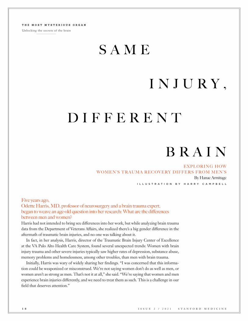
Five years ago, Odette Harris, MD, professor of neurosurgery and a brain trauma expert, began to weave an age-old question into her research: What are the differences between men and women?Harris had not intended to bring sex differences into her work, but while analyzing brain trauma data from the Department of Veterans Affairs, she realized there’s a big gender difference in the aftermath of traumatic brain injuries, and no one was talking about it.
In fact, in her analysis, Harris, director of the Traumatic Brain Injury Center of Excellence at the VA Palo Alto Health Care System, found several unexpected trends: Women with brain injury trauma and other severe injuries typically saw higher rates of depression, substance abuse, memory problems and homelessness, among other troubles, than men with brain trauma.
Initially, Harris was wary of widely sharing her findings. “I was concerned that this informa-tion could be weaponized or misconstrued. We’re not saying women don’t do as well as men, or women aren’t as strong as men. That’s not it at all,” she said. “We’re saying that women and men experience brain injuries differently, and we need to treat them as such. This is a challenge in our field that deserves attention.”
1 8 I S S U E 2 / 2 0 2 1 S T A N F O R D M E D I C I N E
T H E M O S T M Y S T E R I O U S O R G A N
Unlocking the secrets of the brain
S A M E
I N J U R Y ,
D I F F E R E N T
B R A I NEXPLORING HOW
WOMEN’S TRAUMA RECOVERY DIFFERS FROM MEN’SBy Hanae Armitage
I L L U S T R A T I O N B Y H A R R Y C A M P B E L L


To better understand the nature of brain trauma in wom-en — physiologically, psychologically and socially — Har-ris teamed up with colleagues, including Maheen Adamson, PhD, a clinical scientific research director for Rehabilitation Services at the VA Palo Alto and a clinical associate professor of neurosurgery at Stanford School of Medicine. Using data from surveys, neuropsychological testing and brain imag-ing, they have conducted matched analyses comparing male and female patients, meaning that, sex aside, the comparison groups’ specifics — age, severity of injury and time since the injury — were equal.
Their work has so far revealed some big differences in the brains and behavior of men and women with post-trauma in-juries — insights that could guide treat-ment for women who have suffered de-bilitating injuries to the head.
Lisette Meylan is grateful for the new direction. In 2004, her daughter, Mariela, who was on duty in Kuwait, suffered se-vere head and other injuries when a car hit her and four other soldiers as they changed a flat on their truck. She survived the accident but ended up in a coma, re-ceiving care in a nursing home for veter-ans in Washington, D.C. “Her doctors told me I needed to be prepared for my daughter to never wake up,” Meylan said.
But Meylan could not give up on her daughter, so she moved her closer to home, in Livermore, California, to the VA’s Livermore division. There, Meylan and her daughter’s care team tried differ-ent therapies to wake her from a vegeta-tive state. It seemed all but hopeless. Two years passed. Then, one day, Meylan saw a light blinking on her phone’s message machine, indicating a new voicemail.
She played the recording: “This is Mari-ela, I’m your daughter, and I love you.”
“Those were the first words she’d spo-ken in two years,” said Meylan. Since then, her daughter’s recovery has been chal-
lenged by physical and mental hurdles, such as learning to walk again, but she has progressed immensely.
“My biggest challenge is my memory,” said Mariela Meylan. That’s more common for women who have expe-rienced multiple traumatic injuries, compared with men, according to Adamson. “My short-term memory has been affected the most. But through the support of my family and my team of practitioners, I’m able to continue to heal and show up for my life.”
In 2014, she participated in a storytelling workshop run by Harris for women who’ve experienced traumatic brain injury to share their stories with other women who have the diagno-sis and health care professionals. Through intensive physical therapy at the Livermore VA, she now regularly practices yoga, rides horses and swims. She lives with her mother, who helps her navigate other day-to-day activities, like making meals.
2 0 I S S U E 2 / 2 0 2 1 S T A N F O R D M E D I C I N E
STUDIES BY NEUROSCIENTISTS ODETTE HARRIS, LEFT,
AND MAHEEN ADAMSON REVEAL KEY DIFFERENCES IN HOW BRAIN
TRAUMA AFFECTS WOMEN WHEN COMPARED WITH MEN
WHO HAVE SIMILAR INJURIES. THE NEUROSURGERY PROFESSORS
HOPE THEIR INSIGHTS LEAD TO BETTER TREATMENT AND
RECOVERY FOR FEMALE PATIENTS.
LE
SL
IE W
ILL
IAM
SO
N

“Patients like Mariela are the reason we do this,” said Ad-amson. “The stories of their strength, perseverance and mo-tivation give my research a purpose and motivate me to never stop discovering.”
Surveys and analysis of health record data by the Stanford researchers and others continue to find stark differences in how men and women experience severe brain injury. But there’s also a physical clue: The imaging research suggests a link between a physical trait of women’s brains — a thinning of part of the cortex — and the tendency to experience a different array of post-brain injury symptoms than men do.
Their analysis will help fill in research gaps. “Females ac-count for 15% of the traumatic brain cases we see, yet the studies investigating TBI comprise data almost exclusively from men,” said Adamson.
SETTING WOMEN UP TO SUCCEEDIN HER DEEP DIVE into the Armed Forces Health Surveil-lance Center data from 2000 to 2010, Harris found several key differences in the aftermath of severe head trauma for men and women, including that women are four times more likely to abuse drugs, seven times more likely to be homeless and about three times more likely to be unemployed.
Women with traumatic brain injury are also 30% more likely than males to suffer from post-traumatic stress disor-der. And they experience higher rates of vertigo — the feeling that the environment is moving (often spinning) around you.
Part of the research goal is to figure out how best to set wom-en up for success after brain trauma. It’s not always the same as what’s best for men. “For instance, when we see unemployment in males with traumatic brain injury, our approach is to assist in education and skills training,” said Harris.
“So the knee-jerk reaction is to find ways to in-crease education and training when we see unem-ployment in women with traumatic brain injury. But we found that female veterans were better edu-cated and more likely to have a college degree than their male counterparts.”
So education and skills training might not be as helpful for women as it is for men.
BRINGING IT BACK TO THE BRAINWHAT’S CAUSING THE differences in the impact of brain in-jury trauma on women and men?
In 2016, Adamson began investigating, using neuropsy-chological testing and brain imaging. The tests gauged gen-eral brain function and memory, among other abilities. The imaging portion of the study, which comprised 70 veterans (28 women and 42 men) used MRI to measure the thickness of the cortex, the thin outer layer of the brain’s cerebrum.
“Scientists have looked at how cortical thickness changes in a variety of neurological diseases, such as schizophrenia, and we thought it made sense to start there for this research, too,” said Adamson.
Under healthy conditions, women’s cortex is about 6% thicker than men’s. In the MRI study, injured brains of all veterans exhibited signs of cortical thinning, only for women it was significantly worse.
The brains of the women she studied had more patches of cortical thinning, especially in regions that regulate emotion and decision-making. Scientists know cortical thinning is not good, but it’s too early to say how the condition impacts be-havior or overall health of the brain.
Researchers are recruiting more participants to further explore how cortical thinning impacts symptoms and post-brain injury outcomes for women, said Adamson. “We’re just hitting the tip of the iceberg here.”
She and Harris are also considering other populations of brain trauma survivors and how their experiences differ.
“I see our research as aligning well with a shift we’re see-ing at the national level — incorporating gender, race, abil-ity and other differences into science and patient health,”
said Harris. “We’re seeing a shift toward looking at differ-
ences between male and female traumatic brain injury more deeply, and my hope is that that trend will extend to other groups within the traumatic brain injury patient population. That’s what will enable us to improve outcomes and ensure equi-table care for all people, not just women.” — Contact Hanae Armitage at [email protected]
2 1S T A N F O R D M E D I C I N E I S S U E 2 / 2 0 2 1
W E B E X T R A
Watch a video about a woman’s experi-
ence with brain trauma: stan.md/
neurotrauma
‘ M Y B I G G E S T C H A L L E N G E I S M Y M E M O R Y . ’ T H A T ’ S M O R E C O M M O N F O R W O M E N W H O
H A V E E X P E R I E N C E D M U L T I P L E T R A U M A T I C I N J U R I E S , C O M P A R E D W I T H M E N .
‘ M Y S H O R T - T E R M M E M O R Y H A S B E E N A F F E C T E D T H E M O S T . ’

S T A N F O R D M E D I C I N E I S S U E 2 / 2 0 2 1 2 3
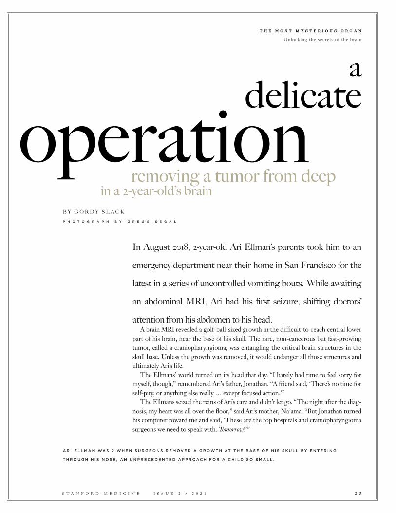
In August 2018, 2-year-old Ari Ellman’s parents took him to an
emergency department near their home in San Francisco for the
latest in a series of uncontrolled vomiting bouts. While awaiting
an abdominal MRI, Ari had his first seizure, shifting doctors’
attention from his abdomen to his head. A brain MRI revealed a golf-ball-sized growth in the difficult-to-reach central lower
part of his brain, near the base of his skull. The rare, non-cancerous but fast-growing tumor, called a craniopharyngioma, was entangling the critical brain structures in the skull base. Unless the growth was removed, it would endanger all those structures and ultimately Ari’s life.
The Ellmans’ world turned on its head that day. “I barely had time to feel sorry for myself, though,” remembered Ari’s father, Jonathan. “A friend said, ‘There’s no time for self-pity, or anything else really … except focused action.’”
The Ellmans seized the reins of Ari’s care and didn’t let go. “The night after the diag-nosis, my heart was all over the floor,” said Ari’s mother, Na’ama. “But Jonathan turned his computer toward me and said, ‘These are the top hospitals and craniopharyngioma surgeons we need to speak with. Tomorrow!’”
A R I E L L M A N WA S 2 W H E N S U R G E O N S R E M O V E D A G R O W T H AT T H E B A S E O F H I S S K U L L B Y E N T E R I N G
T H R O U G H H I S N O S E , A N U N P R E C E D E N T E D A P P R O A C H F O R A C H I L D S O S M A L L .
T H E M O S T M Y S T E R I O U S O R G A N
Unlocking the secrets of the brain
removing a tumor from deep in a 2-year-old’s brain
a delicateoperation
B Y G O R D Y S L A C KP H O T O G R A P H B Y G R E G G S E G A L

A D A R I N G A P P R O A C H
m ILLIONS OF YEARS of evolution buried such essential brain struc-tures as the pituitary gland and hy-pothalamus in the bottom middle of the human head where they
would be well-protected from a world full of sharp and heavy dangers. So, it is no accident that the same important part of the brain, known as the skull base area, is notoriously difficult for surgeons to reach. For the first century of modern brain surgery, the only way to get there was by opening the top of the head, spreading the brain’s hemispheres apart, and tun-neling down between them to the core.
Because the optic nerves, which connect the vision-pro-cessing part of the brain to the eyes, stand between the skull base area and that cranial opening, surgeons often had to work around those delicate and vulnerable structures, too. Collateral damage to essential brain tissue on the way down could be devastating, and further damage could be imposed when surgeons were pulling a tumor up and out.
In the past decade, though, advances in imaging, surgical anatomy and surgical tools have enabled surgeons to use a less destructive approach. Instead of entering the skull from above, they enter through the nose and sinus area, just be-low the hardest-to-reach skull base structures. This method, known as transnasal endoscopic skull base surgery, has be-come the preferred method for removing tumors in this part of the brain — but only in adults. Children have much smaller sinus cavities, and at the time Ari became ill, surgeons still approached pediatric skull base tumors the old-fashioned way — open surgery from above.
However, as the Ellmans would soon discover, an extraor-dinary team of Stanford neurosurgeons and rhinologists be-lieved a transnasal approach would be feasible even in small children. They just needed the right case to prove it.
If successful, they would not only have an opportunity to save the life of a dangerously ill patient but also to pro-vide pediatric neurosurgeons with a technique for skull base surgeries in small children for generations to come. Their
method would give them direct access to essential parts of the young brain that have been excruciatingly hard to reach. A failure would make that path much more difficult for fu-ture surgeons to take — or even consider.
In the first week after learning Ari’s diagnosis, his parents sent his case to tumor boards — multidisciplinary groups of specialists — at 15 leading medical centers. Some suggested old-school open-brain surgery, which, in addition to the ob-stacles described above, often fails to remove the entire tu-mor, partly because the roots of craniopharyngiomas, at the bottom of the brain, may be inaccessible from above.
Other surgical groups suggested radiation, but that can cause devastating and lasting side effects in a young child. It was the third and rarest option suggested, transnasal endoscopic skull base surgery, that really caught the family’s attention. For this method, surgeons slide endoscopes — thin tubes with a light and camera, through which surgical tools can pass — into the brain via the patient’s nose. Unfortunately, only a handful of endoscopic skull base craniopharyngioma surgeries had been conducted on young children, and none of those children was younger than 5. Ari was only 2. It wasn’t just Ari’s age and size that made the surgery an extraordinary challenge, but it was also his tumor’s relatively large size and specific characteristics. It consisted of multiple cysts, and portions of it were calcified.
Most surgeons the Ellmans contacted wouldn’t even consider endoscopic skull base surgery for a huge craniopharyngioma in a child like Ari. It would be an unprecedented operation requiring extraordinary degrees of both expertise and technology that were available at only a few surgical centers around the world.
But not only were doctors at Lucile Packard Children’s Hospital Stanford willing to try it, they also recognized it as an opportunity to advance surgical knowledge, said Juan Fernan-dez-Miranda, MD, a skull base surgeon who was recruited to Stanford from the University of Pittsburgh just a couple months before Ari’s family approached Stanford. It was a case with the right patient, the right surgeons, the right family and the right technology, all coming together in one place. “I felt like I’d been preparing for this surgery for 15 years,” said Fernandez-Miran-da, professor of neurosurgery and surgical director of the Stan-ford Brain Tumor, Skull Base and Pituitary centers.
2 4 I S S U E 2 / 2 0 2 1 S T A N F O R D M E D I C I N E
‘This same tumor in an adult patient would still be very difficult to remove. ... Now add a 2-year-old
patient to the picture and you get a truly unique case...’

The Stanford surgical team also included Gerald Grant, MD, Stanford’s chief of pediatric neurosurgery, and Peter Hwang, MD, professor of otolaryngology and a world-re-nowned endoscopic otolaryngologist who has been conduct-ing adult and pediatric endonasal sinus surgery for over 20 years. Hwang is also division chief of rhinology and endo-scopic skull base surgery.
“We have a unique combination of endonasal skull base surgery expertise and pediatric neurosurgery experience. Both are essential for a resection like Ari’s,” said Grant, the Botha Chan Endowed Professor.
When the Ellmans met Grant, Hwang and Fernandez-Miranda, they knew they had found their team. Their de-cision was reinforced by the group’s record of surgical ex-cellence, their focus on pediatrics, their attentiveness and warmth, and the advanced technology dedicated to neuroen-doscopy in Lucile Packard Children’s Hospital’s surgical suites.
“This same tumor in an adult patient would still be very difficult to remove, even for very experienced neurosur-geons,” said Fernandez-Miranda, explaining the challenges of Ari’s case. “Now add a 2-year-old patient to the picture and you get a truly unique case, never done before — not just difficult, but thought by many to be impossible.”
TA P P I N G T E C H N O L O G Y T O H E L P
P R E PA R E F O R S U R G E R Y
pREPARATIONS BEGAN WEEKS before the surgery. A single high-resolution 3-D digital image of Ari’s brain was created by combining several simpler digital images — such as MRIs and CT scans —
then loaded into a virtual reality tool called Surgical Theater. The tool’s users wear virtual reality headsets that turn the 3-D brain image into what video game players call an “immersive environment” — a “landscape” through which users seem to be moving around and exploring at will. Except, instead of moving around inside a digitally constructed room, as they might in a game, this tool allows users to move around in-side their patient’s brain and to closely study the geography of that brain — its nerves, ventricles, arteries and other essential structures, as well as tumors. They can plan the trajectories their surgery could take and the effects different approaches could have on nearby brain tissue. Using the tool, Ari’s sur-gical team carefully mapped and rehearsed the best possible path to his tumor and the best way to remove it while protect-ing essential brain structures.
“Clearly visualizing the brain structures surrounding the tu-
mor in advance is key,” said Fernandez-Miranda. In addition to the virtual digital modeling, a resin scale model of Ari’s skull base was 3-D printed so the team could take it to the Stanford Neurosurgical Training and Innovation Center, which Fernan-dez-Miranda directs, to plan and practice for several hours with actual surgical tools and “to make sure we had enough space in the nasal cavity to get into the skull base safely.”
“A 2-year-old’s sinuses are only 20 millimeters wide or nar-rower. And you’re removing a tumor that may be wider than the nasal passage itself,” said Hwang. “It’s like getting a ship out of a bottle. You have to figure out how to take it apart and bring it out through this very narrow corridor. You don’t want to wait until it is game time to iterate and innovate — you re-ally need to have your plans in place well before the day of sur-gery. That’s why these additional technologies can play such an important role in pediatric skull base surgery in particular.”
M O V I N G T H R O U G H T H E B R A I N W I T H
E X T R E M E C A R E
bY THE DAY OF THE OPERATION, the Ellmans felt they had done everything they could to ensure Ari had the best place, the best doctors and the very best chances of success, Ari’s mother said. Still, when they left their
home at 5 a.m. on Feb. 8, 2019, they began “by far the hardest drive we’d ever taken,” she said. “At the end of it, we knew we’d be handing him over and it would be out of our control.”
Ari’s prep for surgery began at dawn, with the setup of the same 3-D digital modeling and navigation system used to rehearse the operation, but this time it was anchored to actual landmarks in Ari’s brain so surgeons could see on the monitor exactly where their tools stood relative to both the tumor and to critical brain structures.
By the beginning of the second hour in the OR, Hwang was slipping his endoscopes into each of Ari’s nostrils as he began to navigate the space between Ari’s nose and brain, “creating corridors through the nasal passages by opening sinuses, combining nasal passages, removing bone, and con-verting two separate nasal chambers into one chamber with more access to — and better visualization of — what will be the avenue to Ari’s brain,” said Hwang.
Two hours in, Grant and Fernandez-Miranda enlarged the corridor into the base of the skull using high-speed drills and opened the dura, the thick membrane that seals off the brain from the rest of the world. The tumor was close to the point of entry, but this part of the brain is
S T A N F O R D M E D I C I N E I S S U E 2 / 2 0 2 1 2 5
C O N T I N U E S O N P A G E 4 2


T H E M O S T M Y S T E R I O U S O R G A N
Unlocking the secrets of the brain
S T A N F O R D M E D I C I N E I S S U E 2 / 2 0 2 1 2 7
AROUND 10:30 P.M. ON APRIL 23, 2017, Cindi Dodd ended a phone call with her best friend, turned off her bedside light, and settled in for a night’s sleep in her Salinas, California, home ahead of a surgery the next day.
The scheduled operation was a breast reconstruction following a bout with cancer, but when Dodd’s hus-band, Rick, woke her at 5 a.m., something else was terribly wrong. She seemed to be having a seizure. She couldn’t move her left arm or leg, and when she spoke, only gibberish came out.
Rick Dodd called 911 and yelled to their son to turn on the porch light for an ambulance. At the hospital, the family found out that a blood clot had lodged in Cindi Dodd’s middle cerebral artery, preventing blood and nutrients from flowing to her brain. While she was asleep, the 46-year-old graphic designer had experi-enced an acute ischemic stroke. Cells in her brain had begun to die.
Doctors told Dodd’s family they could do nothing to remove the blockage: Too much time had lapsed since she was last known to be well. According to standard guidelines, after six hours from the onset of a stroke, the risk that treatment would cause dangerous bleeding in the brain and elsewhere in the body outweighed the benefits of restoring blood flow to damaged tissue.
O P E N I N G
S T R O K E ’ S
W I N D O W
Treating stroke more than a few hours after the crisis is no longer considered absurd
By Amy Jeter HansenI L L U S T R A T I O N B Y J O N A T H O N R O S E N

Her best option, an emergency physician told her hus-band, was to get to Stanford Health Care. There, doctors were leading a large clinical trial to determine if the narrow window of time for stroke treatment could be widened for patients under some circumstances.
Rick Dodd didn’t hesitate. Within an hour, a helicopter ar-rived, and Cindi Dodd was on her way to Stanford Hospital.
“That’s my baby. Take care of her,” Dodd’s mother told the flight nurse through tears.
E X T E N D I N G
T R E AT M E N T T I M E L I N E
ON THE OTHER SIDE OF THAT FLIGHT, at Stanford Hospi-tal, Gregory Albers, MD, was leading the most critical clini-cal trial of his 30-year career.
He became fascinated with the human brain when he was a teenager, in California’s San Fernando Valley. Since com-pleting a stroke research fellowship at the Stanford Univer-
sity School of Medicine in 1988, he had dedicated his work to improving the lives of stroke patients.
Whenever stroke patients arrived at the hospital too late for a dismal outcome to be prevented, Albers always felt dismayed. He wondered if some patients who arrived many hours after their stroke began might still benefit if blood flow was restored to their brain tissue. And if so, was there a way to identify which patients could be helped during this longer period?
These questions would frame his professional efforts for nearly three decades, beginning in the 1990s.
During that time, through a methodical progression of research, Albers and his colleagues chipped away at the status quo of stroke treatment. They tested — then refined — a new imaging technique that could quickly reveal the damage wrought by a stroke, as well as the at-risk tissue that could still be saved. Gradually winning over skeptics, they crafted a protocol that added precious hours to the timeline for stroke care and carried immense potential to help countless future patients avoid severe disability or worse.
“It was a huge challenge,” Albers said of his quest to improve outcomes for stroke patients. “We were dealing with the No. 1 cause of disability worldwide, and there was no treatment.”
A D VA N C I N G
B R A I N I M A G I N G
THE STANFORD STROKE CENTER opened in 1992, led by Albers, neuroradiologist Michael Marks, MD, and neuro-surgeon/neuroscientist Gary Steinberg, MD, PhD. The center’s aim was to develop new therapies and bring them to patients. “We were not satisfied with this nihilistic approach, that stroke is something you can never treat,” said Steinberg, who is the Bernard and Ronni Lacroute-William Randolph Hearst Professor in Neurosurgery and Neurosciences.
Within a year of establishing the center, its leaders were approached by Michael Moseley, PhD, a physicist who had recently joined Stanford Medicine’s radiology department. Moseley was studying ways to capture images of brain dam-age from a stroke, and he’d made an intriguing discovery.
At the time, because of limitations of conventional mag-netic resonance imaging and computed tomography, evi-dence of injury from a stroke didn’t appear on a scan until four or more hours after a patient’s symptoms began. When brain cells begin to die during a stroke, they lose the ability to maintain a balance of water inside and outside the cell. Conventional techniques could capture images of dead cells swamped with water hours later, but Moseley wanted an ear-
2 8 I S S U E 2 / 2 0 2 1 S T A N F O R D M E D I C I N E
WHEN CINDI DODD WOKE UP AFTER HER STROKE,
SHE REALIZED THAT BEING IN A STANFORD TRIAL TO EXTEND
THE TREATMENT TIMELINE GAVE
HER ‘A CHANCE TO FIGHT FOR MY LIFE.’ LE
SL
IE W
ILL
IAM
SO
N
lier indication of what was happening.In a late-night laboratory insight, he discovered that he
could track a stroke’s damage to brain tissue as it happened through an MRI technique that was more sensitive to the movement of water in cells. Scans created through diffusion-weighted imaging showed a clear difference between cells that had no active transport of water and those with normal water-shuttling activities.
In short, Moseley had found a way to watch brain cells die in real time.
To Albers, “It seemed like the holy grail.”With Moseley, Marks and other Stanford colleagues, Al-
bers embarked upon a series of studies that used diffusion-weighted imaging to track stroke patients for weeks. The resulting images — with bright white smudges showing dead tissue, in contrast to healthy gray tissue — depicted a process that happened at different rates for different people, but often took more than a day to reach maximum size. This discovery ran counter to the prevailing belief that a stroke completed its path of destruction within one to two hours.
Watching the progression of images brought the re-searchers to the next logical step: “I could see dead brain, but I couldn’t predict what was destined to die,” Moseley, a professor of radiology, said.
They decided to follow the blood. Using a technique called perfusion-weighted imaging, they injected a contrast agent into a patient’s bloodstream and monitored how long it took the contrast to reach distinct areas of the brain. If a major blood vessel was blocked, the contrast agent’s journey to that side of the brain was delayed.
It was hard to tell from a brightly colored perfusion-weighted image whether tissue was dying or already dead. However, when the researchers compared it with a diffusion-weighted image, which clearly depicted dead tissue, they could determine whether there was a mismatch area — brain tissue that had not died but was marked for demise if the artery’s blockage wasn’t cleared.
They realized they could determine whether a person’s stroke was finished. If it was ongoing, they could predict how it would play out if physicians couldn’t open the blood vessel.
Their rough data suggested that about three-quarters of stroke patients could still benefit from treatment after six hours; after 12 hours, treatment could still help about half of them. This was radically different from what animal models had shown: In rats, a stroke would typically complete within 90 minutes. In monkeys, three hours was the upper limit for avoiding brain damage, according to a pivotal study.
In their 1999 paper, Albers and his colleagues predicted
that late-window therapy could improve stroke outcomes, but their study was met with ambivalence from other neu-rologists. Some were intrigued, while many others thought this approach had no chance of success.
“Neurologists in general tend to be skeptical people,” said Albers, the Coyote Foundation Professor, “and there had been a number of stroke trials in the past — many, many stroke trials that had been done in the ’80s and ’90s — that neurologists were initially excited about. They all failed.”
A P R O M I S I N G
N E W D R U G
BEFORE THE 1990s, physicians routinely let a patient’s stroke run its course, convinced there was nothing they could do. This changed as scientists gained a better understanding of the rate at which human brain cells disintegrate and as doctors tested tools for restoring blood flow to oxygen-starved tissue.
A clot-dissolving drug called tissue plasminogen activator — known as tPA — showed promise. It works by activating plasmin, an enzyme responsible for breaking down blood clots. However, because tPA can increase the risk of bleeding every-where in the body, it can cause serious bleeding complications.
In 1996, the U.S. Food and Drug Administration ap-proved tPA for treating ischemic strokes — those caused by a blocked artery — but only within three hours of the first symptoms. After three hours, the thinking went, it was too late to save the dying brain tissue, and the risk of the drug causing further harm outweighed any potential benefit. Because many patients are unable to seek medical care im-mediately, the time constraint strictly limited the number of patients who could be treated with tPA.
Albers believed more people could be helped.In 2001, after he, Moseley and their team published sev-
eral preliminary studies about the new imaging technique, they were funded by the National Institutes of Health to per-form a study of stroke patients who had not arrived in time to receive tPA. The patients would undergo the new imaging techniques and be given tPA between three and six hours af-ter their first stroke symptoms.
Because tPA works for only about half of the patients who receive it, that meant the researchers could test their imag-ing technique in two ways. For patients with a mismatch and whose blockage persisted, they could see whether the stroke proceeded as predicted. For patients with restored blood flow, they could see if the intervention prevented damage in the brain areas predicted to be salvageable, and if those pa-tients had better outcomes.
S T A N F O R D M E D I C I N E I S S U E 2 / 2 0 2 1 2 9

T H E S T A N F O R D S T U D I E S S U G G E S T E D T H A T F O R P A T I E N T S F L A G G E D T H R O U G H I M A G I N G ,
T R E A T M E N T C O U L D S U C C E E D U P T O 2 4 H O U R S A F T E R S Y M P T O M S B E G A N .
The results, published in 2006 as the Diffusion and Perfu-sion Imaging Evaluation for Understanding Stroke Evolu-tion (DEFUSE) study, turned out exactly as Albers and the Stanford team had hoped and predicted.
Patients with a mismatch whose arterial blockage was dis-solved showed improvement in functioning, such as regaining control of their limbs, as measured on the NIH Stroke Scale. This suggested that the imaging had successfully identified salvageable tissue in individual patients and that restoring blood flow by administering tPA had saved the tissue.
What’s more, the study indicated that more than half of the patients still had brain cells that could be saved five to six hours after a stroke.
“Clearly,” Albers thought, “we’re on to something here.”
E X PA N D I N G T H E
L I M I T S HIS TEAM’S NEXT STUDY pushed the envelope further.
Though the design of the second DEFUSE trial was similar to the first, there were two crucial differences. First, the time window was doubled to 12 hours. Second, patients would be treated with a new technique called thrombecto-my, in which a physician threads a catheter through the ar-tery to the clot, then deploys a mechanical device to extract it. Additionally, the team had developed an imaging software called RAPID that automatically analyzed the diffusion and perfusion images and calculated the volume of salvageable tissue; the nine medical centers participating in the study all used the new software in the trial.
Some of the researchers’ peers expressed doubts about the study’s premise — 12 hours was so much longer than the ac-cepted window for stroke treatment. But when the second DEFUSE study was published in 2012, six years after the first, the findings were undeniable.
“It worked beautifully,” Albers said.Their results suggested that the window for successful
treatment could be extended to 12 hours for some stroke pa-tients. But they had not yet conclusively shown that patients selected with their imaging technique benefited from late treatment. Doing so required a randomized trial, which is what Albers wanted to do next.
‘ B R A I N I S
O F T H E E S S E N C E ’
IN THEORY, thrombectomy devices, with their capability to clear blockages, were the perfect instrument to help physi-cians save brain tissue in stroke patients. However, studies of early versions showed disappointing results. Although the devices could reliably restore blood flow to oxygen-deprived brain tissue, stroke patients’ ability to function often didn’t improve much.
In the years after the DEFUSE 2 study was published in 2012, manufacturers improved the devices. Meanwhile, Albers wondered if many of the patients being treated already had ir-reversible injury. He wanted to find out if patients who under-went a thrombectomy fared better when the Stanford team’s imaging had shown salvageable tissue prior to the procedure.
Before the team could secure funding for a third DE-FUSE trial, however, several studies were published that seemed to confirm Albers’ theory, widening the stroke treat-ment window to six hours in certain cases. Two had used the RAPID software, which was now owned by a company Al-bers had co-founded, and these studies produced the greatest treatment benefits. Among patients with scans that showed salvageable tissue, those who received a thrombectomy with-in six hours of a stroke showed better outcomes than those who had not undergone the procedure. In 2015, the Ameri-can Heart Association/American Stroke Association changed treatment guidelines to reflect what the studies had found.
But the Stanford researchers did not believe the issue was settled, as six hours was not long enough to get to the major-ity of stroke patients; many live far from a stroke center or have the stroke while they’re sleeping. The Stanford studies suggested that for patients flagged through imaging, treat-ment could succeed up to 24 hours after symptoms began. The team was finding that “time is not of the essence. Brain is of the essence,” said Maarten Lansberg, MD, PhD, a Stan-ford professor of neurology who joined the effort in 1997.
The third DEFUSE trial, also funded by the NIH, be-gan in 2016 with more than three dozen medical centers participating. The randomized study focused on patients whose stroke was caused by a blood clot obstructing one of two large arteries in the brain — the middle cerebral artery or the internal carotid artery. This happens in about 1 in 4
3 0 I S S U E 2 / 2 0 2 1 S T A N F O R D M E D I C I N E

Albers thought back across the decades — his entire ca-reer. He thought about the times he’d had to tell patients, “I’m sorry. You came in too late. We can’t treat you.” About how, for some future patients, the likelihood of death and disability was now cut in half.
“This is going to change the world,” he thought.The publication of the DEFUSE 3 study in the New
England Journal of Medicine was timed to coincide with the American Heart Association’s International Stroke Confer-ence in January 2018. On the day Albers presented the results of DEFUSE 3, the association announced changes to treat-ment guidelines for acute ischemic stroke, recommending a treatment window for mechanical clot removal up to 24 hours after onset in certain patients with clots in large vessels.
William J. Powers, MD, chair of the neurology department at the University of North Carolina at Chapel Hill at the time, headed the committee in charge of recommending guide-lines. He knew that many researchers had conducted similar
ischemic strokes and accounts for the most disabling strokes. If the RAPID software showed potentially salvageable
brain tissue six to 16 hours after a stroke’s onset, a patient was randomly assigned to have either standard medical treatment or an alternative that included both the standard treatment and a thrombectomy. Participants were tracked for three months, the period when stroke patients typically experience most of their recovery.
Meanwhile, another study using the Stanford-designed software had been launched in 2014 by Stryker Corp., which manufactures devices for stroke care. The design of Stryker’s trial, called the DAWN study, was almost identical to the DEFUSE 3 study with one significant difference: It pushed the treatment window to 24 hours after a stroke’s onset, com-pared with 16 hours in DEFUSE 3.
After an analysis of interim data, the data safety and moni-toring committee stopped enrollment in the DAWN trial ear-ly, in February 2017. Among 206 stroke patients whose scans showed salvageable brain, those who underwent a thrombecto-my experienced less disability than the control group. Because the DEFUSE 3 study was so similar, the NIH placed it on hold soon afterward, in June 2017, to evaluate preliminary findings.
Those weeks were nerve-wracking for the Stanford team, who waited to see what would come next for the study they’d worked so long to begin.
“We were nervous,” said Stephanie Kemp, the Stanford stroke center’s program manager. “They could have said, ‘You don’t need any more patients. You’ve proven what you need to prove.’ But they also could have said, ‘Oh, no, you’re not there yet, and you’re not going to get there.’”
C H A N G E S I N
T R E AT M E N T G U I D E L I N E S
IN FALL 2017, when Albers finally saw the data from 182 par-ticipants in the Stanford-led trial, he couldn’t sleep.There was no need to restart enrollment. The data showed that the imaging technique Albers and his colleagues had re-fined over so many years could help physicians determine when they could do more for a stroke patient and substan-tially improve the patient’s recovery. Three months after a stroke, 45% of the patients who received a thrombectomy six to 16 hours after their first symptom were functionally independent, compared with 17% who received standard care. Among patients receiving the thrombectomy, 14% died within three months of having a stroke, compared with 26% in the control group. The team’s findings underscored the results of the DAWN trial.
S T A N F O R D M E D I C I N E I S S U E 2 / 2 0 2 1 3 1
C O N T I N U E S O N P A G E 4 2
A QUEST TO EXTEND THE TREATMENT WINDOW TO HELP
STROKE VICTIMS AVOID DEBILITATING
OUTCOMES HAD DEFINED GREGORY ALBERS’ CAREER
FOR THREE DECADES.
LE
SL
IE W
ILL
IAM
SO
N

A C O N V E R S A T I O N W I T H F L U T E V I R T U O S O E U G E N I A Z U K E R M A N O N A L Z H E I M E R ’ S , M U S I C , P O E T R Y A N D J O Y
“What seems to have saved me from crumbling and falling apart,” she has said, “was music, love, poetry and, oddly, laughter.”
Classical music fans know her, The Boston Globe exclaimed, “as one of the finest flutists of our times.” Enthusiasts of the CBS Sunday Morning show recognize her as the show’s sharp-minded arts correspondent, a role she held for more than 25 years. For the millions of people living with Alzheimer’s, her poems — heart-breaking and honest — may offer hope or solace.
Contributing editor Paul Costello spoke with Zukerman at the end of 2019 when her book of poetry was first published. He checked in with her recently, just as she received her second dose of the Moderna COVID-19 vaccine. Her husband, Dick Novick, joined her on the phone and helped when she struggled for a word. This Q&A was edited and condensed from those conversations. COSTELLO Although the Delta variant is wreaking havoc in many parts of the United States, for many of us who’ve been vaccinated there’s a new normal. How are you doing?ZUKERMAN I feel really good. I most look forward to seeing my grandchildren in Washington, D.C. It’s torment not to be able to see and hold them.
As far as Alzheimer’s, I am towards the end of the first part, but I feel I am very functional. I know what I want to do, and I am lucky that I’m able to do those things. I start my day by rolling out of bed and doing floor exercises. Of course, I do play my flute a lot. My flute is my best friend, my other. For me, it’s a very positive time in my life. I feel very blessed and I try to simply live every day.
COSTELLO You said you feel you’re at the end of part one? ZUKERMAN I don’t feel that I am in stage two, when one needs more help, but I know it’s getting significantly closer for me. Stage three is hospitalization, and I feel I’m nowhere near that. I’m lucky. I do a lot of exercise. I have friends. I love to play with the animals. I am still
3 2
RIC
CA
RD
O V
EC
CH
IO
performing — virtually — so for me, it has not been a terrible experi-ence. In fact, I’ve learned a great deal through this disease.
COSTELLO What have you learned? ZUKERMAN I think the important lesson is that you try your best. I want people to understand I am not suffering. It’s so important to stay positive. I would say one of the most important things is to be as energetic as you can. What’s been important is to also accept the fact that I’m not perfect. I am flawed.
COSTELLO Since you’ve written your book of poetry, what have you heard from readers? ZUKERMAN Before the pandemic, I was doing [struggling for the words, she turns to her husband] signings, yes, signings at book-stores.
There was one very elegant man in line who bought six books. When he reached me, he said, “I can’t thank you enough, because my family has never understood what I’m going through as a per-son who has Alzheimer’s. What you told us here tonight made me understand, and I’ll be able to go back to my family and they will understand more what I am going through.”
That was a wonderful feeling.
COSTELLO Many people diagnosed with Alzheimer’s are filled with fear and anxiety. Were you? ZUKERMAN I was with my younger daughter when I had the diagnosis; I was not afraid. I knew that I had the condition. For some reason, I didn’t cry. I think I just took it for what it was. I brought it home with me.
When I got up to the apartment and sat down at my desk, I stared at the wall for a while and, for some reason, put some paper down and started to write. That’s how the book came about.
COSTELLO What were you thinking as you stared at the wall?
I S S U E 2 / 2 0 2 1 S T A N F O R D M E D I C I N E
a way through the brambles
Nearly four years ago, internationally renowned flutist Eugenia Zukerman was diagnosed with Alzheimer’s disease. Almost as soon as she heard the verdict after a neuro-psychiatric exam, she sat down to write. What emerged was Like Falling Through a Cloud, a lyrical book of poetry that unfurls her journey through gradual cognitive impairment and memory loss.

A C O N V E R S A T I O N W I T H F L U T E V I R T U O S O E U G E N I A Z U K E R M A N O N A L Z H E I M E R ’ S , M U S I C , P O E T R Y A N D J O Y
T H E M O S T M Y S T E R I O U S O R G A N
Unlocking the secrets of the brain
ZUKERMAN I wasn’t really thinking about what I had just gone through. I was just thinking that this is a new set of circumstances. It’s sudden … but I am not afraid. I haven’t been afraid throughout this whole process.
COSTELLO One of your poems is entitled “Marbles.” Why marbles?ZUKERMAN Marbles are beautiful. They make noise. They make mu-sic. I don’t know why the marbles metaphor came to me in such a strong way, but it seemed to me that what had been a very stable life was now pulling apart and rolling around like marbles.
COSTELLO The title of your book of poetry is so beautiful, Like Falling Through a Cloud. What meaning does that have for you? ZUKERMAN My mother died at the age of 103. She often talked, even in the last days, of how she was float-ing. I had the idea of me floating and just lying up in the sky, and there’s a moment in which you must fall.
I think once you have fallen through a cloud, there is a certain moment of clarity. The clarity for me was the un-derstanding that yes, I had a condition, and yes, it was a death sentence, but I wasn’t afraid. I think it’s a gift. I don’t know where that comes from. Because I’m not a terribly brave person, I feel as if I’ve handled this pretty well. The book, I think, is rather joyful.
COSTELLO You wrote the book for yourself, as a way to channel your Alzheimer’s, but you also wrote the book for other people who struggle with cognitive difficulties.ZUKERMAN I absolutely did. I had met people who were having tre-mendous problems; they were terrified. I felt that it was not good to be terrified. I wanted people to understand that not only can they find their way through this, but they can come out, in many ways, stronger.
COSTELLO Speaking as a classical musician, how is poetry similar to music? ZUKERMAN It’s extremely similar. They are one and the same. I have always written poetry. Since I was a little girl, it just flowed. I thought it was magical the way you could put words on a page and make them feel really alive.
COSTELLO What is it about the flute that has so moved you?
ZUKERMAN It’s given me a best friend for life. At the age of 10, I heard the flute in the local orchestra. I literally ran home and said, “I have to play the flute.” I would go to school and come home and knew that my best friend was at home. I think that I have practiced every day of my life.
COSTELLO You still play professionally. As a self-described perfec-tionist, how do you accept those days when you’re not your best because of your illness?ZUKERMAN I think you deal with it by knowing that every day you want your best to be there, and every day it can’t be there. Not for anyone. I am very clearly listening to myself now, and I know there will be a moment when I play only for myself, for my family or for my grandchildren.
I don’t think I have anything to prove or win anymore. I adore mu-sic every day. I listen to it every day. It is a great strength in my life.
COSTELLO When people meet you at book signings, what do they tell you about memory loss and cogni-tive difficulties? ZUKERMAN Everyone is afraid. They’re afraid because they don’t know what comes next. This is where music helps me a lot.
A few years ago, I was giving a talk and decided to talk about mem-ory loss. I just started talking and telling them what was going on with me. I looked up and every woman was weeping. Weeping. I thought, “Whoa, this is amazing.” Afterwards, I spoke to people individually. Some
were crying because they were afraid of getting it. Others were crying because it just didn’t seem right. It’s a very complicated disease. You know there’s going to be a definite end.
COSTELLO Since your diagnosis, what have you discovered about yourself? ZUKERMAN That’s a tough one. I’ve discovered that I am stronger than I thought. I’ve discovered I want to try, in the time I have left, to write more, keep performing, be with my loved ones. I want to live every day to the clearest of my abilities.
If anyone were to ask me or say to me, “I’ve just been tested, and it seems that I have cognitive impairment. What should I do?” I would say, “Live your life and live it with joy. Live with as much vibrancy that you have.” I only know, for me, I felt it was really important to not give in to this disease, but to figure my way through the brambles. SMR
ICC
AR
DO
VE
CC
HIO
S T A N F O R D M E D I C I N E I S S U E 2 / 2 0 2 1 3 3

On June 17, 2018, Kanwarjit Bhutani stepped out of an elevator with his wife, unaware his life was about to change.A woman followed the couple from the elevator to the door of their condominium in New York City. Out of the blue, she recommended that Bhutani see Stanford Medicine researcher Peter Tass, MD, PhD, about his promising treatment for Parkinson’s — a vibrating glove.
Bhutani was still processing what had happened when he realized the mystery woman was gone. He had been diagnosed with Parkinson’s disease nearly a decade before, but only his close family and friends knew.
“I felt that Parkinson’s was something old people had. I don’t want to be associated with that. I’m not old, and I was very young — only 39 — when I got the disease,” he said.
For years he’d been managing Parkinson’s while juggling a career as president of several companies, in-cluding Tupperware U.S., Avon and Jeunesse. Then, the disease worsened without warning.
“All of a sudden, I couldn’t work,” Bhutani said. “I basically went into hiding.”Bhutani scoured the internet for information on Tass’ research and introduced himself over email. Within
minutes, Tass replied. In August 2018 Bhutani and his wife flew to the Stanford campus in Palo Alto, Califor-nia, to meet Tass, who assessed Bhutani’s condition and explained the concept behind the glove.
“Most of it went over my head,” Bhutani said. “It was all la la land, to be honest with you. I didn’t under-stand much, but he said, ‘It’s noninvasive.’”
“It was noninvasive and it couldn’t hurt him,” added Bhutani’s wife, Suhkpreet Bhutani. “We had noth-ing to lose.”
Parkinson’s disease attacks brain cells that make dopamine, a chemical that is key to nerve communication for functions like movement, mood and behavior. Drugs that mimic dopamine are common treatments for the condition.
If the symptoms stop responding to drugs, deep brain stimulation is the gold standard treatment. The technique targets abnormal brain patterns with electrodes that are implanted into the brain and linked to a pacemaker-like device. Because of the risks of brain surgery, not all patients are eligible for or choose the treatment.
3 4 I S S U E 2 / 2 0 2 1 S T A N F O R D M E D I C I N E
T H E M O S T M Y S T E R I O U S O R G A N
Unlocking the secrets of the brain
Good vibrations
CAN PARKINSON’S SYMPTOMS BE STOPPED?
By Holly Alyssa MacCormickI L L U S T R A T I O N B Y H A R R Y C A M P B E L L

Yet, neither therapy is perfect. Drugs and deep brain stim-ulation are expensive and both can have serious side effects. They also don’t always work and, even when they do, their benefits can wane. So it might be hard to imagine that a vi-brating glove could be much help.
But a recent study of a small group of patients found that wearing the glove for two hours, twice a day does just that, alleviating the tremor, stiffness, abnormal walking, slow body movement and balance problems associated with Parkinson’s. Although the researchers didn’t set out to study other symptoms, they were sur-prised to find patients re-ported the glove also allevi-ated mood swings, behavior changes, depression and the loss of smell and taste.
“It seemed like magic,” said Stanford Medicine neu-robiologist Bill Newsome, PhD, recalling the first time he saw videos show-ing improvements for Par-kinson’s patients before and after using the glove. “But Tass’ modeling studies sug-gest a plausible mechanism whereby fingertip stimula-tion could alter abnormally synchronous activity in the central nervous system.”
Convincing the research community the seemingly “magic” vibrating glove has real therapeutic effects will require further testing, explained Newsome, who holds the Harman Family Provostial Professorship and di-rects Stanford’s Wu Tsai Neurosciences Institute.
AN OLD IDEA REFINEDTHE IDEA OF USING VIBRATIONS TO TREAT Parkinson’s is not new, Tass explained. In the 19th century, neurologist Jean-Martin Charcot created a vibrating chair after learning that his patients’ symptoms briefly improved after long, jos-tling carriage and horseback rides.
Charcot’s vibrating chair, and the vibrating platforms and therapies developed by researchers who followed, alleviated
some symptoms of Parkinson’s, but the results were incon-clusive and temporary.
When Tass was a medical student, he became intrigued with self-organization — the seemingly spontaneous assembly of pat-terns and structures, such as clouds and snowflakes. He went on to earn a doctorate in physics and a master’s in mathematics for his research on self-organization, which revealed potential appli-cations for neurological diseases, including Parkinson’s.
“My goal is to create treatments that are more effective and less brutal on the body by simply utilizing the self-organization power within the body,” Tass said.
HOW A BUZZING GLOVE COULD TREAT PARKINSON’ST H E S Y M P T O M S of Par-kinson’s arise when large groups of neurons abnor-mally fire in unison. Using computer simulations, Tass and his team discovered that a patterned stimulus that vibrates at a frequency of 100 to 300 hertz (cycles per second) can desynchro-nize neuron-firing. They called this coordinated re-set stimulation. Further, Tass discovered how to make the benefits of vibra-tory stimulus last, some-thing that eluded Charcot and others who used vibra-tions to treat Parkinson’s: Pauses are crucial between
treatments and within stimulus patterns.The body needs to unlearn abnormal neural connectivity
patterns, Tass explained. Just as taking small breaks increases the effectiveness of study or exercise, pauses improve the treatment’s effectiveness.
Tass explored possible therapeutic effects of the treatment by applying it directly to the brain with electrical stimuli via deep brain electrodes in studies in monkeys with Par-kinson’s symptoms (Annals of Neurology, 2012) and later in a study of six Parkinson’s patients (Movement Disorders, 2014).
S T A N F O R D M E D I C I N E I S S U E 2 / 2 0 2 1 3 5
C O N T I N U E S O N P A G E 4 3
A B O V E : A R T I S T ’ S C O N C E P T I O N O F A F I N G E R T I P - V I B R AT I N G G L O V E

3 6 I S S U E 2 / 2 0 2 1 S T A N F O R D M E D I C I N E
T H E M O S T M Y S T E R I O U S O R G A N
Unlocking the secrets of the brain
“Why am I here tonight?” Mateo asked. He took his glasses off and set them carefully on the gurney. “Because I don’t know why I can’t cry.”Looking at his hands, open in his lap, he considered each palm in turn, seemingly puzzled by its emptiness. Then his eyes came back up to mine, and his story began to slowly drain out, passively, by force of gravity.
He had been brought to the ER by his three brothers, who were surging about in the tiny waiting room down the hall; I was the psychiatrist summoned to evaluate him. My first impression, on stepping into the room, was that he seemed childlike — just 26 but appearing much younger, with smooth skin and rich brown eyes framed by thick black glasses; he looked as though he had lost his backpack, or perhaps was worried about his homework. And yet that impression lasted only an eyeblink.
Eight weeks earlier, he told me, his wife of a year — his pregnant bride — had been crushed and killed in their car. She was stolen from beside him late one night, as they drove in darkness on a country highway. They were returning from a weekend bed-and-breakfast getaway in Mendocino, when a white-panel van cut across their lane. Mateo jerked the wheel hard left, and their little car flipped into the median, finding a small tree stooped there that had been waiting quietly 50 years for this moment. They hung upside down for an hour, Mateo trapped beside his wife’s broken body, the young family swinging quietly in their seat belts — along with the little one too, deep within her, cooling slowly along with her, unsafe in her soft embrace.
He stared at the wall now, arms empty. Two months later, there was still visceral horror in his heart — but also a relentless dry isolation. As we talked, I learned this was a man whose inner self, his emotions, had been projecting out into the world — tears had come before, in his adult life, in many moments of joy as well as sadness — but this dimensionality was now reduced, his expressions flat and colorless. He seemed set aside, set apart in time, sighted in one direction only. When I asked about his plans, there was only nothingness. Mateo could not see even a few minutes into the future, which was invisible, impossible, a featureless white wall.
Crying is significant in psychiatry; our patients experience extreme emotions, and we work with the ex-pression of these emotions. But the reason for tears is a mystery; pure emotional tears are not clearly present elsewhere in the great ape lineage, and with all the risks of revealing true feelings in complex social environ-ments, the poor controllability of this emotional signal seems a handicap rather than an advantage. Yet value may lie in a signal remaining largely involuntary, and thus mostly true.
Every innovation in evolution is accidental at first. Our neurons are guided during brain development by a vast diversity of path-setting molecules as strong as thread-guides on a loom — tiny signposts that send a slowly growing bundle of nerve fibers, called axons, on to the next brain region, or turn it back if it has come too far. Mutation in genes governing any of these steps, redirecting axons from emotion-regulation regions
The man who couldn’t cryBy Karl Deisseroth
ADAPTED FROM PROJECTIONS: A STORY OF HUMAN EMOTIONS

of the brain, would be enough to bring into the world a new way of being human, with a new way of expressing feeling.
The new target here, for tears, would have been deep in the brainstem: the cells of the seventh cranial nerve, grandmaster of facial expressions but also of the lacrimal gland — the store-house of tears. The lacrimal system likely evolved for flush-ing irritants from the eye, washing away particulate nuisances. With an almost-trivial rewiring, seventh-nerve control of tears would have become accessible by floods of emotion — with a tweaking of fibers already present, already projecting from emotion-control regions in the upper forebrain down to the brainstem parabrachial cells that regulate emotional changes in breathing (next-door neighbors of the seventh-nerve lacrimal cells) — and so finally wrenching, from within, the full cathartic, diaphragmatic contraction of the sob.
Mateo never did cry for his family — not that I saw, nor that he could ever tell me. In considering this, and the reasons we have for crying, it seemed to me that an odd unity links tears of sadness, when they happen, and the more mysterious tears of joy. Tears come when we feel hope and frailty together, as one. I man-aged to keep myself from writing this in the medical chart — and that Mateo had no hope left to cry for.
Mild improvements in material out-come that do not require a new model of self and circumstance — as with just making a bit more money in accord with known probabilities of the world — will not cause most peo-ple to cry. But when we do cry for joy — as when we feel the sudden warmth and promise of human connection, or when we see an unexpected depth of empathy in a young child — we seem to signal a flickering of hope, for the future of a vul-nerable community, for humanity against the cold. We can cry at a wedding or a birth, seeing heartfelt aspiration but knowing deeply the fragility of life and love: I hope that the joy I see here will never die, I hope that the world will be kind enough to let this last forever, I hope that these feelings will survive — but I know very well they may not.
At the other, truly negative, pole of value, tears of sadness in adults also come not with mild losses from known risks but with sudden adverse personal realizations that must be ad-dressed, and signaled (recruiting support from the self as well
as from others) — like a shock of betrayal, when the hope we had for the future is shaken and our model of the world, our map of possible paths in life, must be redrawn. Large brains can contemplate many such possible actions and outcomes, ruminating and worrying, mapping out decision trees thickly ramified with possibilities projecting far into the future. But in situations where no positive outcome is possible, a passiv-ity not only of body but also of mind can be adaptive — a deep discounting of hope, which would otherwise drain re-sources from our attentional and emotional budgets. Perhaps it is best to save the striving and the struggle, and to spare the trouble of tears when hope is gone.
Mateo was not suicidal, but among other symptoms of de-pression he had prominent hopelessness, an inability to look forward in time. With-out hope for the future, Mateo could only look back. There was no point in signal-ing for help; his arms held nothing.
After talking it all over with Mateo and his brothers, we ended up sending him home with them — and with an appoint-ment for follow-up care and medication — but not before I took the time to carry out an hour of predawn psychotherapy with him, right there in the ER, laying groundwork. When we can, we often steal the time to do this in psychiatry, almost in-stinctively, even during the besieged rush of an on-call shift. It can be hard to hold us back, as hard as it is to hold back surgeons from cutting to heal. We do this even
knowing we will never see the patient again. I was discharging Mateo to the care of his family, and to outpatient treatment; in all likelihood our paths would never intersect again.
But that night, I had thought I could do something — not much, but something. And that matters — realizing at a place and moment you have been called to be whatever it is that humanity can be for a person. That is not nothing.
KARL DEISSEROTH is a Stanford professor of bioengineering and of psychiatry. He received the 2021 Albert Lasker Basic Medical Research Award for research on light-activated proteins, which led to optogenetics, a technology for studying the brain. This essay is adapted from Projections: A Story of Human Emotions. Copyright 2021 by Karl Deisseroth. Published by Random House, an imprint and division of Penguin Random House LLC. All rights reserved.
S T A N F O R D M E D I C I N E I S S U E 2 / 2 0 2 1 3 7

plus
MOST BIOENGINEERS will likely tell you the basic goal of those in the field is to make new, useful stuff — yeast imbued with the power to produce medicines,
synthetic tissue to help repair injuries or burns, fur-niture made from the fibers of fungi, that sort of thing. Drew Endy, PhD, a bioengineer at Stanford University, has a different goal:
“To create a planetary-scale civilization that har-nesses bioengineering to flourish in partnership with nature,” he said. “That, and a renewal of liberal de-mocracy for the 21st century.”
Mushroom furniture be damned.The aim of Endy’s bioengineering specialty,
synthetic biology, is to refine the underlying fun-damentals of life — like the genetic code — so that it is possible for more biotechnologies to be made real, including things that nature itself wouldn’t dream of. Put simply, one of the main goals of syn-thetic biology is to “make the making of things” easier, said Endy.
“We tend to think of biology as something that happens to us,” said Endy. “But more and more, we are happening to biology. We’re in an era, scientifically, where we can express our intentions into the very kernel of life to allow for possibilities that are simply never going to exist otherwise.”
Maybe that’s an organism that glows in the presence of poison; crops that are suited for harsh conditions; or cells engineered to seek and destroy tumors, only to self-destruct when the cancer is cleared. The idea is to enable new solutions to
3 8 I S S U E 2 / 2 0 2 1 S T A N F O R D M E D I C I N E
DA
VID
PL
UN
KE
RT
our world’s biggest problems — medi-cal crises, environmental threats, hu-manitarian conundrums — through a means that would be infeasible through nature or improbable through more traditional laboratory techniques. He
How synthetic biology could save us O N E B I O E N G I N E E R ’ S N U T S - A N D - B O LT S A P P R O A C H T O A B I O T E C H - B A S E D U T O P I A
By Hanae Armitage
E X P L O R I N G T H E R E A L M S O F M E D I C I N E A N D H E A L I N G

hopes that in a few decades, biotechnol-ogy will emerge as a core pillar of soci-ety — not only as an economic power-house for supplying food, materials and medicines but as an intrinsic aspect of our culture. “I’m talking about a civili-zation in which we don’t think of bio-technology products as distinct but just a normal part of life because they’re ev-erywhere and they work reliably.”
Endy’s current work ventures into a field of biology that’s still under con-struction, and he’s leading the effort to build the foundation by creating bioengineering-friendly organisms and systems that, quite literally, cannot fail. Every bioengineer’s dream.
APARTNERINBIOLOGY
AN ENGINEER by training, Endy began dabbling in biology in the
early 1990s, before synthetic biology came into its own. At the Massachusetts Institute of Technology, where he began his career, he helped launch the biologi-cal engineering major. Since coming to Stanford, where he is an associate profes-sor of bioengineering, he’s been recog-nized by the White House for his con-tributions to open-source biotechnology and has made a name for himself as a synthetic biology pioneer and leader. But it’s not just the science that drives Endy.
“Drew is incredibly socially con-scious,” said George Church, PhD, professor of genetics at Harvard Uni-versity and a longtime colleague of Endy’s. “There’s a fairly small subset of engineers in each field who are not only hacking physics, chemistry and biology but are also doing so with social struc-tures in mind. That’s Drew.”
“My greatest wish is that the culture surrounding bioengineering is one of love,” said Endy. If this came true, “all the good things that could be done in partnership with biology become pos-sible.” Because at its core, said Endy, love between two entities (yes, in this case, society and bioengineering) often
relies on trust and partnership.In his efforts to manifest this vision,
Endy has served on the National Sci-ence Advisory Board for Biosecurity and the National Academies of Scienc-es, Engineering, and Medicine’s Com-mittee on Science, Technology and Law; he currently serves on the World Health Organization Advisory Com-mittee on Variola Virus (Smallpox) Re-search and the International Union for the Conservation of Nature’s Synthetic Biology Task Force. He and others also founded a public-benefit charity, the BioBricks Foundation, the mission of which is “building with biology to benefit all people and the planet.”
It’s big talk — especially because Endy admits that even he is unsure whether scientists know enough about biology, specifically genetics, to build entirely new organisms. Most every organism, including humans, contains some DNA that’s basically useless; it serves no life-supporting function. It may be residual, left over from our ancestors, redundant or just a random genetic scribble. So, which are the vital bits?
The core of Endy’s team’s research explores taking away the guess work. The basic premise is twofold: first, parse the genetic elements critical to an organism’s survival, then use that information to create organisms built only from their “essen-tial” parts. For scientists manipulating biology to print organs from scratch or to engineer drought-resistant crops, for example, that total understanding of a living system is a be-all-end-all goal.
“If you want to build an organism, you want to definitively know what you’re working with, and right now part of what bioengineers are working with is ambiguity,” he said.
What bioengineering really needs, according to Endy, is certainty as to which genes are needed for a particu-
lar organism to survive along with what each gene is doing. So, he’s working on that, aiming to establish a bare-bones version of a genome, which he’s dubbed a “cleanome.”
Establishing a cleanome for key or-ganisms would allow bioengineers to build and create with more certainty and safety, he said. It could even support the adoption of bioengineering as com-mon practice throughout society, but that’s a vision of a more-distant future.
PHI-X174
TO ADVANCE HIS ambitious idea, Endy started small — with a sim-
ple, well-studied, bacteria-infecting vi-rus called phi-X174.
Scientists can tell where genes start and stop by looking at patterns in DNA sequences. But determining which genes are essential can be diffi-cult. Strategies include looking across related species to spot conserved genes; searching for mutant (but still viable)
versions of the organism to see how their gene patterns differ; and identifying evi-dence that the critical genes are making proteins.
The next step is to turn off those genes one by one, mon-itoring how the organism fares. If it can’t survive with-out the gene, the researchers deem the gene essential and
mark it on the organism’s genomic an-notation — the map charting significant elements along its DNA sequence.
“After that, most research moves on to asking what the obviously important genes do. But we’re saying, ‘Are we sure we’ve found and labeled everything that’s functional in the first place?’” Endy said. In 2017, the researchers in his lab took it upon themselves to an-swer that question for phi-X174 and found that its genome encodes up to 315 potential genes.
3 9
‘If you want to build an
organism, you want to de-
finitively know what you’re
working with, and right now part of what
bioengineers are working with is
ambiguity,’
C O N T I N U E S O N P A G E 4 4
S T A N F O R D M E D I C I N E I S S U E 2 / 2 0 2 1

W HEN STANFORD UNIVERSI-
TY SCHOOL OF MEDICINE
science writer Tracie White first saw Whitney Dafoe’s face, she was peeking at him through the keyhole in his bedroom door. White sat, not too quietly, outside the room — something his family does to gently let him know it’s time to begin his day.
Dafoe, 37, has suffered for more than a decade from what was eventually diagnosed as myalgic encephalomyeli-tis/chronic fatigue syndrome. The ill-ness robbed the young photographer of his ability to work and travel, something he loved. It left him weak and unable to eat, talk or tolerate sound, vibration or touch. Even getting out of bed is painful.
On the day White met Dafoe, his family members — his father, Ron Davis, PhD, a renowned Stanford professor of biochemistry and of genetics; his mother, Janet Dafoe; and his sister, Ashley Haugen — were preparing him for a ride to a local hospital to have a new feeding tube inserted. The preparations included giv-ing him Ativan, an anti-anxiety drug, to lessen the stress of the change in routine.
White had first visited Dafoe’s home while reporting a Stanford Medicine maga-zine article about his father’s search for a cure. White and Davis detailed the fam-ily’s journey in a new book, The Puzzle Solver: A Scientist’s Quest to Cure the Illness That Stole His Son, from which the following excerpts were selected.
A FAMILY WORKING AS ONE TO FIND A CURE
RON LOOKED UP AS JANET entered the kitchen; she patted him on the shoulder and took the three filled syringes. I heard her shuffle down the hall to Whitney’s bedroom. The couple, married almost fifty years, often move in tandem like this. One picking up where the other leaves off. A team.
Ashley arrived dressed in black yoga pants, her long blonde hair pulled back in
4 0 I S S U E 2 / 2 0 2 1 S T A N F O R D M E D I C I N E
TIM
OT
HY
AR
CH
IBA
LD
A scientist’s quest to save his son from a mystifying illnessE X C E R P T S F R O M T H E P U Z Z L E S O LV E R
By Tracie White with Ron Davis
plus
a ponytail. With the grace of the ballet dancer she once was, she too crossed through the kitchen to the closed hall-way door that leads to Whitney’s room. Her tiny, white fluff-ball of a dog, Frankie, was tucked under her arm and started to yap. Janet’s head popped out suddenly from behind the hallway door. She grinned and reached for the dog. She too was dressed in black, as were Ron and I because of Whitney’s sensi-tivity to colors. That was our uniform for the day. Janet squeezed Frankie tight, laughing as the dog’s long pink tongue washed her face. Ashley set Frankie down on the kitchen floor, and together they walked back to Whitney’s bedroom, with Ron following. Then
GENETICIST RON DAVIS SITS OUTSIDE THE BEDROOM OF HIS SON, WHITNEY DAFOE, IN THIS 2016 PHOTO.
IT’S A FAMILY RITUAL THAT LETS DAFOE KNOW THEY ARE WAITING FOR HIS SIGNAL TO ENTER THE ROOM.

Janet waved to me to follow. “Stay outside of the bedroom in the
hallway,” she instructed me. She’d told me earlier they’d discovered that Ativan did more than calm Whitney’s nerves. It also seemed to calm some of his sensory processing difficulties, making sounds and movement more bearable. When he took the drug, he could communi-cate with his eyes and pantomime using his hands and arms and facial expres-sions. I watched carefully from the door as the family, huddled together in Whit-ney’s small bedroom, gathered around his bed as if in prayer. These moments, I knew, were so rare, they were almost sacred, and emotions ran raw. Ashley sat cross-legged on the floor in front of her brother’s bed, her eyes rimmed in red from tears. Ron, smiling softly, reclined on the carpeted floor just behind her, his long legs stretched out straight. His gaze drifted outside to the backyard. The winter sun sat on the leaves of an oak tree, painting the room’s carpet with reds and gold. Janet stood next to the bed, beaming down at her beloved boy, waiting for him to acknowledge them. After the drug worked its way into his blood system, Whitney emerged from his comatose-like state.
His eyes opened. The family had told me that usually, at this point, he would look up at his dad with his eyebrows raised, asking, Is there a cure yet? His father would shake his head no, then make fist pumps with his hands, mean-ing he’s working hard. And then Whit-ney would begin to pump his fists too, asking for his dad to work harder. Then they joined together, fists pumping out like boxers. A few hours later, as the drug began to wear off, Whitney’s new-found energy would slip away. In tears, he would head back to the abyss. Ron, Janet, and Ashley all would begin to sob. And then, suddenly, he was gone, alone again in his comatose-like state.
But today was different. He raised
his hands to his face and clicked an in-visible camera. Ashley sprinted from the room to get a real camera, her high-end Nikon. I couldn’t believe what I was witnessing. I had expected to see Whitney alert for a only few minutes; now he was sitting, smiling and inter-acting with his family. Tears began to fill my eyes. He was still unable to speak, but through pantomime he told them that he wanted a photograph of the family together. He could not hold the camera, but from his bed with two ice packs covering his concave stomach, he gave instructions to Ashley on just how to stage each photograph. His hands motioned his parents to move in together for a shot. The professional photographer still knew how to command a room. I could feel a hope fill the room. Ashley even laughed.
Early on, before he got too sick, Whitney had begun to document his illness with plans of someday making his own film about the disease. Now it appeared he still wanted to tell his own story. Maybe I could help him.
THIS EXCERPT TAKES
READERS BACK TO BEFORE DAVIS
BEGAN INVESTIGATING:
RON WOULD COME HOME in the af-ternoons and help Whitney put on
compression socks to ease the pain in his legs. And continue to add the endless numbers of supplements and antibiotics and other drugs to his IV line. Whit-ney was diagnosed with sleep apnea and tried using a CPAP machine to help him breathe. After months of trying, he gave up. He just didn’t have enough en-ergy to deal with it. Ron began to panic. He watched his son wasting away and couldn’t do anything to stop it.
Whitney still managed to find bits of joy listening to his music: He filled the
empty hours by creating playlists on his iPod from bed. He liked to curate the lists to tell a story and named them things like Long Road Mix and Bummer Mix. He could eat some foods, although that was getting more difficult each day. Ice cream had been replaced by yogurt and constant stomach pain. But Ron was busy hatch-ing new plans. He couldn’t stand by and watch his son’s endless suffering.
And so he got started on his own. I imagine him one night, shuffling out to his toolshed out back after set-ting up Whitney’s IV, fill-ing his water containers, and changing his socks. Ron had set up a sort of makeshift sci-ence lab on his tool bench. It was cluttered out there, so he cleared out a space on
the workbench, pushing aside his old woodworking tools. He showed me the centrifuge — a tool used for blood separation and analysis. It’s still there, small and round, rather old, but func-tional. It reminded me of one I’d used in a high school chemistry class.
“Is this where you built Whitney that beautiful oaken cradle that you guys keep inside the house?” I asked him.
“No, we hadn’t moved to this house yet, but these are the same tools,” Ron said, looking at one of the hammers nos-talgically. He hadn’t been out here for a while and was sort of embarrassed by the clutter. Plus he was always hesitant to talk about himself, so he laughed nervously when he added, “My dad was a carpenter. I’m good with my hands like he was.”
That’s how his scientific investiga-tion first began. Once a week, after chat-ting with Whitney in his bedroom, he’d take a vial of his son’s blood, then carry it with him to the work shed out back, curious to see if he could find any mo-lecular clues to the mystery. He’d watch it spin around fast in the centrifuge, separating into its different parts, and
S T A N F O R D M E D I C I N E I S S U E 2 / 2 0 2 1 4 1
‘His hands motioned his
parents to move in
together fora shot. The
professional photographer
still knew how to
command a room.’

I S S U E 2 / 2 0 2 1 S T A N F O R D M E D I C I N E
then he or Janet would get into the car and
make the twenty-minute drive over to his lab
for processing.
This is the way Ron has worked through-
out his life. He would find the right tools to
tinker with, in settings where he felt free to dis-
appear into the imaginary three-dimensional
worlds of scientific exploration. A place that
feels safe to him, that feels like home.
… As Ron began to study his son’s blood
cells, he also began to worry about how he
would fund any future research. He set to
work making plans for experiments and more
advanced testing, getting his lab involved. He
knew it would take a lot of money. He made
plans to run every kind of available test in his
high-tech Stanford lab on Whitney’s blood,
searching for clues of what had gone so badly
wrong in the cells’ molecular pathways that
could lead to treatments or even cures. The
list was long and complicated. Testing would
include things like cytokine analysis, genome
sequencing, microbiome sequencing, me-
tabolomics, magnetic levitation profiling, PCR
assays for any viruses, antibody assays for my-
cotoxins, and much more.
Over the years, Ron’s lab had developed
a wealth of biotech inventions and advanced
diagnostic testing tools. In 1989, Ron co-
founded the Stanford Genome Technology
Center with a large government grant to
help build tools for the $3.8 billion Human
Genome Project, the same project called by
President Bill Clinton at its completion “the
most wondrous map ever produced by hu-
mankind.” Ron became director of the lab in
1992 and has remained there since. The lab
made a name for itself as a think tank for the
creation of diagnostic tools to help battle
human illness and pinpoint disease. It also
became the launching pad for biotech scien-
tists who would go on to develop successful
new startups to advance medical care.
But now he was thinking about chang-
ing the course of his research. Exactly how to
launch this new project kept him up nights.
He needed a plan. SM
From The Puzzle Solver: A Scientist’s
Desperate Hunt to Cure the Illness that
Stole His Son by Tracie White
with Ronald W. Davis, published by
Hachette Books. Copyright © 2021.
— Contact Tracie White at
F E AT U R E
Opening stroke’s windowC O N T I N U E D F R O M P A G E 3 1
investigations, but the Stanford team’s con-
tribution stood out.
“There’s plenty of papers out there that
say, ‘Oh, we think we can, with 60% or 80% ac-
curacy, identify this tissue that will live or die,’”
Powers said. “But what distinguishes them —
and what they really should get credit for — is
being the ones who came up with a practical
way to do this. They took it on, did the clinical
trial, and proved that it worked.”
Powers said he has integrated the ap-
proach into his own practice, as have untold
numbers of health care providers. As of July
2021, the software originally developed at
Stanford was being used in more than 1,800
medical centers around the world, and more
than 2 million scans had been performed. At
Stanford Health Care, about five stroke pa-
tients undergo a mechanical thrombectomy
after perfusion imaging every week.
‘A FIGHTING CHANCE’
After Cindi Dodd arrived at Stanford Hos-
pital on that April day in 2017, a CT scan
showed salvageable tissue in her brain. As
part of the DEFUSE 3 clinical trial, Dodd un-
derwent a thrombectomy to remove the clot
blocking her artery.
When she woke up in the intensive care
unit, her family told her what had happened.
Her first thought was that she was too young
for a life-threatening health scare. Then she
realized the significance of the clinical trial: “It
gave me the opportunity to fight for my life.”
Dodd’s rehabilitation was gradual, but
after a year, she was back to walking, talking,
driving and working.
She never met Albers in person, but on
Thanksgiving Day 2018, Dodd looked up
his email address and started to type. She
told him about herself, that she was a wife
with dreams of traveling and a mother with
a fierce desire to be there when her two chil-
dren graduated from college, married and
had children of their own.
“I thank you for your study that gave me a
fighting chance at living as a functional human
being, a contributing member of society!”
she wrote. “I will forever be thankful for you.”
The next day, Albers replied.
“It has been such an amazing year for our
group to see the dream that we have had for
two decades finally come true,” he wrote.
“We are so grateful to patients like you, who
were willing to take a chance on a new ap-
proach to treating stroke.”
— Contact Amy Jeter Hansen at
F E AT U R E
A delicate operationC O N T I N U E D F R O M P A G E 2 5
chock-full of essential structures, so they
moved with extreme deliberation. Hwang
stood alongside the other two surgeons, serv-
ing as the cameraman, doing what is called
dynamic endoscopy: Directing the camera
and light source by hand, he could change the
angle slightly, anticipating what the neurosur-
geons would need to see next. “The way we
work together, and that Dr. Hwang anticipates
our moves, feels almost telepathic,” said Grant.
“Three surgeons and the scrub tech were
all surrounding this tiny head,” said Hwang.
Also in the room were a neurophysiologist, a
neuro-anesthesiologist and a couple of circu-
lating nurses, and equipment specialists were
coming in and out. “We really have to coordi-
nate our movements. It’s like a dance,” but in
an extremely confined space and one where
even a small mistake could be catastrophic.
For the next 10 hours, the surgeons pains-
takingly removed tiny pieces of the tumor un-
til, finally, they were shaving the last portions
of it off the hypothalamus, which, among oth-
er functions, links the brain to the endocrine
system. When the surgeons were satisfied
that they had removed nearly all the tumor,
they prepared to withdraw and close up.
Suddenly, Fernandez-Miranda saw blood
leaking from an artery that must have been
disturbed while they were removing the tu-
mor. He tried closing the leak with forceps
that have a bipolar electrical charge that can
gently cauterize bleeding. But the effort in-
creased the flow instead of staunching it. So
the surgeon slipped in a curved aneurysm
clip, which he used to clamp the side wall of
the tiny leaking vessel, but without narrowing
it so significantly that the change would im-
pede blood flow. Amazingly, the clip slipped
perfectly over the injury point, and Fernandez-
Miranda used surgical glue to secure a muscle
patch over the area. Catastrophe averted.
The Ellmans received reports on the
4 2
surgery’s progress every few hours. From
the waiting room, Jonathan Ellman posted
updates to a WhatsApp group of hundreds
of friends and family members around the
world. After 16 hours of waiting on pins and
needles, Ellman pressed “send” on a 9:41
p.m. post that read: “The final report from
Fernandez-Miranda: ‘We preserved all struc-
tures while completely removing the giant
craniopharyngioma.’”
But Ari’s ordeal wasn’t over. After the tu-
mor was removed, a flap of tissue was placed
over the hole between the nasal passage
and the brain to lock air and infection out
and to keep cerebrospinal fluid in. “It’s like
closing a leaky roof from the inside; you’re
borrowing things from inside the house to
try to patch it up,” said Hwang.
Unfortunately, when Ari was almost ready
to go home, he became somnolent and
stopped talking. It turned out that because
Ari was so small, the first flap hadn’t fully
sealed, which led to a cerebrospinal leak,
meningitis and air within his brain. In another
exceptional move, the surgeons performed
a more elaborate repair with a bigger flap
made of soft tissue taken from underneath
the scalp and tunneled through the sinuses
to create a strong seal.
This leak repair procedure, which had
never before been tried on a child Ari’s size,
worked. Ari soon began to eat, talk and laugh
again. Six weeks after his admission, he was
sent home to restart his toddler life. He ex-
perienced a small regrowth of his tumor in
2020, and a transnasal reoperation at Pack-
ard Children’s was successful, with Ari leaving
the hospital after only five days. The Stanford
team has since performed transnasal skull
base surgeries in several other children.
Ari, now 5, continues to do well. His fami-
ly has moved to Los Angeles, but he remains
under the care of the Stanford team.
“The world of a 5-year-old who has gone
through such a crazy health journey is a lot
more complex and nuanced than any other
5-year-old’s,” his mother said recently. For
instance, because the original tumor com-
promised Ari’s pituitary gland, which normally
produces growth hormone, he receives daily
injections of the hormone to compensate.
This is not his favorite activity, though he is ex-
cited about the foot of height he has gained in
the last year. His parents continue helping him
manage the challenges of his medical situa-
tion (such as by fielding questions like, “Why
do I need shots when my sister doesn’t?”)
while also reveling in Ari’s enjoyment of nor-
mal kid stuff. He’s a big fan of building intri-
cate Lego sets, trying new sports and showing
off his favorite dance moves.
“One of Ari’s attributes is how commit-
ted he gets to something he’s passionate
about,” said his father. He’s recently become
enamored of tennis and loves hitting balls
from the ball machine on the tennis court
near his grandparents’ house. In fact, he’ll
happily swing at 500 tennis balls in a row.
“He’s super determined,” his mother
said. “It’s hard to say whether his determina-
tion was built up while he was in and out of
the hospital. Or was he born this way, and the
diagnosis made it even more of his thing, his
skill? It’s probably some of both.” SM
Erin Digitale contributed to this article.
— Contact the authors at
F E AT U R E
Good vibrationsC O N T I N U E D F R O M P A G E 3 5
In the 2014 study in humans, coordinated
reset stimulation was applied for three con-
secutive days in two daily sessions of up to
two hours. The researchers found that the
stimulation reduced the neural synchrony
associated with Parkinson’s and this corre-
lated with improvement of motor function.
Next, Tass and his team set out to find a
way to deliver the stimulation without implant-
ing electrodes in the brain. The solution was
to replace electrical bursts delivered through
electrodes embedded in the brain with vibra-
tory bursts delivered through mechanical stim-
ulators to the fingertips.
Fingertips have many sensory neurons,
which means a large portion of the sensory
cortex of the brain is dedicated to receiv-
ing signals from them. This is important
because a noninvasive therapy must act on
a sufficiently large portion of the brain to
have similar benefits as deep brain stimula-
tion. (This is also why fingertips are ideal for
Braille, but not tattoos.)
The outcome of this research is a strappy,
skin-exposing glove that looks like something
out of a sci-fi film. The glove is lightweight and
can be worn while performing regular daily
activities. It’s attached to a device that deliv-
ers bursts of 250 hertz (a buzz slightly stronger
than a cat’s purr) through pin-sized openings
on plastic pads strapped to the index, middle,
ring and pinky fingertips.
Each glove collectively stimulates a patch
of skin smaller than a dime.
WHAT’S NEXT FOR THE RESEARCH?
In April 2021, Tass and his team published
the results of pilot studies of patients — in-
cluding Bhutani — with mild to moderate
Parkinson’s disease in Frontiers in Physi-
ology. In these studies, eight Parkinson’s
patients received vibrotactile coordinated
reset stimulation daily for at least three
months (three of those patients received the
therapy for six or more months).
The researchers assessed patients’ mo-
tor function and obtained at-rest electroen-
cephalographs before and after the three
months of glove therapy using four subcat-
egories — tremor, rigidity, bradykinesia (slow
body movement) and axial (balance). They
used EEGs, which measure brain activity, to
investigate the therapy’s possible effects on
the abnormal, synchronous neural patterns
associated with Parkinson’s.
The researchers assessed the patients’
movements and brain activity off medication
at the start of the study, at three months, and
during follow-up visits approximately every
three months thereafter.
These pilot studies revealed that the vi-
brations were well-tolerated, produced no
side effects, improved the patient’s motor
performance and reduced Parkinson-relat-
ed neuronal synchrony in the brain.
“There’s currently no middle ground
between drugs and invasive treatments for
Parkinson’s patients,” said Leila Montaser
Kouhsari, MD, PhD, a movement disorders
neurologist at Stanford Medicine.
“Parkinson’s patients are often really suf-
fering but symptoms, such as tremor, can
vary with stress and medication fluctuations,
so they may not be ready to go all in with
invasive procedures. Or, because of other
health problems, they may not be able to
get surgery,” said Montaser Kouhsari, clini-
cal assistant professor of neurology and
neurological sciences.
“Depending on how the clinical trial
goes, the glove could expand what we have
S T A N F O R D M E D I C I N E I S S U E 2 / 2 0 2 1 4 3

to offer patients. It could be huge if it helps
a lot of patients with no side effects.”
For now, the glove treatment is available
only to Parkinson’s patients participating in a
clinical trial of the device that started Aug. 1.
Tass is also working with an industry partner
to gain U.S. Food and Drug Administration
clearance for the treatment, which he hopes
to have by summer 2023.
Newsome said the new trial is one of many
important next steps: “The therapeutic effects
need to be documented in a larger group of
patients.” More research will be needed to
identify which Parkinson’s patients are likely to
benefit from the therapy, he said.
“Although much painstaking research
remains to be done, this therapy is poten-
tially game-changing because it is com-
pletely noninvasive,” Newsome said.
Before Bhutani used the glove as part of
the study published this year in Frontiers, his
Parkinson’s symptoms included muscle con-
tractions, loss of taste and smell, inability to
speak above a whisper, mood swings, and
obsessive-compulsive buying behaviors.
Each day he took 25 medications — some
to treat Parkinson’s and others to alleviate
the side effects of the other drugs.
At the beginning of his treatment, Bhutani
wore the glove for two hours every morning,
and two hours in the afternoon or evening.
Within three weeks, he said, his sense of
taste and smell returned, and he was able to
work in the garden again. Bhutani also re-
duced the drugs he was taking to 10 medi-
cations a day, and his muscles became less
rigid and stiff, which restored his ability to
show emotion with his face.
Bhutani still uses the vibrating glove, but
not as often as he did initially because the
benefits are lasting longer.
“In November 2018, I ran my first marathon,”
Bhutani said. “It was a dream come true.”
His mood has also improved. “I feel my
quality of life has come back. And I’ve got a
very strong caregiver,” Bhutani said, smiling
at his wife. “She has been by my side … I’m
grateful to her.”
He’s also grateful to the mystery woman
who suggested he contact Tass in the first
place. Bhutani tried to discover her identity
to thank her, but he never saw or heard from
her again.
“I don’t know who she was, but she
changed my life,” Bhutani said. SM
— Contact the Tass lab about clinical trials
Contact Holly Alyssa MacCormick at
F E AT U R E
How synthetic biology could save usC O N T I N U E D F R O M P A G E 3 9
Gabrielle Dotson, an undergraduate re-
searcher in Endy’s lab at the time, helped
lead that effort. Their experiment involved
building a new virus with only the 11 genes
scientists had thought were functional. The
idea was, if 11 genes are all phi-X174 needs,
the cleaned-up virus should grow at the same
rate as a normal phi-X174.
It did not: The cleaned-up phi-X174 grew
at half the rate of the normal variant. So,
something was missing from the annotation.
The researchers discovered that “something”
wasn’t a gene but a genetic modifier — a
stretch of DNA that influences a gene’s activ-
ity. Without that modifier, the gene’s function
was stunted. In variants that contained the
modifier, the organism grew normally.
The team published a study in the Pro-
ceedings of the National Academy of Sci-
ences in 2019 providing all the DNA details
that make phi-X174 tick, creating what Endy
believes is the first truly validated annota-
tion of a genome.
“With our phi-X paper, we were trying to
set up a new way of thinking, showing how
the genetics of natural living systems can be
formally completed by building the thing from
scratch,” said Endy.
For years, living systems big and small
have been repurposed for commercial use,
like bacteria engineered to produce biofu-
els, said Dotson, who is pursuing her PhD at
the University of Michigan, Ann Arbor.
“Applying this approach to get a com-
plete genome annotation for organisms of in-
terest could allow us to more rigorously rede-
sign genomes for a new beneficial purpose,”
she said. “This simple virus is a proof of princi-
ple that we can build off of as we start thinking
about other more complex systems.”
PUTTING UP (GENETIC) GUARD RAILS
The cleanome could provide bioengineers with
the materials to more easily build new “syn-
thetic friends,” as Endy puts it. But for every
gas pedal, there must be brakes, so Endy and
graduate student Jonathan Calles are pushing
on yet another aspect of life’s foundations —
evolution — but from an engineer’s perspective.
Conceptually, a fail-safe system could be
a self-destruct button triggered when some-
thing goes wrong. Think of a rocket headed
for the moon. If halfway into the stratosphere
the rocket takes a sharp turn toward a big city,
that’s a problem. The fail-safe system in the
rocket flags the dangerous reroute and ex-
plodes the rocket midair, saving the town.
That’s what Endy and Calles want to do
for synthetic organisms. This would be a cru-
cial feature for say, cells engineered to seek
out and attack tumor cells. You certainly
wouldn’t want those cells to evolve inside a
patient to have new cell-killing abilities.
“If we want to be practical and moral bio-
engineers, which we do, and we anticipate
needing to deploy bioengineering to help
solve the world’s problems, we need some-
thing that actively preserves whatever bio-
logical solution we’ve devised,” said Calles.
In other words, you want whatever you’ve
engineered to do exactly what you’ve built
it to do and nothing more.
In all organisms, DNA serves as the in-
structive template for proteins; any changes
to those instructions, or mutations, can
change the resulting proteins. In nature, mu-
tations are common, and some help organ-
isms evolve with new adaptations to survive.
Other mutations reduce the organism’s fit-
ness, and while one mutation might not bring
down the whole organism, as more pile up,
the organism is less likely to survive and pass
on the changes it acquired. Because evolu-
tion depends on the passing on of newly
acquired mutations, a fail-safe system would
significantly slow evolution down.
In a fail-safe organism, the idea is to rejig-
ger life’s underpinnings so that any and all
mutations are inherently detrimental and thus
leave the organism more susceptible to death.
“I have to qualify this by saying it’s kind
of a crazy idea,” Endy said. “We’re quite
literally exploring reconstructing biology’s
central dogma to limit evolution.”
WHAT’S IN A FAIL-SAFE?
To understand how this fail-safe system should
work — and why it could impede evolution —
it may be helpful to put yourself in the shoes
4 4 I S S U E 2 / 2 0 2 1 S T A N F O R D M E D I C I N E

of a ribosome, a tiny machine that operates
inside cells to make proteins.
As a ribosome, your job is to gobble up
the amino acid molecules specified in your
genetic instructions, link them together in
the prescribed order and spit out the result:
one of the organism’s many proteins. But
you don’t work alone. You have a helper,
and its name is transfer RNA. Transfer RNAs
hunt down amino acids and hand them to
you so that you can turn them into proteins.
Transfer RNAs pair with specific seg-
ments of your genetic instructions, called
codons, which code for a given amino acid.
And just to make things nice and com-
plicated, several codons can represent
one amino acid — and there are almost as
many types of transfer RNAs as there are
codons. That’s how the natural biological
system works.
In the fail-safe system, the team weeds
out the transfer RNA genes so there’s only
one transfer RNA per amino acid.
As a result, there’s only one viable in-
struction code per amino acid, and if a mu-
tation occurs, it’s more than likely that no
transfer RNA will be available to keep the
protein-making process rolling. When that
happens, the ribosome is stalled and can’t
contribute to the cell’s life. The idea is, the
more mutations that accumulate, the more
ribosomes will stall and the less likely the
mutant cell will persist in a survival-of-the-
fittest competition.
Endy’s team’s first fail-safe model was an
engineered version of E. coli protein synthe-
sis. Now, they’ve turned to the trusty phi-
X174 to show how a fail-safe system would
play out with a genome that has some com-
plexities not found in other lab models —
like genes that overlap.
Now, after two years, Calles and Endy are
about halfway to making a fail-safe version of
phi-X174. But if there’s one thing they know,
it’s that there’s more than one way that evolu-
tion could evade a system meant to stall it.
“There’s this famous quip from evolu-
tionary biologist Leslie Orgel: ‘Evolution is
cleverer than you are,’” said Calles. “It keeps
me up at night.”
THE BIG PICTURE
While the researchers spend most of their
time mulling over molecules and toying with
transfer RNAs, Endy keeps the grand vision:
to empower the public to engineer biology
in the same way they’re empowered to read
and write.
To what end? It all comes back to part-
nership with nature, democracy and helping
humanity flourish in synergy with biology
and the planet. The key to a synthetic biol-
ogy-fueled democracy, Endy said, is having
the option to change biology yourself.
He draws on history to exemplify the
idea. In the early 1800s, Thomas Jefferson
wrote John Adams a letter about access to
land ownership for citizens.
“They could have land to labor, from
which they could derive a satisfactory liveli-
hood and eventually retire,” said Endy. “This
kind of option and access to a means of pro-
duction is often a defense against political
oppression — if the government or some-
one else is going to oppress me, but I can
provide for myself elsewhere, I’ll just leave.”
Endy ponders what it means to have
those sorts of options in the 21st century.
The idea isn’t to create an upheaval of the
global economy, where everyone engineers
their own food and medicine.
“I still think the power of the market
should be in full play. But I also think the
option to access bioengineering capacities
can be a safeguard against being exploited.
Another way to think of this is as an intrin-
sic defense against monopolies.” It’s also a
never-ending source of problem solving.
Endy hopes for a future for society that’s
rooted in building a new kind of infrastruc-
ture in which it’s possible to “parts kit” ge-
nomes. As in, you could buy sets of genes or
the cleanome version of organisms to build
your own synthetic organisms.
In concrete terms, Endy sees bioengi-
neering as a necessary staple of any soci-
ety. So, it should be possible to design and
make things like food, drugs and other bio-
based solutions anywhere.
“That implies we’re going to have to
implement a fully disaggregated workflow,
where the DNA designs you come up with
in one place operate reliably in another,” he
said. “It also implies we’re going to need
to fundamentally understand how biology
works, through efforts like the cleanome, so
that we’re not doing endless tinkering and
testing but designing and deploying.”
It’s an ambitious, unwieldy vision. But
he’s determined. Every email he sends ends
in the same way. In place of a signature is a
promise: “Our victory inevitable, our timing
uncertain.” SM
— Contact Hanae Armitage at
S T A N F O R D M E D I C I N E I S S U E 2 / 2 0 2 1 4 5
Editor: R O S A N N E S P E C T O R
Associate Editor: PAT R I C I A H A N N O N
Art/Design Direction: D AV I D A R M A R I O D E S I G N
Chief Communications Officer:
C E C I L I A A R R A D A Z A
Contributing Editor:PA U L C O S T E L L O
Writers: H A N A E A R M I TA G E
K R I S TA C O N G E R
K A R L D E I S S E R O T H
E R I N D I G I TA L E
B R U C E G O L D M A N
A M Y J E T E R H A N S E N
H O L LY A LY S S A M A C C O R M I C K
R U T H A N N R I C H T E R
G O R D Y S L A C K
N I C H O L A S W E I L E R
T R A C I E W H I T E
Copy Editor:M A N D Y E R I C K S O N
Circulation Manager:A L I S O N P E T E R S O N
Stanford Medicine is published by the Stanford Health Care|School of Medicine Office of Communications as part of an ongoing programof public information and education.
© 2021 by Stanford University Board of Trustees. Letters to the editor, subscriptions, address changes and correspondence for permission to copy or reprint should be addressed to Stanford Medicine magazine, Stanford Health Care|School of Medicine Office of Communications, 455 Broadway St., 4th floor, MC 5471, Redwood City, CA 94063. We can be reached by phone at (650) 723-6911, by fax at (650) 723-7172 and by email at [email protected].
To read the online version of Stanford Medicine and to get more news about Stanford University School of Medicine, visit stanmed.stanford.edu. For information from the Stanford University Medical Center Alumni Association, visit http://med.stanford.edu/alumni/.

LE
SL
IE W
ILL
IAM
SO
N
NOURISHING THE BRAIN WITH BLOOD FROM THE BELLY
When Hope Kim was 6, a debilitating stroke forced her to spend a month in Seattle Children’s Hospital, then years in physical and occupational therapy. Though it’s rare for
someone so young to have a stroke, Kim has a brain condition called moyamoya that upped the odds.
The disease is caused by blocked arteries at the base of the brain, where a tangle of small vessels forms to com-
pensate for the low blood flow. On a scan, the tangle resembles a puff of smoke (moyamoya is a form of moya, or
“haze” in Japanese).
One of Kim’s uncles, a medical student at the time, recommended Gary Steinberg, MD, PhD, a professor of
neurosurgery at Stanford and the director of the Stanford Moyamoya Center. Steinberg is world famous for his
success in treating moyamoya.
In August 2006, five months after Kim’s stroke, Steinberg performed two
bypass procedures, a week apart, in which he harvested a scalp artery and
sewed it to a brain artery to restore blood flow to her brain. Five years later, he
operated on her again, this time using blood vessels in the omentum, a sheet
of fatty tissue that covers the abdomen, to supply the brain. Steinberg, one of
only a handful of surgeons skilled in the technique, recently performed it on his
40th moyamoya patient, a 46-year-old woman from Florida.
“It’s a good option when you’re out of scalp blood vessels,” he said. “The
omentum is really a miraculous organ. It has a luxuriant blood supply.”
After her first surgery, all was well with Kim until the summer before she
began seventh grade. “My arm and leg would have spasms,” she said. “And
every now and then I would feel weak on that side. At first I thought it was anxi-
ety about starting junior high.”
Her mother, a nurse, suspected her neurological problems had resurfaced
and brought her back to Steinberg. Kim was one of the less than 1% of Stein-
berg’s moyamoya patients to require a second procedure. He suspects that
as she grew and her brain developed, the bypass was no longer able to
supply an adequate amount of blood to the right side of her brain.
The problem was that she was out of blood vessels in her scalp that he
could use for another bypass. He and a pediatric laparoscopic surgeon cut a
3-foot-long flap of the omentum — still attached to the blood supply in the abdomen — and stretched it under her
chest, along her neck and into the surface of her brain. Her abdomen now supplies part of her brain with blood.
Steinberg said he started using the omentum to treat moyamoya and stroke patients in the 1990s, after learning
that cardiac surgeons use it to help heal chest infections or heart problems. But the large incision needed to access
the omentum meant that patients endured a long and painful recovery, so he shelved the procedure for 10 years.
He resurrected it in 2000 when “the pediatric surgeons had become wizards at operating laparoscopically.” With
the aid of a laparoscope — a long, thin tube with a camera at the end — they could conduct surgeries through small
incisions, cutting and stretching the omentum using tools inserted through the incisions.
Kim, who had her second bypass in November 2011, bounced back quickly enough to return to junior high before
winter break. “Everyone was telling me to take it easy, but I felt totally healed,” she said.
Kim is now 22 and this fall is starting a master’s program in school counseling at Seattle University. Though she sees
Steinberg every 10 years for a follow-up, she has had no more moyamoya symptoms since her second bypass, and
Steinberg projects that won’t change.
“I get migraines every so often,” Kim said. “But that’s from lack of sleep or stress, not moyamoya.”
— B Y M A N D Y E R I C K S O N
B A C K S T O R Y
A STRANGE AND WONDERFUL SURGERY FOR A RARE NEUROLOGICAL DISORDER
Hope Kim’s health is back as result of an unusual surgery for moyamoya disease that uses blood vessels from the omentum, in the abdomen, to bypass a tangle in the brain.

Stanford Health Care | School of Medicine Office of CommunicationsStanford University455 Broadway Street, 4th floor, MC 5471Redwood City, CA 94063
Change Service Requested
S T A N F O R D
M E D I C I N E
Nonprofit Org.U.S. Postage PaidPermit No. 2989San Francisco, CA
The combination of mental effort and state-of-the-art technology have enabled a man in his 60s with immobilized limbs to communicate by text at speeds rivaling those achieved by his able-bodied peers texting on a smartphone.
In a study of the communication method, a brain-computer interface implanted in the man’s brain sent signals to a computer with software that quickly converted his thoughts about handwriting into text on a computer screen. The man, who lost practically all move-ment below his neck because of a spinal-cord injury in 2007, produced text at a rate of about 18 words per minute. By comparison, able-bodied people of the same age can punch out about 23 words per minute on a smartphone.
For the study, Jaimie Henderson, MD, profes-sor of neurosurgery, placed two brain-computer-interface chips, each the size of a baby aspirin, in the left side of the man’s brain. The man then concentrated on writing individual letters of the alphabet on an imaginary legal pad with an imagi-nary pen. The chips picked up signals from neurons firing in the part of the motor cortex — a region of the brain’s outermost surface — that governs hand movement.
Those neural signals were sent via wires to a computer, where artificial-intelligence algo-rithms decoded the signals and surmised the man’s intended hand and finger motions. The algorithms were designed in Stanford’s Neural Prosthetics Translational Lab, which Henderson co-directs with electrical engineering professor Krishna Shenoy, PhD.
Call it mindwriting A WAY TO COMMUNICATE FOR THOSE WHO CAN’T SPEAK OR PRODUCE TEXT IN AN ORDINARY WAY
TO SUBSCRIBE TO STANFORD MEDICINE
email [email protected] or call (650) 723-6911.
Using this approach, the man was able to write more than twice as quickly as he could using a previous method developed by the Stanford researchers, who reported those findings in 2017 in the journal eLife.
The new findings, published May 12 in Nature, could spur further advances ben-efiting hundreds of thousands of Ameri-cans, and millions globally, who’ve lost the use of their upper limbs or their abil-ity to speak as the result of spinal-cord injuries, strokes or amyotrophic lateral sclerosis, also known as Lou Gehrig’s dis-ease, said Henderson.
Shenoy and Henderson are the study’s senior co-authors. The lead author is Frank Willett, PhD, a research scientist in the lab.
“We’ve learned that the brain retains its ability to prescribe fine movements a full decade after the body has lost its ability to execute those movements,” Willett said.
— BRUCE GOLDMAN
FR
AN
K W
ILL
ET
T