Embed Size (px)
Citation preview
Stable Incorporation of ATPase Subunits into 19 S RegulatoryParticle of Human Proteasome Requires Nucleotide Bindingand C-terminal Tails*□S
Received for publication, October 21, 2011, and in revised form, January 4, 2012 Published, JBC Papers in Press, January 24, 2012, DOI 10.1074/jbc.M111.316208
Seung-Hoon Lee‡, Joo-Hong Moon‡, Sungjoo Kim Yoon§, and Jong-Bok Yoon‡1
From the ‡Department of Biochemistry and Translational Research Center for Protein Function Control, Yonsei University, Seoul120-749, Korea and the §Department of Biomedical Sciences, Catholic University of Korea, Seoul 137-040, Korea
Background: Assembly of the 26 S proteasome is a complex process involving formation of ring structures from homolo-gous subunits.Results: Mutations in the Walker A and Walker B motifs and C-terminal tails of Rpt subunits differentially affect cellularproteasome assembly.Conclusion: The cellular assembly of the regulatory particle requires nucleotide binding and the C termini of Rpt subunits.Significance: This study provides insight into the mechanism of the cellular proteasome assembly.
The 26 S proteasome is a large multi-subunit protein complexthat degrades ubiquitinated proteins in eukaryotic cells. Protea-some assembly is a complex process that involves formation of six-and seven-membered ring structures from homologous subunits.Herewe report that the assembly of hexameric Rpt ring of the 19 Sregulatory particle (RP) requires nucleotide binding but not ATPhydrolysis. Disruption of nucleotide binding to an Rpt subunit bymutation in the Walker A motif inhibits the assembly of the Rptring without affecting heterodimer formation with its partner Rptsubunit. Coexpression of the base assembly chaperones S5b andPAAF1withmutantRpt1 andRpt6, respectively, relieves assemblyinhibitionofmutantRptsby facilitating their interactionwithadja-cent Rpt dimers. The mutation in the Walker B motif whichimpairs ATP hydrolysis does not affect Rpt ring formation. Incor-poration of aWalker Bmutant Rpt subunit abrogates the ATPaseactivity of the 19 S RP, suggesting that failure of themutant Rpt toundergo the conformational transition from an ATP-bound to anADP-boundstate impairs conformational changes in theother fivewild-typeRpts in theRpt ring. Inaddition,wedemonstrate that theC-terminal tailsofRptsubunitspossessingcoreparticle (CP)-bind-ingaffinities facilitate thecellularassemblyof the19SRP, implyingthat the 20 S CP may function as a template for base assembly inhuman cells. Taken together, these results suggest that the ATP-bound conformational state of an Rpt subunit with the exposedC-terminal tail is competent for cellular proteasome assembly.
The 26 S proteasome is a large proteolytic complex that func-tions in the ATP-dependent degradation of ubiquitinated pro-
teins in eukaryotic cells (1). The 26 S proteasome consists of a20 S core particle (CP)2 and a 19 S regulatory particle (RP, alsoknown as PA700) (2). The 20 SCPhas a barrel-shaped structurecomposed of two outer � rings and two inner � rings, whereeach ring contains seven homologousmembers (3). The central� rings possess proteolytically active sites, whereas the outer �rings provide attachment sites for the 19 S RP and control theaccess of substrates to the catalytic chamber by serving as agated channel (4, 5). The 19 S RP can be further divided into thebase and the lid subcomplexes. The base subcomplex containsthree non-ATPase subunits and six homologous ATPase sub-units, Rpt1-Rpt6, which assemble into a six-membered ringwith the specific order of Rpt1-Rpt2-Rpt6-Rpt3-Rpt4-Rpt5 (6).The hexameric Rpt ring directly touches the heptameric � ringof the 20 SCP and opens the� ring channel. ATP binding to theRpt subunits is required for the association of the RP to the CPand the opening of the gated channel (7, 8). ATP hydrolysis bythe Rpt ring drives the unfolding of the substrate prior to itspassage through the � ring channel. The lid subcomplex islocated distally to the CP and contains nine non-ATPase sub-units, where Rpn11 removes ubiquitin from the substrate forrecycling of ubiquitin molecules (9, 10).The assembly of the proteasome appears to involve chaper-
one-assisted formation of CP and ATPase ring structures aswell as self-association between specific subunits. Thus, CPassembly is mediated by dedicated chaperones Pba1–4 andUmp1 in yeast and by PAC1–4 and UMP1/POMP in mamma-lian cells (11, 12). Recent studies have revealed that the assem-bly of the base subcomplex is supported by four specific chap-erones, Hsm3, Rpn14, Nas6, and Nas2, in yeast, and by theirrespective orthologs, S5b, PAAF1, p28/gankyrin, and p27, inmammalian cells (13–20). The assembly of the base subcom-plex starts with the formation of ATPase heterodimers, Rpt1-Rpt2, Rpt6-Rpt3, and Rpt4-Rpt5, each of which binds at least
* This work was supported in part by a mid-career Researcher Programthrough an NRF Grant funded by the MEST (No. 2009-0081317), by grantsfrom the Korean Ministry of Science and Technology through the Studieson Ubiquitome Functions Project, and from the Translational ResearchCenter for Protein Function Control, NSF.
□S This article contains supplemental Figs. S1 and S2.1 To whom correspondence should be addressed: Department of Biochemis-
try, College of Life Science & Biotechnology, Yonsei University, 262 Seong-sanno, Seodaemun-Gu, Seoul 120-749, Korea. Tel.: 82-2-2123-2704; Fax:82-2-392-3488; E-mail: [email protected].
2 The abbreviations used are: CP, core particle; RP, regulatory particle; AMC,7-amino-4-methylcoumarin; AMP-PNP, adenosine 5�-(�,�-imino)triphos-phate; PAN, proteasome-activating nucleotidase; CC, coiled coil; OB,oligonucleotide/oligosaccharide-binding.
THE JOURNAL OF BIOLOGICAL CHEMISTRY VOL. 287, NO. 12, pp. 9269 –9279, March 16, 2012© 2012 by The American Society for Biochemistry and Molecular Biology, Inc. Published in the U.S.A.
MARCH 16, 2012 • VOLUME 287 • NUMBER 12 JOURNAL OF BIOLOGICAL CHEMISTRY 9269
by guest on March 30, 2018
http://ww
w.jbc.org/
Dow
nloaded from
one specific chaperone. Structural analysis of the archaeal RP,the hexameric ATPase ring with identical subunits called pro-teasome-activating nucleotidase (PAN), has indicated that thearchaeal RP has a trimer of dimers structure (21). PAN and Rptsubunits contain a coiled coil (CC) domain and an oligonucleo-tide/oligosaccharide-binding (OB) domain in the N terminus.Dimerization is promoted partly by a dimeric coiled coil formedby the CC domains of two subunits. PAN and three Rpt sub-units, Rpt2, Rpt3, and Rpt5, contain a critical proline residuebetween CC and OB domains, which can assume a cis configu-ration enabling dimer formation with a partner subunit pos-sessing a residue in a trans configuration at the correspondingsite (21). The ATPase domain of proteasomal ATPases is com-posed of a nucleotide-binding subdomain followed by a smallerC-terminal helical subdomain called the C-domain, to whichRP chaperones bind (13, 14, 22, 23). How each RP chaperonefunctions in the assembly of the RP ATPase ring is not fullyunderstood.C-terminal tails of Rpt subunits have been shown to play
important roles in binding of 19 S RP to 20 S CP and gateopening (24–26). Rpt2, Rpt3, and Rpt5 contain hydrophobic-tyrosine-X (HbYX)motifs at their C-terminal ends (27). Recentstudies have shown that the HbYX motif-containing C-termi-nal peptides of Rpt2 and Rpt5 bind to the 20 S proteasome andstimulate its activity (27, 28). Deletion of the HbYX motifs inRpt2 and Rpt5 by carboxypeptidase A inhibited the associationof the 19 S RP to the 20 S CP and caused the loss of proteasomeactivation (27, 28). In addition, the C-terminal HbYX motif ofRpt3was shown to be required for the association of the 19 SRPwith the 20 S CP (29). The C termini of Rpt1, Rpt4, and Rpt6 donot possess HbYX motifs and have no appreciable bindingaffinity to the 20 S CP. Although HbYX motifs have an impor-tant function in the stable association of the RP and CP, it is notclear whether they play any role in 19 S RP assembly in mam-malian cells. In yeast, deletion of the last C-terminal residuefrom Rpt4 or Rpt6 resulted in severe defects in proteasomeassembly (30). Recently, CP-assisted base assembly was pro-posed based on biochemical and genetic studies in yeast andmammalian cells (12, 30, 31, 33).In this study, to better understand the proteasome assembly
pathway, we investigated the effects of nucleotide binding andATP hydrolysis of Rpt subunits on the cellular formation of theproteasome. We also evaluated the role of the C termini of Rptsubunits in the proteasome assembly process. We show thatinhibition of nucleotide binding to Rpts resulted in impairmentof ATPase ring assembly, whichwas rescued by the overexpres-sion of base-specific chaperones in some cases. In addition, wedemonstrate that the C-terminal tails of Rpt2, Rpt3, and Rpt5are required for the efficient cellular assembly of the 19 S RP.
EXPERIMENTAL PROCEDURES
Plasmids—The cDNAs encoding human proteasome sub-units were obtained from MRC geneservice (Cambridge, UK).To construct plasmids for the expression of epitope-taggedproteins, cDNAs were amplified by PCR with appropriateprimers and ligated into pcDNA3.1 (Invitrogen, Carlsbad, CA)or pYR vectors (34).
Site-directed Mutagenesis—The lysine residues (K222 ofRpt1, K232 of Rpt2, K212 of Rpt3, K194 of Rpt4, K233 of Rpt5,and K196 of Rpt6) in theWalker Amotif and the glutamic acidresidues (E276 of Rpt1, E286 of Rpt2, E276 of Rpt3, E248 ofRpt4, E287 of Rpt5, and E250 of Rpt6) in theWalker B motif ofRpt subunits were mutagenized to serine and lysine, respec-tively. The mutations were introduced with the QuikChangeSite-directed Mutagenesis Kit (Stratagene, La Jolla, CA)according to the manufacturer’s protocol using appropriatemutagenesis primers. All constructs were sequenced to ensurecorrectness.Western Blot Analysis—Transfection was performed with
Lipofectamine 2000 (Invitrogen). After 36 h, cells were lysed in50mMTris-HCl (pH 7.4) buffer containing 150mMNaCl, 1mM
EDTA, 1 mM dithiothreitol, 0.2 mM PMSF, and 1.0% NonidetP-40. Proteins were separated by sodiumdodecyl sulfate (SDS)-polyacrylamide gel electrophoresis (PAGE), transferred tonitrocellulose membranes (Whatman, Kent, UK), and visual-ized by Western blotting with enhanced chemiluminescencereagents (Amersham Biosciences Pharmacia, Uppsala, Swe-den). For Western blotting, we used antibodies against FLAG(Sigma), hemagglutinin (HA) (Roche, Basel, Switzerland), T7(Novagen, Gibbstown, NJ), and proteasome subunits (Biomol,Plymouth Meeting, NJ).Affinity Purification of the Proteasome from Cell Lines
Expressing a FLAG-tagged Proteasome Subunit—Cells derivedfrom a HeLa Tet-Off cell line (Clontech, Mountain View, CA)that stably expressed EBNA-1 were transfected with the epi-somal vector pYR for expression of FLAG-tagged proteasomesubunits. These vectors contained the gene of interest under atetracycline-regulated promoter, oriP for episome replication,and a selection marker for hygromycin B. The cells wereselected and maintained in Dulbecco modified Eagle’s medium(Invitrogen, Carlsbad, CA) supplementedwith 10% fetal bovineserum (GIBCO BRL, Carlsbad, CA), 100 �g/ml G418 (Sigma),300 �g/ml hygromycin B (Clontech), 100 units/ml penicillin,100 �g/ml streptomycin, 1 mM L-glutamine, and 2 �g/ml tetra-cycline (Sigma). Cells were grown without tetracycline for 3days to induce the expression of the FLAG-taggedproteins. Thecells were lysed in buffer A containing 20 mM Tris-HCl at pH7.4, 150 mM NaCl, 1 mM EDTA, 1 mM dithiothreitol, 0.2 mM
PMSF, and 0.5% Nonidet P-40. The lysates were centrifuged at20,000 � g for 15 min to remove cell debris. The supernatantwas incubated with anti-FLAG M2 agarose (Sigma) over-night at 4 °C. After extensive washes with buffer A withoutNonidet P-40, the proteins were eluted with 0.3 mg of FLAGpeptide per ml in buffer A without Nonidet P-40.Peptidase Activity Assay—All proteasome peptidase assays
were performed in 200 �l reaction mixtures containing 50 mM
Tris-HCl, pH 7.5, 40 mM KCl, 5 mMMgCl2, 0.5 mM ATP, 1 mM
dithiothreitol (DTT), 100 �M fluorogenic substrates, and 1 �gof proteasome. Proteasome samples were assayed for chymot-rypsin-like activity using the 7-amino-4-methylcoumarin(AMC)-labeled peptide Suc-LLVY-AMC as a fluorogenic sub-strate. Peptidase activity was measured at 37 °C for 30 min bycontinuously monitoring AMC production with a Gemini EMMicroplate spectrofluorometer (MolecularDevices, Sunnyvale,
Requirements for 19 S RP Assembly
9270 JOURNAL OF BIOLOGICAL CHEMISTRY VOLUME 287 • NUMBER 12 • MARCH 16, 2012
by guest on March 30, 2018
http://ww
w.jbc.org/
Dow
nloaded from

CA) using 380 nm excitation and 460 nm emission filters andquantified with reference to an AMC calibration curve.Native Polyacrylamide Gel Electrophoresis and Fluorescence
Substrate Overlay Assay—Non-denaturing PAGE was con-ducted with 3.5% polyacrylamide gels as described (35). Protea-some samples were visualized with 50 �M Suc-LLVY-AMC indeveloping buffer containing 50 mM Tris-Cl (pH 7.4), 5 mM
MgCl2, and 1 mM ATP for 20 min at 37 °C.ATPase Assay—ATPase activity was assayed by a modified
Malachite Green method (36). Proteasome samples were incu-bated in 80 �l of reaction buffer (20 mM HEPES, pH 7.2, 1 mM
DTT, and 5 mM MgCl2) containing 200 �M ATP for 30 min at37 °C. After incubation, the reactionswere quenchedwith 20�lof Malachite Green reagent (0.034% Malachite Green oxalate,1.1% ammonium molybdate, 1 M HCl, and 0.04% Tween 20).The quenched reaction mixtures were incubated for 5 min atroom temperature, and samples weremeasured at 655 nmwitha Benchmark microplate reader (Bio-Rad).In VitroUbiquitination andDegradationAssay—Auto-ubiq-
uitination of the BIR domain-deleted C-terminal half of cIAP2(cIAP2-C)was performed in a reaction buffer containing 25mM
Tris-HCl, pH 7.5, 50mMNaCl, 5 mMATP, 10mMMgCl2, 1 mM
DTT, 1 �g of ubiquitin, 250 ng of Uba1, 500 ng of UbcH5a, and100 ng of T7-cIAP2-C (37). After incubation at 25 °C for 1 h, thereaction was terminated by the addition of SDS sample bufferand analyzed by Western blotting analysis with anti-T7 anti-body. The in vitro degradation assay was performed usingpolyubiquitinated cIAP2-C as a substrate. PolyubiquitinatedcIAP2-C was incubated with the purified proteasome in a reac-tion buffer containing 50mMTris-HCl, pH 7.5, 100mMNaCl, 1mM DTT, 2 mM ATP, and 5 mM MgCl2 at 37 °C. The reactionwas terminated by adding SDS sample buffer and analyzed byWestern blotting with anti-T7 antibody to assess the degree ofdegradation.
RESULTS
Rpt Ring Formation Requires Nucleotide Binding but NotATP Hydrolysis—To examine the effects of nucleotide bindingandATP hydrolysis of Rpt subunits on the cellular formation ofthe proteasome, we have generated point mutants of Rpt sub-units that are incapable of nucleotide binding or ATP hydroly-sis. Because the invariable lysine residue of theWalker A motifand the conserved glutamate residue of theWalker B motif arecrucial in nucleotide binding and ATP hydrolysis in otherATPases, respectively, we replaced the Walker A lysine andWalker B glutamate in Rpt subunits with serine and lysine bysite-directed mutagenesis. We then tested whether theWalkerA and Bmutations have the expected effect on nucleotide bind-ing and ATP hydrolysis by analyzing purified recombinantwild-type andmutant Rpt6 proteins. As shown in supplementalFig. S1, wild-type and Walker B mutant Rpt6 were capable ofbinding radiolabeled ATP, whereasWalker Amutant Rpt6 wasnot. Recombinant wild-type Rpt6 as well as two mutant formsof Rpt6 did not display anyATPase activities (supplemental Fig.S1B), indicating that the isolated Rpt subunit by itself does notfunction as an ATPase. This finding is consistent with therecent study from DeMartino group showing that RP subcom-plexes containing two different Rpt subunits does not show any
ATPase activity although the reconstituted RP from thethree subcomplexes possesses ATPase activity (38).Although we were not able to directly show the effect ofWalker B mutation on ATPase activity of the isolated Rptsubunit, the data obtained with the RP containing aWalker Bmutant Rpt strongly suggested that Walker B mutation ren-ders the Rpt subunit nonfunctional in ATP hydrolysis asdescribed below (Fig. 3).To investigate the incorporation of Rpt subunits into 26 S
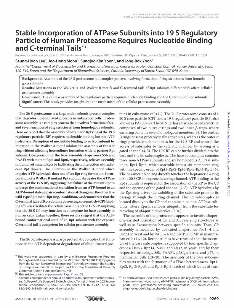
proteasome, FLAG-tagged versions of wild-type or Walker Amutant Rpt subunits were transiently expressed in HeLa cellsand immunoprecipitated with anti-FLAG antibody. Westernblotting analysis of immunoprecipitated samples revealed thatthe 20 S proteasome and lid subunits Rpn7 and Rpn12 werecopurified with wild-type Rpt subunits but not with theWalkerAmutant subunits (Fig. 1A), suggesting that the lysine to serinemutation in the Walker A motif impaired the incorporation ofthe mutant Rpts into the intact 26 S proteasome. To furtherstudy defects of Walker A mutant Rpts in the assembly of theproteasome, HeLa-derived cell lines were established whichconditionally expressed FLAG-tagged wild-type or Walker Amutant Rpt subunits, and the proteasome was affinity purifiedfrom these cells. SDS-PAGE and immunoblotting analysis ofpurified FLAG-Rpt3 and FLAG-Rpt5 samples confirmed thatthe assembly of mutant Rpts into the proteasome was impaired(Fig. 1, B–E). Although mutant Rpt3 and Rpt5 were readilyassociated with their dimer partner subunits Rpt6 and Rpt4,respectively, they did not interact with other Rpt subunits asefficiently as wild-type ones, indicating that the hexameric Rptring was not formed properly. Taken together, these resultssuggest that nucleotide binding to Rpt subunits is a prerequisitefor stable assembly of the proteasomal ATPase ring.We next investigated the influence of ATP hydrolysis by Rpt
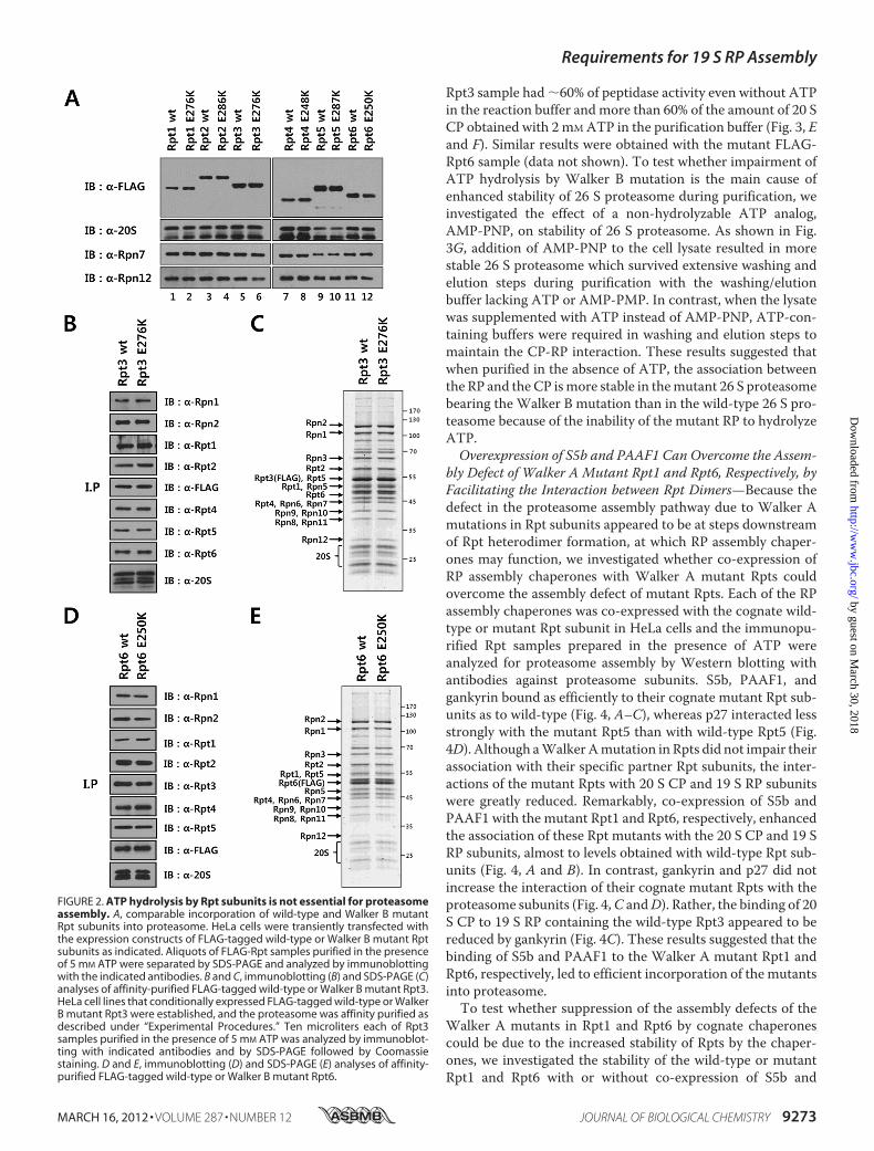
subunits on the assembly of the proteasome. Walker B motifmutants of Rpt subunitswere tested for their incorporation intothe 26 S proteasome by transient transfection and immunopre-cipitation of FLAG-taggedmutants. As shown in Fig. 2A,West-ern blotting analysis revealed that both the wild-type and theWalker B mutant Rpt subunits bound to the 20 S proteasomeand lid subunits Rpn7 and Rpn12 to a similar extent, suggestingthat Walker B mutations did not cause any negative effect onthe assembly. To confirm the proper assembly of Walker Bmutants into 26 S proteasome, we established tetracycline-in-ducible cell lines expressing FLAG-tagged Walker B mutantRpts and analyzed purified FLAG-Rpt3 and FLAG-Rpt6 sam-ples by SDS-PAGE and immunoblotting. The protein patternsof wild-type andWalker Bmutant Rpt3 and Rpt6were basicallyidentical, showing that the mutant Rpts were readily incorpo-rated into the intact 26 S proteasome (Fig. 2,B–E). These resultsindicated that ATP hydrolysis by Rpt subunits was not requiredfor 26 S proteasome assembly.Incorporation of a SingleWalker BMutant Rpt Subunit Com-
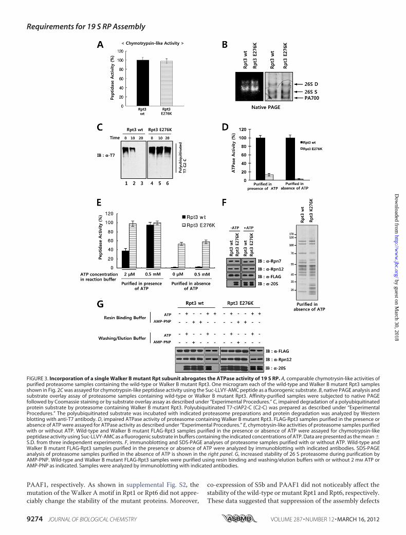
pletely Abrogates ATPase Activity of the 19 S RP—To study theeffect of incorporation of a Walker B mutant Rpt subunit onproteasome function, we analyzed activities of purified protea-some samples containingwild-type orWalker Bmutant FLAG-Rpt3 shown in Fig. 2C. Peptidase activities of proteasomesamples were assessed by solution assays using Suc-Leu-Leu-
Requirements for 19 S RP Assembly
MARCH 16, 2012 • VOLUME 287 • NUMBER 12 JOURNAL OF BIOLOGICAL CHEMISTRY 9271
by guest on March 30, 2018
http://ww
w.jbc.org/
Dow
nloaded from
Val-Tyr-AMC as a substrate. The magnitudes of peptidaseactivities of wild-type and mutant proteasomes were compara-ble to each otherwhenmeasured in the presence of 0.5mMATP(Fig. 3A). Native gel electrophoresis of the purified proteasomesrevealed that both wild-type and mutant proteasome prepara-tions contained similar amounts of singly and doubly capped 26S proteasome and 19 S RP (Fig. 3B, right panel). Substrate over-lay assays confirmed that the peptidase activity of wild-typeproteasome was similar to that of mutant proteasome (Fig. 3B,left panel), showing that theWalker Bmutation in Rpt3 did notimpair the gate opening and peptidase activities of the 26 Sproteasome. Since the major function of the proteasome is thedegradation of polyubiquitinated proteins, we prepared apolyubiquitinated form of cIAP2-C as a proteasome substrateand measured protein degradation activities of wild-type andmutant proteasomes. As shown in Fig. 3C, whereas the wild-type proteasome completely digested the polyubiquitinatedproteinwithin 20min, the proteasome containing theWalker Bmutant Rpt3 failed to efficiently digest the substrate. To exam-ine whether the impaired degradation of the polyubiquitinatedsubstrate by the mutant proteasome was caused by its de-creased ATPase activity, we analyzed the ATPase activities ofthe wild-type and Walker B mutant proteasome. The ATPaseactivity of the proteasome containing the Walker B mutantRpt3 was 13% of that of wild-type proteasome (Fig. 3D).Because the affinity-purified proteasome samples prepared inthe presence of 2 mM ATP contained the doubly capped 26 Sproteasome (Fig. 3B), it was likely that some of the purifiedproteasome complex possessed endogenous untagged Rpt3 inaddition to the FLAG-tagged mutant Rpt3. To prevent the co-purification of 19 S RP containing endogenous Rpt3, we puri-fied FLAG-Rpt3 and its associated proteins in the absence ofATP and determined the ATPase activity. Remarkably, whilethe 19 S RP preparation containingwild-type Rpt3was active inATPase assays, the sample containing the Walker B mutantFLAG-Rpt3 did not show any ATPase activity (Fig. 3D), indi-cating that aWalker Bmutation in a single Rpt subunit resultedin a complete loss of ATPase activity of the hexameric Rpt ring.Interestingly, when we measured the peptidase activities of
the purified 26 S proteasome samples in reaction buffer con-taining 2 �M ATP, the peptidase activity of wild-type protea-some was reduced to �40% of the activity obtained in buffercontaining 0.5 mM ATP, whereas the peptidase activity of pro-teasome containing Walker B mutant Rpt3 was reduced onlyslightly (Fig. 3E). Given that the activation of peptidase activityby ATP is closely related to association of the 19 S RP with the20 S CP, the higher activity of the mutant proteasome mightreflect a more stable association of the CP with the mutant RPthan with wild-type RP under low-ATP conditions. To test thispossibility, we purified the proteasome containing either wild-type or Walker B mutant FLAG-Rpt3 in the presence andabsence of ATP, and examined the peptidase activity (Fig. 3E)and the presence of the 20 S CP by Western blot analysis (Fig.3F). In the presence of 2 mM ATP, the 20 S CP as well as the 19S RP was efficiently co-purified with wild-type and mutantFLAG-Rpt3. When purified in the absence of ATP, the wild-type FLAG-Rpt3 sample possessed practically no peptidaseactivity and very little 20 S CP. In contrast, the mutant FLAG-
FIGURE 1. Nucleotide binding to Rpt subunits is required for the assemblyof proteasome. A, impaired incorporation of Walker A mutant Rpt subunitsinto proteasome. HeLa cells were transiently transfected with the expressionconstructs of FLAG-tagged wild-type or Walker A mutant Rpt subunits asindicated. Aliquots of FLAG-Rpt samples purified in the presence of 5 mM ATPwere separated by SDS-PAGE and analyzed by immunoblotting with indi-cated antibodies. B–C, immunoblotting (B) and SDS-PAGE (C) analyses ofaffinity-purified FLAG-tagged wild-type or Walker A mutant Rpt3. HeLa celllines that conditionally expressed FLAG-tagged wild-type or Walker A mutantRpt3 were established, and the proteasome was affinity purified as describedunder “Experimental Procedures.” Ten microliters each of Rpt3 samples puri-fied in the presence of 5 mM ATP were analyzed by immunoblotting with theindicated antibodies and by SDS-PAGE followed by Coomassie staining. Dand E, immunoblotting (D) and SDS-PAGE (E) analyses of affinity-purifiedFLAG-tagged wild-type or Walker A mutant Rpt5.
Requirements for 19 S RP Assembly
9272 JOURNAL OF BIOLOGICAL CHEMISTRY VOLUME 287 • NUMBER 12 • MARCH 16, 2012
by guest on March 30, 2018
http://ww
w.jbc.org/
Dow
nloaded from
Rpt3 sample had �60% of peptidase activity even without ATPin the reaction buffer and more than 60% of the amount of 20 SCP obtained with 2 mMATP in the purification buffer (Fig. 3, Eand F). Similar results were obtained with the mutant FLAG-Rpt6 sample (data not shown). To test whether impairment ofATP hydrolysis by Walker B mutation is the main cause ofenhanced stability of 26 S proteasome during purification, weinvestigated the effect of a non-hydrolyzable ATP analog,AMP-PNP, on stability of 26 S proteasome. As shown in Fig.3G, addition of AMP-PNP to the cell lysate resulted in morestable 26 S proteasome which survived extensive washing andelution steps during purification with the washing/elutionbuffer lacking ATP or AMP-PMP. In contrast, when the lysatewas supplemented with ATP instead of AMP-PNP, ATP-con-taining buffers were required in washing and elution steps tomaintain the CP-RP interaction. These results suggested thatwhen purified in the absence of ATP, the association betweenthe RP and the CP ismore stable in themutant 26 S proteasomebearing the Walker B mutation than in the wild-type 26 S pro-teasome because of the inability of the mutant RP to hydrolyzeATP.Overexpression of S5b and PAAF1 Can Overcome the Assem-
bly Defect of Walker A Mutant Rpt1 and Rpt6, Respectively, byFacilitating the Interaction between Rpt Dimers—Because thedefect in the proteasome assembly pathway due to Walker Amutations in Rpt subunits appeared to be at steps downstreamof Rpt heterodimer formation, at which RP assembly chaper-ones may function, we investigated whether co-expression ofRP assembly chaperones with Walker A mutant Rpts couldovercome the assembly defect of mutant Rpts. Each of the RPassembly chaperones was co-expressed with the cognate wild-type or mutant Rpt subunit in HeLa cells and the immunopu-rified Rpt samples prepared in the presence of ATP wereanalyzed for proteasome assembly by Western blotting withantibodies against proteasome subunits. S5b, PAAF1, andgankyrin bound as efficiently to their cognate mutant Rpt sub-units as to wild-type (Fig. 4, A–C), whereas p27 interacted lessstrongly with the mutant Rpt5 than with wild-type Rpt5 (Fig.4D). Although aWalkerAmutation in Rpts did not impair theirassociation with their specific partner Rpt subunits, the inter-actions of the mutant Rpts with 20 S CP and 19 S RP subunitswere greatly reduced. Remarkably, co-expression of S5b andPAAF1 with the mutant Rpt1 and Rpt6, respectively, enhancedthe association of these Rpt mutants with the 20 S CP and 19 SRP subunits, almost to levels obtained with wild-type Rpt sub-units (Fig. 4, A and B). In contrast, gankyrin and p27 did notincrease the interaction of their cognate mutant Rpts with theproteasome subunits (Fig. 4,C andD). Rather, the binding of 20S CP to 19 S RP containing the wild-type Rpt3 appeared to bereduced by gankyrin (Fig. 4C). These results suggested that thebinding of S5b and PAAF1 to the Walker A mutant Rpt1 andRpt6, respectively, led to efficient incorporation of the mutantsinto proteasome.To test whether suppression of the assembly defects of the
Walker A mutants in Rpt1 and Rpt6 by cognate chaperonescould be due to the increased stability of Rpts by the chaper-ones, we investigated the stability of the wild-type or mutantRpt1 and Rpt6 with or without co-expression of S5b and
FIGURE 2. ATP hydrolysis by Rpt subunits is not essential for proteasomeassembly. A, comparable incorporation of wild-type and Walker B mutantRpt subunits into proteasome. HeLa cells were transiently transfected withthe expression constructs of FLAG-tagged wild-type or Walker B mutant Rptsubunits as indicated. Aliquots of FLAG-Rpt samples purified in the presenceof 5 mM ATP were separated by SDS-PAGE and analyzed by immunoblottingwith the indicated antibodies. B and C, immunoblotting (B) and SDS-PAGE (C)analyses of affinity-purified FLAG-tagged wild-type or Walker B mutant Rpt3.HeLa cell lines that conditionally expressed FLAG-tagged wild-type or WalkerB mutant Rpt3 were established, and the proteasome was affinity purified asdescribed under “Experimental Procedures.” Ten microliters each of Rpt3samples purified in the presence of 5 mM ATP was analyzed by immunoblot-ting with indicated antibodies and by SDS-PAGE followed by Coomassiestaining. D and E, immunoblotting (D) and SDS-PAGE (E) analyses of affinity-purified FLAG-tagged wild-type or Walker B mutant Rpt6.
Requirements for 19 S RP Assembly
MARCH 16, 2012 • VOLUME 287 • NUMBER 12 JOURNAL OF BIOLOGICAL CHEMISTRY 9273
by guest on March 30, 2018
http://ww
w.jbc.org/
Dow
nloaded from
PAAF1, respectively. As shown in supplemental Fig. S2, themutation of theWalker A motif in Rpt1 or Rpt6 did not appre-ciably change the stability of the mutant proteins. Moreover,
co-expression of S5b and PAAF1 did not noticeably affect thestability of the wild-type ormutant Rpt1 and Rpt6, respectively.These data suggested that suppression of the assembly defects
FIGURE 3. Incorporation of a single Walker B mutant Rpt subunit abrogates the ATPase activity of 19 S RP. A, comparable chymotrysin-like activities ofpurified proteasome samples containing the wild-type or Walker B mutant Rpt3. One microgram each of the wild-type and Walker B mutant Rpt3 samplesshown in Fig. 2C was assayed for chymotrypsin-like peptidase activity using the Suc-LLVY-AMC peptide as a fluorogenic substrate. B, native PAGE analysis andsubstrate overlay assay of proteasome samples containing wild-type or Walker B mutant Rpt3. Affinity-purified samples were subjected to native PAGEfollowed by Coomassie staining or by substrate overlay assay as described under “Experimental Procedures.” C, impaired degradation of a polyubiquitinatedprotein substrate by proteasome containing Walker B mutant Rpt3. Polyubiquitinated T7-cIAP2-C (C2-C) was prepared as described under “ExperimentalProcedures.” The polyubiquitinated substrate was incubated with indicated proteasome preparations and protein degradation was analyzed by Westernblotting with anti-T7 antibody. D, impaired ATPase activity of proteasome containing Walker B mutant Rpt3. FLAG-Rpt3 samples purified in the presence orabsence of ATP were assayed for ATPase activity as described under “Experimental Procedures.” E, chymotrysin-like activities of proteasome samples purifiedwith or without ATP. Wild-type and Walker B mutant FLAG-Rpt3 samples purified in the presence or absence of ATP were assayed for chymotrypsin-likepeptidase activity using Suc-LLVY-AMC as a fluorogenic substrate in buffers containing the indicated concentrations of ATP. Data are presented as the mean �S.D. from three independent experiments. F, immunoblotting and SDS-PAGE analyses of proteasome samples purified with or without ATP. Wild-type andWalker B mutant FLAG-Rpt3 samples purified in the presence or absence of ATP were analyzed by immunoblotting with indicated antibodies. SDS-PAGEanalysis of proteasome samples purified in the absence of ATP is shown in the right panel. G, increased stability of 26 S proteasome during purification byAMP-PNP. Wild-type and Walker B mutant FLAG-Rpt3 samples were purified using resin binding and washing/elution buffers with or without 2 mM ATP orAMP-PNP as indicated. Samples were analyzed by immunoblotting with indicated antibodies.
Requirements for 19 S RP Assembly
9274 JOURNAL OF BIOLOGICAL CHEMISTRY VOLUME 287 • NUMBER 12 • MARCH 16, 2012
by guest on March 30, 2018
http://ww
w.jbc.org/
Dow
nloaded from
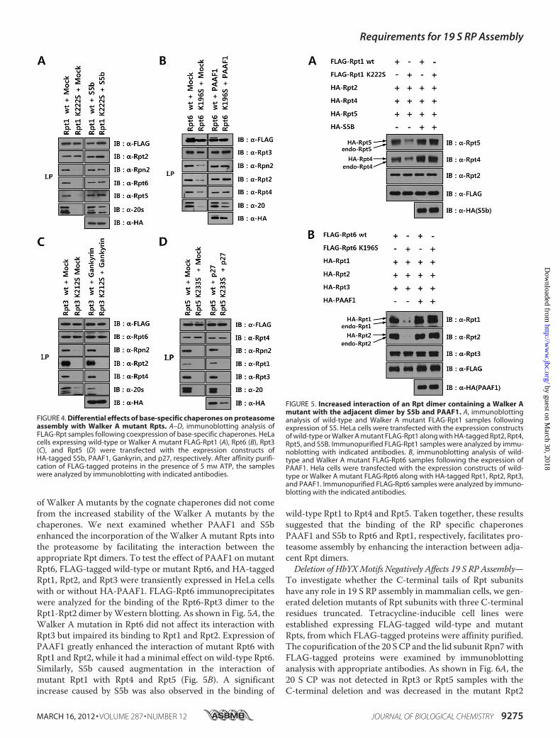
of Walker A mutants by the cognate chaperones did not comefrom the increased stability of the Walker A mutants by thechaperones. We next examined whether PAAF1 and S5benhanced the incorporation of the Walker A mutant Rpts intothe proteasome by facilitating the interaction between theappropriate Rpt dimers. To test the effect of PAAF1 onmutantRpt6, FLAG-tagged wild-type or mutant Rpt6, and HA-taggedRpt1, Rpt2, and Rpt3 were transiently expressed in HeLa cellswith or without HA-PAAF1. FLAG-Rpt6 immunoprecipitateswere analyzed for the binding of the Rpt6-Rpt3 dimer to theRpt1-Rpt2 dimer byWestern blotting. As shown in Fig. 5A, theWalker A mutation in Rpt6 did not affect its interaction withRpt3 but impaired its binding to Rpt1 and Rpt2. Expression ofPAAF1 greatly enhanced the interaction of mutant Rpt6 withRpt1 and Rpt2, while it had a minimal effect on wild-type Rpt6.Similarly, S5b caused augmentation in the interaction ofmutant Rpt1 with Rpt4 and Rpt5 (Fig. 5B). A significantincrease caused by S5b was also observed in the binding of
wild-type Rpt1 to Rpt4 and Rpt5. Taken together, these resultssuggested that the binding of the RP specific chaperonesPAAF1 and S5b to Rpt6 and Rpt1, respectively, facilitates pro-teasome assembly by enhancing the interaction between adja-cent Rpt dimers.Deletion of HbYXMotifs Negatively Affects 19 S RP Assembly—
To investigate whether the C-terminal tails of Rpt subunitshave any role in 19 S RP assembly in mammalian cells, we gen-erated deletion mutants of Rpt subunits with three C-terminalresidues truncated. Tetracycline-inducible cell lines wereestablished expressing FLAG-tagged wild-type and mutantRpts, from which FLAG-tagged proteins were affinity purified.The copurification of the 20 S CP and the lid subunit Rpn7withFLAG-tagged proteins were examined by immunoblottinganalysis with appropriate antibodies. As shown in Fig. 6A, the20 S CP was not detected in Rpt3 or Rpt5 samples with theC-terminal deletion and was decreased in the mutant Rpt2
FIGURE 4. Differential effects of base-specific chaperones on proteasomeassembly with Walker A mutant Rpts. A–D, immunoblotting analysis ofFLAG-Rpt samples following coexpression of base-specific chaperones. HeLacells expressing wild-type or Walker A mutant FLAG-Rpt1 (A), Rpt6 (B), Rpt3(C), and Rpt5 (D) were transfected with the expression constructs ofHA-tagged S5b, PAAF1, Gankyrin, and p27, respectively. After affinity purifi-cation of FLAG-tagged proteins in the presence of 5 mM ATP, the sampleswere analyzed by immunoblotting with indicated antibodies.
FIGURE 5. Increased interaction of an Rpt dimer containing a Walker Amutant with the adjacent dimer by S5b and PAAF1. A, immunoblottinganalysis of wild-type and Walker A mutant FLAG-Rpt1 samples followingexpression of S5. HeLa cells were transfected with the expression constructsof wild-type or Walker A mutant FLAG-Rpt1 along with HA-tagged Rpt2, Rpt4,Rpt5, and S5B. Immunopurified FLAG-Rpt1 samples were analyzed by immu-noblotting with indicated antibodies. B, immunoblotting analysis of wild-type and Walker A mutant FLAG-Rpt6 samples following the expression ofPAAF1. Hela cells were transfected with the expression constructs of wild-type or Walker A mutant FLAG-Rpt6 along with HA-tagged Rpt1, Rpt2, Rpt3,and PAAF1. Immunopurified FLAG-Rpt6 samples were analyzed by immuno-blotting with the indicated antibodies.
Requirements for 19 S RP Assembly
MARCH 16, 2012 • VOLUME 287 • NUMBER 12 JOURNAL OF BIOLOGICAL CHEMISTRY 9275
by guest on March 30, 2018
http://ww
w.jbc.org/
Dow
nloaded from
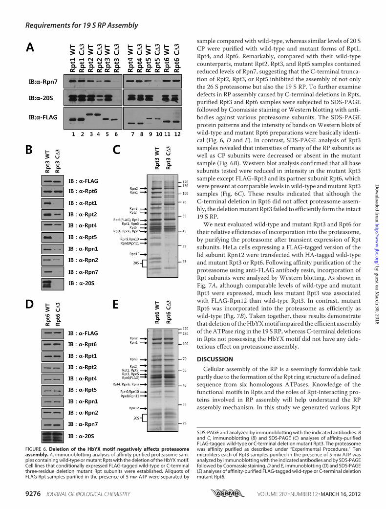
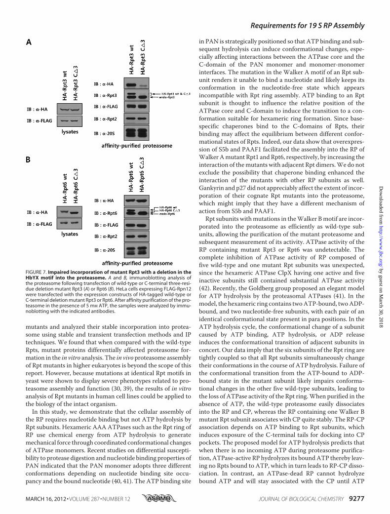
sample compared with wild-type, whereas similar levels of 20 SCP were purified with wild-type and mutant forms of Rpt1,Rpt4, and Rpt6. Remarkably, compared with their wild-typecounterparts, mutant Rpt2, Rpt3, and Rpt5 samples containedreduced levels of Rpn7, suggesting that the C-terminal trunca-tion of Rpt2, Rpt3, or Rpt5 inhibited the assembly of not onlythe 26 S proteasome but also the 19 S RP. To further examinedefects in RP assembly caused by C-terminal deletions in Rpts,purified Rpt3 and Rpt6 samples were subjected to SDS-PAGEfollowed by Coomassie staining or Western blotting with anti-bodies against various proteasome subunits. The SDS-PAGEprotein patterns and the intensity of bands onWestern blots ofwild-type and mutant Rpt6 preparations were basically identi-cal (Fig. 6, D and E). In contrast, SDS-PAGE analysis of Rpt3samples revealed that intensities of many of the RP subunits aswell as CP subunits were decreased or absent in the mutantsample (Fig. 6B). Western blot analysis confirmed that all basesubunits tested were reduced in intensity in the mutant Rpt3sample except FLAG-Rpt3 and its partner subunit Rpt6, whichwere present at comparable levels inwild-type andmutant Rpt3samples (Fig. 6C). These results indicated that although theC-terminal deletion in Rpt6 did not affect proteasome assem-bly, the deletionmutant Rpt3 failed to efficiently form the intact19 S RP.We next evaluated wild-type and mutant Rpt3 and Rpt6 for
their relative efficiencies of incorporation into the proteasome,by purifying the proteasome after transient expression of Rptsubunits. HeLa cells expressing a FLAG-tagged version of thelid subunit Rpn12 were transfected with HA-tagged wild-typeand mutant Rpt3 or Rpt6. Following affinity purification of theproteasome using anti-FLAG antibody resin, incorporation ofRpt subunits were analyzed by Western blotting. As shown inFig. 7A, although comparable levels of wild-type and mutantRpt3 were expressed, much less mutant Rpt3 was associatedwith FLAG-Rpn12 than wild-type Rpt3. In contrast, mutantRpt6 was incorporated into the proteasome as efficiently aswild-type (Fig. 7B). Taken together, these results demonstratethat deletion of theHbYXmotif impaired the efficient assemblyof theATPase ring in the 19 S RP, whereas C-terminal deletionsin Rpts not possessing the HbYX motif did not have any dele-terious effect on proteasome assembly.
DISCUSSION
Cellular assembly of the RP is a seemingly formidable taskpartly due to the formation of the Rpt ring structure of a definedsequence from six homologous ATPases. Knowledge of thefunctional motifs in Rpts and the roles of Rpt-interacting pro-teins involved in RP assembly will help understand the RPassembly mechanism. In this study we generated various Rpt
FIGURE 6. Deletion of the HbYX motif negatively affects proteasomeassembly. A, immunoblotting analysis of affinity purified proteasome sam-ples containing wild-type or mutant Rpts with the deletion of the HbYX motif.Cell lines that conditionally expressed FLAG-tagged wild-type or C-terminalthree-residue deletion mutant Rpt subunits were established. Aliquots ofFLAG-Rpt samples purified in the presence of 5 mM ATP were separated by
SDS-PAGE and analyzed by immunoblotting with the indicated antibodies. Band C, immunoblotting (B) and SDS-PAGE (C) analyses of affinity-purifiedFLAG-tagged wild-type or C-terminal deletion mutant Rpt3. The proteasomewas affinity purified as described under “Experimental Procedures.” Tenmicroliters each of Rpt3 samples purified in the presence of 5 mM ATP wasanalyzed by immunoblotting with the indicated antibodies and by SDS-PAGEfollowed by Coomassie staining. D and E, immunoblotting (D) and SDS-PAGE(E) analyses of affinity-purified FLAG-tagged wild-type or C-terminal deletionmutant Rpt6.
Requirements for 19 S RP Assembly
9276 JOURNAL OF BIOLOGICAL CHEMISTRY VOLUME 287 • NUMBER 12 • MARCH 16, 2012
by guest on March 30, 2018
http://ww
w.jbc.org/
Dow
nloaded from
mutants and analyzed their stable incorporation into protea-some using stable and transient transfection methods and IPtechniques. We found that when compared with the wild-typeRpts, mutant proteins differentially affected proteasome for-mation in the in vitro analysis. The in vivo proteasome assemblyof Rpt mutants in higher eukaryotes is beyond the scope of thisreport. However, because mutations at identical Rpt motifs inyeast were shown to display severe phenotypes related to pro-teasome assembly and function (30, 39), the results of in vitroanalysis of Rpt mutants in human cell lines could be applied tothe biology of the intact organism.In this study, we demonstrate that the cellular assembly of
the RP requires nucleotide binding but not ATP hydrolysis byRpt subunits. Hexameric AAA ATPases such as the Rpt ring ofRP use chemical energy from ATP hydrolysis to generatemechanical force through coordinated conformational changesof ATPase monomers. Recent studies on differential suscepti-bility to protease digestion and nucleotide binding properties ofPAN indicated that the PAN monomer adopts three differentconformations depending on nucleotide binding site occu-pancy and the bound nucleotide (40, 41). The ATP binding site
in PAN is strategically positioned so that ATP binding and sub-sequent hydrolysis can induce conformational changes, espe-cially affecting interactions between the ATPase core and theC-domain of the PAN monomer and monomer-monomerinterfaces. The mutation in the Walker A motif of an Rpt sub-unit renders it unable to bind a nucleotide and likely keeps itsconformation in the nucleotide-free state which appearsincompatible with Rpt ring assembly. ATP binding to an Rptsubunit is thought to influence the relative position of theATPase core and C-domain to induce the transition to a con-formation suitable for hexameric ring formation. Since base-specific chaperones bind to the C-domains of Rpts, theirbinding may affect the equilibrium between different confor-mational states of Rpts. Indeed, our data show that overexpres-sion of S5b and PAAF1 facilitated the assembly into the RP ofWalker Amutant Rpt1 andRpt6, respectively, by increasing theinteraction of themutants with adjacent Rpt dimers.We do notexclude the possibility that chaperone binding enhanced theinteraction of the mutants with other RP subunits as well.Gankyrin and p27 did not appreciably affect the extent of incor-poration of their cognate Rpt mutants into the proteasome,which might imply that they have a different mechanism ofaction from S5b and PAAF1.Rpt subunits withmutations in theWalker Bmotif are incor-
porated into the proteasome as efficiently as wild-type sub-units, allowing the purification of the mutant proteasome andsubsequent measurement of its activity. ATPase activity of theRP containing mutant Rpt3 or Rpt6 was undetectable. Thecomplete inhibition of ATPase activity of RP composed offive wild-type and one mutant Rpt subunits was unexpected,since the hexameric ATPase ClpX having one active and fiveinactive subunits still contained substantial ATPase activity(42). Recently, the Goldberg group proposed an elegant modelfor ATP hydrolysis by the proteasomal ATPases (41). In themodel, the hexameric ring contains twoATP-bound, twoADP-bound, and two nucleotide-free subunits, with each pair of anidentical conformational state present in para positions. In theATP hydrolysis cycle, the conformational change of a subunitcaused by ATP binding, ATP hydrolysis, or ADP releaseinduces the conformational transition of adjacent subunits inconcert. Our data imply that the six subunits of the Rpt ring aretightly coupled so that all Rpt subunits simultaneously changetheir conformations in the course of ATP hydrolysis. Failure ofthe conformational transition from the ATP-bound to ADP-bound state in the mutant subunit likely impairs conforma-tional changes in the other five wild-type subunits, leading tothe loss of ATPase activity of the Rpt ring.When purified in theabsence of ATP, the wild-type proteasome easily dissociatesinto the RP and CP, whereas the RP containing one Walker Bmutant Rpt subunit associates with CP quite stably. The RP-CPassociation depends on ATP binding to Rpt subunits, whichinduces exposure of the C-terminal tails for docking into CPpockets. The proposed model for ATP hydrolysis predicts thatwhen there is no incoming ATP during proteasome purifica-tion, ATPase-active RP hydrolyzes its boundATP thereby leav-ing no Rpts bound to ATP, which in turn leads to RP-CP disso-ciation. In contrast, an ATPase-dead RP cannot hydrolyzebound ATP and will stay associated with the CP until ATP
FIGURE 7. Impaired incorporation of mutant Rpt3 with a deletion in theHbYX motif into the proteasome. A and B, immunoblotting analysis ofthe proteasome following transfection of wild-type or C-terminal three-resi-due deletion mutant Rpt3 (A) or Rpt6 (B). HeLa cells expressing FLAG-Rpn12were transfected with the expression constructs of HA-tagged wild-type orC-terminal deletion mutant Rpt3 or Rpt6. After affinity purification of the pro-teasome in the presence of 5 mM ATP, the samples were analyzed by immu-noblotting with the indicated antibodies.
Requirements for 19 S RP Assembly
MARCH 16, 2012 • VOLUME 287 • NUMBER 12 JOURNAL OF BIOLOGICAL CHEMISTRY 9277
by guest on March 30, 2018
http://ww
w.jbc.org/
Dow
nloaded from
dissociates from the Rpt subunits. Our data with Walker Bmutants are consistent with this scenario and imply that disso-ciation of ATP from ATPase-dead RP is a slow process.The C-terminal tails of Rpt2, Rpt3, and Rpt5 containing the
HbYXmotifs bind to the 20 SCPwith varying affinities (28, 29).There appears a correlation between the CP binding affinity ofthe C-terminal tails and the deleterious effect of their deletionon RP assembly. Thus, the deletion of the Rpt3 or Rpt5 C ter-minus with a higher CP binding affinity had a greater effect onRP assembly than that of the Rpt2 tail, whereas the C-terminaltruncation of Rpts not possessing theHbYXmotif did not affectRP assembly. Data from the differential incorporation of wild-type and mutant Rpts into the proteasome shown here by bothstable and transient transfections is consistent with the ideathat CP may function as a template for base assembly. We notethat although deletion of the HbYXmotif inhibits RP assembly,it does not completely impair the incorporation of mutant Rptsinto the proteasome. Therefore, CP-independent RP assemblymust be in operation as well. Kim et al. (32) very recentlyreported that the HbYX motifs of Rpt3 and Rpt5 are requiredfor 26 S proteasome assembly, which is in agreement with ourdata. However, they failed to detect the effect of the HbYXmotif deletion on RP assembly. The basis of the discrepancy isnot clear but may come from differences in experimentaldesign; it could be due to the fact that conditional rather thanconstitutive expression of Rpt mutants was employed in ourexperiments which enabled expression of the mutants only fora short period to minimize potential cellular adaptation to theexpression of mutant proteins. Unlike their human counter-parts, the C termini of yeast Rpt4 and Rpt6 appear to playimportant roles in 26 S proteasome assembly, since deletion ofthe last residue from each Rpt impaired efficient formation of26 S proteasome (30). The proteasome assembly pathway mayhave diverged between yeast and human. The data presentedhere suggest that RP assembly in human cells can be drivennormally by two different pathways: the CP-assisted pathwayrequiring the HbYX motifs and the CP-independent pathway.
REFERENCES1. Schwartz, A. L., and Ciechanover, A. (2009) Targeting proteins for de-
struction by the ubiquitin system: implications for human pathobiology.Annu. Rev. Pharmacol. Toxicol. 49, 73–96
2. Finley, D. (2009) Recognition and processing of ubiquitin-protein conju-gates by the proteasome. Annu. Rev. Biochem. 78, 477–513
3. Groll, M., Ditzel, L., Löwe, J. Stock, D., Bochtler, M., Bartunik, H. D., andHuber, R. (1997) Structure of 20 S proteasome from yeast at 2.4 A resolu-tion. Nature. 386, 463–471
4. Whitby, F. G., Masters, E. I., Kramer, L., Knowlton, J. R., Yao, Y., Wang,C. C., and Hill, C. P. (2000) Structural basis for the activation of 20 Sproteasomes by 11S regulators. Nature. 408, 115–120
5. Groll, M., Bajorek, M., Köhler, A., Moroder, L., Rubin, D. M., Huber, R.,Glickman, M. H., and Finley, D. (2000) A gated channel into the protea-some core particle. Nat. Struct. Biol. 7, 1062–1067
6. Tomko, R. J., Jr., Funakoshi,M., Schneider, K.,Wang, J., andHochstrasser,M. (2010) Heterohexameric ring arrangement of the eukaryotic protea-somal ATPases: implications for proteasome structure and assembly.Mol.Cell 38, 393–403
7. Liu, C. W., Li, X., Thompson, D., Wooding, K., Chang, T. L., Tang, Z., Yu,H., Thomas, P. J., and DeMartino, G. N. (2006) ATP binding and ATPhydrolysis play distinct roles in the function of 26 S proteasome.Mol. Cell24, 39–50
8. Stadtmueller, B.M., andHill, C. P. (2011) Proteasome activators.Mol. Cell41, 8–19
9. Verma, R., Aravind, L., Oania, R., McDonald, W. H., Yates, J. R., 3rd,Koonin, E. V., and Deshaies, R. J. (2002) Role of Rpn11 metalloprotease indeubiquitination and degradation by the 26 S proteasome. Science 298,611–615
10. Yao, T., and Cohen, R. E. (2002) A cryptic protease couples deubiquitina-tion and degradation by the proteasome. Nature 419, 403–407
11. Murata, S., Yashiroda, H., and Tanaka, K. (2009) Molecular mechanismsof proteasome assembly. Nat. Rev. Mol. Cell Biol. 10, 104–115
12. Kusmierczyk, A. R., andHochstrasser,M. (2008) Some assembly required:dedicated chaperones in eukaryotic proteasome biogenesis. Biol. Chem.389, 1143–1151
13. Roelofs, J., Park, S., Haas,W., Tian, G.,McAllister, F. E., Huo, Y., Lee, B. H.,Zhang, F., Shi, Y., Gygi, S. P., and Finley, D. (2009) Chaperone-mediatedpathway of proteasome regulatory particle assembly. Nature 459,861–865
14. Saeki, Y., Toh-E, A., Kudo, T., Kawamura, H., and Tanaka, K. (2009) Mul-tiple proteasome-interacting proteins assist the assembly of the yeast 19 Sregulatory particle. Cell 137, 900–913
15. Funakoshi, M., Tomko, R. J., Jr., Kobayashi, H., and Hochstrasser, M.(2009) Multiple assembly chaperones govern biogenesis of the protea-some regulatory particle base. Cell 137, 887–899
16. Kaneko, T., Hamazaki, J., Iemura, S., Sasaki, K., Furuyama, K., Natsume,T., Tanaka, K., and Murata, S. (2009) Assembly pathway of the mamma-lian proteasome base subcomplex is mediated by multiple specific chap-erones. Cell 137, 914–925
17. Le Tallec, B., Barrault, M. B., Guérois, R., Carré, T., and Peyroche, A.(2009) Hsm3/S5b participates in the assembly pathway of the 19 S regu-latory particle of the proteasome.Mol. Cell 33, 389–399
18. Bedford, L., Paine, S., Sheppard, P. W., Mayer, R. J., and Roelofs, J. (2010)Assembly, structure, and function of the 26 S proteasome. Trends CellBiol. 20, 391–401
19. Park, S., Tian,G., Roelofs, J., and Finley, D. (2010)Assemblymanual for theproteasome regulatory particle: the first draft. Biochem. Soc. Trans. 38,6–13
20. Tomko, R. J., Jr., and Hochstrasser, M. (2011) Order of the proteasomalATPases and eukaryotic proteasome assembly.Cell Biochem. Biophys. 60,13–20
21. Zhang, F., Hu, M., Tian, G., Zhang, P., Finley, D., Jeffrey, P. D., and Shi, Y.(2009) Structural insights into the regulatory particle of the proteasomefromMethanocaldococcus jannaschii. Mol. Cell 14, 473–484
22. Nakamura, Y., Umehara, T., Tanaka, A., Horikoshi, M., Padmanabhan, B.,and Yokoyama, S. (2007) Structural basis for the recognition between theregulatory particles Nas6 and Rpt3 of the yeast 26 S proteasome. Biochem.Biophys. Res. Commun. 359, 503–509
23. Nakamura, Y., Nakano, K., Umehara, T., Kimura, M., Hayashizaki, Y.,Tanaka, A., Horikoshi, M., Padmanabhan, B., and Yokoyama, S. (2007)Structure of the oncoprotein gankyrin in complex with S6 ATPase of the26 S proteasome. Structure 15, 179–189
24. Rabl, J., Smith, D. M., Yu, Y., Chang, S. C., Goldberg, A. L., and Cheng, Y.(2008) Mechanism of gate opening in the 20 S proteasome by the protea-somal ATPases.Mol. Cell 30, 360–368
25. Yu, Y., Smith, D. M., Kim, H. M., Rodriguez, V., Goldberg, A. L., andCheng, Y. (2010) Interactions of PAN’s C termini with archaeal 20 S pro-teasome and implications for the eukaryotic proteasome-ATPase interac-tions. EMBO J. 29, 692–702
26. Stadtmueller, B. M., Ferrell, K., Whitby, F. G., Heroux, A., Robinson, H.,Myszka, D. G., and Hill, C. P. (2010) Structural models for interactionsbetween the 20 S proteasome and its PAN/19 S activators. J. Biol. Chem.285, 13–17
27. Smith, D. M., Chang, S. C., Park, S., Finley, D., Cheng, Y., and Goldberg,A. L. (2007) Docking of the proteasomal ATPases’ carboxyl termini in the20 S proteasome’s alpha ring opens the gate for substrate entry.Mol. Cell27, 731–744
28. Gillette, T. G., Kumar, B., Thompson, D., Slaughter, C. A., andDeMartino,G. N. (2008) Differential roles of the COOH termini of AAA subunits ofPA700 (19 S regulator) in asymmetric assembly and activation of the 26 S
Requirements for 19 S RP Assembly
9278 JOURNAL OF BIOLOGICAL CHEMISTRY VOLUME 287 • NUMBER 12 • MARCH 16, 2012
by guest on March 30, 2018
http://ww
w.jbc.org/
Dow
nloaded from
proteasome. J. Biol. Chem. 283, 31813–3182229. Kumar, B., Kim, Y. C., and DeMartino, G. N. (2010) The C terminus of
Rpt3, an ATPase subunit of PA700 (19 S) regulatory complex, is essentialfor 26 S proteasome assembly but not for activation. J. Biol. Chem. 285,39523–39535
30. Park, S., Roelofs, J., Kim,W., Robert, J., Schmidt,M., Gygi, S. P., and Finley,D. (2009) Hexameric assembly of the proteasomal ATPases is templatedthrough their C termini. Nature 459, 866–870
31. Kusmierczyk, A. R., Kunjappu,M. J., Funakoshi, M., and Hochstrasser, M.(2008) Amultimeric assembly factor controls the formation of alternative20 S proteasomes. Nat. Struct. Mol. Biol. 15, 237–244
32. Kim, Y. C., and DeMartino, G. N. (2011) C termini of proteasomalATPases play nonequivalent roles in cellular assembly of mammalian 26 Sproteasome. J. Biol. Chem. 286, 26652–26666
33. Hendil, K. B., Kriegenburg, F., Tanaka, K., Murata, S., Lauridsen, A. M.,Johnsen, A. H., andHartmann-Petersen, R. (2009) The 20 S proteasome asan assembly platform for the 19 S regulatory complex. J. Mol. Biol. 394,320–328
34. Min, K.W., Hwang, J.W., Lee, J. S., Park, Y., Tamura, T. A., and Yoon, J. B.(2003) TIP120A associates with cullins and modulates ubiquitin ligaseactivity. J. Biol. Chem. 278, 15905–15910
35. Elsasser, S., Schmidt, M., and Finley, D. (2005) Characterization of theproteasome using native gel electrophoresis. Methods Enzymol. 398,
353–36336. Hoffman, L., and Rechsteiner,M. (1996)Nucleotidase activities of the 26 S
proteasome and its regulatory complex. J. Biol. Chem. 271, 32538–3254537. Park, S. M., Yoon, J. B., and Lee, T. H. (2004) Receptor interacting protein
is ubiquitinated by cellular inhibitor of apoptosis proteins (c-IAP1 andc-IAP2) in vitro. FEBS Lett. 566, 151–156
38. Thompson, D., Hakala, K., and DeMartino, G. N. (2009) Subcomplexes ofPA700, the 19 S regulator of the 26 S proteasome, reveal relative roles ofAAA subunits in 26 S proteasome assembly and activation and ATPaseactivity. J. Biol. Chem. 284, 24891–24903
39. Rubin D. M., GlickmanM. H., Larsen, C. N., Dhruvakumar, S., and FinleyD. (1998) Active site mutants in the six regulatory particle ATPases revealmultiple roles for ATP in the proteasome. EMBO J. 17, 4909–4919
40. Horwitz, A. A., Navon, A., Groll, M., Smith, D. M., Reis, C., and Goldberg,A. L. (2007) ATP-induced structural transitions in PAN, the proteasome-regulatory ATPase complex in Archaea. J. Biol. Chem. 282, 22921–22929
41. Smith, D.M., Fraga, H., Reis, C., Kafri, G., and Goldberg, A. L. (2011) ATPbinds to proteasomal ATPases in pairs with distinct functional effects,implying an ordered reaction cycle. Cell 144, 526–538
42. Martin, A., Baker, T. A., and Sauer, R. T. (2005) Rebuilt AAA � motorsreveal operating principles for ATP-fueled machines. Nature 437,1115–1120
Requirements for 19 S RP Assembly
MARCH 16, 2012 • VOLUME 287 • NUMBER 12 JOURNAL OF BIOLOGICAL CHEMISTRY 9279
by guest on March 30, 2018
http://ww
w.jbc.org/
Dow
nloaded from
Seung-Hoon Lee, Joo-Hong Moon, Sungjoo Kim Yoon and Jong-Bok YoonProteasome Requires Nucleotide Binding and C-terminal Tails
Stable Incorporation of ATPase Subunits into 19 S Regulatory Particle of Human
doi: 10.1074/jbc.M111.316208 originally published online January 24, 20122012, 287:9269-9279.J. Biol. Chem.
10.1074/jbc.M111.316208Access the most updated version of this article at doi:
Alerts:
When a correction for this article is posted•
When this article is cited•
to choose from all of JBC's e-mail alertsClick here
Supplemental material:
http://www.jbc.org/content/suppl/2012/01/24/M111.316208.DC1
http://www.jbc.org/content/287/12/9269.full.html#ref-list-1
This article cites 42 references, 10 of which can be accessed free at
by guest on March 30, 2018
http://ww
w.jbc.org/
Dow
nloaded from


![V-ATPase · From Wiki: Vacuolar-type H+ -ATPase (V-ATPase) is a highly conserved evolutionarily ancient enzyme with remarkably diverse functions in eukaryotic organisms.[1] membranes](https://img.pdfslide.net/doc/110x75/5fa3fb056ad5ca477269e2ce/v-atpase-from-wiki-vacuolar-type-h-atpase-v-atpase-is-a-highly-conserved-evolutionarily.jpg)