Embed Size (px)
Citation preview
Proc. Nati. Acad. Sci. USAVol. 74, No. 9, pp. 3937-3941, September 1977Genetics
Stable association of the human transgenome and host murinechromosomes demonstrated with trispecific microcell hybrids
(somatic cell genetics/gene transfer/chromosome transfer)
R. E. K. FOURNIER AND F. H. RUDDLE
Department of Biology, Kline Biology Tower, Yale University, New Haven, Connecticut 06520
Contributed by Frank H. Ruddle, June 13, 1977
ABSTRACT Trispecific microcell hybrids were preparedby transferring limited numbers of chromosomes from ahuman/mouse gene-transfer cell line to a Chinese hamster re-cipient line. The donor cells employed were murine L-cells thatstably expressed the human form of the enzyme hypoxanthinephosphoribosyltransferase. Karyotypic, zymographic, andback-selection tests of the resultin uman/mouse/Chinesehamster microcell hybrids provided strong genetic evidence fora stable association of the human transgenome with host murinechromosomes in stable gene-transfer cell lines. This association,which may represent physical integration of the transgenomeinto the host cell genome, occurred at multiple chromosomalsites.
Chromosome-mediated gene transfer is a technique that allowssmall pieces of genetic material to be transferred from onemammalian cell to another (1-6). This process, which wasconvincingly demonstrated by McBride and Ozer in 1973 (1),utilizes isolated metaphase chromosomes as vectors for geneticexchange. According to current views (7), chromosomes enterthe recipient cells by phagocytosis and are subsequently de-graded by lysosomal enzymes. By using appropriate selectiveconditions, transformant clones that express a particulardonor-derived phenotype can be isolated at low frequency.Both intra- and interspecific gene transfer experiments havebeen reported using mouse, Chinese hamster, and humansomatic cells, and a variety of selectable markers have beentransferred (1-8).The transformant cell lines generated by chromosome-
mediated gene transfer share the following general properties.First, no donor chromosomes or chromosome fragments aredetectable cytologically. Second, isozyme analyses of trans-formant cell lines unambiguously demonstrate expression ofthe complementing gene derived from the donor cells (1, 3-6,8), and, in some cases (9, 10), coexpression of tightly linked loci.Unlinked markers characteristic of the donor cells are notpresent in the transformant clones (3). Third, expression of thetransferred marker may be stable or unstable. Unstable lineslose the transferred genetic element ("transgenome") at dif-ferent rates (usually 1-10% per cell generation) (1, 3, 10), andcan give rise to stably transformed lines upon prolonged culti-vation (6, 9). Such stable lines seem to express the transferredmarker as an integral genetic element of the host cell.
Little is known concerning the state, physical nature, orlocation of the transgenome in either stable or unstable genetransfer clones. The properties of unstable versus stable trans-formants are consistent with the view that the transgenome isan autonomous genetic entity in unstable lines. According tothis view such cells could give rise to stably transformed lines
by the physical insertion of the transgenome into host cellchromosomes. This working hypothesis predicts a stable asso-
ciation of the transgenome with recipient chromosome(s) instable transformants. In fact, indirect evidence supporting thisview has recently been obtained in serial gene transfer exper-iments (8, 11). In this report we describe studies employingrecently developed techniques for microcell-mediated chro-mosome transfer (12) which provide direct evidence for a stableassociation of the transgenome with host cell chromosomes.Furthermore, we show that this association is not a site-specificprocess and does not occur at the homologous locus of the re-cipient-cell genome. Rather, the transgenome can become as-sociated with a variety of chromosomes of the host cell.
MATERIALS AND METHODSAll cell lines were maintained in monolayer culture at 370 under10% C02/90% air in Dulbecco's modified, Eagle's medium(Gibco) supplemented with 10% fetal bovine serum (Interna-tional Biological Laboratories). Parental cell lines and microcellhybrids were found to be free of mycoplasma by the culturemethod of Hayflick as modified by Barile (13).CT1IC is one of the three independent gene transfer clones
originally isolated by Willecke and Ruddle (3). These lines wereprepared by transferring the human gene for hypoxanthinephosphoribosyltransferase (HPRT; IMP:pyrophosphate phos-phoribosyltransferase, EC 2.4.2.8) into HPRT- murine L-cells(A9) using donor metaphase chromosomes derived from HeLaS3. All three clones expressed only the human form of HPRTas judged by electrophoretic and immunochemical tests. Uponprolonged cultivation one of these unstable lines (CT1 C) gaverise to a stable transformant which'is designated CT1 IC1. Thisstabilized population was used without subeloning in the ex-periments reported here.
Microcell hybrids were prepared by transferring limitednumbers of chromosomes from CT11C, into HPRT- Chinesehamster E36 cells (14) and selecting for the expression of(human) HPRT using the hypoxanthine/aminopterin/thy-midine (HAT) selective system (15). The generation of suchmicrocell hybrids has been described elsewhere (12). Inde'pendent microcell hybrid clones were picked by the ringtechnique and expanded. Electrophoretic analyses of hybridcell extracts were performed using published procedures (16,17). Karyotyping was accomplished using a sequential stainingtechnique (18) involving Giemsa/viokase banding followed byIjoechst 33258 staining. The distribution of introduced murinechromosomes in each clone, was determined by scoringHoechst-stained metaphase spreads and interphase nuclei forthe presence of bright chromocenters diagnostic of the centricheterochromatin of murine chromosomes.
Abbreviations: HPRT, hypoxanthine hponeytasrs;hypoxanthine/aminopterin/thymidine.
3937
HAT,
The costs of publication of this article were defrayed in part by thepayment of page charges. This article must therefore be hereby marked"aduertisement" in accordance with 18 U. S. C. §1734 solely to indicatethis fact.
Dow
nloa
ded
by g
uest
on
Dec
embe
r 1,
202
1
3938 Genetics: Fournier and Ruddle
lOOr
50_
5
u4-
0
c
05'0.
Ih
ECm-2- N- 50M N 50
u E- I IIECm 2AT1- N=100
0 - E N=50
0o1 2 3 4 5 6
0
°o°r
50k
50k
10o0r
501-k
H,IECm-1
N- 16
iiaI~I1.4
F10I:jri
I IECmlAT2
A I A4*
ECm-2N- 15
m-0 I 2 3 4 5 6
-1
ECm-4e- N= 1000 N=50
u -
v
ECm4eAT5- N- 100
0 _ 9 I
5
0 1 2 3 4 5 6No. mouse chromosomes/chromocenters per cell
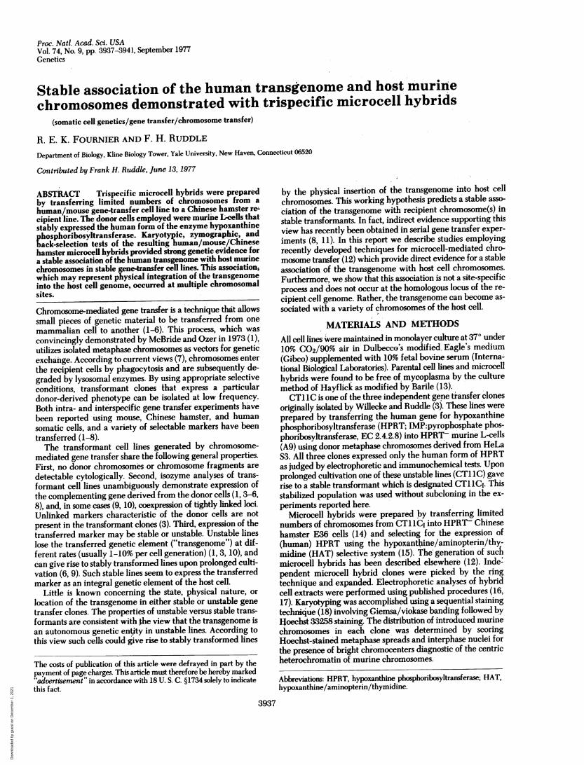
FIG. 1. Distribution of introduced murine chromosomes intrispecific microcell hybrids and their back-selectants (indicated byAT in the name of the cell line). The data were obtained by scoringHoechst 33258-stained interphase nuclei (solid bars) or metaphasespreads (hatched bars) for the presence of high fluorescence intensitychromocenters or by karyotyping Giemsa/viokase-banded prepara-
tions (stippled bars).
All hybrid clones were subject to back-selection in mediumcontaining 6-thioguanine (10 sg/ml) and 8-azaguanine (30jig/ml) and the resulting HPRT- populations were charac-terized as described above without subcloning.
RESULTSThe microcell hybrids generated by fusion of CT1 1CI micro-cells with intact E36 recipients had either a single (IS hybrids)or a double (2S hybrids) input of Chinese hamster chromosomescharacteristic of E36 (IS modal chromosome number = 21) andalso contained from one to four mouse chromosomes derivedfrom CT1 1CI. In addition, electrophoretic analyses of extractsprepared from each of the microcell hybrids demonstrated thatthe cells expressed the human and only the human form ofHPRT. We have used the descriptive term "tribrid" to em-phasize the trispecific nature of these cells. The characterizationof individual tribrid clones is considered in detail below.
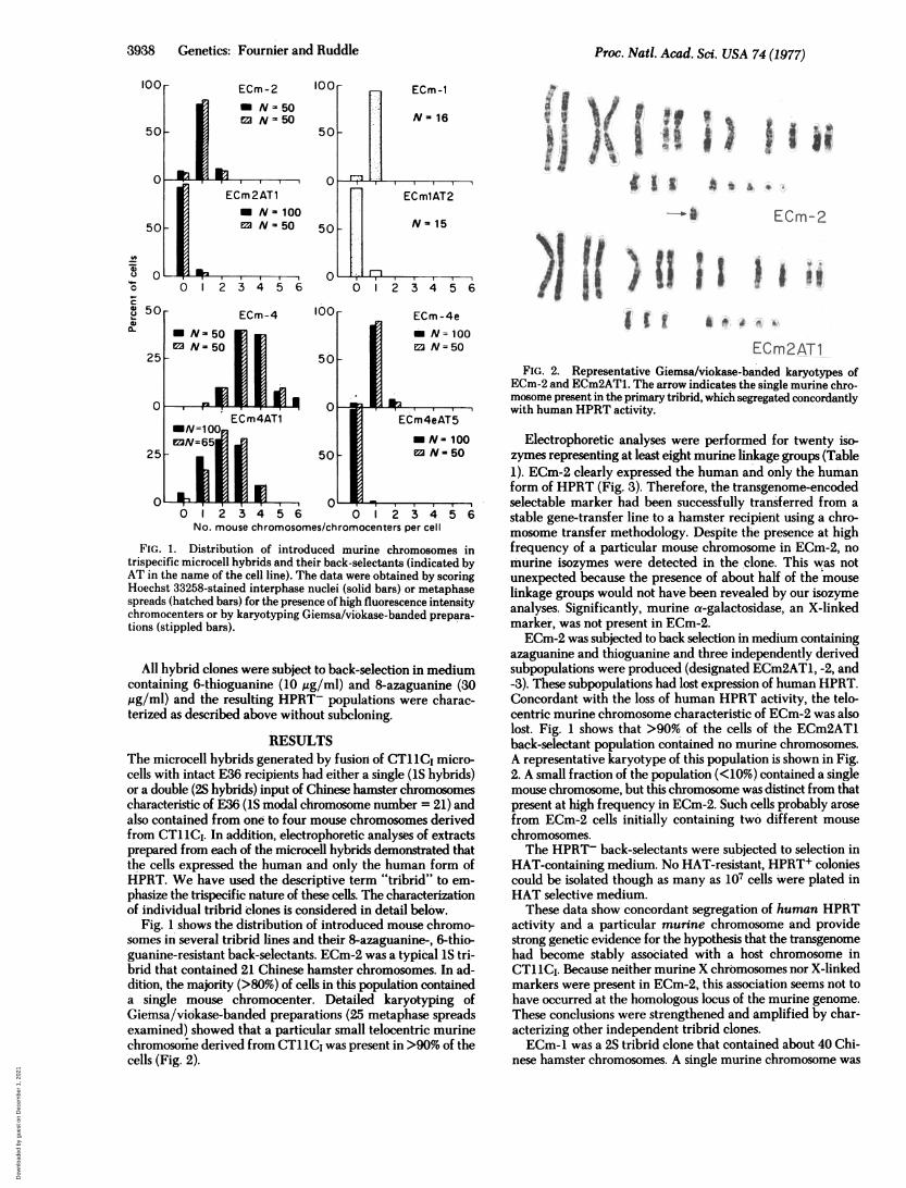
Fig. 1 shows the distribution of introduced mouse chromo-somes in several tribrid lines and their 8-azaguanine-, 6-thio-guanine-resistant back-selectants. ECm-2 was a typical IS tri-brid that contained 21 Chinese hamster chromosomes. In ad-dition, the majority (>80%) of cells in this population containeda single mouse chromocenter. Detailed karyotyping ofGietusa/viokase-banded preparations (25 metaphase spreadsexamined) showed that a particular small telocentric murinechromosomrie derived from CT11C, was present in >90% of thecells (Fig. 2).
I ii
ECm2AT1FIG. 2. Representative Giemsa/viokase-banded karyotypes of
ECm-2 and ECm2AT1. The arrow indicates the single murine chro-mosome present in the primary tribrid, which segregated concordantlywith human HPRT activity.
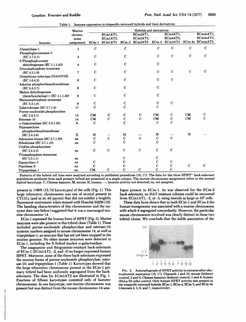
Electrophoretic analyses were performed for twenty iso-zymes representing at least eight murine linkage groups (Table1). ECm-2 clearly expressed the human and only the humanform of HPRT (Fig. 3). Therefore, the transgenome-encodedselectable marker had been successfully transferred from astable gene-transfer line to a hamster recipient using a chro-mosome transfer methodology. Despite the presence at highfrequency of a particular mouse chromosome in ECm-2, nomurine isozymes were detected in the clone. This was notunexpected because the presence of about half of the mouselinkage groups would not have been revealed by our isozymeanalyses. Significantly, murine a-galactosidase, an X-linkedmarker, was not present in ECm-2.ECm-2 was subjected to back selection in medium containing
azaguanine and thioguanine and three independently derivedsubpopulations were produced (designated ECm2AT1, -2, and-3). These subpopulations had lost expression of human HPRT.Concordant with the loss of human HPRT activity, the telo-centric murine chromosome characteristic of ECm-2 was alsolost. Fig. 1 shows that >90% of the cells of the ECm2AT1back-selectant population contained no murine chromosomes.A representative karyotype of this population is shown in Fig.2. A small fraction of the population (<10%) contained a singlemouse chromosome, but this chromosome was distinct from thatpresent at high frequency in ECm-2. Such cells probably arosefrom ECm-2 cells initially containing two different mousechromosomes.The HPRT- back-selectants were subjected to selection in
HAT-containing medium. No HAT-resistant, HPRT+ coloniescould be isolated though as many as 107 cells were plated inHAT selective medium.
These data show concordant segregation of human HPRTactivity and a particular murine chromosome and providestrong genetic evidence for the hypothesis that the transgenomehad become stably associated with a host chromosome inCT1 1CI. Because neither murine X chromosomes nor X-linkedmarkers were present in ECm-2, this association seems not tohave occurred at the homologous locus of the murine genome.These conclusions were strengthened and amplified by char-acterizing other independent tribrid clones.ECm-1 was a 2S tribrid clone that contained about 40 Chi-
nese hamster chromosomes. A single murine chromosome was
I
Proc. Nati. Acad. Sci. USA 74 (1977)
n I
% I0
i I
Dow
nloa
ded
by g
uest
on
Dec
embe
r 1,
202
1
Proc. Nati. Acad. Sci. USA 74(1977) 3939
Table 1. Isozyme expression in trispecific microcell hybrids and their derivatives
Murine Hybrids and derivativeschromo- ECmlAT1, ECm2AT1, ECm4AT1, ECm4eAT1,some ECm1AT2, ECm2AT2, ECm4AT2, ECm4eAT2,
Isozyme assignment ECm-1 ECm1AT3 ECm-2 ECm2AT3 ECm-4 ECm4AT3 ECm-4e ECm4eAT3
Dipeptidase-1 1 C C C C C CPhosphoglucomutase-2
(EC2.7.5.1) 4 C C C C C C6-Phosphogluconatedehydrogenase (EC 1.1.1.43) 4 C C C
Glucosephosphate isomerase(EC 5.3.1.9) 7 C C C C C C
Glutathione reductase [NAD(P)H](EC 1.6.4.2) 8 C C C C
Adenine phosphoribosyltransferase(EC 2.4.2.7) 8 C C C
Malate dehydrogenase(decarboxylating)-l (EC 1.1.1.40) 9 C C C
Mannosephosphate isomerase(EC 5.3.1.8) 9 C C C C
Galactokinase (EC 2.7.1.6) 11 C C CPurine-nucleoside phosphorylase(EC 2.4.2.1) 14 CM C C C CM C CM C
Esterase-10 14 CM C C C CM C CM Ca-Galactosidase (EC 3.2.1.22) X C C C CHypoxanthine
phosphoribosyltransferase(EC2.4.2.8) X H H H H
Adenosine kinase (EC 2.7.1.20) na C C C C C CRibokinase (EC 2.7.1.15) na C C CUridine phosphorylase(EC 2.4.2.3) na C C C C C C
Triosephosphate isomerase(EC 5.3.1.1) na C C
Dipeptidase-2 na C C C CPeptidase-S na C C C C C CTripeptidase-1 na CM C C C C C
Extracts of the hybrid cell lines were analyzed according to published procedures (16, 17). The data for the three HPRT- back-selectantpopulations produced from each primary tribrid are presented in a single column. The murine chromosome assignment refers to the normaldiploid karyotype. C, Chinese hamster; M, mouse; H, human; -, isozyme activity not detected; na, not assigned.
present in >90% (15/16 karyotypes) of the cells (Fig. 1). Thislarge telocentric chromosome was one of several present inCT 1CI (and in its A9 parent) that did not exhibit a brightlyfluorescent centromere when stained with Hoechst 33258 (19).The banding characteristics of this chromosome and the iso-zyme data (see below) suggested that it was a rearranged mu-rine chromosome 14.ECm-1 expressed the human form of HPRT (Fig. 3). Murine
isozymes were also present in this tribrid clone (Table 1). Theseincluded purine-nucleoside phosphorylase and esterase-10,syntenic markers assigned to mouse chromosome 14, as well astripeptidase-1, an isozyme that has not yet been mapped in themurine genome. No other mouse isozymes were detected inECm-1, including the X-linked marker a-galactosidase.The azaguanine and thioguanine-resistant back-selectants
of ECm-1 (ECmIATi, -2, and -3) no longer expressed humanHPRT. Moreover, none of the three back-selectants expressedthe murine forms of purine-nucleoside phosphorylase, ester-ase-10, and tripeptidase-I (Table 1). Karyotypes showed thatthe large telocentric chromosome present in the ECm-1 pri-mary tribrid had been uniformly segregated from the back-selectants. The data for ECm1AT2 are illustrated in Fig. 1.Fourteen of fifteen karyotypes consisted only of hamsterchromosomes. In one karyotype, one murine chromosome waspresent but was distinct from the mouse chromosome-14-ana-
logue present in ECm-1. As was observed for the ECm-2back-selectants, no HAT-resistant colonies could be recoveredfrom ECmiATi, -2, or -3, using inocula as large as 107 cells.
These data have shown that in both ECm-1 and ECm-2 thehuman transgenome was associated with a murine chromosomewith which it segregated concordantly. However, the particularmouse chromosome involved was clearly distinct in these twotribrid clones. We conclude that the stable association of the
(±)
origin -_
2 3 4 5 6 7 8 9 10
FIG. 3. Autoradiograph ofHPRT activity in extracts after elec-trophoretic separation (16, 17). Channels 1 and 10, mouse (kidney)control; 2 and 9, Chinese hamster (kidney) control; 3 and 8, human(HeLa S3 cells) control. Only human HPRT activity was present inthe trispecific microcell hybrids ECm-1, ECm-4, ECm-2, and ECm-4e(channels 4, 5, 6, and 7, respectively).
Genetics: Fournier and Ruddle
Dow
nloa
ded
by g
uest
on
Dec
embe
r 1,
202
1
3940 Genetics: Fournier and Ruddle
ECm-1 ECm-2 ECm-4e ECm4eAT1
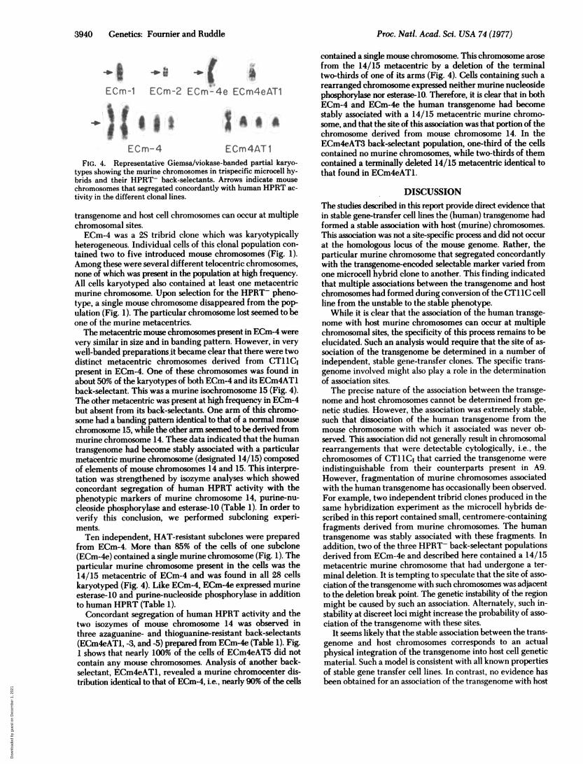
ECm-4 ECm4AT1FIG. 4. Representative Giemsa/viokase-banded partial karyo-
types showing the murine chromosomes in trispecific microcell hy-brids and their HPRT- back-selectants. Arrows indicate mouse
chromosomes that segregated concordantly with human HPRT ac-
tivity in the different clonal lines.
transgenome and host cell chromosomes can occur at multiplechromosomal sites.ECm-4 was a 2S tribrid clone which was karyotypically
heterogeneous. Individual cells of this clonal population con-
tained two to five introduced mouse chromosomes (Fig. 1).Among these were several different telocentric chromosomes,none of which was present in the population at high frequency.All cells karyotyped also contained at least one metacentricmurine chromosome. Upon selection for the HPRT- pheno-type, a single mouse chromosome disappeared from the pop-ulation (Fig. 1). The particular chromosome lost seemed to beone of the murine metacentrics.The metacentric mouse chromosomes present in ECm-4 were
very similar in size and in banding pattern. However, in verywell-banded preparations it became clear that there were twodistinct metacentric chromosomes derived from CT1 1Cipresent in ECm-4. One of these chromosomes was found inabout 50% of the karyotypes of both ECm-4 and its ECm4AT1back-selectant. This was a murine isochromosome 15 (Fig. 4).The other metacentric was present at high frequency in ECm-4but absent from its back-selectants. One arm of this chromo-some had a banding pattern identical to that of a normal mouse
chromosome 15, while the other arm seemed to be derived frommurine chromosome 14. These data indicated that the humantransgenome had become stably associated with a particularmetacentric murine chromosome (designated 14/15) composedof elements of mouse chromosomes 14 and 15. This interpre-tation was strengthened by isozyme analyses which showedconcordant segregation of human HPRT activity with thephenotypic markers of murine chromosome 14, purine-nu-cleoside phosphorylase and esterase-10 (Table 1). In order toverify this conclusion, we performed subeloning experi-ments.Ten independent, HAT-resistant subclones were prepared
from ECm-4. More than 85% of the cells of one subclone(ECm-4e) contained a single murine chromosome (Fig. 1). Theparticular murine chromosome present in the cells was the14/15 metacentric of ECm-4 and was found in all 28 cellskaryotyped (Fig. 4). Like ECm-4, ECm-4e expressed murineesterase-10 and purine-nucleoside phosphorylase in additionto human HPRT (Table 1).
Concordant segregation of human HPRT activity and thetwo isozymes of mouse chromosome 14 was observed inthree azaguanine- and thioguanine-resistant back-selectants(ECm4eAT1, -3, and -5) prepared from ECm-4e (Table 1). Fig.1 shows that nearly 100% of the cells of ECm4eAT5 did notcontain any mouse chromosomes. Analysis of another back-selectant, ECm4eATl, revealed a murine chromocenter dis-tribution identical to that of ECm-4, i.e., nearly 90% of the cells
contained a single mouse chromosome. This chromosome arosefrom the 14/15 metacentric by a deletion of the terminaltwo-thirds of one of its arms (Fig. 4). Cells containing such arearranged chromosome expressed neither murine nucleosidephosphorylase nor esterase-10. Therefore, it is clear that in bothECm-4 and ECm-4e the human transgenome had becomestably associated with a 14/15 metacentric murine chromo-some, and that the site of this association was that portion of thechromosome derived from mouse chromosome 14. In theECm4eAT3 back-selectant population, one-third of the cellscontained no murine chromosomes, while two-thirds of themcontained a terminally deleted 14/15 metacentric identical tothat found in ECm4eAT1.
DISCUSSIONThe studies described in this report provide direct evidence thatin stable gene-transfer cell lines the (human) transgenome hadformed a stable association with host (murine) chromosomes.This association was not a site-specific process and did not occurat the homologous locus of the mouse genome. Rather, theparticular murine chromosome that segregated concordantlywith the transgenome-encoded selectable marker varied fromone microcell hybrid clone to another. This finding indicatedthat multiple associations between the transgenome and hostchromosomes had formed during conversion of the CT IIC cellline from the unstable to the stable phenotype.While it is clear that the association of the human transge-
nome with host murine chromosomes can occur at multiplechromosomal sites, the specificity of this process remains to beelucidated. Such an analysis would require that the site of as-sociation of the transgenome be determined in a number ofindependent, stable gene-transfer clones. The specific trans-genome involved might also play a role in the determinationof association sites.The precise nature of the association between the transge-
nome and host chromosomes cannot be determined from ge-netic studies. However, the association was extremely stable,such that dissociation of the human transgenome from themouse chromosome with which it associated was never ob-served. This association did not generally result in chromosomalrearrangements that were detectable cytologically, i.e., thechromosomes of CT11C1 that carried the transgenome wereindistinguishable from their counterparts present in A9.However, fragmentation of murine chromosomes associatedwith the human transgenome has occasionally been observed.For example, two independent tribrid clones produced in thesame hybridization experiment as the microcell hybrids de-scribed in this report contained small, centromere-containingfragments derived from murine chromosomes. The humantransgenome was stably associated with these fragments. Inaddition, two of the three HPRT- back-selectant populationsderived from ECm-4e and described here contained a 14/15metacentric murine chromosome that had undergone a ter-minal deletion. It is tempting to speculate that the site of asso-ciation of the transgenome with such chromosomes was adjacentto the deletion break point. The genetic instability of the regionmight be caused by such an association. Alternately, such in-stability at discreet loci might increase the probability of asso-ciation of the transgenome with these sites.
It seems likely that the stable association between the trans-genome and host chromosomes corresponds to an actualphysical integration of the transgenome into host cell geneticmaterial. Such a model is consistent with all known propertiesof stable gene transfer cell lines. In contrast, no evidence hasbeen obtained for an association of the transgenome with host
Proc. Natl. Acad. Sci. USA 74 (1977)
Dow
nloa
ded
by g
uest
on
Dec
embe
r 1,
202
1
Proc. Nati. Acad. Sci. USA 74 (1977) 3941
chromosomes in unstable lines. Using microcells derived fromvarious unstable gene-transfer clones in chromosome transferstudies similar to those reported here, we have been unable tointroduce the transgenome-encoded selectable marker intoHPRT- hamster cells despite repeated attempts (six hybrid-ization experiments).
Irrespective of the exact mode of interaction of the transge-nome with host chromosomes, our results indicate that this as-sociation is sufficiently stable to allow the construction of newgenetic tools. Specifically, it is now possible to introduce atransgenome-encoded selectable marker into a variety ofchromosomes of any mammalian species. Using a chromosometransfer approach, these marked chromosomes can be trans-ferred to a third species which would serve as a carrier. Thus,a series of hamster cell lines could be constructed, each con-taining a single and unique murine chromosome carrying thehuman transgenome and maintained in the population at highfrequency by selection. Such novel gene assignment panelswould be precisely defined and would eliminate the karyotypicinstability that has been encountered in more conventional geneassignment panels. By appropriate experimental design, suchpanels could be constructed for any mammalian species ofchoice.
We gratefully acknowledge the valuable technical assistance of Ms.C. Colmenares, Ms. J. Lawrence, and Ms. E. Nichols, and the prepa-ration of the manuscript by Ms. M. Reger. R.E.K.F. is a LeukemiaSociety of America Fellow. These studies were also supported by GrantGM 9966 from the National Institutes of Health.
1. McBride, 0. W. & Ozer, H. L. (1973) Proc. Nati. Acad. Sci. USA70, 1258-1262.
2. Shani, M. E., Huberman, E., Aloni, Y. & Sachs, L. (1974) Virology61,303-S05.
3. Willecke, K. & Ruddle, F. H. (1975) Proc. Nati. Acad. Sci. USA72, 1792-1796.
4. Burch, J. W. & McBride, 0. W. (1975) Proc. Nati. Acad. Sci. USA72, 1797-1801.
5. Wullems, G. J., Van der Horst, J. & Bootsma, D. (1975) SomaticCell Genet. 1, 137-152.
6. Degnen, G. E., Miller, I. L., Eisenstadt, J. M. & Adelberg, E. A.(1976) Proc. Natl. Acad. Sci. USA 73,2838-2842.
7. McBride, 0. W. & Athwal, R. S. (1976) In Vitro 12,777-786.8. Spandidos, D. A. & Siminovitch, L. (1977) Proc. Natl. Acid. Sci.
USA 74,3480-3484.9. Willecke, K., Lange, R., Kruger, A. By Reber, T. (1976) Proc. Natl.
Acad. Sci. USA 73,1274-1278.10. Ruddle, F. H. & McBride, 0. W. (1976) in The Molecular Biology
of the Mammalian Genetic Apparatus, ed. T'so, P. (Assoc. Sci-entific Publ., Amsterdam), in press.
11. Athwal, R. S. & McBride, 0. W. (1977) Proc. Nati. Acad. Sci.. USA74,2943-2947.
12. Fournier, R. E. K. & Ruddle, F. H. (1977) Proc. Natl. Acad. Sci.USA 74, 319-323.
13. Barile, M. F., Bodey, G. P., Snyder, J., Riggs, D. B. & Grabowski,M. W. (1966) J. Natl. Cancer Inst. 38,155-168.
14. Gillan, F. D., Roufa, D. J., Beaudet, A. L. & Caskey, C. T. (1972)Genetics 72,239-252.
15. Littlefield, J. W. (1964) Science 145,709-710.16. Nichols, E. A. & Ruddle, F. H. (1973)J. Histochem. Cytochem.
21, 1066-1081.17. Tischfield, J. A., Bernhard, H. P. & Ruddle, F. H. (1973) Anal.
Biochem. 53,545-554.18. Kozak, C. A., Lawrence, J. B. & Ruddle, F. H. (1977) Exp. Cell
Res. 105, 109-117.19. Kucherlapati, R. S., Hilwig, I., Gropp, A. & Ruddle, F. H. (1975)
Hurnangenetik 27,9-14.
Genetics: Fournier and Ruddle
Dow
nloa
ded
by g
uest
on
Dec
embe
r 1,
202
1