Embed Size (px)
Citation preview
Vol. 6, 693-697, September /997 Cancer Epidemiology, Biomarkers & Prevention 693
3 The abbreviations used are: MSI, microsatellite instability; HNPCC, hereditary
nonpolyposis colorectal cancer.
Stage-dependent Evaluation of Microsatellite Instability in Gastric
Carcinoma with Familial Clustering’
Kazuya Shinmura, Wang Yin, Jun Isogaki, Ken Saitoh,Kyotaro Kanazawa, Kenji Koda, Jun Yokota,Isamu Kino, Tomio Arai, and Haruhiko Sugimura2
First Department of Pathology, Hamamatsu University School of Medicine,
Hamamatsu 431-31 [K. Sh., W. Y., J. I., I. K., T. A., H. 5.1; First Department
of Pathology, Jichi Medical School [K. Sal; Department of Gastrointestinal
Surgery, Jichi Medical School, Minami Kawachi, 329-04 [K. Ka.l; Department
of Pathology, Fujieda Municipal Hospital. Fujieda. 426 [K. Ko.]; and Biology
Division, National Cancer Center Research Institute, Tokyo, 104 [J. Yb, Japan
Abstract
Familial clustering of gastric cancer is probably causedby multifactorial processes, both environmental andgenetic. In this report, the incidence of microsatelliteinstability (MSI) in 31 cases of gastric cancer in Japanese(33 lesions) with familial clustering (two or more gastriccancers within second-degree relatives) was compared toMSI in Japanese cases without a family history of any
cancer in an age (±10 years)-, stage-, and histologicalsubtype-matched case-control study. Although thedifference noted was not significant, we noted a strongtrend for MSI at any of up to seven boci of CA repeats tooccur more frequently in the patients with a familyhistory of gastric cancer than in the control patients inearly cancer (intramucosal and submucosal), whereas theprevalence of MSI was similar in both groups in moreadvanced cases, in which the tumor invaded beyond theproper muscle layer of the gastric wall. Because thecontribution of a family history of gastric cancer to MSIapparently differs in early and advanced gastric cancer,interpretation of MSI in familial gastric cancer casespublished previously requires reevaluation in terms ofstage and proper controls. An acquisition of CA repeatalterations in the early stage rather than in the late stageof gastric carcinogenesis may have in common etiologicalfactors, at least in some cases, with the familial clusteringof gastric cancer.
Introduction
Familial clustering of gastric cancer has been webb documented(1), and various reasons, including shared environmental car-
Received 1 2/2/96; revised 4/3/97; accepted 4/9/97.
The costs of publication of this article were defrayed in part by the payment of
page charges. This article must therefore be hereby marked advertisement in
accordance with 18 U.S.C. Section 1734 solely to indicate this fact.
I This work is supported in part by a Grant-in-Aid from the Ministry of Education.
Science, Sports and Culture of Japan; the Uehara Memorial Foundation; and the
Smoking Research Foundation.
2 To whom requests for reprints should be addressed. at First Department of
Pathology, Hamamatsu University School of Medicine, 3600 Handa-cho,
Hamamatsu 431-31 Japan. Phone: 81-53-435-2218: Fax: 81-53-435-2225; E-
mail: [email protected].
cinogens such as He!icobacter pv!ori and food mutagens, havebeen proposed, as well as genetic factors such as blood type andcancer family syndrome, but analysis of such cases has been
limited to date. In contrast, studies of familial colorectal cancerhave revealed many markers that provide evidence of a genetic
predisposition. MSI3 detected by PCR spanning CA repeatswas identified originally in tumors of HNPCC, which is asso-
ciated with defects in mismatch repair genes (2-4). MSI hasbeen found in many human cancers, including gastric cancer,
without regard to familial clustering. Although MSI in gastriccancers with familial clustering suggests a “familial” gastriccancer situation analogous to that of HNPCC (5-7), it is diffi-
cult to interpret this as evidence for genetic predisposition,because of the frequent occurrence of MSI in gastric cancerswithout any evidence of family history (8-I I). Clustering of
gastric cancer has been observed in familial cancers such asLi-Fraumeni syndrome caused by germ-line p53 mutations(12), and defects other than mismatch repair gene defects may
be the cause of familial clustering of gastric cancer. Therefore,the significance of MSI in gastric cancer in terms of its asso-ciation with familial history requires further evaluation.
Because previous reports indicate that histopathological
categories and stages may influence the detectability of MSI in
gastric tumors (1 1, 13, 14), we evaluated the contribution offamily history to detectability of MSI in gastric cancer bymatching histopathological subtypes and stages of gastric can-cer invasion in the stomach wall.
Subjects and Methods
Pathological Examination. Resected stomachs were exam-med independently by two or more pathologists, and the find-ings were reported in standardized form by one of the authors(I. K.). The subject of pathological examination included the
entire area covered by the cancer, the deepest layer of thegastric wall into which the tumor had invaded, and all regional
lymph nodes.
All lesions were resected stomach cancers in which thedeepest layer of the gastric wall involved was identified by
studying multiple sections covering the entire mucosal tumorarea.
All sections were classified according to the General Rules
of the Japanese Research Society for Gastric Cancer (15). Thisclassification system was converted to the Lauren classificationsystem (16), in which each category, diffuse or intestinal,corresponded to one or more categories in the Japanese classi-fication system. The papillary structure of gastric cancer was
used to determine the category “papillary carcinoma of thestomach” (pap), and well and moderately differentiated adeno-carcinomas were subtyped “tub I “ and “tub2” based on the
on February 18, 2020. © 1997 American Association for Cancer Research. cebp.aacrjournals.org Downloaded from
694 MSI in Familial Gastric Carcinoma
Table I MSI of gastric can cer with and without family h istory: early gastric cancer
No. of pairs
With family history.
of gastric cancerMatched parameters
Age and sex
Without family.
history of any cancer
MSI” LOH”MSI” LOH” Subtype’ Subtype” Depth’ Stage1
I
23
4
5
6
7
8
9
10
I I
12
13
14
15
16
17
18
0/5 -
1/7 -
4/7 +
0/3 -
0/3 -
0/3 -
2/6 -
0/3 +
2/6 -
2/6 -
0/6 -
0/7 -
0/7 -
0/5 -
1/5 -
0/6 -
0/7 -
2fl -
Intestinal
Intestinal
Intestinal
Intestinal
Intestinal
Intestinal
Intestinal
Intestinal
Intestinal
Intestinal
Intestinal
Diffuse
Diffuse
Diffuse
Diffuse
Diffuse
Diffuse
Diffuse
pap ru/sm
pap m
pap m
pap m
pap m
tubl m
tubl m
tubl m
tubl m
tubl m
tub2 m
sig m
sig m
por m
por sm
por sm
por sm
sig sm
t1
t1
t1
t1
t�
t1
t1
t1
t�
t1
t1
t�
t1
t�
t�
t1
t1
t�
72/69F
69/66F
62F1M
60M
60M
64F
69M
MM
78177F
58M
56M
62M
44M
55M
7SF
56M
77M
70M
2/7 -
0/7 -
0/7 -
0/4 -
0/7 -
0/7 -
1/6 -
0/7 -
0/5 -
0/7 -
0/1 -
0/7 -
0/6 -
0/7 -
0/1 -
0/3 -
0/3 -
0/7 -
a Number of loci showing MSllnumber of loci for which data were available.
I, Presence ( + ) or absence ( - ) of the loss of heterozygosity at any locus studied.
‘. Subtypes according to the Lauren classification.
(/ Subtypes according to the Japanese classification; see “Subjects and Methods.”
� The deepest layer of gastric wall involved by tumor. according to the Japanese classification; see “Subjects and Methods.”I The tnf classification (pathologically verified TNM) according to the Japanese system.
formation of well differentiated and moderately differentiated
glands. Poorly differentiated carcinoma and signet ring cellcarcinoma are categorized separately in the Japanese classifi-
cation system. In this report, we did not separately classify“porl” and “por2,” i.e., solid and scirrhous subtype of poorly
differentiated adenocarcinoma, respectively. The first three cat-egories by the Japanese classification system (pap, tubb, and
tub2) correspond to the intestinal subtype in the Lauren clas-sification system, and the Japanese categories “por” and “sig”
correspond to Lauren’s “diffuse” subtype.The depth of tumors was recorded according to the rules in
the Japanese classification system, which state that “depth”should be the deepest layer of gastric wall involved by the
tumor. Identification of the deepest layer affected was achievedby examining multiple sections of the whole tumor and re-
corded as invasion to the lamina muscularis mucosae (m),submucosal loose connective tissue (sm), proper muscle layer
(mp), subserosa (ss), invasion through the serosa with the tumorexposed to the abdominal coelom (se), or direct infiltration toadjacent organs through adherent serosa (si).
The “t” parameters of the staging of gastric cancer werealso classified based on the Japanese system.
Gastric Cancer with Familial Clustering. Thirty-one casesof gastric cancer with familial clustering were retrieved fromthe pathological and clinical records for 1985-1995, at
Hamamatsu University Hospital, Fujieda Municipal Hospital,and Jichi Medical School. The retrieval of the cases was based
on family records in which at least two members within second-degree relatives had gastric cancer in addition to the proband.The patients consisted of 22 males and 9 females, and their ages
ranged from 44 to 77 years old. Two of the 3 1 patients haddouble primary cancers pathologically identified in the resected
stomach, and thus 33 gastric cancer tissues were investigated.The 33 lesions consisted of 5 cases of papillary adenocarcinoma
(pap), 14 tubular adenocarcinomas [8 well differentiated (tub I)
and 6 moderately differentiated tubular carcinomas (tub2)], 10
poorly differentiated (por) adenocarcinomas, and 4 signet ringcell carcinomas (sig). As for depth, the deepest involvement ofthe tumor was to the mucosab layer (m), including muscularismucosae, in 14 lesions, and 4 were to the submucosal layer(sm); the other cases were in advanced stage with the tumor inthe proper muscle (mp) of the gastric wall in 2 cases, tumor
invasion rupturing through the muscularis propria to subserosa(ss) in 3 cases, and tumor invasion observed on the serosal
surface (se) or in adjacent structures (si) in the remaining cases.This classification system is described in detail elsewhere (15).
Matched Control Group. We selected control cases with nofamily history of any cancer from the pathological and clinical
records of Hamamatsu University School of Medicine andFujieda Municipal Hospital (1985-1995). The areas fromwhich the cases and controls were collected were within 200
km of Tokyo. No endemic gastric cancer is known in any of theareas from which the cases and controls were collected. All ofthe subjects were Japanese and neither consanguinity nor par-
ticubar exposure to chemical carcinogens was recorded in any ofthe cases or the controls. No record of occupational history of
exposure to high levels of carcinogens was found for thesubjects of this study. We matched age (± 10 years old), depth
(early versus advanced: m, sm, mp, ss, Se, and si), and his-topathobogicab subtypes (pap, tubb, tub2, por, and sig) in the
control group, as explained above. When matching histologicalsubtypes, we first collected cases so that the histological sub-
types according to the Japanese classification system matched,
and then we included cases that belonged to the same typeaccording to the Lauren classification system, whenever a strictmatch based on the Japanese classification system was impos-sibbe. In collecting depth-matched controls, the deepest layers
involved were matched with equivalent layers of involvement,or, where this was not possible, with the closest more advancedcase. The family records of the controls as well as the patients
on February 18, 2020. © 1997 American Association for Cancer Research. cebp.aacrjournals.org Downloaded from
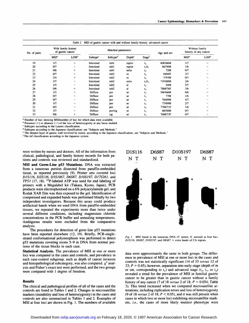
Table 2 MSI of gastric cancer with and without family history: advanced cancer
No. of pairs
With family history.
of gastric cancer
MSI� LOH”
Matched parametersAge and sex
Without family.
history of any cancer
Subtype� Subtype” Depth� Stage1 MSI” LOH”
19
20
21
22
23
24
25
26
27
28
29
30
3 1
32
33
1/7 -
0/7 -
0/6 +
0/7 -
2/4 +
3/7 -
1/5 -
0/6 +
3/5 -
0/7 -
0/7 -
1/7 -
0/5 -
3/7 -
0/6 +
Intestinal
Intestinal
Intestinal
Intestinal
Intestinal
Intestinal
Intestinal
Intestinal
Diffuse
Diffuse
Diffuse
Diffuse
Diffuse
Diffuse
Diffuse
tubl mp/ss
tub I mp/se
tubl se/ss
tub2 ss
tub2 ss
tub2 ss/se
tub2 si
tub2 si
por se
por se
por se
por se
por se
por/sig se
sig se
t2
t2/t3
t2
t2
t2
t,/t3
t4
t4
t3
t3
13
t3
t3
t3
t3
60F/66M
66/76M
72M
MM/F
57F/M
7 1F/68M
54M
78M/76F
56F/66M
77M
76/69M
77/69M
77Mfl 1F
60/58M
76M173F
1/7 -
1/6 -
0/7 -
2/7 -
0/3 -
2/6 -
5/7 -
3/6 -
0/6 -
1/6 -
4/7 -
2/7 -
1/6 -
0/7 -
0/7 -
a Number of loci showing MSI/number of loci for which data were available.
b Presence (+) or absence (-) of the loss of heterozygosity at any locus studied.
� Subtypes according to the Lauren classification.
d Subtypes according to the Japanese classification; see “Subjects and Methods.”
� The deepest layer of gastric wall involved by tumor, according to the Japanese1The tnf classification according to the Japanese system.
classification; see “Subjects and Methods.”
D1S116 D6S87 D10S197 D6S87
NT NT NT NT
a
Fig. 1. MSI found in the tumorous DNA (T, tumor; N, normal) at four loci,
DISJJ6, D6587, DJOS/97, and D6587. *, extra bands of CA repeats.
Cancer Epidemiology, Biomarkers & Prevention 695
were written by nurses and doctors. All of the information from
clinical, pathological, and family history records for both pa-tients and controls was reviewed and standardized.
MSI and Germ-Line p53 Mutations. DNA was extractedfrom a tumorous portion dissected from paraffin-embeddedtissue, as reported previously (9). Primer sets covered lociDJSJJ6, D2S136, D3S1067, D6S87, D10S197, D17S261, and
TP53 ( I 7, 1 8). 32P-labeled ATP was used for end labeling ofprimers with a Megalabel kit (Takara, Kyoto, Japan), PCRproducts were electrophoresed on a 6% polyacrylamide gel, and
Kodak XAR film was then exposed to the gel. Identification ofcompressed and expanded bands was performed blindly by twoindependent investigators. Because this assay could produceartifactual bands when we used DNA from paraffin-embedded
tissues, we repeated the experiments more than twice underseveral different conditions, including magnesium chloride
concentrations in the PCR buffer and annealing temperatures.Ambiguous results were excluded from the comparative
analysis.
The procedures for detection of germ-line p53 mutationshave been reported elsewhere (12, 19). Briefly, PCR-singbe-strand conformational polymorphism was performed to detect
p53 mutations covering exons 5-9 in DNA from normal por-tions of the tissue blocks in each case.
Statistical Analysis. The prevalence of MSI at one or moreboci was compared in the cases and controls, and prevalence ineach case-control subgroup, such as depth of cancer invasionand histopathological subcategory, was also compared. x� anal-
ysis and Fisher’s exact test were performed, and the two groups
were compared with 1 degree of freedom.
Results
The clinical and pathological profiles of all of the cases and thecontrols are listed in Tables 1 and 2. Changes in microsatellite
markers (instability and loss of heterozygosity) in the cases andcontrols are abso summarized in Tables 1 and 2. Examples ofMSI at four boci are shown in Fig. 1 . The numbers of available
data were approximately the same in both groups. The differ-ence in prevalence of MSI at one or more loci in the cases andcontrols was not statistically significant (14 of 33 versus 12 of
33; P = 0.45); however, separation into early stage (depth of mor sm, corresponding to t1) and advanced stage (t2, t3, or t4)
revealed a trend for the prevalence of MSI in familial gastriccancer to be greater than in gastric cancer without a family
history of any cancer (7 of 18 versus 2 of 18; P 0.054; Table3). This trend increased when we compared microsatellite ab-terations, including replication errors and boss of heterozygosity(9 of 18 versus 2 of b8; P < 0.05), and it was still present whencases in which two or more loci exhibiting microsatellite mark-ers, i.e., the cases of more likely mutator phenotype were
on February 18, 2020. © 1997 American Association for Cancer Research. cebp.aacrjournals.org Downloaded from
696 MSI in Familial Gastric Carcinoma
Table 3 Prevalence of MSI in case and control according to stage of disease
Group MSI” With family history of ,�, Without family historygastric cancer of any cancer
Total
Early - I 1 of 18 16 of 18 27 of 36
�l 7ofl8 0.05” 2of18 9of 36’
�2 5of18 0.07” 1 ofl8 60f36d
Advanced - 8ofls Sofl5 13 of 30
�l 7ofls 0.27” lOoflS 17of30’
�2 4 of 15 0.43” 6 of 15 10 of 30”
Total - 19of33 21 of 33
�l 14of33 0.45” 12 of 33
�2 9 of 33 0.57” 7 of 33
“ Number of loci with MSI. - , no MSI at any loci.1� p� are the result of � analysis (without Yates correction). The values become greater when Fisher’s exact test is applied.
‘ Frequency of MSI at one or more loci between early and advanced cancer is significantly different (P = 0.009).,, Frequency of MSI at two or more loci between early and advanced cancer is not statistically different (P = 0. 1 1).
Table 4 Prevalence of MSI in case and control according to histological subtype
Group MSI” With family history �,,, Without family historyof gastric cancer of any cancer
Total
Intestinal - lOof 19 II of 19 21 of38
�l 9ofl9 0.74” 8of19 l7of 38’
�2 6of19 0.72” 5ofI9 11 of 38”
Diffuse - 8of14 9of14 17of28
�l 6ofl4 0.90” 4of13 lOof 28’(0rLOH�l) (8ofl4) (0.25)” (4ofI4)
�2 3ofl4 1.00” 2ofl4 5of28”
“ Number of loci with MSI. - , no MSI at any loci. LOH, loss of heterozygosity.
1� Ps are the result of � analysis (without Yates correction). The values become greater when Fisher’s exact test is applied.
‘ Frequency of MSI at one or more loci between intestinal and diffuse type is not statistically different (P - 0.46).
,1 Frequency of MSI at two or more loci between intestinal and diffuse type is not statistically different (P = 0.29).
compared (5 of 18 versus 1 of 18). In contrast, prevalence wasthe same in the study cases and controls in advanced gastric
cancer (8 of 13 versus 10 of 13; P = 0.6). The overall preva-lence of MSI was greater in advanced-stage cancer (17 of 30)than early-stage cancer (9 of 36; P 0.009), which is consist-
ent with previous reports (10, 20). Furthermore, when dividedaccording to histological subtype, no differences between the
two groups in prevalence of MSI were seen (Table 4). In
addition, overall prevalence of MSI did not differ in the intes-tinal and diffuse histological subtypes ( I 7 of 38 versus 10 of 28;
P = 0.46; Table 4).No p53 mutations in exons 5-9 were detected in the DNA
from normal portions of any of the cases or controls whensingle-strand conformational polymorphism screening was per-formed.
Discussion
Because MSI in colorectal tumors in HNPCC patients reflects
mismatch repair gene defects in the germ line of these patients,genomic instability detected as MSI in tumors is believed to beassociated with individual genetic predisposition to cancer (4,2 1 ). However, not all MSI found in tumors represents germ-linemismatch repair gene defects, and somatic mutations of those
genes have also been reported. Gastric cancer exhibits a varietyof histological findings and is one of the common epithelialtumors in which MSI is observed frequently. MSI in gastrictumors has frequently been correlated with stage, location,prognosis, and erbB-2 expression (4, 10, 13, 14, 22-25). It isalso associated with multiple tumors and familial history (8, 9,26). Recently, Akiyama ci’ a!. (5) reported MSI in the gastric
cancer tissue of six patients with a family history of gastric
cancer, two of which were early-stage cancers. On the basis of
their own data on the four families and data on three probandsreported by others (27), they speculated that MSI is more
frequent in gastric cancer with familial clustering and that the
frequent MSI in the tumor may mean that its pathogenesis
might be similar to that of HNPCC. However, none of these
previous reports have examined MSI in sporadic gastric can-
cers, and little is known about the association of MSI in gastric
tumors and a family history of gastric cancer (22). Our report
is the largest study of MSI in gastric cancer with a family
history and addresses the importance of stage-dependent com-
parison for the first time. We adopted a histological subclassi-
fication system that included more specific morphologicab char-acteristics than a simple dichotomy such as “diffuse” and“intestinal” in selecting controls. Furthermore, taking advan-
tage of information on tumor depth, which is standardized bythe Japanese classification system, we matched each case to the
control on the basis of depth of invasion. We believe that this
strictly matched evaluation is necessary to assess the associa-
tion between familial clustering and the presence of MSI ingastric cancer. Actually, a trend for increased prevalence of
MSI in familial cases had only been noted in early cancer,whereas our data clearly showed that the prevalence of MSI
was similar in advanced gastric cancer, with or without familial
clustering ofgastric cancer. This MSI, more frequently found in
advanced cancer, appears to be acquired during the progressionof gastric cancer. It is more probable that the MSI in early-stage
cancer is associated with familial clustering, but the numbers ofcases and controls were small, and these results should be
on February 18, 2020. © 1997 American Association for Cancer Research. cebp.aacrjournals.org Downloaded from
Cancer Epidemiology, Biomarkers & Prevention 697
interpreted with caution. In particular, cases exhibiting MSI at
more than two boci, potentially mutator phenotype tumors,which are too small in number to allow a statistically significant
conclusion in this data set, are the most important candidates
for further investigation for genetic predisposition to gastriccancer. Possible targets such as the transforming growth factor
13receptor type II gene (TGFf3RIT) or the MSH3 gene must alsobe examined to confirm that the MSI in these familial gastriccancers represents the mutator phenotype of the tumor. Natu-rally, the germ-line alterations of mismatch repair genes in ourcases are of interest, but because most of the patients in thisstudy were older than 50 years old, the situation differs from
that of HNPCCs, which have germ-line mutations of mismatchrepair genes. Akiyama et a!. (5) found a somatic hMSH2
mutation in tumor DNA but not in the germ-line DNA of the
family members, suggesting a contribution of somatic changesto MSI in gastric cancer with familial clustering. Although wehave not yet examined germ-line changes in our cases, the
causal factors in familial gastric cancer, including environmen-
tab and genetic factors, may also possibly be associated with CArepeat alterations in early stages of carcinogenesis. Among thecases of gastric cancer with familial clustering, which areobviously heterogeneous entities, the cases that exhibited MSIfrom the early stage would be good candidates in which to
pursue the genetic predisposition of gastric cancer. Early ac-
quisition of MSI in HNPCC has been reported very recently(28). The mechanism of early acquisition of MSI in familialgastric cancer is intriguing from the standpoint of genetic
defects in the mismatch repair system. Target molecule changes
now under investigation will resolve this issue.
Acknowledgments
We thank Dr. Yo Kato (Cancer Institute, Tokyo. Japan) for constructive com-
ments. This work was motivated and supported by the members of the Japanese
Gastric Cancer Study Group (President, Dr. M. Nishi. Cancer Institute, Tokyo).
References
1 . Sherlock, P., Morson, B. C., Barbara, L., and Veronesi, U. (eds.). Precancerous
Lesions of the Gastrointestinal Tract. New York: Raven Press, 1983.
2. Aaltonen, L. A., Peltomaki, P., Leach, F. S., Sistonen, P., Pylkkanen, L.,
Mecklin, J. P., Jarvinen, H., Powell, S. M., Jen, J.. Hamilton, S. R., Petersen. G.,Kinzler, K., Vogelstein, B., and de Ia Chapelle, A. Clues to the pathogenesis of
familial colorectal cancer. Science (Washington DC), 260: 812-816. 1993.
3. Leach, F. S., Nicolaides, N. C., Papadopoulos, N., Liu, B., Jen, J., Parsons, R.,
Peltomaki, P., Sistonen, P., Aaltonen, L. A., Nystrom-Lahti, M.. Guan, X-Y.,Zhang, J., Meltzer, P., Yu, J., Kao, F., Chen, D., Cerosaletti, K., Foumier, R.,
Todd, S., Lewis, T., Leach, R., Naylor, S.. Weissenbach, J., Mecklin, J., J#{228}rvinen,
H., Petersen, G., Hamilton, S., Green, J., Jass, J., Watson, P., Lynch, H., Trent,J.. de Ia Chapelle, A., Kinzler, K., and Vogelstein, B. Mutations of a mutShomolog in hereditary nonpolyposis colorectal cancer. Cell, 75: 1215-1225.
1993.
4. lonov, Y., Peinado, M. A., Malkhosyan, S., Shibata, D.. and Perucho, M.
Ubiquitous somatic mutations in simple repeated sequences reveal a new mech-
anism for colonic carcinogenesis. Nature (Lond.), 363: 558-561, 1993.
5. Akiyama, Y., Nagasaki, H., Nikei, Z., Iwama, T., Nomizu, T., Utsunomiya. J..
and Yuasa, Y. Frequent microsatellite instabilities and analyses of the related
genes in familial gastric cancers. Jpn. J. Cancer Res., 87: 595-601, 1996.
6. Sasaki, A., Nagashima, M., Shiseki, M., Katai, H., Maruyama, K.. Iwanaga,
R., Akiyama, Y., Yuasa, Y., and Yokota, J. Microsatellite instability in gastriccancer prone families. Cancer Lett., 99: 169-175, 1996.
7. Lothe, R. A., Peltom#{228}ki, P., Meling, G. I., Aaltonen, L. A., Nystrom-Lahti, M.,
Pylkkanen, L., Heimdal, K., Andersen, T. I., M#{248}ller,P., Rognum, T. 0., Foss&S. D., Haldorsen, T., Langmark, F., Br#{248}gger,A., de Ia Chapelle, A., and B#{248}rresen,A-L. Genomic instability in colorectal cancer: relationship to clinicopathological
variables and family history. Cancer Res., 53: 5849-5852, 1993.
8. Nakashima, H., Honda, M., Inoue, H., Shibuta, K., Arinaga, S., Era, S., Ueo.
H., Mon. M., and Akiyoshi, T. Microsatellite instability in multiple gastric
cancers. Int. J. Cancer, 64: 239-242, 1995.
9. Shinmura, K., Sugimura, H.. Naito. Y., Shields, P. G., and Kino, I. Frequent
co-occurrence of mutator phenotype in synchronous. independent multiple can-
cers of the stomach. Carcinogenesis (Lond.). 16: 2989-2993. 1995.
10. Rhyu. M. G., Park, W. S., and Meltzer, S. J. Microsatellite instability occurs
frequently in human gastric carcinoma. Oncogenc. 9: 29-32. 1994.
1 1 . Seruca, R., Santos. N. R.. David. L.. Consiancia. M., Barroca. H.. Cameiro,
F., Seixas. M., Peltomaki, P.. Lothe. R., and Sobrinho-Simocs. M. Sporadic
gastric carcinomas with microsatellite instability display a particular clinicopath-
ologic profile. Int. J. Cancer. 64: 32-36. 1995.
12. Sameshima, Y., Matsuno, Y., Hirohashi, S., Shimosato, Y.. Mizoguchi, H..
Sugimura. T., Terada, M., and Yokota. J. Alterations of the p53 gene are common
and critical events for the maintenance of malignant phenotypes in small-cell lung
carcinoma. Oncogene. 7: 451-457, 1992.
13. Han, H. J., Yanagisawa, A., Kato, Y., Park, J. G.. and Nakamura. Y. Genetic
instability in pancreatic cancer and poorly differentiated type of gastric cancer.
Cancer Res., 53: 5087-5089, 1993.
14. dos Santos, N. R., Seruca. R.. Constancia, M.. Seixas. M.. and Sobrinho-
Simoes. M. Microsatellite instability at multiple loci in gastric carcinoma: din-
icopathologic implications and prognosis. Gastroenterology. I/O: 38-44. 1996.
15. Japanese Research Society for Gastric Cancer. Japanese Classification of
Gastric Carcinoma. Tokyo: Kanehara, 1995.
16. Lauren, P. The two histological main types ofgastric carcinoma-diffuse and
so-called intestinal type carcinoma: an attempt at a histological classification.
Acta Pathol. Microbiol. Scand., 64: 31-39, 1965.
17. Weissenbach, J. A second generation linkage map of the human genome
based on highly informative microsatellite Id. Gene, 135: 275-278. 1993.
18. Breukel, C., Tops, C.. van Leeuwen, C., van der Klift, H., Nakamura, Y..
Fodde, R.. and Khan, P. M. CA repeat polymorphism at the D5S82 locus,
proximal to adenomatous polyposis coli (APC). Nucleic Acids Res., 19: 5804.
1991.
19. Shiseki. M.. Nishikawa, R., Yamamoto, H., Ochiai, A., Sugimura. H.,
Shitara. N., Sameshima, Y., Mizoguchi. H.. Sugimura, 1., and Yokota, J. Germ-
line p53 mutation is uncommon in patients with triple primary cancers. Cancer
Lett., 73: 51-57. 1993.
20. Lin, J. T., Wu, M. S., Shun, C. T., Lee, W. J., Wang, J. T.. Wang. T. H.. and
Sheu, J. C. Microsatellite instability in gastric carcinoma with special references
to histopathology and cancer stages. Eur. J. Cancer, 3/A: 1879-1882, 1995.
21. Peltom#{228}ki. P., Lothe. R. A., Aaltonen, L. A.. Pylkkanen. L.. Nystrom-Lahti.
M., Seruca, R.. David, L., HoIm, R., Ryberg, D.. Haugen. A.. Bragger. A..
B#{248}rresen, A-L., and de Ia Chapelle, A. Microsatellite instability is associated with
tumors that characterize the hereditary non-polyposis colorectal carcinoma syn-
drome. Cancer Res., 53: 5853-5855, 1993.
22. Chong. J. M., Fukayama. M.. Hayashi. Y.. Takizawa. T.. Koike. M., Konishi,
M., Kikuchi-Yanoshita, R., and Miyaki, M. Microsatellite instability in the
progression of gastric carcinoma. Cancer Res., 54: 4595-4597. 1994.
23. Gleeon, C. M., Sloan, J. M., McGuigan. J. A.. Ritchie, A. J.. Weher. J. L.. and
Russell. S. H. Widespread microsatellite instability occurs infrequently in ade-
nocarcinoma of the gastric cardia. Oncogene. 12: 1653-1662. 1996.
24. Lin, J. T., Wu, M. S., Shun, C. T., Lee, W. J., Sheu. J. C., and Wang. T. H.
Occurrence of microsatellite instability in gastric carcinoma is associated with
enhanced expression of erbB-2 oncoprotein. Cancer Res., 55: 1428-1430, 1995.
25. Mironov, N. M.. Aguelon, M. A., Potapova. G. I.. Oman, Y.. Gorhunov.
0. V., Klimenkov, A. A., and Yamasaki, H. Alterations of (CA),, DNA repeats
and tumor suppressor genes in human gastric cancer. Cancer Res., 54: 4 1-44,
1994.
26. Keller, G., Rotter. M., Vogelsang. H.. Bischoff. P.. Becker, K. F.. Mueller, J..
Brauch, H., Siewert. J. R., and Holler, H. Microsatellite instability in adenocar-
cinomas ofthe upper gastrointestinal tract: relation to clinicopathological data and
family history. Am. J. Pathol., /47: 593-600, 1995.
27. Sasaki, K., Bertrand. 0., Nakazawa, H., Fitzgerald. D. J., Mironov, N., and
Yamasaki, H. Cell-type-specific ras mutations but no microsatellite instability in
chemically induced mouse skin tumors and transformed 3T3 cells. Cancer Res.,
55: 3513-3516. 1995.
28. Konishi, M.. Kikuchi-Yanoshita, R., Tanaka. K.. Muraoka. M.. Onda, A..
Okumura, Y., Kishi, N.. Iwama, T.. Mon. T., Koike, M.. Ushio, K., Chiba, M..
Nomizu, S.. Konishi, F.. Utsunomiya, J.. and Miyaki. M. Molecular nature of
colon tumors in hereditary nonpolyposis colon cancer, familial polyposis, and
sporadic colon cancer. Gastrocnterology. Ill: 307-317. 1996.
on February 18, 2020. © 1997 American Association for Cancer Research. cebp.aacrjournals.org Downloaded from
1997;6:693-697. Cancer Epidemiol Biomarkers Prev K Shinmura, W Yin, J Isogaki, et al. gastric carcinoma with familial clustering.Stage-dependent evaluation of microsatellite instability in
Updated version
http://cebp.aacrjournals.org/content/6/9/693
Access the most recent version of this article at:
E-mail alerts related to this article or journal.Sign up to receive free email-alerts
Subscriptions
Reprints and
To order reprints of this article or to subscribe to the journal, contact the AACR Publications
Permissions
Rightslink site. Click on "Request Permissions" which will take you to the Copyright Clearance Center's (CCC)
.http://cebp.aacrjournals.org/content/6/9/693To request permission to re-use all or part of this article, use this link
on February 18, 2020. © 1997 American Association for Cancer Research. cebp.aacrjournals.org Downloaded from


![A National Cancer Institute Workshop on Microsatellite ...ICANCER RESEARCH 58. 5248-5257, November 15. 1998] Meeting Report A National Cancer Institute Workshop on Microsatellite Instability](https://img.pdfslide.net/doc/110x75/6023dabb0b4d3d4b71328e59/a-national-cancer-institute-workshop-on-microsatellite-icancer-research-58.jpg)