Embed Size (px)
Citation preview
Copyright 0 1993 by the Genetics Society of America
Stage-Specific Effects of X-Irradiation on Yeast Meiosis
Leigh W. Thorne' and Breck Byers Department of Genetics, University of Washington, Seattle, Washington 981 95
Manuscript received July 3 1 , 1992 Accepted for publication January 8, 1993
ABSTRACT Previous work has shown that cdcl3 causes meiotic arrest of Saccharomyces cerevisiae following DNA
replication by a RAD9-dependent mechanism. In the present work, we have further investigated the implicit effects of chromosomal lesions on progression through meiosis by exposing yeast cells to X- irradiation at various times during sporulation. We find that exposure of RAD9 cells to X-irradiation early in meiosis prevents sporulation, arresting the cells at a stage prior to premeiotic DNA replication. rad9 meiotic cells are much less responsive to X-irradiation damage, completing sporulation after treatment with doses sufficient to cause arrest of RAD9 strains. These findings thereby reveal a RAD9- dependent checkpoint function in meiosis that is distinct from the GP arrest previously shown to result from cdcl3 dysfunction. Analysis of the spores that continued to be produced by either RAD9 or rad9 cultures that were X-irradiated in later stages of sporulation revealed most spores to be viable, even after exposure to radiation doses sufficient to kill most vegetative cells. This finding demonstrates that the lesions induced by X-irradiation at later times fail to trigger the checkpoint function revealed by cdcl3 arrest and suggests that the lesions may be subject to repair by serving as intermediates in the recombination process. Strains mutant for chromosomal synapsis and recombination, and therefore defective in meiotic disjunction, were tested for evidence that X-ray-induced lesions might alleviate inviability by promoting recombination. Enhancement of spore viability when spoll (but not hopl) diploids were X-irradiated during meiosis indicates that induced lesions may partially substitute for SPOll-dependent functions that are required for the initiation of recombination.
S PORULATION in Saccharomyces cerevisiae re- quires many genetically controlled functions that
are shared with the vegetative cell cycle, but the order in which these functions are required with respect to various cytological landmarks often differs strikingly between the two pathways of cell division. For exam- ple, mutations that affect START-such as cdc28, cdc?6 and cdc39-cause mitotic arrest at a stage prior to DNA replication (HARTWELL 1973) and spindle pole duplication (BYERS and GOETSCH 1974; REED 1980) [reviewed in PRINGLE and HARTWELL ( 1 98 l)]. In meiosis, on the other hand, these mutations permit the execution of both of these functions but then cause arrest at the stage of chromosomal synapsis, as evidenced by the persistent presence of synaptonemal complexes (SHUSTER and BYERS 1989). As a further example, mutations primarily affecting chromosomal DNA replication cause mitotic arrest in a state follow- ing spindle formation, whereas the common state of their arrest in meiosis precedes both spindle formation and synapsis (SCHILD and BYERS 1978). In light of the recent recognition that many such mitotic arrests re- flect the action of checkpoint functions (HARTWELL and WEINERT 1989), it was of interest to ask how checkpoints may affect meiosis and how they may be
sity, Box 7614, Raleigh, North Carolina 27695.
Genetics 134: 29-42 (May, 1993)
' Present address: Department of Genetics, North Carolina State Univer-
ordered with respect to other meiotic functions. In this regard, we had previously tested whether the mutational defect in cdcl3, which leads to a RAD9- dependent mitotic arrest (WEINERT and HARTWELL 1988) presumably due to the induction of chromo- somal lesions (HARTWELL and SMITH 1985), could be used to generate an arrest of meiosis. We found that the mutation does, indeed, cause meiotic arrest in a RAD9-dependent manner, cdcl? RAD9 cells failing to undergo either synapsis or the meiotic divisions at the restrictive temperature, while cdcl3 rad9 cells con- tinue through meiosis and produce inviable progeny (WEBER and BYERS 1992). This being the case, we then wished to establish whether chromosomal lesions imposed at other stages of meiosis would result in a similar arrest, but found no suitable genetic deficien- cies for this purpose. We therefore turned to the use of X-ray damage, which could be imposed at any stage of meiosis and which has been shown to cause mitotic arrest in GP in a manner similar to cdcl? (WEINERT a n d HARTWELL 1988).
The impact of ultraviolet (UV) irradiation on yeast cells during meiosis had been investigated previously. SIMCHEN, SALTS and PINON (1973) found that cells heavily irradiated during premeiotic DNA synthesis were severely affected, undergoing meiotic arrest and suffering a reduction in meiotic recombination
30 L. W. Thorne and B. Byers
(SALTS, SIMCHEN and PINON 1976; RESNICK, GAME and STASIEWICZ 1983). At 30-fold lower doses of irradiation, MACHIDA and NAKAI (1980) detected no significant effect on intragenic recombination but found that intergenic recombination was enhanced. The effects of X-irradiation on meiotic yeast have also been examined, but in less detail. ROMAN (1984) de- scribed striking effects on gene conversion both in mitosis and in meiosis, but the doses tested (up to 3 krad) were insufficient to cause clear-cut effects on levels of sporulation. Using higher doses and a return- to-mitotic-growth procedure, KELLY, MERRILL and PARRY (1 983) clearly demonstrated that reciprocal recombination, as measured by an ade2 heteroallele system, is stimulated by X-irradiation. The effects of X-irradiation on ongoing meiosis merit further atten- tion because the well-characterized lesions that are generated-single- and double-strand breaks (Mc- GRATH and WILLIAMS 1966; HO 1975; RESNICK and MARTIN 1976)-can be induced independently of cel- lular functions, such as the excision repair system, which might be required to transform the primary lesions of UV-irradiation and some types of chemical mutagenesis into interruptions of strand continuity.
X-irradiation has been shown to affect meiotic re- combination in other organisms. Progeny testing in Drosophila has shown that X-irradiation leads to the induction of recombination, translocations, and mu- tations in meiotic cells not only of females but also in males, which normally display no recombination dur- ing meiosis [reviewed in SANKARANARAYANAN and SOBELS (1 976) and HANNAH-ALAVA (1 964)) Without any effective way to stage the time of treatment with respect to the complex biology of reproduction in Drosophila, it has been difficult to establish whether it was meiosis per se that was being affected. Better timing has been achieved in the irradiation of Lilium (LAWRENCE 196 1 a) and Tradescantia (LAWRENCE 196 1 b) meiocytes, wherein increased frequencies of chiasmata were found at metaphase I. Significantly increased meiotic recombination in response to X- irradiation has also been reported in Chlamydomonas reinhardi (LAWRENCE and HOLT 1970) and Caenor- habditis elegans (KIM and ROSE 1987; MCKIM, HOWELL and ROSE 1988).
It seemed reasonable to expect an induction of meiotic recombination by X-irradiation because the lesions caused by X-irradiation-single-strand and dou- ble-strand breaks in DNA-are thought to be inter- mediates in meiotic recombination. Most models for the mechanisms of meiotic recombination invoke the formation of such breaks as a crucial initiating event and many experiments are interpreted as consistent with this concept (HOLLIDAY 1964; MESELSON and RADDINC 1975; ORR-WEAVER, SZOSTAK and ROTH- STEIN 198 1 ; RESNICK 1976; RESNICK et al. 198 1 , 1984;
SYMINCTON 1991; SZOSTAK et al. 1983) [for review see ORR-WEAVER and SZOSTAK (1985)l. Moreover, meiosis-specific double-strand breaks have been dem- onstrated by physical assays not only as hotspots for meiotic recombination (CAO, ALANI and KLECKNER 1990; NICOLAS et al. 1989; SUN et al. 1989) but also more generally in meiotic chromosomes (GAME et al. 1989). Furthermore, double-strand breaks induced by HO cutting stimulate meiotic recombination both in S. cereuisiae (KOLODKIN, KLAR and STAHL 1986) and in Schizosaccharomyces pombe (KLAR and MICLIO 1986).
In the present work, we provide evidence in S. cereuisiae that X-ray-induced lesions can stimulate re- combination if the treatment is imposed after the initiation of premeiotic DNA synthesis, whereas the induction of X-ray damage prior to this point causes arrest in the GI phase of meiosis in a RADY-dependent manner. In addition, assays for spore viability in $01 1 meiosis provide evidence that the lesions induced by X-irradiation at the later stage may partially substitute for this mutational deficiency in meiotic recombina- tion and lead to improved meiotic disjunction.
MATERIALS AND METHODS
Strains: The genotypes of the strains used in these exper- iments are described in Table 1. Standard procedures for strain construction and genetic analysis were employed (MORTIMER and HAWTHORNE 1969). Strains LW3501 and LW3502 were generated from a diploid of 5837-2-1 and 5821-c-16, which were obtained from N. HOLLINGSWORTH. Strains LW3201, LW3202, LW3203 and LW3204 were made by crossing spore clones of 7845-8-4 and 3985-4-1b and are in the A364a background; LW3205 is a spore clone of this same diploid. Strains LW3605, LW3606, LW3607 and LW3608 were generated by crossing spore clones of a diploid of LW3501 and 5819-24-3 to LW3502, and strain LW3604 is a spore clone of this same diploid. Spore clones of a diploid of LW3604 and CAY63 were crossed to each other to generate LW3802, which (presumably because of slight differences in background) proceeds through meiosis slightly faster than the other strains used, as confirmed by fluorescence-activated cell sorter (FACS) analysis (data not shown). These strain constructions were designed to main- tain a hybrid background of A364a and 131-20 while cre- ating marked intervals that could be used to monitor recip- rocal exchange. Strain LW3901 was generated by crossing spore clones from a diploid of LW3205 and LW3502 to 13 1-20, thus also retaining the hybrid background of A364a and 131-20. No significant differences in the rates of vege- tative growth or sporulation were found among any of the strains used for the irradiation experiments (data not shown).
Genetic procedures: Materials and methods used in this study have been described, including liquid and solid media (HARTWELL 1967; WOOD 1982), sporulation procedures (SCHILD and BYERS 1978), assays of viability and commit- ment to recombination (SHUSTER and BYERS 1989), and the assay for haploidization (SHERMAN and ROMAN 1963; HOL- LINGSWORTH and BYERS 1989). Sporulation medium (SPM) contains 3.0 g of potassium acetate and 0.2 g of raffinose per liter of water, while -N medium contains 1.61 g of yeast
Radiation Response of Meiotic Yeast
TABLE 1
Saccharomyces cerevisiae strains
Strain Genotype
CAY63 MATa ade2 ura? canl his3 leu2 trpt atr::HIS3 spoll-D?
13 1-20 MATa ade2-R8 ura? leul canl cyh2
3985-4-1b MATa his7 ade2
58 19-24-3 MATa ade2 trpl ura?
5821-C-16 MATa ade2 canl leul leu2 trp5 cyh2 lys5 hopl::LEU2 his7
5837-2-1 MATa his1 leu.? trpl ura3 spol l
7845-8-4 MATa cdcl?-1 rad9::LEU2 his7 trpl ura3 canl cyh2
L W 3 0 0 3 M A T a c d c l 3 - 1 ade2 ural LEU1 CYH2 his7 tyrl CANl URAS lys2 MATa cdc l3 - l ade2 URA1 leul cyh2 HIS7 TYRl canl ura3 LYS2 ""
LW3201 MATa rad9::LEU2 " ade2 trpl URA3 CANl " CYH2 his7 MATa RAD9 ADE2 TRPl ura3 canl cyh2 his7
L W 3 2 0 2 MATa rad9::LEU2 ade2 trpl URAS CANl CYH2 his7 MATa RAD9 ade2 TRPl ura3 canl cyh2 his7 "
LW3203 MATa rad9::LEUP ade2 trpl URA3 CANl CYH2 his7 MATa rad9::LEUS ade2 trpl ura3 canl cyh2 his7
~~ "
LW3204 MATa rad9::LEUS ~- ade2 trpl URA3 CANl -- CYH2 his7 MATa rad9::LEU2 ADE2 TRPl ura? canl cyh2 his7
LW3205 MATa ade2 his7 ura? cdcl3-1 rad9::LEUZ
LW3501 MATa can l cyh2 hisl hopI::LEU2 leul leu2 lys5 spa1 I trp5
LW3502 MATa ade2 canl cyh2 hisl hopl::LEU2 leu1 leu2 lys5 sf1011 trp5
LW3604 MATa ade2 ura3
LW3605"
LW3606"
MATa ade2 hop1 spoll canl URA3 hisl leul trp5 cyh2 lys5 MATa ADE2 hopl spol 1 CANl ufa3 HIS1 LEUl TRP5 CYH2 LYS.5 LEU2 ""
MATa ade2 hopl spoll canl URA3 hisl leu1 trp5 cyh2 lys5 MATa ADE2 HOPl spol 1 CANl ura3 HIS1 LEUl TRP5 CYH2 LYS5 LEU2 ~ " -
LW3607" ~-__- MATa ade2 hopl spoll canl URA3 hisl leu1 trp5 cyh2 lys5 x MATa ADE2 hopl SPOl1 CANl ura3 HISl LEUl TRP5 CYH2 LYs5 LEU2
MATa ade2 hopl spoll canl URA3 hisl leu1 trp5 ~ $ 2 1 ~ ~ 5 leu2 MATa ADE2 HOPl SPOll CANl ura3 HISl LEU1 TRP5 CYH2 Ly.5 LEU:'
LW3608" ""
LW3802b
L W 3 9 0 1
MATa CANl ura3 HISl TRP5 CYH2 LYS5 trpl spol1-D3 ade2 MATa canl URA3 hisl trp5 cyh2 lys5 T R P l $01 1-03 ADE2
- ~ -
MATa ade2 cdcl3-1 LEUl CYH2 lys5 URAS CANl rad9::LEUP MATa ade2 cdcl3-1 leul cqh2 E ura3 canl rad9::LEU2 "-
C A Y 6 3 was provided by R. E. ESPOSITO. 131-20 is described in HOPPER and HALL (1975). 5819-24-3, 5821-c-16 and 5837-2-1 were obtained from N. HOLLINGSWORTH. 58 19-24-3 is a hybrid of A364a and 13 1-20 backgrounds. 3985-4-1 b and 7845-8-4 were obtained from L. HARTWELL. L W 3 0 0 3 is described in WEBER and BYERS (1992) .
a hopl allele is hopl::LEU2; $011 allele is spol l -1 . spol 1-03 is a deletion of SPOl I and was kindly provided b y R. E. ~ P O S I T O .
nitrogen base without amino acids (Difco), 1 1.1 g of succinic acid, and 6.7 g of sodium hydroxide per liter. All experi- ments were conducted at room temperature (23") unless otherwise indicated.
X-irradiation procedures: Vegetative cultures were grown to cell densities of 1.3 X 10' cells/ml to 1.9 X 10' cells/ml in YPA medium, washed with -N medium, resus- pended in SPM, and incubated with shaking at room tem- perature. Subsequently cells were pelleted from 5-10-ml aliquots of larger cultures by centrifugation, resuspended in
a small amount of the supernatant liquid, and plated on an agar plate of the same medium as that used for liquid growth (KAC, YEPD or minimal medium) to generate a monolayer of cells that would not be shielded from irradiation by liquid or by each other. These plated cultures were X-irradiated (with lids removed) at a dose rate of 108 rad/sec in a Picker X-ray machine set at 50 kilovolts and 20 milliamps. Unir- radiated cultures were similarly subjected to replating and transport to the X-ray machine to guard against inadvertant effects of these manipulations. Treated and control cells
32 L. W. Thorne and B. Byers
were then washed from the plates with appropriate liquid media and either returned to the previous growth conditions or immediately diluted for plating on test media. These plating and resuspension procedures had little effect on progression of unirradiated cells through meiosis except for those replated near the time of meiosis I (about 12- 15 hr in these strains), beyond which stage responses to replating varied erratically both within and between experiments. All data presented here were gathered using cells treated at stages prior to the time of meiosis I (from 0 to 1 1 hr). Every experiment reported was repeated at least three times, yielding nearly identical results in each repetition. Because individual cultures varied slightly in their time of entry into the meiotic process (typically f 3 0 min), data are presented from single experiments rather than being pooled. FAGS analysis on control meiotic time courses of these strains confirmed that cells accumulated in GI after transfer to SPM and later entered into S phase within about 5 hr. All values reported represent at least 100 colony-forming units (cfu) in each experiment and appropriately account for dilution factors prior to plating.
Cytological procedures: Preparation and procedures for electron microscopy were as described in BYERS and GOETSCH (1 99 1). Serial sections were examined with a Phil- ips EM300 electron microscope. Fluorescence microscopy procedures were as described in ADAMS and PRINCLE (1 984) and BAUM, GOETSCH and BYERS (1988). DNA was stained with 4’,6-diamidino-2-phenylindole (DAPI) (1 .O pg/ml, Sigma) for examination with a Nikon Microphot microscope equipped with epifluorescence optics and filters. Flow cy- tometry analysis was as described in HUTTER and EIPEL (1979) using propidium iodide to stain DNA (Sigma). A Becton-Dickinson FACScan flow cytometer and the soft- ware packages CELLFIT and LYSYS were used to collect and analyze these data. For the sake of clarity in comparison with the vegetative cell cycle parameters, we will refer to premeiotic S phase, the following interval, and meiosis I as S, G:! and M, respectively.
Liquid holding recovery procedure: A vegetative culture was grown to the point of readiness for meiosis in YPA medium and cells were harvested by centrifugation. Half of the cells were resuspended in -N liquid medium (a starvation medium lacking any metabolic nitrogen source); the other cells were resuspended in SPM. After 5 hr of incubation with shaking, aliquots were irradiated. Serial dilutions were plated on complete solid medium at this time to assess culture viability. Dilutions were also plated from both cul- tures after control sporulation reached maximal levels (about 48 hr).
RESULTS
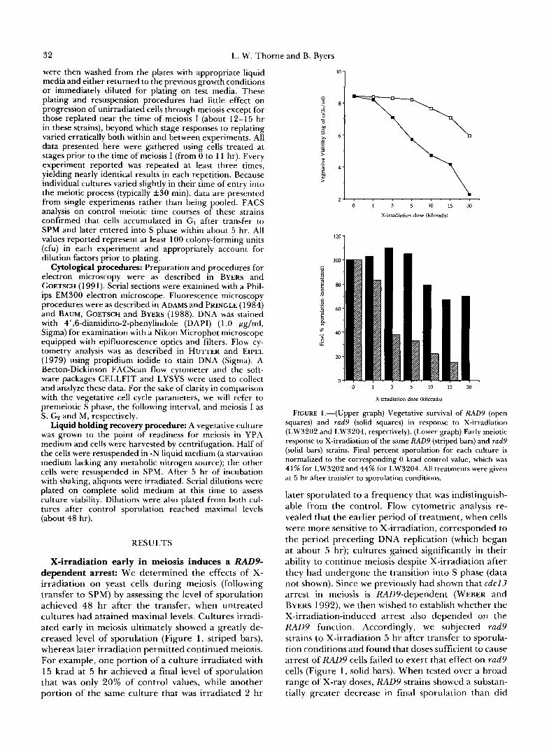
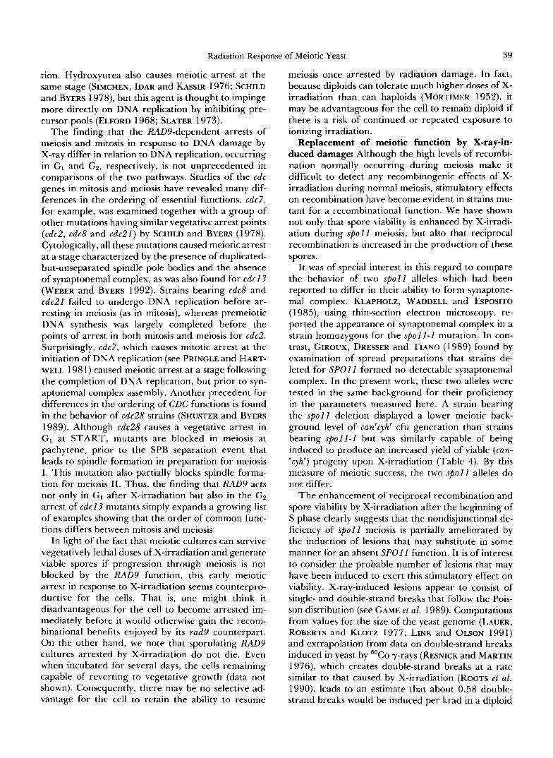
X-irradiation early in meiosis induces a RADP dependent arrest: We determined the effects of X- irradiation on yeast cells during meiosis (following transfer to SPM) by assessing the level of sporulation achieved 48 hr after the transfer, when untreated cultures had attained maximal levels. Cultures irradi- ated early in meiosis ultimately showed a greatly de- creased level of sporulation (Figure l , striped bars), whereas later irradiation permitted continued meiosis. For example, one portion of a culture irradiated with 15 krad at 5 hr achieved a final level of sporulation that was only 20% of control values, while another portion of the same culture that was irradiated 2 hr
’*O 1
0 1 3 5 1 0 1 5 3 0
X-irradiation dose (kilorads)
0 1 3 5 10 15 30
X-irradiation dose (kilorads)
FIGURE 1.-(Upper graph) Vegetative survival of RAD9 (open squares) and rad9 (solid squares) in response to X-irradiation (LW3202 and LW3204, respectively). (Lower graph) Early meiotic response to X-irradiation of the same RAD9 (striped bars) and rad9 (solid bars) strains. Final percent sporulation for each culture is normalized to the corresponding 0 krad control value, which was 41% for LW3202 and 44% for LW3204. All treatments were given at 5 hr after transfer to sporulation conditions.
later sporulated to a frequency that was indistinguish- able from the control. Flow cytometric analysis re- vealed that the earlier period of treatment, when cells were more sensitive to X-irradiation, corresponded to the period preceding DNA replication (which began at about 5 hr); cultures gained significantly in their ability to continue meiosis despite X-irradiation after they had undergone the transition into S phase (data not shown). Since we previously had shown that c d c l 3 arrest in meiosis is RAD9-dependent (WEBER and BYERS 1992), we then wished to establish whether the X-irradiation-induced arrest also depended on the RAD9 function. Accordingly, we subjected rad9 strains to X-irradiation 5 hr after transfer to sporula- tion conditions and found that doses sufficient to cause arrest of RAD9 cells failed to exert that effect on rad9 cells (Figure 1, solid bars). When tested over a broad range of X-ray doses, RAD9 strains showed a substan- tially greater decrease in final sporulation than did
Radiation Response of Meiotic Yeast 33
35 1
.- = I J
I
5 7 9 11 13 15
Time after transfer to SPM (hours)
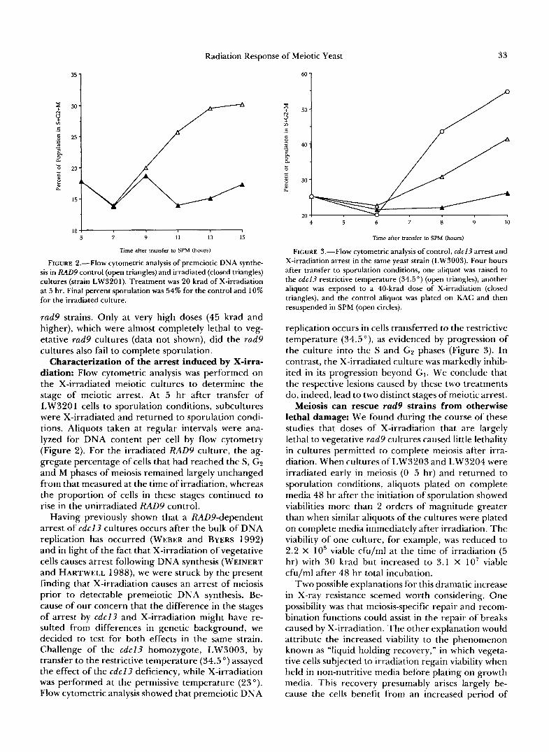
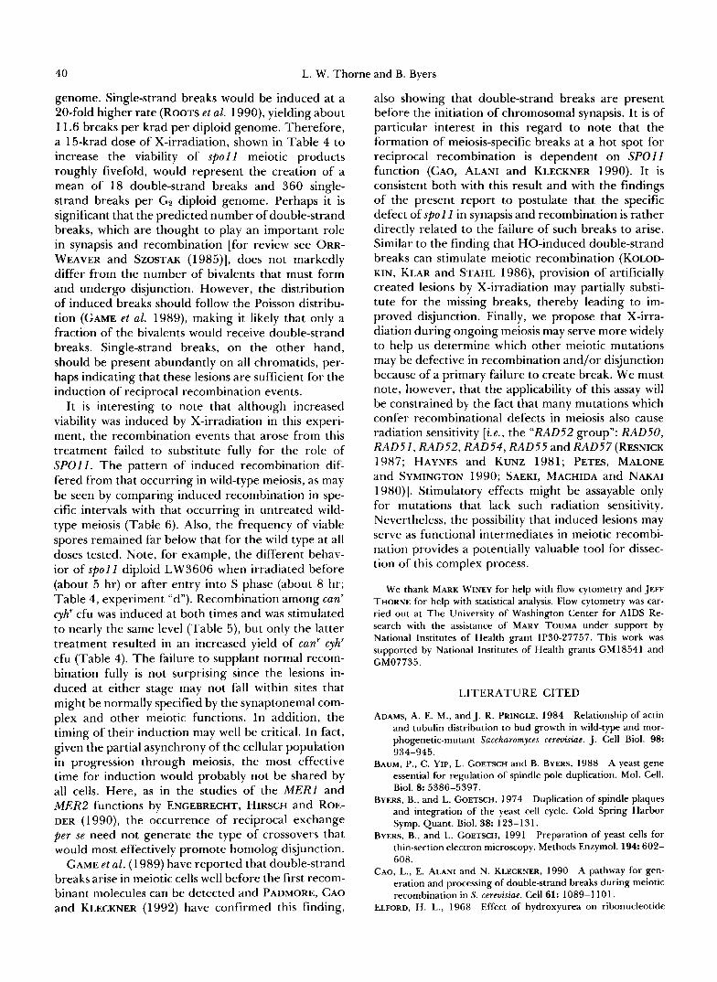
FIGURE 2.-Flow cytometric analysis of premeiotic DNA synthe- sis in RAD9 control (open triangles) and irradiated (closed triangles) cultures (strain LW3201). Treatment was 20 krad of X-irradiation at 5 hr. Final percent sporulation was 54% for the control and 10% for the irradiated culture.
rad9 strains. Only at very high doses (45 krad and higher), which were almost completely lethal to veg- etative rad9 cultures (data not shown), did the rad9 cultures also fail to complete sporulation.
Characterization of the arrest induced by X -1rra- '
diation: Flow cytometric analysis was performed on the X-irradiated meiotic cultures to determine the stage of meiotic arrest. At 5 hr after transfer of LW320 1 cells to sporulation conditions, subcultures were X-irradiated and returned to sporulation condi- tions. Aliquots taken at regular intervals were ana- lyzed for DNA content per cell by flow cytometry (Figure 2). For the irradiated RAD9 culture, the ag- gregate percentage of cells that had reached the S, GZ and M phases of meiosis remained largely unchanged from that measured at the time of irradiation, whereas the proportion of cells in these stages continued to rise in the unirradiated RAD9 control.
Having previously shown that a RADPdependent arrest of cdcl3 cultures occurs after the bulk of DNA replication has occurred (WEBER and BYERS 1992) and in light of the fact that X-irradiation of vegetative cells causes arrest following DNA synthesis (WEINERT and HARTWELL 1988), we were struck by the present finding that X-irradiation causes an arrest of meiosis prior to detectable premeiotic DNA synthesis. Be- cause of our concern that the difference in the stages of arrest by cdcl3 and X-irradiation might have re- sulted from differences in genetic background, we decided to test for both effects in the same strain. Challenge of the cdcl3 homozygote, LW3003, by transfer to the restrictive temperature (34.5") assayed the effect of the cdcl3 deficiency, while X-irradiation was performed at the permissive temperature (23"). Flow cytometric analysis showed that premeiotic DNA
6ol Y N
m c 1 .-
/
20 2 4 5 6 8 9 10
Time after transfer to SF" (hours)
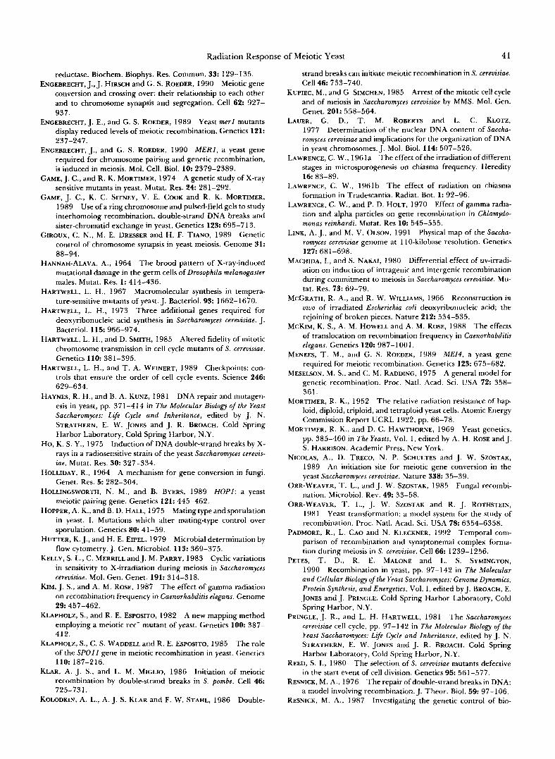
FIGURE 3.-Flow cytometric analysis of control, cdc l3 arrest and X-irradiation arrest in the same yeast strain (LW3003). Four hours after transfer to sporulation conditions, one aliquot was raised to the cdc l3 restrictive temperature (34.5") (open triangles), another aliquot was exposed to a 40-krad dose of X-irradiation (closed triangles), and the control aliquot was plated on KAC and then resuspended in SPM (open circles).
replication occurs in cells transferred to the restrictive temperature (34.5"), as evidenced by progression of the culture into the S and GZ phases (Figure 3). In contrast, the X-irradiated culture was markedly inhib- ited in its progression beyond GI. We conclude that the respective lesions caused by these two treatments do, indeed, lead to two distinct stages of meiotic arrest.
Meiosis can rescue rad9 strains from otherwise lethal damage: We found during the course of these studies that doses of X-irradiation that are largely lethal to vegetative rad9 cultures caused little lethality in cultures permitted to complete meiosis after irra- diation. When cultures of LW3203 and LW3204 were irradiated early in meiosis (0-5 hr) and returned to sporulation conditions, aliquots plated on complete media 48 hr after the initiation of sporulation showed viabilities more than 2 orders of magnitude greater than when similar aliquots of the cultures were plated on complete media immediately after irradiation. The viability of one culture, for example, was reduced to 2.2 X lo5 viable cfu/ml at the time of irradiation (5 hr) with 30 krad but increased to 3.1 X lo7 viable cfu/ml after 48 hr total incubation.
Two possible explanations for this dramatic increase in X-ray resistance seemed worth considering. One possibility was that meiosis-specific repair and recom- bination functions could assist in the repair of breaks caused by X-irradiation. The other explanation would attribute the increased viability to the phenomenon known as "liquid holding recovery," in which vegeta- tive cells subjected to irradiation regain viability when held in non-nutritive media before plating on growth media. This recovery presumably arises largely be- cause the cells benefit from an increased period of
34 L. W. Thorne and B. Byers
TABLE 2
Survival of rad9 cultures after X-irradiation: LHR us. sporulation
Time Viable cells/ml sampled
Medium ( W + 15 krad Control
-N 5 1.1 X IO6 2.3 X 10' 48 2.5 x IO6 4.9 x 10'
SPM 5 1.5 X lo6 2.6 X 10' 48 31.0 x IO6 3.2 x 10'
LW3203 cells in log phase were transferred to -N medium or SPM and incubated at room temperature with shaking. At 5 hr, aliquots were irradiated and returned to previous culture condi- tions. Serial dilutions were plated both immediately after the 5-hr treatment and after 48-hr total incubation to quantitate cell viabil- ity.
LHR is liquid holding recovery; see text for discussion.
time to execute the type of repair functions that are typical of nonmeiotic cells before growth is resumed [for review, see HAYNES and KUNZ (1 98 l)]. Wishing to establish whether the recovery of viability in irra- diated rad9 cultures resulted from meiotic functions or simply from liquid holding recovery, we also as- sayed survival of cells that were irradiated during incubation in starvation conditions (-N media), thus providing a prolonged period for repair in the pre- sumed absence of meiotic functions (Table 2). Cells suffering a 200-fold loss of viability immediately after irradiation with 15 krad recovered only about twofold in plating efficiency upon continued incubation in -N medium. This twofold increase is nearly equal to the extent of increased plating efficiency that was seen in the unirradiated controls and may simply reflect a delay in separation of cells that had already undergone cytokinesis. Liquid holding recovery per se may there- fore be less than twofold. In contrast, the culture that was returned to sporulation medium showed a 2 1 -fold increase in viability between the time of irradiation at 5 hr and the 48-hr final time point. Even if the SPM cultures had experienced a twofold increase due to delayed cell separation, as was evident for the -N cultures, a full order of magnitude in increased via- bility would still be attributable to the return to spor- ulation conditions and therefore, presumably, to the resumption of meiosis.
The viability of spores derived from asci formed upon the meiotic rescue of irradiation in rad9 strains was assayed by plating for recessive drug resistance phenotypes. The yield of drug-resistant segregants was found to be high, even after X-irradiation at dosages (e.g., 20 krad) that would kill more than 99% of vegetative rad9 cells. Tetrad dissection revealed, for example, that a dose of 30 krad reduced the viability of spores to only about half that shown by the unirradiated control. (Among 21 tetrads dis- sected, one had four viable 'spores, seven had three viable spores, four had two viable spores, eight had
one viable spore, and one had no viable spores, for a total spore viability of 47%.) We also monitored recip- rocal recombination between scored markers in this experiment, but did not detect any significant differ- ence from the extent of recombination seen in unir- radiated rad9 cultures (Table 3).
X-irradiation during the meiosis of mutant strains: The failure of X-irradiated rad9 strains to undergo G2 arrest or to suffer significant lethality during meiosis suggested that X-ray-induced chro- mosomal lesions may be subject to processing by the same mechanisms that act on recombination inter- mediates during meiosis. We wished to test this hy- pothesis by asking whether such lesions might serve as recombination intermediates. Expecting that any contribution to overall recombination by these lesions would be undetectable among a predominance of normal meiotic recombination events, we sought to assay strains in which recombination was severely in- hibited by relevant mutations. Necessarily, of course, any other meiotic functions that were required for the resolution of these potential intermediates would have to remain functional in the mutant strains.
We chose to test for behavior of this sort among mutants defective either in pairing ( h o p l ) or in an- other recombination-specific function (spol I ). s p o l l , like many other meiotic mutations conferring defects on recombination [ e .g . , hop1 (HOLLINGSWORTH and BYERS 1989), mei4 (MENEES and ROEDER 1989), mer1 (ENGEBRECHT and ROEDER 1989, 1990), red1 (ROCK- MILL and ROEDER 1988, 1990), and rad50 (GAME and MORTIMER 1974)] causes production of spores that rarely are viable (KLAPHOLZ, WADDELL and ESPOSITO 1985). Representative viable spore colonies could be selected by the use of two recessive drug resistance markers, can1 and cyh2, that are heterozygous in the parent diploid. Preliminary tests confirmed that hap- loid (spore) progeny recovered from the meiosis of unirradiated spol I diploids by plating for resistance to both drugs displayed levels of reciprocal recombi- nation substantially less than 10% of wild-type levels (e.g., see Table 5) . This confirmed previous findings (KLAPHOLZ and ESPOSITO 1982) and is consistent with the proposition that recombination is sufficiently re- duced by s p o l l that the resulting nondisjunction is largely responsible for the prevalent inviability of spores.
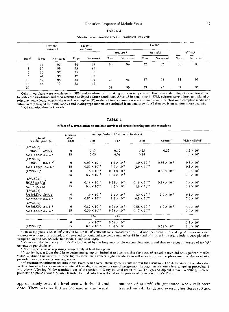
Treatment of s p o l l diploids in sporulating condi- tions with X-rays enhanced the production of can'cyh' meiotic products nearly sixfold (Table 4). The mini- mal levels of X-irradiation capable of causing a de- tectable increase in the frequency of viable spores left the viability of vegetative cultures virtually unaf- fected. A higher dose (30 krad), which decreased the viability of vegetative cultures by approximately half, increased the number of canrcyhr cfu generated to
Radiation Response of Meiotic Yeast
TABLE 3
Meiotic recombination ( r e ) in irradiated rad9 cells
35
LW3203 LW3204 LW3901 canl-ura3 canl-ura3
canl-ura3 leuI-cyh2 cyhFlys5
Dosea % rec No. scored % rec No. scored % rec No. scored % rec No. scored % rec No. scored
0 34 93 44 91 30 93 32 93 33 93 1 30 95 33 93 3 33 92 35 88 5 41 93 42 93
10 37 95 33 94 34 95 37 95 38 95 15 34 77 35 46 20 31 95 33 95 27 95
Cells in log phase were transferred to SPM and incubated with shaking at room temperature. Five hours later, aliquots were transferred to plates for irradiation and then returned to liquid culture conditions. After 48 hr total time in SPM, cultures were diluted and plated on selective media (-arg +can+cyh) as well as complete (D) media. Colonies arising on selective media were patched onto complete media and subsequently assayed for auxotrophies and mating type (nonmaters excluded from data shown). All data are from random spore analysis.
X-irradiation dose in kilorads.
TABLE 4
Effect of X-irradiation on meiotic survival of strains bearing meiotic mutations
Radiation can' cyh'lviahle cella at time of treatment (Strain),
relevant genotype dose (krad) 5 hr 0 hr 10 hr
(LW3608) HOPl SPOl l 0 0.17 0.17 0.23
hop1::LEUZ spol l -1 15 0.05 0.08 0.14 (LW3606)
H O P l s p 0 1 l - l ~ 0 0.93 X 10⁻³ 1.0 X 10⁻³ 1.0 X
h0pl::LEVZ ~ p 0 1 l - l 15 0.81 x 1 0 - ~ 5.9 x lo+ 4.6 X (LW3606)c 0 1.3 x 10⁻³ 0.54 X 10⁻³
15 6.7 X lo-' 10.6 X IO-' (LW3802) HOPI s p o l l d 0 0.13 x 10⁻³ 0.13 X 10⁻³ 0.16 X 10⁻³ HOPI s p o l l d 15 5.4 x 10⁻³ 3.6 X 10⁻³ 1.8 X 1 0 - ~ (LW3607) hop1::LEUZ SPOl I 0 1.6 X lo-' 1.2 X 1 0 - 3 1.5 X
hopl::LEU2 spoll-1 15 0.95 x 10⁻³ 1.6 X 10⁻³ 0.5 X 1 0 - ~ (LW3605) hop1::LEUZ s p o l l - 1 0 0.82 x 10⁻³ 0.71 X 10⁻³ 0.58 X 10⁻³
hopl::LEU2 s p o l l - I 15 0.36 X IO-' 0.38 X IO-' 0.17 X lo-'
"
5 hr 7 hr
0 1.3 x 10⁻³ 0.54 X 10⁻³ (LW3606Y 15 6.7 X IO-' 10.6 X IO-'
Controlb Viable cells/mlc
0.27 1.9 x 10' 1.3 x 10'
0.86 X 1 0 - 3 9.3 X 10⁷ 9.1 X lo7
0.58 X 1 0 - ~ 1.3 X lo8 1.0 x los
0.18 x 1.3 X 10' 1.4 X 10'
2.0 X 10⁻³ 6.1 X 10⁷ 7.0 X 10⁷
1.2 X 10⁻³ 4.4 X 10⁷ 5.0 X lo7
1.3 x 10' 0.58 x 10⁻³ 1.0 x 10'
Cells in log phase (1.3 X I O 7 cells/ml to 1.9 X lo7 cells/ml) were transferred to SPM and incubated with shaking. At times indicated, aliquots were plated, irradiated, and returned to liquid culture conditions. After 48 hr total of incubation, serial dilutions were plated on complete (D) and can'cyh"se1ective media (-arg+can+cyh).
a Values are the frequency of can'cyh' cfu divided by the frequency of cfu on complete media and thus represent a measure of can'cyh' generation per viable cell.
No resuspensions or replatings; assayed only at final time point. Viability figures from the 5-hr experimental group are included to illustrate that the doses of radiation used did not significantly affect
viability. Minor fluctuations in these figures most likely reflect slight variability in cell recovery from the plates used for the irradiation procedure (see MATERIALS AND METHODS).
d*efSeparate experiments fell into these classes, which were internally consistent; see text for discussion. The differences in the 5-hr values in these two sets of experiments is attributable to slight variation in rates of progression through meiosis, some 5-hr samplings preceding ( d ) and others following ( e ) the transition out of the period of X-ray induced arrest in G I . The s p o l l d diploid strain LW3802 entered premeiotic S phase about 3 hr after transfer to SPM, which is reflected in the pattern of induction of canrcyh' cfu.
approximately twice the level seen with the 15-krad number of can'cyh' cfu generated when cells were dose. There was no further increase in the overall treated with 45 krad, and even higher doses (60 and
36 L. W. Thorne and B. Byers
90 krad) led to a decrease in the overall yield (see Table 6). In spite of these decreases in overall yield, the proportion of sporulating cells surviving the treat- ment (scored as the ratio of can'cyh' cfu to total cfu) continued to rise, further implicating the entry into meiosis in the development of resistance to X-irradi- ation.
In contrast to the behavior of the s p o l l diploids, no significant increase in can'cyh" cfu generation was seen in hop1 diploids or hop1 spo l l diploids (Table 4). Therefore, hop1 strains seem incapable of being stim- ulated to higher survival by X-irradiation, and this deficiency of hop1 is epistatic to the effect shown for
A crucial parameter in the induction of increased spore viability during s p o l l meiosis was the time of X-irradiation. If irradiated before the beginning of premeiotic DNA synthesis (about 5 hr in these strains), spo l l cultures failed to show an increase in can'cyh' cfu. This is consistent with the observation (data not shown) that spo l l diploids subjected to irradiation early in meiosis become arrested to the same extent as wild-type cells. Similarly, delaying the time of X- irradiation until a late stage of meiosis also prevented the treatment from effectively increasing the fre- quency of can'cyh' cfu (Table 4). A maximally respon- sive stage was found to lie in the interval of about 5- 8 hr after transfer to sporulation medium. In these strains, this represents a period extending from early in premeiotic S phase until the latter part of meiotic prophase. The greatest induction of canrcyhr cfu de- tected in the course of these experiments was obtained for strain LW3606 after irradiation at 7 hr. It remains to be resolved whether this is indicative of there being only a narrow span of time in which a culture is maximally susceptible to induction.
Induction of meiotic survival and recombination by X-irradiation: The origin of the can'cyh' progeny obtained upon X-irradiation of spo l l strains was ex- amined by undertaking further genetic characteriza- tion. Having found sporulation generally to be incom- plete in these strains, we wished to ascertain first whether these drug-resistant colonies represented spore colonies or arose from unsporulated diploids that had simply been rendered homozygous for the drug resistance markers by virtue of high levels of gene-centromere recombination in response to the irradiation treatment (for review see HAYNES and KUNZ 198 1). Furthermore, genotypic analysis of hap- loid colonies would serve to assess the level of recip- rocal recombination that had occurred during the meiosis leading to their formation. To permit evalu- ation of mating capability and reciprocal exchange, canrcyhr cfu were patched individually onto complete medium and assayed for mating ability.
Many of the nonmaters were prototrophic for un-
s p o l l .
selected markers and were therefore presumed to be either hyperploid products of meiotic nondisjunction or diploids that had not undergone meiosis. The frequency of nonmaters was higher among the can' cyh' products of the recombination-deficient mutants than those from wild type (Table 5), as had previously been seen for hop1 strains (HOLLINGSWORTH and BYERS 1989), and this frequency failed to show a dosage-dependent response to X-irradiation. The ab- sence of dosage dependency might be explained by positing that meiosis had occurred but that full levels of recombination (as indicated by marker segregation) had not been achieved upon irradiation, so the prog- eny were still largely subject to the pattern of nondis- junction that is typical of recombination-deficient meiosis. It also seemed possible that some of the observed recombination events occurred during veg- etative growth rather than meiosis, but the fact that an increased production of can' cyh' cfu could be induced only during a limited time after transfer to SPM argued in favor of their origin in meiosis. Re- gardless, nonmating cfu were eliminated from the recombination analysis in order to focus specifically on those progeny that most likely were true haploids.
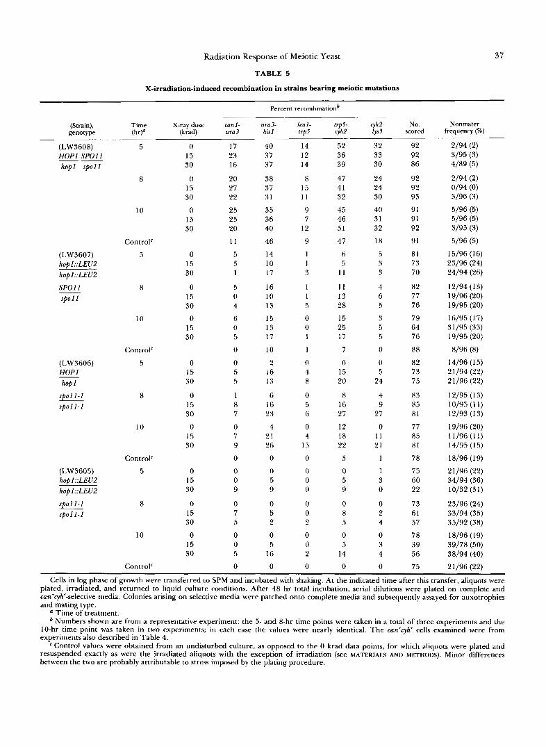
The haploid maters were assayed for reciprocal exchange in five intervals: canl-ura3 and ura3-his1 on chromosome V; and leul-trp5, trp5-cyh2 and cyh2-lys5 on chromosome VI1 (Table 5 ) . X-irradiation caused an increased incidence of reciprocal exchange in every interval assayed in the spo l l diploid. Little or no induction was detected in the wild-type strain or the hop1 homozygote. Limited induction detected in cer- tain intervals in the hop1 $01 1 diploid was largely restricted to the later times. Further, this induced recombination was not accompanied by the pattern of increase in viable can'cyh" cfu that was seen for spo l 1 diploids (Table 4). The failure of irradiation to cause increased spore viability in the doubly mutant strain suggested that the limited induction of recombination was ineffective in promoting improved disjunction.
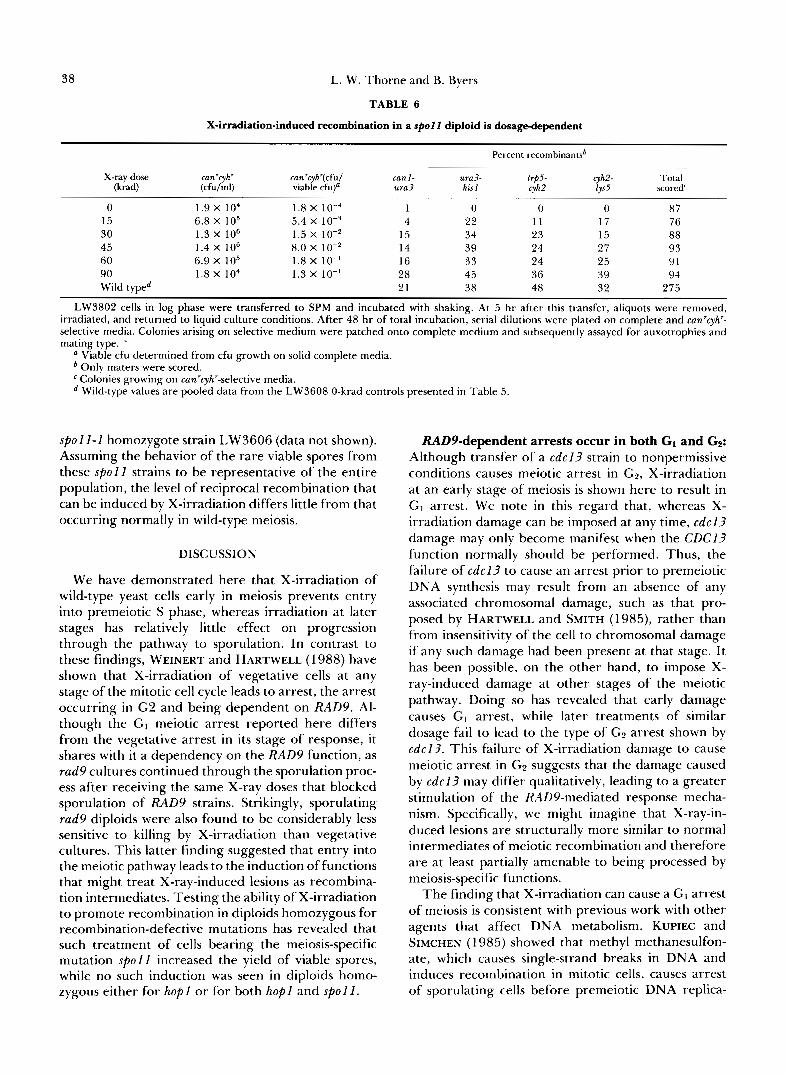
Having found that X-ray-induced lesions are capa- ble of enhancing recombination in spo l l strains, we then wished to ask whether one might attain normal meiotic levels of recombination at sufficiently high doses. Having tested several strains and culture con- ditions for a combination yielding the highest level of induction, we chose for further study spo l l A homo- zygote LW3802, which proceeds rapidly through meiosis and becomes quite susceptible to induction (and insensitive to RADPdependent arrest) 5 hr after transfer to SPM. High dose irradiation treatments of LW3802 at this time (Table 6) yielded a percentage of recombinants among the can'cyh' segregants that was at least as high in most intervals tested as that found among spores produced by an unirradiated wild-type strain. Similar results were obtained with
Radiation Response of Meiotic Yeast
TABLE 5
X-irradiation-induced recombination in strains bearing meiotic mutations
37
Percent recombinationb
(Strain), Time X-ray dose canl - genotype W a (krad) u r a 3
(LW3608) H O P 1 S P O l l hop1 s p o l l "
(LW3607) hop1::LEUZ hopl::LEU2
S P O l l __ sp01 I
(LW3606) HOPI hop 1 -
sp011-I spor 1-1
(LW3605) hop1::LEUZ hop1::LEUZ
s p 0 l l - I SPOll-1
5
8
10
ControlC
5
8
10
Controlc
5
8
10
ControlC
5
8
10
ControlC
0 15 30
0 15 30
0 15 30
0 15 30
0 15 30
0 15 30
0 15 30
0 15 30
0 15 30
0 15 30
0 15 30
0 15 30
17 23 16
20 27 22
25 25 20
11
5 3 1
5 0 4
6 0 5
0
0 5 5
1 8 7
0 7 9
0
0 0 9
0 7 5
0 0 5
0
ura3. his1
40 37 37
38 37 31
35 36 40
46
14 10 17
16 10 13
15 13 17
10
2 16 13
6 16 23
4 21 26
0
0 5 9
0 5 2
0 5
16
0
~
leul- trps- trp5 cyh2
14 12 14
8 15 11
9 7
12
9
1 1 3
1 1 5
0 0 1
1
0 4 8
0 5 6
0 4
15
0
0 0 0
0 0 2 0 0 2
0
52 36 39
47 41 32
45 46 51
47
6 5
11
11 13 28
15 25 17
7
6 15 20
8 16 27
12 18 22
5
0 5 9
0 8 5
0 5
14
0
q h 2 - lys5
32 33 30
24 24 30
40 31 32
18
5 3 3
4 6 5
3 5 5
0
0 5
24
4 9
27
0 11 21
1
1 3 0
0 2 4
0 3 4
0
~
scored No.
92 92 86
92 92 93
91 91 92
91
81 73 70
82 77 76
79 64 76
88
82 73 75
83 85 81
77 85 81
78
75 60 22
73 61 57
78 39 56
75
~
frequency (%) Nonmater
2/94 (2) 3/95 (3) 4/89 (5)
2/94 (2) 0194 (0) 3/96 (3)
5/96 (5) 5/96 (5) 3/95 (3)
5/96 (5) 15/96 (1 6) 23/96 (24) 24/94 (26)
12/94 ( 1 3) 19/96 (20) 19/95 (20)
16/95 (1 7) 31/95 (33) 19/95 (20)
8/96 (8) 14/96 (1 5) 2 1 /94 (22) 21/96 (22)
12/95 ( 1 3) 10/95 ( 1 1) 12/93 (1 3)
19/96 (20) 11/96 (11) 14/95 (1 5)
18/96 (1 9)
2 1 /96 (22) 34/94 (36) 10/32 (31)
23/96 (24)
35/92 (38)
18/96 (19) 39/78 (50) 38/94 (40)
33/94 (35)
21/96 (22)
Cells in log phase of growth were transferred to SPM and incubated with shaking. At the indicated time after this transfer, aliquots were plated, irradiated, and returned to liquid culture conditions. After 48 hr total incubation, serial dilutions were plated on complete and cun'cyh'-selective media. Colonies arising on selective media were patched onto complete media and subsequently assayed for auxotrophies and mating type.
a Time of treatment. Numbers shown are from a representative experiment: the 5- and 8-hr time points were taken in a total of three experiments and the
10-hr time point was taken in two experiments; in each case the values were nearly identical. The can'cyh' cells examined were from experiments also described in Table 4.
Control values were obtained from an undisturbed culture, as opposed to the 0 krad data points, for which aliquots were plated and resuspended exactly as were the irradiated aliquots with the exception of irradiation (see MATERIALS AND METHODS). Minor differences between the two are probably attributable to stress imposed by the plating procedure.
38 L. W. Thorne and B. Byers
TABLE 6
X-irradiation-induced recombination in a spol l diploid is dosage-dependent
Percent recombinantsb
X-ray dose can'cyh' can'cyh'(cfu1 (krad) (cfu/ml)
can I - ura3- viable cfu)a ura3
trp5- cyh2- his I cyh2 lys5
Total scored'
0 1.9 X lo4 1.8 X 10⁻⁴ 1 0 0 0 87 15 6.8 X IO5 5.4 X 1 0 - ~ 4 22 11 17 76 30 1.3 X lo6 1.5 X IO-' 15 34 23 15 88 45 1.4 X I O 6 8.0 X IO-* 14 39 24 27 93 60 6.9 X lo5 1.8 X IO-' 16 33 24 25 91 90 1.8 X 10⁴ 1.3 X 10" 28 45 36 39 94 Wild typed 21 38 48 32 275
LW3802 cells in log phase were transferred to SPM and incubated with shaking. At 5 hr after this transfer, aliquots were removed, irradiated, and returned to liquid culture conditions. After 48 hr of total incubation, serial dilutions were plated on complete and can'cyh" selective media. Colonies arising on selective medium were patched onto complete medium and subsequently assayed for auxotrophies and mating type. .
a Viable cfu determined from cfu growth on solid complete media. Only maters were scored. Colonies growing on can'cyh'-selective media. Wild-type values are pooled data from the LW3608 0-krad controls presented in Table 5.
spoll-1 homozygote strain LW3606 (data not shown). Assuming the behavior of the rare viable spores from these spoll strains to be representative of the entire population, the level of reciprocal recombination that can be induced by X-irradiation differs little from that occurring normally in wild-type meiosis.
DISCUSSION
We have demonstrated here that X-irradiation of wild-type yeast cells early in meiosis prevents entry into premeiotic S phase, whereas irradiation at later stages has relatively little effect on progression through the pathway to sporulation. In contrast to these findings, WEINERT and HARTWELL (1 988) have shown that X-irradiation of vegetative cells at any stage of the mitotic cell cycle leads to arrest, the arrest occurring in G2 and being dependent on RAD9. Al- though the G I meiotic arrest reported here differs from the vegetative arrest in its stage of response, it shares with it a dependency on the RAD9 function, as rad9 cultures continued through the sporulation proc- ess after receiving the same X-ray doses that blocked sporulation of RAD9 strains. Strikingly, sporulating rad9 diploids were also found to be considerably less sensitive to killing by X-irradiation than vegetative cultures. This latter finding suggested that entry into the meiotic pathway leads to the induction of functions that might treat X-ray-induced lesions as recombina- tion intermediates. Testing the ability of X-irradiation to promote recombination in diploids homozygous for recombination-defective mutations has revealed that such treatment of cells bearing the meiosis-specific mutation spoll increased the yield of viable spores, while no such induction was seen in diploids homo- zygous either for hop1 or for both hop1 and spoll.
RADPdependent arrests occur in both GI and Gp: Although transfer of a cdcl3 strain to nonpermissive conditions causes meiotic arrest in G2, X-irradiation at an early stage of meiosis is shown here to result in GI arrest. We note in this regard that, whereas X- irradiation damage can be imposed at any time, cdcl? damage may only become manifest when the CDC13 function normally should be performed. Thus, the failure of cdcl? to cause an arrest prior to premeiotic DNA synthesis may result from an absence of any associated chromosomal damage, such as that pro- posed by HARTWELL and SMITH (1 985), rather than from insensitivity of the cell to chromosomal damage if any such damage had been present at that stage. I t has been possible, on the other hand, to impose X- ray-induced damage at other stages of the meiotic pathway. Doing so has revealed that early damage causes GI arrest, while later treatments of similar dosage fail to lead to the type of G2 arrest shown by cdcl?. This failure of X-irradiation damage to cause meiotic arrest in GS suggests that the damage caused by cdc l3 may differ qualitatively, leading to a greater stimulation of the RADY-mediated response mecha- nism. Specifically, we might imagine that X-ray-in- duced lesions are structurally more similar to normal intermediates of meiotic recombination and therefore are at least partially amenable to being processed by meiosis-specific functions.
The finding that X-irradiation can cause a GI arrest of meiosis is consistent with previous work with other agents that affect DNA metabolism. KUPIEC and SIMCHEN (1 985) showed that methyl methanesulfon- ate, which causes single-strand breaks in DNA and induces recombination in mitotic cells, causes arrest of sporulating cells before premeiotic DNA replica-
Radiation Response of Meiotic Yeast 39
tion. Hydroxyurea also causes meiotic arrest at the same stage (SIMCHEN, IDAR and KASSIR 1976; SCHILD and BYERS 1978), but this agent is thought to impinge more directly on DNA replication by inhibiting pre- cursor pools (ELFORD 1968; SLATER 1973).
The finding that the RADY-dependent arrests of meiosis and mitosis in response to DNA damage by X-ray differ in relation to DNA replication, occurring in GI and G2, respectively, is not unprecedented in comparisons of the two pathways. Studies of the cdc genes in mitosis and meiosis have revealed many dif- ferences in the ordering of essential functions. cdc7, for example, was examined together with a group of other mutations having similar vegetative arrest points (cdc2, cdc8 and cdc21) by SCHILD and BYERS (1978). Cytologically, all these mutations caused meiotic arrest at a stage characterized by the presence of duplicated- but-unseparated spindle pole bodies and the absence of synaptonemal complex, as was also found for c d c l 3 (WEBER and BYERS 1992). Strains bearing cdc8 and cdc21 failed to undergo DNA replication before ar- resting in meiosis (as in mitosis), whereas premeiotic DNA synthesis was largely completed before the points of arrest in both mitosis and meiosis for cdc2. Surprisingly, cdc7, which causes mitotic arrest at the initiation of DNA replication (see PRINGLE and HART- WELL 198 1) caused meiotic arrest at a stage following the completion of DNA replication, but prior to syn- aptonemal complex assembly. Another precedent for differences in the ordering of CDC functions is found in the behavior of cdc28 strains (SHUSTER and BYERS 1989). Although cdc28 causes a vegetative arrest in GI at START, mutants are blocked in meiosis at pachytene, prior to the SPB separation event that leads to spindle formation in preparation for meiosis I. This mutation also partially blocks spindle forma- tion for meiosis 11. Thus, the finding that RAD9 acts not only in G1 after X-irradiation but also in the G2 arrest of c d c l 3 mutants simply expands a growing list of examples showing that the order of common func- tions differs between mitosis and meiosis.
In light of the fact that meiotic cultures can survive vegetatively lethal doses of X-irradiation and generate viable spores if progression through meiosis is not blocked by the RADY function, this early meiotic arrest in response to X-irradiation seems counterpro- ductive for the cells. That is, one might think it disadvantageous for the cell to become arrested im- mediately before it would otherwise gain the recom- binational benefits enjoyed by its rad9 counterpart. On the other hand, we note that sporulating RADY cultures arrested by X-irradiation do not die. Even when incubated for several days, the cells remaining capable of reverting to vegetative growth (data not shown). Consequently, there may be no selective ad- vantage for the cell to retain the ability to resume
meiosis once arrested by radiation damage. In fact, because diploids can tolerate much higher doses of X- irradiation than can haploids (MORTIMER 1952), it may be advantageous for the cell to remain diploid if there is a risk of continued or repeated exposure to ionizing irradiation.
Replacement of meiotic function by X-ray-in- duced damage: Although the high levels of recombi- nation normally occurring during meiosis make it difficult to detect any recombinogenic effects of X- irradiation during normal meiosis, stimulatory effects on recombination have become evident in strains mu- tant for a recombinational function. We have shown not only that spore viability is enhanced by X-irradi- ation during s p o l l meiosis, but also that reciprocal recombination is increased in the production of these spores.
It was of special interest in this regard to compare the behavior of two s p o l l alleles which had been reported to differ in their ability to form synaptone- mal complex. KLAPHOLZ, WADDELL and ESPOSITO (1 985), using thin-section electron microscopy, re- ported the appearance of synaptonemal complex in a strain homozygous for the s p o l l - 1 mutation. In con- trast, GIROUX, DRESSER and TIANO (1 989) found by examination of spread preparations that strains de- leted for S P O I l formed no detectable synaptonemal complex. In the present work, these two alleles were tested in the same background for their proficiency in the parameters measured here. A strain bearing the s p o l l deletion displayed a lower meiotic back- ground level of can'cyh' cfu generation than strains bearing s p o l l - I but was similarly capable of being induced to produce an increased yield of viable (can- 'cyh') progeny upon X-irradiation (Table 4). By this measure of meiotic success, the two s p o l l alleles do not differ.
The enhancement of reciprocal recombination and spore viability by X-irradiation after the beginning of S phase clearly suggests that the nondisjunctional de- ficiency of s p o l l meiosis is partially ameliorated by the induction of lesions that may substitute in some manner for an absent SPOl I function. It is of interest to consider the probable number of lesions that may have been induced to exert this stimulatory effect on viability. X-ray-induced lesions appear to consist of single- and double-strand breaks that follow the Pois- son distribution (see GAME et al. 1989). Computations from values for the size of the yeast genome (LAUER, ROBERTS and KLOTZ 1977; LINK and OLSON 1991) and extrapolation from data on double-strand breaks induced in yeast by 6oCo y-rays (RESNICK and MARTIN 1976), which creates double-strand breaks at a rate similar to that caused by X-irradiation (ROOTS et al. 1990), leads to an estimate that about 0.58 double- strand breaks would be induced per krad in a diploid
40 L. W. Thorne and B. Byers
genome. Single-strand breaks would be induced at a 20-fold higher rate (ROOTS et al. 1990), yielding about 1 1.6 breaks per krad per diploid genome. Therefore, a 15-krad dose of X-irradiation, shown in Table 4 to increase the viability of s p o l l meiotic products roughly fivefold, would represent the creation of a mean of 18 double-strand breaks and 360 single- strand breaks per G2 diploid genome. Perhaps it is significant that the predicted number of double-strand breaks, which are thought to play an important role in synapsis and recombination [for review see ORR- WEAVER and SZOSTAK (1985)], does not markedly differ from the number of bivalents that must form and undergo disjunction. However, the distribution of induced breaks should follow the Poisson distribu- tion (GAME et al. 1989), making it likely that only a fraction of the bivalents would receive double-strand breaks. Single-strand breaks, on the other hand, should be present abundantly on all chromatids, per- haps indicating that these lesions are sufficient for the induction of reciprocal recombination events.
It is interesting to note that although increased viability was induced by X-irradiation in this experi- ment, the recombination events that arose from this treatment failed to substitute fully for the role of SPOI I. The pattern of induced recombination dif- fered from that occurring in wild-type meiosis, as may be seen by comparing induced recombination in spe- cific intervals with that occurring in untreated wild- type meiosis (Table 6). Also, the frequency of viable spores remained far below that for the wild type at all doses tested. Note, for example, the different behav- ior of spo l I diploid LW3606 when irradiated before (about 5 hr) or after entry into S phase (about 8 hr; Table 4, experiment “d”). Recombination among canr cyh‘ cfu was induced at both times and was stimulated to nearly the same level (Table 5), but only the latter treatment resulted in an increased yield of can‘ cyh’ cfu (Table 4). The failure to supplant normal recom- bination fully is not surprising since the lesions in- duced at either stage may not fall within sites that might be normally specified by the synaptonemal com- plex and other meiotic functions. In addition, the timing of their induction may well be critical. In fact, given the partial asynchrony of the cellular population in progression through meiosis, the most effective time for induction would probably not be shared by all cells. Here, as in the studies of the MER1 and MER2 functions by ENGEBRECHT, HIRSCH and ROE- DER (1990), the occurrence of reciprocal exchange p e r se need not generate the type of crossovers that would most effectively promote homolog disjunction.
GAME et al. (1 989) have reported that double-strand breaks arise in meiotic cells well before the first recom- binant molecules can be detected and PADMORE, CAO and KLECKNER (1992) have confirmed this finding,
also showing that double-strand breaks are present before the initiation of chromosomal synapsis. It is of particular interest in this regard to note that the formation of meiosis-specific breaks at a hot spot for reciprocal recombination is dependent on SPOl1 function (CAO, ALANI and KLECKNER 1990). It is consistent both with this result and with the findings of the present report to postulate that the specific defect of s p o l l in synapsis and recombination is rather directly related to the failure of such breaks to arise. Similar to the finding that HO-induced double-strand breaks can stimulate meiotic recombination (KOLOD- KIN, KLAR and STAHL 1986), provision of artificially created lesions by X-irradiation may partially substi- tute for the missing breaks, thereby leading to im- proved disjunction. Finally, we propose that X-irra- diation during ongoing meiosis may serve more widely to help us determine which other meiotic mutations may be defective in recombination and/or disjunction because of a primary failure to create break. We must note, however, that the applicability of this assay will be constrained by the fact that many mutations which confer recombinational defects in meiosis also cause radiation sensitivity [ i . e . , the “RAD52 group”: RAD50, RADSI, RAD52, RAD54, RAD55 and RAD57 (RESNICK 1987; HAYNES and KUNZ 1981; PETES, MALONE and SYMINGTON 1990; SAEKI, MACHIDA and NAKAI 1980)l. Stimulatory effects might be assayable only for mutations that lack such radiation sensitivity. Nevertheless, the possibility that induced lesions may serve as functional intermediates in meiotic recombi- nation provides a potentially valuable tool for dissec- tion of this complex process.
We thank MARK WINEY for help with flow cytometry and JEFF THORNE for help with statistical analysis. Flow cytometry was car- ried out at The University of Washington Center for AIDS Re- search with the assistance of MARY TOUMA under support by National Institutes of Health grant IP30-27757. This work was supported by National Institutes of Health grants GM18541 and GM07735.
LITERATURE CITED
ADAMS, A. E. M. , and J. R. PRINGLE, 1984 Relationship of actin and tubulin distribution to bud growth in wild-type and mor- phogenetic-mutant Saccharomyces cereuisiae. J. Cell Biol. 98: 934-945.
BAUM, P., C. YIP, L. GOETSCH and B. BYERS, 1988 A yeast gene essential for regulation of spindle pole duplication. Mol. Cell. Biol. 8: 5386-5397.
BYERS, B., and L. GOETSCH, 1974 Duplication of spindle plaques and integration of the yeast cell cycle. Cold Spring Harbor Symp. Quant. Biol. 38: 123-131.
BYERS, B., and L. GOETSCH, 1991 Preparation of yeast cells for thin-section electron microscopy. Methods Enzymol. 194 602- 608.
CAO, L., E. ALANI and N. KLECKNER, 1990 A pathway for gen- eration and processing of double-strand breaks during meiotic recombination in S. cereuisiae. Cell 61: 1089-1 101.
ELFORD, H. L., 1968 Effect of hydroxyurea on ribonucleotide
Radiation Response of Meiotic Yeast 41
reductase. Biochem. Biophys. Res. Commun. 33: 129-135. ENGEBRECHT, J., J. HIRSCH and G. S. ROEDER, 1990 Meiotic gene
conversion and crossing over: their relationship to each other and to chromosome synapsis and segregation. Cell 62: 927- 937.
ENGEBRECHT, J. E., and G. S. ROEDER, 1989 Yeast mer1 mutants display reduced levels of meiotic recombination. Genetics 121:
ENGEBRECHT, J., and G. S. ROEDER, 1990 M E R ] , a yeast gene required for chromosome pairing and genetic recombination, is induced in meiosis. Mol. Cell. Biol. 10: 2379-2389.
GAME, J. C., and R. K. MORTIMER, 1974 A genetic study of X-ray sensitive mutants in yeast. Mutat. Res. 24: 281-292.
GAME, J. C., K. C. SITNEY, V. E. COOK and R. K. MORTIMER, 1989 Use of a ring chromosome and pulsed-field gels to study interhomolog recombination, double-strand DNA breaks and sister-chromatid exchange in yeast. Genetics 123: 695-71 3.
GIROUX, C. N., M. E. DRESSER and H. F. TIANO, 1989 Genetic control of chromosome synapsis in yeast meiosis. Genome 31: 88-94.
HANNAH-ALAVA, A., 1964 The brood pattern of X-ray-induced mutational damage in the germ cells of Drosophila melanogaster males. Mutat. Res. 1: 414-436.
HARTWELL, L. H., 1967 Macromolecular synthesis in tempera- ture-sensitive mutants of yeast. J. Bacteriol. 93: 1662-1670.
HARTWELL, L. H., 1973 Three additional genes required for deoxyribonucleic acid synthesis in Saccharomyces cerevisiae. J. Bacteriol. 115: 966-974.
HARTWELL, L. H., and D. SMITH, 1985 Altered fidelity of mitotic chromosome transmission in cell cycle mutants of S. cereuisiae. Genetics 110 38 1-395.
HARTWELL, L. H., and T. A. WEINERT, 1989 Checkpoints: con- trols that ensure the order of cell cycle events. Science 2 4 6 629-634.
HAYNES, R. H., and B. A. KUNZ, 1981 DNA repair and mutagen- esis in yeast, pp. 371-414 in The Molecular Biology ofthe Yeast Saccharomyces: Lijie Cycle and Inheritance, edited by J. N. STRATHERN, E. W. JONES and J. R. BROACH. Cold Spring Harbor Laboratory, Cold Spring Harbor, N.Y.
Ho, K. S. Y., 1975 Induction of DNA double-strand breaks by X- rays in a radiosensitive strain of the yeast Saccharomyces cereuis- iae. Mutat. Res. 3 0 327-334.
HOLLIDAY, R., 1964 A mechanism for gene conversion in fungi.
HOLLINGSWORTH, N. M., and B. BYERS, 1989 HOPI: a yeast meiotic pairing gene. Genetics 121: 445-462.
HOPPER, A. K., and B. D. HALL, 1975 Mating type and sporulation in yeast. I. Mutations which alter mating-type control over sporulation. Genetics 8 0 41-59.
HUTTER, K. J., and H. E. EIPEL, 1979 Microbial determination by flow cytometry. J. Gen. Microbiol. 113: 369-375.
KELLY, S. L., C. M E R R I L L ~ ~ ~ J . M. PARRY, 1983 Cyclic variations in sensitivity to X-irradiation during meiosis in Saccharomyces cerevisiae. Mol. Gen. Genet. 191: 314-318.
KIM, J. S., and A. M. ROSE, 1987 The effect of gamma radiation on recombination frequency in Caenorhabditis elegans. Genome
KLAPHOLZ, S., and R. E. ESPOSITO, 1982 A new mapping method employing a meiotic rec- mutant of yeast. Genetics 100: 387- 41 2.
KLAPHOLZ, S., C. S. WADDELL and R. E. ESPOSITO, 1985 The role of the S P O l l gene in meiotic recombination in yeast. Genetics
237-247.
Genet. Res. 5 282-304.
29: 457-462.
1 1 0 187-216. KLAR, A, J. S., and L. M. MIGLIO, 1986 Initiation of meiotic
recombination by double-strand breaks in S. pombe. Cell 4 6
KOLODKIN, A. L., A. J. S. KLAR and F. W. STAHL, 1986 Double- 725-73 1.
strand breaks can initiate meiotic recombination in S. cereuisiae. Cell 46: 733-740.
KUPIEC, M., and G. SIMCHEN, 1985 Arrest of the mitotic cell cycle and of meiosis in Saccharomyces cereuisiae by MMS. Mol. Gen. Genet. 201: 558-564.
LAUER, G. D., T . M. ROBERTS and L. C. KLOTZ, 1977 Determination of the nuclear DNA content of Saccha- romyces cerevisiae and implications for the organization of DNA in yeast chromosomes. J. Mol. Biol. 114 507-526.
LAWRENCE, C. W., 1961a The effect ofthe irradiation of different stages in microsporogenesis on chiasma frequency. Heredity 1 6 83-89.
LAWRENCE, C. W., 1961b The effect of radiation on chiasma formation in Tradescantia. Radiat. Bot. 1: 92-96.
LAWRENCE, C . W., and P. D. HOLT, 1970 Effect of gamma radia- tion and alpha particles on gene recombination in Chlamydo- monas reinhardi. Mutat. Res 1 0 545-555.
LINK, A. J., and M. V. OLSON, 1991 Physical map of the Saccha- romyces cerevisiae genome at 1 10-kilobase resolution. Genetics
MACHIDA, I., and S. NAKAI, 1980 Differential effect of uv-irradi- ation on induction of intragenic and intergenic recombination during commitment to meiosis in Saccharomyces cereuisiae. Mu- tat. Res. 73: 69-79.
MCGRATH, R. A., and R. W. WILLIAMS, 1966 Reconstruction in uivo of irradiated Escherichia coli deoxyribonucleic acid; the rejoining of broken pieces. Nature 212: 534-535.
MCKIM, K. S., A. M. HOWELL and A. M. ROSE, 1988 The effects of translocation on recombination frequency in Caenorhabditis elegans. Genetics 120: 987-1001.
MENEES, T. M., and G. S. ROEDER, 1989 MEI4, a yeast gene required for meiotic recombination. Genetics 123: 675-682.
MESELSON, M. S., and C. M. RADDING, 1975 A general model for genetic recombination. Proc. Natl. Acad. Sci. USA 72: 358- 361.
MORTIMER, R. K., 1952 The relative radiation resistance of hap- loid, diploid, triploid, and tetraploid yeast cells. Atomic Energy Commission Report UCRL 1922, pp. 66-78.
MORTIMER, R. K., and D. C. HAWTHORNE, 1969 Yeast genetics, pp. 385-460 in The Yeasts, Vol. 1 , edited by A. H. ROSE and J. S. HARRISON. Academic Press, New York.
NICOLAS, A., D. TRECO, N. P. SCHULTES and J. W. SZOSTAK, 1989 An initiation site for meiotic gene conversion in the yeast Saccharomyces cereuisiae. Nature 338: 35-39.
ORR-WEAVER, T. L., and J. W. SZOSTAK, 1985 Fungal recombi- nation. Microbiol. Rev. 4 9 33-58.
ORR-WEAVER, T. L., J. W. SZOSTAK and R. J. ROTHSTEIN, 1981 Yeast transformation: a model system for the study of recombination. Proc. Natl. Acad. Sci. USA 78: 6354-6358.
PADMORE, R., L. CAO and N. KLECKNER, 1992 Temporal com- parison of recombination and synaptonemal complex forma- tion during meiosis in S. cereuisiae. Cell 66: 1239-1256.
PETES, T. D., R. E. MALONE and L. S. SYMINGTON, 1990 Recombination in yeast, pp. 97-142 in The Molecular and Cellular Biology of the Yeast Saccharomyces: Genome Dynamics, Protein Synthesis, and Energetics, Vol. I , edited by J. BROACH, E. JONES and J. PRINGLE. Cold Spring Harbor Laboratory, Cold Spring Harbor, N.Y.
PRINGLE, J. R., and L. H. HARTWELL, 1981 The Saccharomyces cerevisiae cell cycle, pp. 97-1 42 in The Molecular Biology of the Yeast Saccharomyces: Lijie Cycle and Inheritance, edited by J. N. STRATHERN, E. W. JONES and J. R. BROACH. Cold Spring Harbor Laboratory, Cold Spring Harbor, N.Y.
REED, S. I . , 1980 The selection of S. cerevisiae mutants defective in the start event of cell division. Genetics 95: 561-577.
RESNICK, M. A., 1976 The repair of double-strand breaks in DNA: a model involving recombination. J. Theor. Biol. 5 3 97-106.
RESNICK, M. A., 1987 Investigating the genetic control of bio-
127: 681-698.
42 L. W. Thorne and B. Byers
chemical events in meiotic recombination, pp. 157-210 in Meiosis, edited by P. B. MOENS. Academic Press, New York.
RFSNICK, M. A., J. C. GAME and S. STASIEWICZ, 1983 Genetic effects of uv irradiation on excision-proficient and -deficient yeast during meiosis. Genetics 104: 603-618.
RESNICK, M. A., and P. MARTIN, 1976 The repair of double- strand breaks in the nuclear DNA of Saccharomyces cerevisiae and its genetic control. Mol. Gen. Genet. 143: 119-129.
RESNICK, M. A,, J. N. KASIMOS, J. C. GAME, R. J. BRAUN and R. M. ROTH, 1981 Changes in DNA during meiosis in a repair- deficient mutant ( rad52) of yeast. Science 212: 543-545.
RESNICK, M. A., T. CHOW, J. NITISS and J. GAME, 1984 Changes in the chromosomal DNA of yeast during meiosis in repair mutants and the possible role of a deoxyribonuclease. Cold Spring Harbor Symp. Quant. Biol. 4 9 639-649.
ROCKMILL, B., and G. S . ROEDER, 1988 REDZ: a yeast gene required for the segregation of chromosomes during the re- ductional division of meiosis. Proc. Natl. Acad. Sci. USA 85: 6057-6061.
ROCKMILL, B., and G. S. ROEDER, 1990 Meiosis in asynaptic yeast.
ROMAN, H., 1984 A comparison of induced and spontaneous gene conversion in mitotic and meiotic cells of Saccharomyces cerevis- iae. Carlsberg Res. Commun. 4 9 351-358.
ROOTS, R., W. HOLLEY, A. CHATTERJEE, M. IRIZARRY and G. KRAFT, 1990 The formation of strand breaks in DNA after high-LET irradiation: a comparison of data from in vitro and cellular systems. Int. J. Radiat. Biol. 5 8 55-59.
SAEKI, T., I . MACHIDA and S. NAKAI, 1980 Genetic control of diploid recovery after y-irradiation in the yeast Saccharomyces cerevisiae. Mutat. Res. 73: 251-265.
SALTS, Y., G. SIMCHEN and R. PINON, 1976 DNA degradation and reduced recombination following uv irradiation during meiosis in yeast (Saccharomyces cerevisiae). Mol. Gen. Genet. 146: 55-59.
SANKARANARAYANAN, K., and F. H. SOBELS, 1976 Radiation ge-
Genetics 123: 563-574.
netics, pp. 1089-1250 in The Genetics and Biology ofDrosophila, Vol. IC, edited by M. ASHBURNER and E. NOVITSKI. Academic Press, New York.
SCHILD, D., and B. BYERS, 1978 Meiotic effects of DNA-defective cell-division-cycle mutations of Saccharomyces cerevisiae. Chro- mosoma 70: 109-130.
SHERMAN, F., and H. ROMAN, 1963 Evidence for two types of allelic recombination in yeast. Genetics 48: 255-261.
SHUSTER, E. O., and B. BYERS, 1989 Pachytene arrest and other meiotic effects of the start mutations in Saccharomyces cerevisiae. Genetics 123: 29-43.
SIMCHEN, G., D. IDAR and Y. KASSIR, 1976 Recombination and hydroxyurea inhibition of DNA synthesis in yeast meiosis. Mol. Gen. Genet. 144: 21-27.
SIMCHEN, G., Y. SALTS and R. PINON, 1973 Sensitivity of meiotic yeast cells to ultraviolet light. Genetics 73: 53 1-54 1.
SLATER, M. L., 1973 Effect of reversible inhibition of deoxyribo- nucleic acid synthesis on the yeast cell cycle. J. Bacteriol. 113:
SUN, H., D. TRECO, N. P. SCHULTES and J. W. Szostak, 1989 Double-strand breaks at an initiation site for meiotic gene conversion. Nature 338: 87-90.
SYMINGTON, L. S., 1991 Double-strand-break repair and recom- bination catalyzed by a nuclear extract of Saccharomyces cerevis- iae. EMBO J. 10: 987-996.
SZOSTAK, J. W., T . L. ORR-WEAVER, R. J. ROTHSTEIN and F. W. STAHL, 1983 The double-strand break repair model for re- combination. Cell 33: 25-35.
WEBER, L., and B. BYERS, 1992 A RADB-dependent checkpoint blocks meiosis of cdc l3 yeast cells. Genetics 131: 55-63.
WEINERT, T. A,, and L. H. HARTWELL, 1988 The RAD9 gene controls the cell cycle response to DNA damage in Saccharo- myces cerevisiae. Science 241: 317-322.
WOOD, J. S., 1982 Genetic effects of methyl benzimidazole-2-yl- carbamate on Saccharomyces cerevisiae. Mol. Cell. Biol. 2: 1064- 1079.
263-270.
Communicating editor: P. J. PUKKILA


![PETl Acts in the 5”Leader of Saccharomyces cereuisiae ... · PDF fileDAUP JJM 194 JJM50 PTH44 PTY 7 DBY947 MCCl18 MATa ade2-101 ura3-52 petl 11-1 1 [rho"] MATa ade2-101 ura3-52 petl](https://img.pdfslide.net/doc/110x75/5a828f9a7f8b9aa24f8e2114/petl-acts-in-the-5leader-of-saccharomyces-cereuisiae-jjm-194-jjm50-pth44.jpg)