Embed Size (px)
Citation preview
Journal of Virological Methods 117 (2004) 19–25
Standardised evaluation of the performance of a simple membranefiltration-elution method to concentrate bacteriophages
from drinking water
Javier Méndeza, Ana Audicanab, Ana Isernc, Julián Llanezad, Belén Morenoe,Marıa Luisa Tarancónf, Juan Jofrea, Francisco Lucenaa,∗
a Departamento de Microbiolog´ıa, Universidad de Barcelona, Avda. Diagonal 645, 08028 Barcelona, Spainb Dirección de Salud Pública del Gobierno Vasco, C/ Mar´ıa Dıaz de Haro, 60, 48010 Bilbao, Spain
c Laboratori Municipal de Barcelona, Institut Municipal de Salut Pública, Avda. de les Drassanes, 13-15, 08001 Barcelona, Spaind Laboratorio de Salud Pública de Asturias, Finca de la Cedellada, Camino de Rub´ın s/n, 33011 Oviedo, Spain
e Laboratorio de Salud Pública de Gipuzkoa, Avda. Navarra, 4, 20013 Donostia-San Sebastián, Spainf Conselleria de Salut i Consum, Plaça Hospital 3, 07012 Palma de Mallorca, Spain
Received 7 July 2003; received in revised form 17 November 2003; accepted 18 November 2003
Abstract
The bacteriophage elution procedure described further after adsorption to acetate–nitrate cellulose membrane filters allows better recoveryof phages concentrated from 1 l of water than elution proceduresm used previously. The improvement is due to the combined effect of theeluent (3% (w/v) beef extract, 3% (v/v) Tween 80, 0.5 M NaCl, pH 9.0) and the application of ultrasound instead of agitation or swirling.Average recovery of somatic coliphages, 82± 7%, was the greatest, and that of phages infectingBacteroides fragilis, 56± 8%, the lowest,with intermediate values for F-specific and F-specific RNA bacteriophages. Thus, the method allowed recovery of over 56% for all the phagessuggested as surrogate indicators. The method was then validated according to an International Standardisation Organisation validationstandard procedure and implemented in routine laboratories, which obtained reproducible results.© 2003 Elsevier B.V. All rights reserved.
Keywords:Bacteriophages; Concentration; Membrane filter; Elution; Validation
1. Introduction
Bacteriophages have been investigated as potential modelorganisms for water quality assessment (IAWPRC, 1991;Grabow, 2001; Jofre, 2002). Phages proposed for waterquality assessment are somatic coliphages (Kott and BenAri, 1968), F-specific RNA bacteriophages (Havelaar et al.,1986) and bacteriophages infectingBacteroides fragilis(Tartera and Jofre, 1987). Although phages are present insurface waters at concentrations high enough to allow di-rect enumeration in small volumes of sample, this is notthe case with groundwater or drinking water. In this case itis necessary to test phages in larger, at least 1 l, volumes.Detecting phages in 1 l volumes can be achieved by two
∗ Corresponding author. Tel.:+34-934-021-484;fax: +34-934-110-592.
E-mail address:[email protected] (F. Lucena).
approaches: the first is a presence/absence method, whichis only qualitative and the second is through concentrationand plaque assay, which is quantitative. Many methodsfor concentrating phages from water samples had beendescribed (Logan et al., 1980; Schulze and Lenk, 1983;Shields and Farrah, 1986; Sobsey et al., 1990; Borregoet al., 1991; Lucena et al., 1995; Sinton et al., 1996). Mostof the methods have only been described for one type ofbacteriophages and generally validated with a laboratorystrain. Among these, we find very feasible a method basedon adsorption to acetate–nitrate cellulose ester membranefilters described bySobsey et al. (1990)that was modifiedfurther bySinton et al. (1996). The method is very efficientfor concentrating coliphages from 100 ml, but results for1 l volumes are below 50% recovery (Hsu et al., 1998). Wetested the method as modified bySinton et al. (1996)forconcentrating somatic coliphages from 1 l of drinking waterand the recovery was also below 50% (data not shown).
0166-0934/$ – see front matter © 2003 Elsevier B.V. All rights reserved.doi:10.1016/j.jviromet.2003.11.013
20 J. Mendez et al. / Journal of Virological Methods 117 (2004) 19–25
It is well known that concentration efficiencies obtainedwhen testing natural samples for viruses are frequently sig-nificantly lower than those reported in the publications de-scribing the method as developed with reference laboratorybacteriophages. Also, Round Robin tests among differentlaboratories show significant differences in concentration ef-ficiencies between the different laboratories (Melnick et al.,1984; Vilaginés et al., 1997). Many factors other than theconcentration method itself seem to influence the recovery:the characteristics of the water sample, the amount of sam-ple, the numbers of bacteriophages spiked and the bacterio-phages groups themselves. Because of the many sources ofuncertainty it is important to establish “standardised” pro-cedures for evaluating concentration procedures, based onboth reliability and efficiency. Recently, a standard proce-dure for validation of methods for concentration of bacte-riophages from water has been adopted by the InternationalStandardisation Organisation (Anonymous, 2002). This pro-cedure does not give specific details of concentration meth-ods, but provides a general framework for the evaluationof the suitability of a particular method for a given typeand volume of water. Secondly, it also discusses how to usethis information in establishing the actual values of bacte-riophages in a given sample taken into account the numbersrecovered after concentration.
We describe modifications introduced to the method firstreported bySobsey et al. (1990)and then modified bySintonet al. (1996). The modifications introduced were examinedfor their impact upon the concentration of phages belongingto the three groups reported above as potential indicatorsfrom 1 l of drinking water. The final modified method wasthen validated according to the international validation stan-dard mentioned above. Finally, the implementation of themethod in routine microbiology laboratories and the repro-ducibility of the results with sewage derived bacteriophagesin these laboratories were assessed.
2. Materials and methods
2.1. Phage assay
Enumeration of bacteriophages was done by determin-ing the number of plaque forming units (pfu) by the dou-ble agar layer technique according to standards ISO10705-1(Anonymous, 1995), ISO10705-2 (Anonymous, 2000) andISO10705-4 (Anonymous, 2002), which describe methodsfor the detection and enumeration of F-specific (F-TOTPH)and F-specific RNA bacteriophages (F-RNAPH), somaticcoliphages (SOMCPH) and phages infecting strains HSP40(BFBHSP40) and RYC2056 (BFBRYC2056) ofB. fragilis,respectively.
2.2. Bacteriophages
Phages�X174, MS2 and B40-8, recommended by theStandard ISO methods indicated above as reference phages
for somatic coliphages, F-specific RNA bacteriophagesand B. fragilis phages, respectively were used as con-trols in enumeration tests and for spiking of water in themethod development experiments indicated further. As well,sewage derived phage stocks for SOMCPH, F-TOTPH,F-RNAPH, BFRPHHSP40 and BFRPHRYC2056 wereprepared as indicated further for method validationstudies.
2.3. Spiking phage suspensions
Two kinds of phage suspensions were used.
2.3.1. For developing the modified methodVolumes of suspensions of phages�X174, MS2 and
B40-8 containing between 1.5 × 10⁴ and 5.0 × 10⁵ pfu perml were added to the water sample to be submitted to con-centration to reach a final concentration of 60–200 plaqueforming units per litre. After vigorous agitation, phagesin the sample were enumerated, either directly or afterdilution.
2.3.2. For validating the modified methodSewage derived phage stocks were prepared as indicated
in the ISO 10705-3 Standard (Anonymous, 2003). Briefly,primary or secondary sewage effluent was either centrifugedfor 10 min at 2000× g or filtered through a 12�m poresize membrane filter as described inMendez et al. (2002).Target bacteriophages were enumerated for initial titre. Thephage suspensions were stored at 4◦C until the enumerationresults were available. If necessary, the suspensions werediluted to obtain concentrations of 60–200 plaque formingunits per ml of each of the phage groups. Since their numbersin sewage are different, these means that three sets of phagespike-stocks were prepared. Glycerol was added to a finalconcentration of 10% (v/v). Then, these suspensions weredistributed in 10 ml volumes in plastic containers, and frozenat −20 or−70◦C.
To ensure that sewage derived phages were distributed atrandom within and between the containers, the within andbetween containers homogeneity was tested. Two contain-ers for each phage group suspension (somatic coliphages,F-specific/F-specific RNA phages and phages infectingB.fragilis infecting strains HSP40 and RYC2056) were thawedat room temperature, and from each container two 0.5 mlaliquots for the target bacteriophages were assayed. Thecounts were then analysed for within (T1) and between con-tainers (T2) homogeneity.
T1 is a measure for the variation within one container ofspiking material (replicate variation).T2 is a measure forthe variation between different vials of one batch of spikingmaterial.
T1 =I∑
i=1
J∑j=1
[(zij − zi+/J)
(zi+/J)
2]
J. Mendez et al. / Journal of Virological Methods 117 (2004) 19–25 21
wherezij is the number of pfu per analytical portion (j) ofvial i, and
zi+ =J∑
j=1
zij
being the sum of numbers of pfu in all containers counted(I, that in this case is 2) andJ (in this case 2) the numberof analytical portions per container. Degrees of freedom forT1 areI(J − 1).
T2 =J∑
j=1
[(zi+ − z++/J)
z++/I
]
where
z++ =I∑
i=1
(∑zij
)
is the total count of plaques over all vials and duplicates.Degrees of freedom forT2 areI − 1.As indicated in the ISO Standard the phage suspensions
were considered as suitable for the validation of the methodif 0.01 < T1 < 5.99 andT2 < 3.84.
2.4. Modifications assayed for the phage concentrationmethod
The experiments to test the effect of changes of protocolin concentration efficiency were done spiking bottled wa-ter samples with suspensions of laboratory reference phagesfollowed by enumeration of phages before and after con-centration. The protocol was applied to the assays describedfurther.
2.4.1. Sample amendmentThe effect on virus adsorption of different MgCl2 concen-
trations (0.005, 0.05 and 0.5 M, the last one being the con-centration proposed bySinton et al. (1996)) in the samplewas assayed. For these tests a 4.14 M MgCl2·6H2O solutionwas prepared and the volumes necessary to reach the aboveindicated final concentrations were added to the referencephage spiked water sample.
2.4.2. Eluting solutionsThe following eluting solutions were tested: (1) 3% (w/v)
beef extract, 3% (v/v) Tween 80, 0.5 M NaCl, pH 9.0; (2)1% (w/v) beef extract, 0.5 M NaCl, pH 9.0; (3) 3% (w/v)beef extract, 0.5 M NaCl, pH 9.0; (4) 3% (w/v) beef extract,0.5 M NaCl, 0.4 M urea, pH 9.0; (5) 1% (w/v) beef extract,0.5 M NaCl, 0.4 M urea, pH 9.0; (6) 1% (w/v) beef extract,0.5 M NaCl, 0.25 M glycine, pH 9.0; (7) 0.5 M NaCl, 0.25 Mglycine, pH 9.0; (8) 1% (w/v) beef extract, 0.25 M glycine,pH 9.5.
2.4.3. Eluting conditionsThe effect on elution efficiency of swirling agitation and
ultrasound was compared as follows. The eluting conditionswere assayed with the eluting solution 1 that was confirmedbe the most efficient as indicated in the results section. Theswirling agitation of the membrane filter submerged in elut-ing solution was applied as indicated bySinton et al. (1996).Ultrasound was applied by submerging the flask or tube con-taining the membrane filter into an ultrasound cleaning bathfor 4 min.
2.5. Concentration method proposed and validated
As indicated above, the method came up as a modificationof a membrane filter phage concentration method previouslydescribed (Sobsey et al., 1990; Sinton et al., 1996) accord-ing to different changes tested herein. The method is recom-mended for volumes ranging from 100 ml to 1 l of waterswith turbidity <2.0 NTU. For concentrating bacteriophagesfrom 1 l of water proceed as follows. First, add MgCl2 to thewater sample to reach a final concentration 0.05 M. Then,filter the sample through an acetate–nitrate cellulose estermembrane filter, 0.22�m pore size, 47 mm diameter. Filterat a flow rate of approximately 1 l in 30 min. Cut the mem-brane filter in eight fragments and place into a glass flaskcontaining 5 ml of eluting solution (1% beef extract, 0.5 MNaCl and 3% Tween 80). Place the flask into an ultrasoundcleaning bath for 4 min. Count the eluted bacteriophagesby the double layer agar method. Count the bacteriophagesretained in the membrane fragments placing the fragmentsface down onto a host monolayer.
2.6. Evaluation and validation of the method with sewagederived phages
This was carried out as indicated in the standardISO10705-3 (Anonymous, 2003).
Briefly, five samples of bottled water or tap waterdechlorinated by adding sodium thiosulphate (Anonymous,1998) were tested. Turbidity was measured after spikingand ranged from 0.1 to 2.0 nephelometric turbidity units(NTUs). Since 1 l is the maximum volume proposed forthis method, containers containing 125, 250, 500, 1000 mlwere prepared. One millilitre of sewage derived spikingmaterial prewarmed at room temperature was added to eachcontainer, and the remainder of the spiking material waskept on melting ice for enumeration.
2.6.1. Calculation of recoveryLet Nc be the total number of plaques recovered from a
concentrate, andNs be the number of plaques from 1 ml ofthe spiking material. The percentage recovery was calculatedasR(%) = Nc/Ns × 100.
The arithmetic average recovery, the standard deviationand the confidence interval were calculated for all assayedvolumes. TheR(%) plus the confidence intervals were
22 J. Mendez et al. / Journal of Virological Methods 117 (2004) 19–25
plotted against the volume of sample and examined forlinearity. If confidence intervals ofR(%) for the differentvolumes overlapped, it was considered that there were nosignificant volume effects.
The results of all volumes tested were combined and thearithmetic mean ofR(%), the standard deviation and the rela-tive standard deviation (standard deviation/arithmetic mean)were calculated. According to the ISO standard the methodcan be considered reliable if the relative standard deviationis <0.5.
2.7. Concentration in different laboratories
Five routine laboratories, which in a previous collabora-tive study aimed to ensure the correct implementation in thelaboratory of the methods for enumerating bacteriophagessucceeded in the implementation of the methods, partici-pated in the trial.
Each laboratory prepared its own spiking material fromsewage as indicated above.
They assayed (4/5 assays for each group of bacterio-phages per laboratory) the concentration of somatic col-iphages, F-specific bacteriophages and phages infectingB.fragilis RYC2056 from 1 l of spiked water.
2.8. Statistical tests
Confidence intervals based on Student’st-tests werecalculated as{X − t × s/
√n; X + t × s/
√n} whereX is
the arithmetic average recovery;s the standard deviation,n the numbers ofR(%) values andt values of a Student’st-distribution (Anonymous, 1999).
Statistical tests (ANOVA, Student’st-test, Multiplerange test analysis using least significant differences be-tween means using Student’st-test) were performed usingthe Statistical Package for Social Sciences (Anonymous,1999). Differences were considered significant whenP < 0.05.
3. Results
3.1. Modifications assayed for the phage concentrationmethod
3.1.1. Amendment of the sampleThe three reference bacteriophages studied (�X174, MS2
and B40-8) adsorbed over 93% in the presence of 0.5 and0.05 M MgCl2. No significant differences (ANOVA,P >
0.05) were observed between the numbers of phages ad-sorbed in the presence of the two concentrations of MgCl2.In contrast, the lower concentration, 0.005 M, of MgCl2tested showed significantly (ANOVA,P < 0.05) lower ad-sorption of bacteriophagesOX174 and MS2. Consequentlyit was decided to amend the samples with 0.05 M MgCl2before the concentration step.
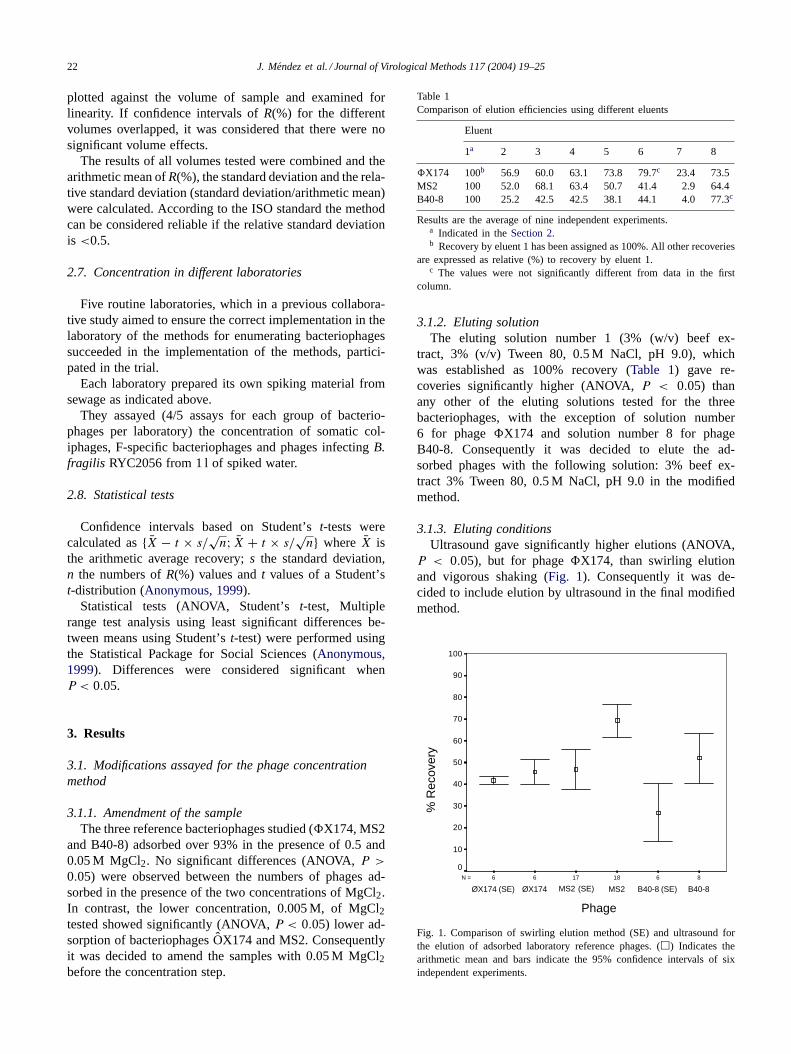
Table 1Comparison of elution efficiencies using different eluents
Eluent
1a 2 3 4 5 6 7 8
�X174 100b 56.9 60.0 63.1 73.8 79.7c 23.4 73.5MS2 100 52.0 68.1 63.4 50.7 41.4 2.9 64.4B40-8 100 25.2 42.5 42.5 38.1 44.1 4.0 77.3c
Results are the average of nine independent experiments.a Indicated in theSection 2.b Recovery by eluent 1 has been assigned as 100%. All other recoveries
are expressed as relative (%) to recovery by eluent 1.c The values were not significantly different from data in the first
column.
3.1.2. Eluting solutionThe eluting solution number 1 (3% (w/v) beef ex-
tract, 3% (v/v) Tween 80, 0.5 M NaCl, pH 9.0), whichwas established as 100% recovery (Table 1) gave re-coveries significantly higher (ANOVA,P < 0.05) thanany other of the eluting solutions tested for the threebacteriophages, with the exception of solution number6 for phage�X174 and solution number 8 for phageB40-8. Consequently it was decided to elute the ad-sorbed phages with the following solution: 3% beef ex-tract 3% Tween 80, 0.5 M NaCl, pH 9.0 in the modifiedmethod.
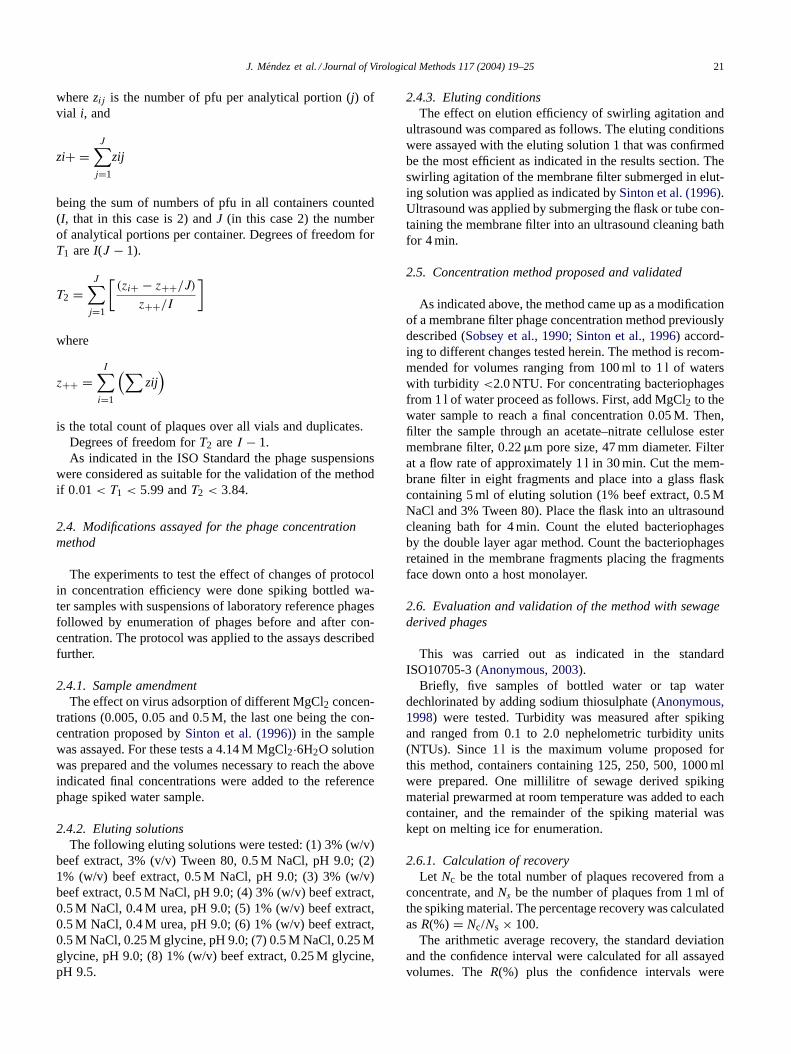
3.1.3. Eluting conditionsUltrasound gave significantly higher elutions (ANOVA,
P < 0.05), but for phage�X174, than swirling elutionand vigorous shaking (Fig. 1). Consequently it was de-cided to include elution by ultrasound in the final modifiedmethod.
86181766N =
B40-8B40-8 (SE)MS2
100
90
80
70
60
50
40
30
20
10
0
Phage
% R
ecov
ery
MS2 (SE)ØX174ØX174 (SE)
Fig. 1. Comparison of swirling elution method (SE) and ultrasound forthe elution of adsorbed laboratory reference phages. (�) Indicates thearithmetic mean and bars indicate the 95% confidence intervals of sixindependent experiments.
J. Mendez et al. / Journal of Virological Methods 117 (2004) 19–25 23
3.1.4. Bacteriophages remaining in the filtersEven applying the best eluting solution and the best elut-
ing conditions, non-negligible numbers of phages, rangingfrom 1 to 28% depending on the experiments and phages,remained in the membrane filter. Consequently, it was rec-ommended in the proposed method to include the count ofphages retained in the filter as part of the total number ofphages recovered as was recommended in the original meth-ods (Sobsey et al., 1990; Sinton et al., 1996).
As a consequence of the results reported in this section,the final modified method was established as indicated intheSection 2.
3.2. Evaluation and validation of the method
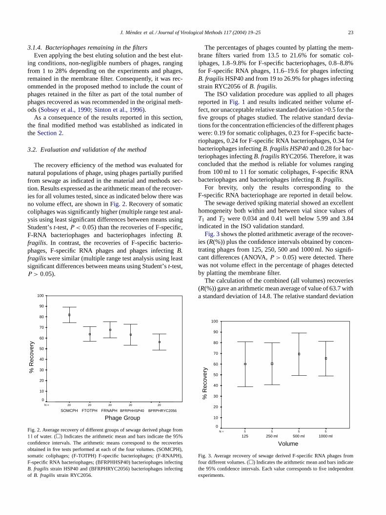
The recovery efficiency of the method was evaluated fornatural populations of phage, using phages partially purifiedfrom sewage as indicated in the material and methods sec-tion. Results expressed as the arithmetic mean of the recover-ies for all volumes tested, since as indicated below there wasno volume effect, are shown inFig. 2. Recovery of somaticcoliphages was significantly higher (multiple range test anal-ysis using least significant differences between means usingStudent’st-test,P < 0.05) than the recoveries of F-specific,F-RNA bacteriophages and bacteriophages infectingB.fragilis. In contrast, the recoveries of F-specific bacterio-phages, F-specific RNA phages and phages infectingB.fragilis were similar (multiple range test analysis using leastsignificant differences between means using Student’st-test,P > 0.05).
20202020N =
FRNAPHFTOTPHSOMCPH
100
90
80
70
60
50
40
30
20
10
0
Phage Group
% R
ecov
ery
20
BFRPHHSP40 BFRPHRYC2056
Fig. 2. Average recovery of different groups of sewage derived phage from1 l of water. (�) Indicates the arithmetic mean and bars indicate the 95%confidence intervals. The arithmetic means correspond to the recoveriesobtained in five tests performed at each of the four volumes. (SOMCPH),somatic coliphages; (F-TOTPH) F-specific bacteriophages; (F-RNAPH),F-specific RNA bacteriophages; (BFRPHHSP40) bacteriophages infectingB. fragilis strain HSP40 and (BFRPHRYC2056) bacteriophages infectingof B. fragilis strain RYC2056.
The percentages of phages counted by platting the mem-brane filters varied from 13.5 to 21.6% for somatic col-iphages, 1.8–9.8% for F-specific bacteriophages, 0.8–8.8%for F-specific RNA phages, 11.6–19.6 for phages infectingB. fragilisHSP40 and from 19 to 26.9% for phages infectingstrain RYC2056 ofB. fragilis.
The ISO validation procedure was applied to all phagesreported inFig. 1 and results indicated neither volume ef-fect, nor unacceptable relative standard deviation >0.5 for thefive groups of phages studied. The relative standard devia-tions for the concentration efficiencies of the different phageswere: 0.19 for somatic coliphages, 0.23 for F-specific bacte-riophages, 0.24 for F-specific RNA bacteriophages, 0.34 forbacteriophages infectingB. fragilis HSP40and 0.28 for bac-teriophages infectingB. fragilis RYC2056. Therefore, it wasconcluded that the method is reliable for volumes rangingfrom 100 ml to 1 l for somatic coliphages, F-specific RNAbacteriophages and bacteriophages infectingB. fragilis.
For brevity, only the results corresponding to theF-specific RNA bacteriophage are reported in detail below.
The sewage derived spiking material showed an excellenthomogeneity both within and between vial since values ofT1 and T2 were 0.034 and 0.41 well below 5.99 and 3.84indicated in the ISO validation standard.
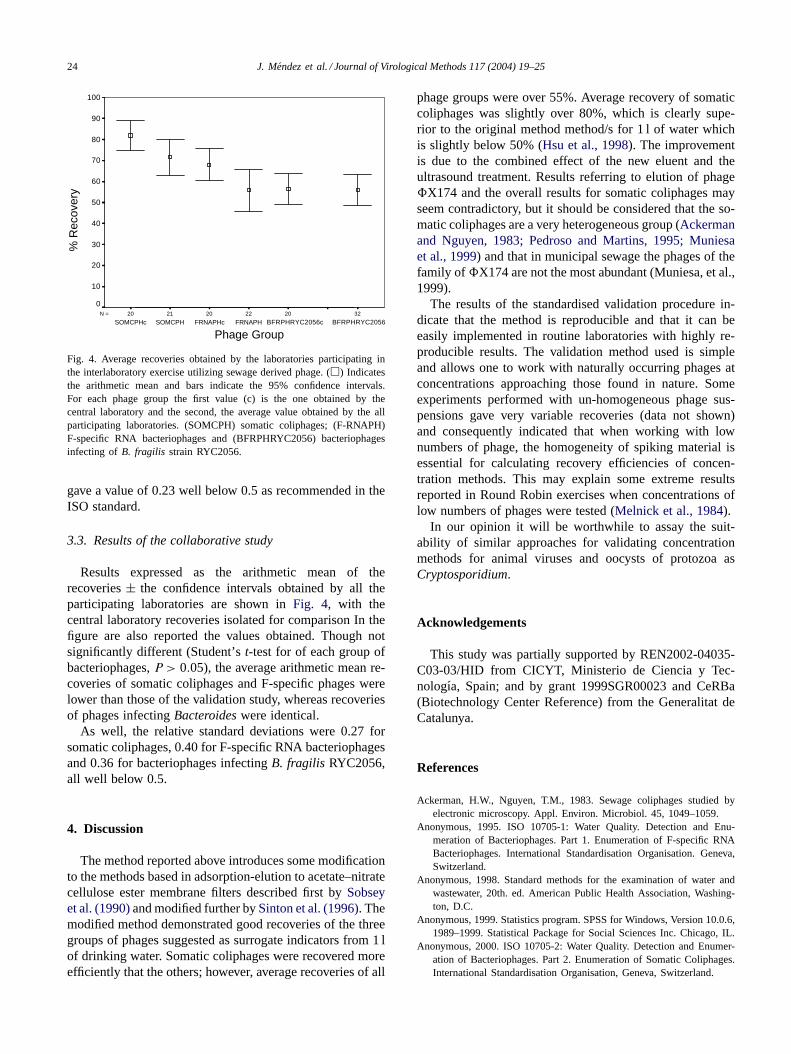
Fig. 3shows the plotted arithmetic average of the recover-ies (R(%)) plus the confidence intervals obtained by concen-trating phages from 125, 250, 500 and 1000 ml. No signifi-cant differences (ANOVA,P > 0.05) were detected. Therewas not volume effect in the percentage of phages detectedby platting the membrane filter.
The calculation of the combined (all volumes) recoveries(R(%)) gave an arithmetic mean average of value of 63.7 witha standard deviation of 14.8. The relative standard deviation
5555N =
1000 ml500 ml250 ml125
100
90
80
70
60
50
40
30
20
10
0
Volume
% R
ecov
ery
Fig. 3. Average recovery of sewage derived F-specific RNA phages fromfour different volumes. (�) Indicates the arithmetic mean and bars indicatethe 95% confidence intervals. Each value corresponds to five independentexperiments.
24 J. Mendez et al. / Journal of Virological Methods 117 (2004) 19–25
2022202120N =
FRNAPHFRNAPHcSOMCPHSOMCPHc
100
90
80
70
60
50
40
30
20
10
0
Phage Group
% R
ecov
ery
32
BFRPHRYC2056BFRPHRYC2056c
Fig. 4. Average recoveries obtained by the laboratories participating inthe interlaboratory exercise utilizing sewage derived phage. (�) Indicatesthe arithmetic mean and bars indicate the 95% confidence intervals.For each phage group the first value (c) is the one obtained by thecentral laboratory and the second, the average value obtained by the allparticipating laboratories. (SOMCPH) somatic coliphages; (F-RNAPH)F-specific RNA bacteriophages and (BFRPHRYC2056) bacteriophagesinfecting of B. fragilis strain RYC2056.
gave a value of 0.23 well below 0.5 as recommended in theISO standard.
3.3. Results of the collaborative study
Results expressed as the arithmetic mean of therecoveries± the confidence intervals obtained by all theparticipating laboratories are shown inFig. 4, with thecentral laboratory recoveries isolated for comparison In thefigure are also reported the values obtained. Though notsignificantly different (Student’st-test for of each group ofbacteriophages,P > 0.05), the average arithmetic mean re-coveries of somatic coliphages and F-specific phages werelower than those of the validation study, whereas recoveriesof phages infectingBacteroideswere identical.
As well, the relative standard deviations were 0.27 forsomatic coliphages, 0.40 for F-specific RNA bacteriophagesand 0.36 for bacteriophages infectingB. fragilis RYC2056,all well below 0.5.
4. Discussion
The method reported above introduces some modificationto the methods based in adsorption-elution to acetate–nitratecellulose ester membrane filters described first bySobseyet al. (1990)and modified further bySinton et al. (1996). Themodified method demonstrated good recoveries of the threegroups of phages suggested as surrogate indicators from 1 lof drinking water. Somatic coliphages were recovered moreefficiently that the others; however, average recoveries of all
phage groups were over 55%. Average recovery of somaticcoliphages was slightly over 80%, which is clearly supe-rior to the original method method/s for 1 l of water whichis slightly below 50% (Hsu et al., 1998). The improvementis due to the combined effect of the new eluent and theultrasound treatment. Results referring to elution of phage�X174 and the overall results for somatic coliphages mayseem contradictory, but it should be considered that the so-matic coliphages are a very heterogeneous group (Ackermanand Nguyen, 1983; Pedroso and Martins, 1995; Muniesaet al., 1999) and that in municipal sewage the phages of thefamily of �X174 are not the most abundant (Muniesa, et al.,1999).
The results of the standardised validation procedure in-dicate that the method is reproducible and that it can beeasily implemented in routine laboratories with highly re-producible results. The validation method used is simpleand allows one to work with naturally occurring phages atconcentrations approaching those found in nature. Someexperiments performed with un-homogeneous phage sus-pensions gave very variable recoveries (data not shown)and consequently indicated that when working with lownumbers of phage, the homogeneity of spiking material isessential for calculating recovery efficiencies of concen-tration methods. This may explain some extreme resultsreported in Round Robin exercises when concentrations oflow numbers of phages were tested (Melnick et al., 1984).
In our opinion it will be worthwhile to assay the suit-ability of similar approaches for validating concentrationmethods for animal viruses and oocysts of protozoa asCryptosporidium.
Acknowledgements
This study was partially supported by REN2002-04035-C03-03/HID from CICYT, Ministerio de Ciencia y Tec-nologıa, Spain; and by grant 1999SGR00023 and CeRBa(Biotechnology Center Reference) from the Generalitat deCatalunya.
References
Ackerman, H.W., Nguyen, T.M., 1983. Sewage coliphages studied byelectronic microscopy. Appl. Environ. Microbiol. 45, 1049–1059.
Anonymous, 1995. ISO 10705-1: Water Quality. Detection and Enu-meration of Bacteriophages. Part 1. Enumeration of F-specific RNABacteriophages. International Standardisation Organisation. Geneva,Switzerland.
Anonymous, 1998. Standard methods for the examination of water andwastewater, 20th. ed. American Public Health Association, Washing-ton, D.C.
Anonymous, 1999. Statistics program. SPSS for Windows, Version 10.0.6,1989–1999. Statistical Package for Social Sciences Inc. Chicago, IL.
Anonymous, 2000. ISO 10705-2: Water Quality. Detection and Enumer-ation of Bacteriophages. Part 2. Enumeration of Somatic Coliphages.International Standardisation Organisation, Geneva, Switzerland.
J. Mendez et al. / Journal of Virological Methods 117 (2004) 19–25 25
Anonymous, 2002. ISO 10705-3/FD: Water Quality. Detection and Enu-meration of Bacteriophages. Part 3. Validation of Methods for Con-centration of Bacteriophages from Water. International StandardisationOrganisation. Geneva, Switzerland.
Anonymous, 2003. ISO 10705-4: Water Quality. Detection and Enumera-tion of Bacteriophages. Part 4. Enumeration of Bacteriophages Infect-ing Bacteroides fragilis. International Standardisation Organisation.Geneva, Switzerland.
Borrego, J.J., Cornax, R., Preston, D.R., Farrah, S.R., McElhaney, B., Bit-ton, G., 1991. Development and application on new positively chargedfilters for recovery of bacteriophages from water. Appl. Environ. Mi-crobiol. 57, 1218–1222.
Grabow, W.O.K., 2001. Bacteriophages: update on application as modelfor viruses in water. Water SA 27, 251–268.
Havelaar, A.H., Furuse, K., Hogeboom, E.H., 1986. Bacteriophages andindicators bacteria in human and animal faeces. J. Appl. Bacteriol.60, 255–262.
Hsu, F.C., Handzel, T.R., Lovelance, G., Stewart, J.R., Sobsey, M.D.,1998. Improved methods to detect low levels of coliphages in wa-ter by enrichment presence-absence and membrane filter methods.In: Proceedings of the 1998 Water Quality Technology Conference,American Water Works Association. Denver, CO.
IAWPRC Study Group on Health Related Water Microbiology, 1991.Bacteriophages as model viruses in water quality control. Water Res.25, 529–545.
Jofre, J., 2002. Bacteriophages as indicators. In: Bitton, G. (Eds.), Encyclo-pedia of Environmental Microbiology. Willey, New York, pp. 354–363.
Kott, Y., Ben Ari, H., 1968. Bacteriophages as marine pollution indicators.Rev. Int. Océanogr. Méd. ix, 207–217.
Logan, K.B., Rees, G.E., Primrose, S.B., 1980. Rapid filtration of bacte-riophages from large volumes of freshwater: evaluation of positivelycharged % microporous filters. J. Virol. Methods 1, 87–97.
Lucena, F., Muniesa, M., Araujo, R., Jofre, J., 1995. Simple concentrationmethods for bacteriophages ofBacteroides fragilisin drinking water.J. Virol. Methods 45, 121–130.
Melnick, J.L., Safferman, R., Rao, V.C., Goyal, S., Berg, G., Dahling,D.R., Wright, B.A., Akin, E., Stetler, R., Sorber, C., Moore, B.,Sobsey, M.D., Moore, R., Lewis, A.L., Wellings, F.M., 1984. RoundRobin investigation of methods for the recovery of poliovirus fromdrinking water. Appl. Environ. Microbiol. 47, 144–150.
Mendez, J., Jofre, J., Lucena, F., Contreras, N., Mooijman, K., Araujo, R.,2002. Conservation of phage reference materials and water simplescontaining bacteriophages of enteric bacteria. J. Virol. Methods 106,215–224.
Muniesa, M., Lucena, F., Jofre, J., 1999. Study of the potential relationshipbetween the morphology of infectious somatic coliphages and theirpresence in the environment. Lett. Appl. Microbiol. 87, 402–409.
Pedroso, D.M.M., Martins, M.T., 1995. Ultra-morphology of coliphagesisolated from water. Water Res. 29, 1190–1202.
Schulze, E., Lenk, J., 1983. Concentration of coliphages from drinkingwater by Mg(OH)2 flocculation. Naturwissenschaften 7, S: 612.
Shields, P.A., Farrah, S., 1986. Concentration of viruses in beef extractby flocculation with ammonium sulphate. Appl. Environ. Microbiol.51, 211–213.
Sinton, L.W., Finlay, R.K., Reid, J.A., 1996. A simple membranefiltration-elution method for the enumeration of F-RNA, F-DNA andsomatic coliphages in 100 ml water samples. J. Microbiol. Methods25, 257–269.
Sobsey, M.D., Schwab, K.J., Handzel, T.R., 1990. A simple membranefilter method to concentrate and enumerate male-specific RNA col-iphages. J. Am. Water Works Assoc. 82, 52–59.
Tartera, C., Jofre, J., 1987. Bacteriophages active againstBacteroidesfragilis in sewage polluted waters. Appl. Environ. Microbiol. 53,1632–1637.
Vilaginés, P., Sarrette, B., Champsaur, H., Hughes, B., Dubrou, S., Joret,J.C., Laveran, H., Lesne, J., Paquin, J.L., Delattre, J.M., Oger, C.,Alame, J., Grateloup, I., Perrollet, H., Serceau, R., Sinegre, F., Vi-laginés, R., 1997. Round Robin investigation of glass wool methodfor poliovirus recovery from drinking water and sea water. Water Sci.Technol. 35, 445–449.