Embed Size (px)
Citation preview

Standardization of Diagnostic Immunohistochemistry Coming of Age
Emina Emilia Torlakovic, MD, PhD, FCAP UHN/Toronto General HospitalCanada

19701971197219731974197519761977197819791980198119821983198419851986198719881989199019911992199319941995199619971998199920002001200220032004
0
100
200
300
400
500
600
700N
umbe
r of
Pub
licat
ions
on
Pub
Med
SARCOMALYMPHOMABONE MARROWUNDIFFERENTIATED

Standardization of IHC IHC has been in use in diagnostic pathology
for about 4 decades It is only in the last 10 years that
technological advances enabled us to claim that:
IHC results are highly reproducible IHC can be finely tuned/calibrated, and IHC is amenable to standardization

As long as tissue processing cannot be fully standardized, diagnostic IHC can be only optimized.
Standardization is possible only if there are so-called “gold standards”for reference values.
Diagnostic IHC has very few “gold standards”at this time.
“Standardization” is greatly misused term in IHC

Standardization vs. Optimization Pre-Analytical variables of IHC tests – Any and all steps in tissue
processing, including intraoperative tissue handling/treatment (prolonged ischemia, delayed fixation, etc.), type and length of fixation, decalcification, and elements of tissue handling. The pre-analytical component is concluded at microtomy and the placement of the tissue section on pre-treated glass slides.
Analytical variables of IHC tests – The analytical variables phase begins with the handling of the cut slides in a clinical IHC laboratory. It is completed with the coverslipping of the stained slides. Antibodies, controls, automation, reagents
Post-Analytical variables of IHC tests – Interpretation and reporting of the results.

What standards are defined so far?
1983: A meter is the distance light travels in a vacuum in 1/299,792,458th of a second.
http://www.popsci.com/technology/article/2010-11/celebrating-international-standard-units-meter

Ag
Amplification and Detection
Tissue

Intensity of Staining
1+ 3+2+
Most of the time relevant for the interpretation, but not reported.
Must be stated if class II guidelines are asking for reporting (ER/PR)

MELANMELAN--AA
A B

QC/QA for High Complexity Testing
Compared with other laboratory disciplines, the state of the art in both, quality control (QC) and quality assurance (QA) practices for high complexity testing including IHC and molecular testing has fallen behind.
IHC and Molecular Testing share similar challenges.

In Common: High Expectations of Accuracy
Sensitivity and specificity of most test is not defined When possible to calculate sensitivity and
specificity, standards are not set or not universally agreed upon
Two types of sensitivity and specificity are applicable and need to be recognized: Clinical (how accurately our test result will detect
clinically relevant parameter) Analytical:
Expected sensitivity and specificity - design of the test (design of prim. Ab, design of primers)
Actual achieved sensitivity and specificity

In Common: New and Rapidly Evolving Technology
New test targets are described almost daily New methodology has a potential to redefine
the entire filed and make all published knowledge obsolete

New methodology requires new approach to validation FDA-approved tests are considered validated for
clinical applications Most IHC and molecular and cytogenetic tests are
performed as those using ASRs and their validation is in hands of board-certified pathologists (LDT)
Validation may not be even possible regarding the cost and time required for each new developed test
Can we put in clinical use tests that were not properly validated?

In Common: Lack of Quality Control Samples and Standardization
Lack of definitions and/or agreement what samples should be used for either positive or negative controls
Lack of actual source of QC samples Lack of funds for appropriate generation of QC
samples Lack of standardized calibrators Lack of knowledge dissemination in QC including
both laboratory physicians, technologists, managers, and users (oncologists, other…)

QA in High Complexity Testing How to generate data on accuracy and precision?
Monitoring outputs in such way to enable application of statistical analysis.
Challenge: Is such traditional QC strategy applicable to immunohistochemistry testing and molecular diagnostics? Produced results of controls can be serially plotted on Levey-Jennings
charts to monitor the test system for shifts or trends in performance. Produce such results so that “Westgard Rules” can be applied o to determine
when corrective action should be taken to prevent test failure. Challenge:
Develop new rules for IHC control monitoring which are more appropriate to data that is generated by IHC?
Develop new controls that are amenable to be plotted on Levey-Jennings charts?


Alternative: Use of Relative Values Introducing LSRMSR Sample for controls is prepared by an inexpensive cell line
(cell block). One slide is sent to reference laboratory to be stained. H-score is determined by image analysis.
Lab H-SCORE / Ref Method or Lab H-SCORE = LSRMSR
LSRMSR can be plotted on the Levy-Jennings charts.

LDT, “Home Brew Tests” All IHC tests except FDA approved kits. All molecular tests except FDA approved kits. All cytogenetic tests and FISH.
“CLIA regulated laboratories qualified to perform high complexity testing have demonstrated expertise and ability to use ASRs in test procedures and analyses”.

FDA Enforcement Discretion for LDTs Starting in 1992, FDA asserted that all LDTs are devices
subject to regulation under the Federal Food, Drug, and Cosmetic Act. Since then, the agency said it was exercising its enforcement discretion and not regulating LDTs.
Thus, the primary federal regulation of laboratories has been under the Clinical Laboratory Improvement Amendments of 1988 (CLIA).
LDTs are also regulated by the states (notably New York) and other bodies (notably the College of American Pathologists) (“CAP”).
Until recently, FDA has departed from this position of enforcement discretion in relatively few instances. However, FDA now took a different stand and will address in much more detail LDTs.

FDA: Upcoming New Regulation of LDTs? Regulating these tests will raise many policy,
regulatory, legal, and public health questions. Elements that need to be outlined by the
agency include: risk categorization, a phase-in period for premarket review and quality systems requirements for new LDTs; registration and listing; and inspections of laboratories.

Requirements for Test Validation The FDA does not specify requirements for
test validation, but it provides guidance for commercial manufacturers that intend to submit validation data for FDA approval or clearance.
When FDA-approved kits are used, laboratory only need to confirm its performance characteristics (verify the test claims).

LDT Validation The laboratory must establish test performance
specifications: Accuracy Precision Reportable range Reference range
The laboratory must develop and plan procedures for calibration and control of the test system.
The laboratory must establish analytical sensitivity and specificity.
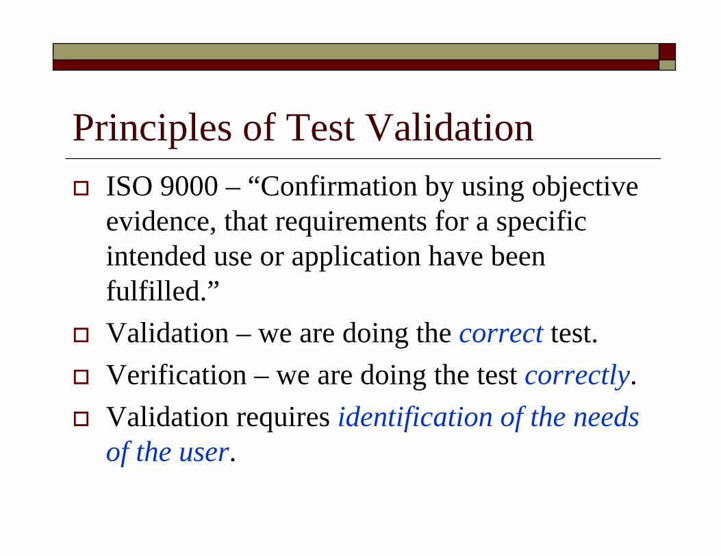
Principles of Test Validation ISO 9000 – “Confirmation by using objective
evidence, that requirements for a specific intended use or application have been fulfilled.”
Validation – we are doing the correct test. Verification – we are doing the test correctly. Validation requires identification of the needs
of the user.

Analytic Performance Characteristics Accuracy: our result – reference value (or
conventional true value) = error Trueness: systematic error/bias Precision (for quantitative tests): measure of random
error (SD) Reproducibility (precision) Repeatability: reproducibility within-run Reference range: range of test values for designated
population

Analytic Performance Characteristics Analytic sensitivity: positive agreement as compared
to reference method Analytic specificity: negative agreement as
compared to reference method Clinical sensitivity: proportion of subjects with a
disorder with positive test result Clinical specificity: proportion of subjects without
disorder with negative test result Limit of detection: the lowest amount of analyte
(Ag) that is statistically distinguishable from background or negative control

Cochrane Collaboration 2003 Standards for the Reporting of Diagnostic
Accuracy Diagnostic accuracy: agreement between test results
and reference standard. Reference standard: the best available method for
establishing the presence or absence of the condition of interests.
Reference standard: single method, combination of methods, imaging, pathology, clinical follow-up, etc.

Clinical and Laboratory Standards Institute (CLSI) Recommendations: Evaluation Protocols
Generally: 10 to 20 operating days 20 to 40 patient samples 50 positive and 50 negative
www.clsi.org/

LDT, ASR, “Home Brew Tests” All IHC tests except FDA approved kits. All molecular tests except FDA approved kits. All cytogenetic tests and FISH.
“CLIA regulated laboratories qualified to perform high complexity testing have demonstrated expertise and ability to use ASRs in test procedures and analyses”.

CBC News in Depth:
Misdiagnosed - Anatomy of Newfoundland's Cancer-Testing Scandal
Of the 1,013 breast cancer patients retested (1997-2005), 383 — more than a third — were found to be false negative. That meant 383 patients were denied a fighting chance against cancer. More than 100 of those wrongly tested patients are now dead.
Not all of those affected were notified that a mistake had been made.
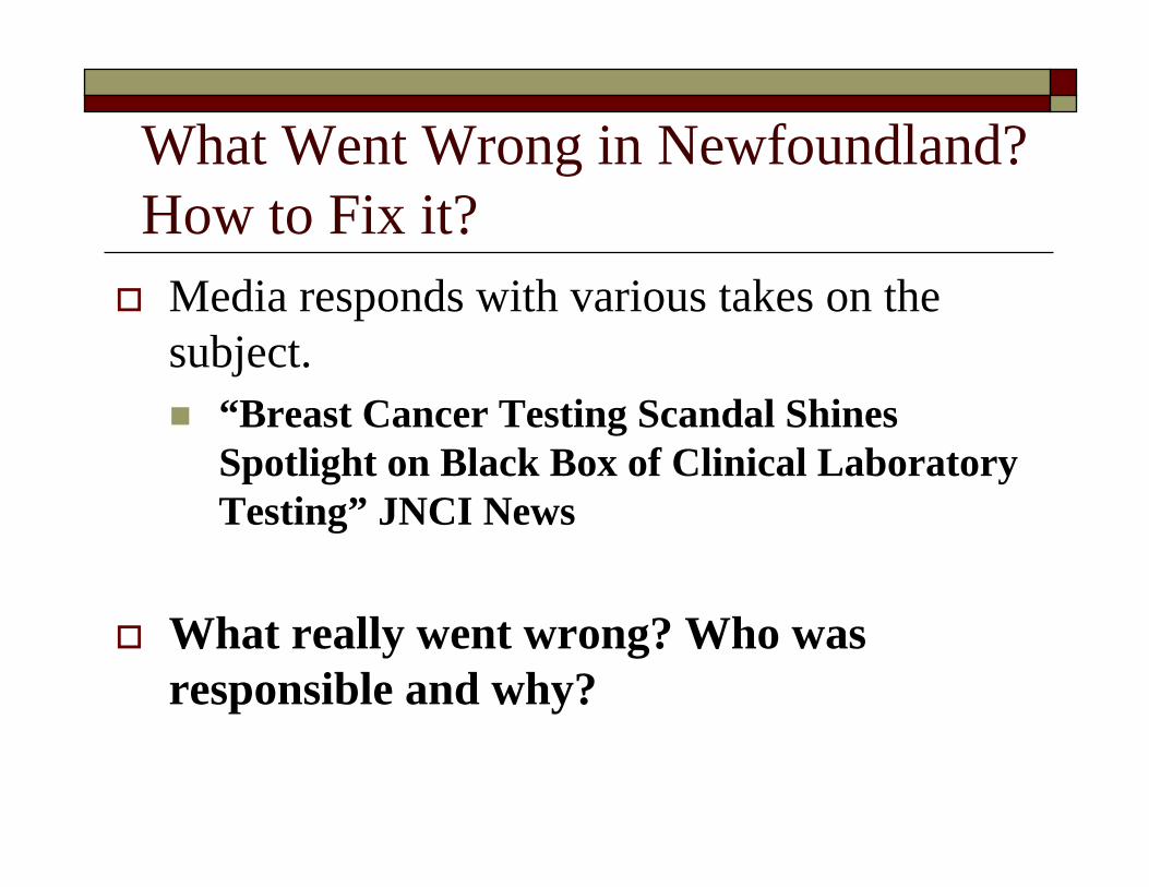
What Went Wrong in Newfoundland? How to Fix it? Media responds with various takes on the
subject. “Breast Cancer Testing Scandal Shines
Spotlight on Black Box of Clinical Laboratory Testing” JNCI News
What really went wrong? Who was responsible and why?

The Commission expressed conclusions and recommendations regarding responsibility of various persons or organizations, and delivered its final report and recommendations to the Minister of Health and Community Services on February 28, 2009.
QA QA QA http://www.cihrt.nl.ca/about.html


Guidelines:Table of Contents Use of Standard Terminology in Clinical
Immunohistochemistry Principles/Best Practices for Quality Assurance of Clinical
IHC Testing Class II Immunohistochemistry Tests Principles/Best
Practices Proficiency testing: Monitoring the quality of laboratory
performance Education and training standards for laboratory personnel References
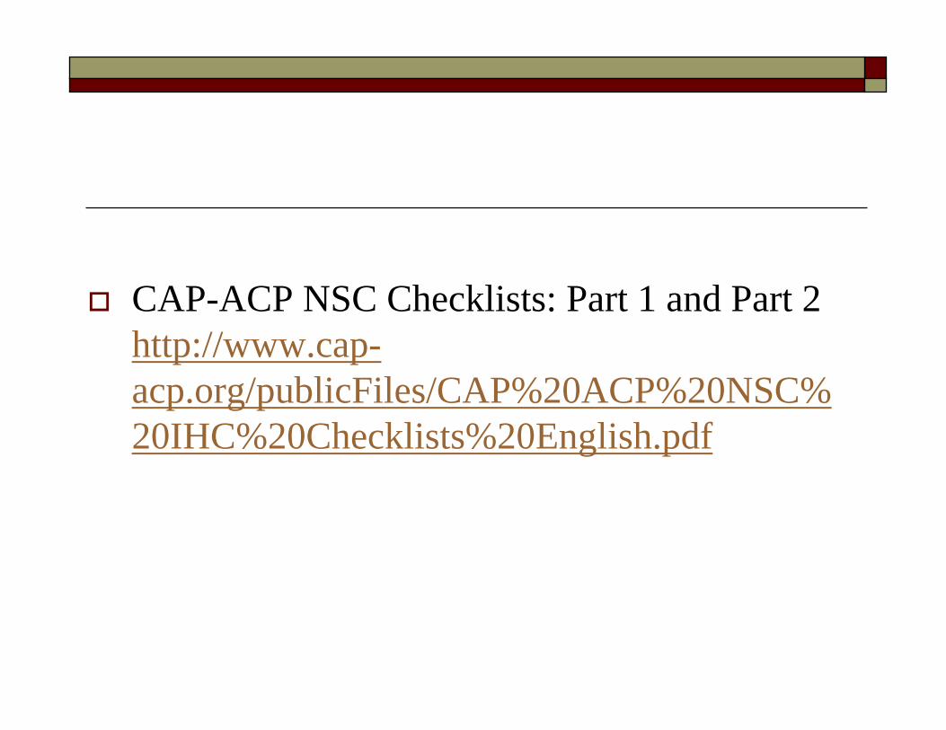
CAP-ACP NSC Checklists: Part 1 and Part 2 http://www.cap-acp.org/publicFiles/CAP%20ACP%20NSC%20IHC%20Checklists%20English.pdf

The Role of Test Classification on Tools for QC/QA in IHC
Class I – results used by pathologists Class II – results used by clinicians QC/QA ideally should be the same for both
types of tests. Class II currently have priority as they are
linked to higher risk for patient safety.

Adjunctive diagnostic information not independently reported by physician
Used after tumor diagnosed by other methods
E.g. cytokeratin differentiation markers
Class I IHC Tests(used by pathologists)

Class I
The results are incorporated into the diagnostic interpretation by the pathologists.
Results NOT to be listed/described in entirety in the pathology reports?
Readily available internal and external controls.
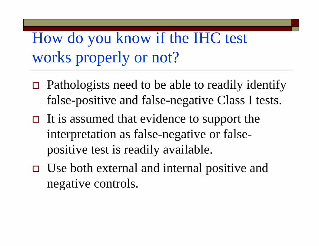
How do you know if the IHC test works properly or not? Pathologists need to be able to readily identify
false-positive and false-negative Class I tests. It is assumed that evidence to support the
interpretation as false-negative or false-positive test is readily available.
Use both external and internal positive and negative controls.

IHC Controls Internal positive control is most important to
exclude false-negative results. Internal negative control is most important to
exclude false-positive results.
External controls monitor system, not individual patient’s sample.
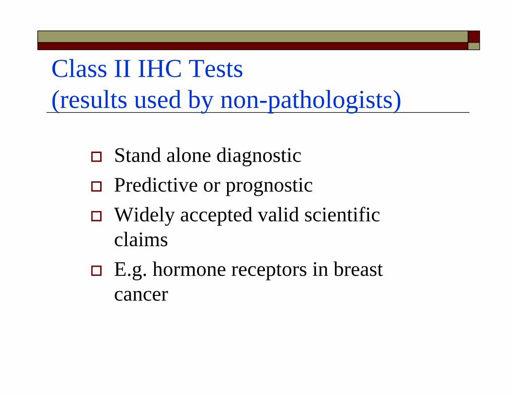
Class II IHC Tests (results used by non-pathologists)
Stand alone diagnostic Predictive or prognostic Widely accepted valid scientific
claims E.g. hormone receptors in breast
cancer

Class II
Prognostic IHC tests – The results of these tests independently forecast clinical outcome. They may be either qualitative or quantitative. HER2/neu if used as prognostic marker.
Predictive IHC tests – The results of these tests independently predict response to a particular therapy. They may either be qualitative or quantitative (e.g., ER/PR, HER2/neu in breast carcinoma, CD117 in gastrointestinal stromal tumor).

CAP-ACP IHC Test Classification: Class II Tests
CD20
c-MycCD117
IgG/IgG4Proliferation Marker Ki-67
GCET1MMRHuman Epidermal Growth Factor Receptor 2 (HER2)
FOXP1DOG1Progesterone Receptor (PR)
NPM1C4dEstrogen Receptor (ER)For DiscussionIn ConsiderationCurrent

Interpretation and Test Classification Class I IHC tests, which have critical
significance for interpretation of overall assessment:
ALK-1, cyclin D1, CD30, TdT, TTF-1, CDX-2, HMB-45, …
New IHC test Class may be necessary to rise awareness and prevent wrong diagnoses. Class IA and IB?

FDA and Health Canada are focused on whether this level of regulation is adequate for the protection of public health
FDA is aware that variability in IHC results may be introduced at every step:
Collection and fixation of the specimen, Automated processing, Embedding and sectioning, Staining of the final slide preparation, and Microscopic interpretation by the pathologist.

FDA (also Health Canada) counts on (counted on):
Ongoing initiatives by professional organizations and manufacturers directed at ensuring that pre- and postanalytic, as well as analytic procedures, are properly performed.

Class I and Class II IHC Tests: Incorporating Appropriate Language in Pathology Reports For analyte specific reagents (ASR), a U.S. Food and Drug
Administration (FDA)-required disclaimer is included in the reports.
The mandatory language is as follows:“These tests were developed and their performance characteristics
determined by the name of institution, Pathology Laboratory. They have not been cleared or approved by the FDA. However, the FDA has
determined that such clearance or approval is not necessary. These tests are used for clinical purposes. They should not be regarded as
investigational or for research. This laboratory is certified under the Clinical Laboratory Improvement Amendments of 1988 (CLIA) as qualified to perform high complexity clinical laboratory testing.”
This does not apply to FDA-approved kits for IHC testing.

Postanalytical: Interpretation of IHC Results
Are there published guidelines for interpretation? Starts with interpretation of results in controls by
technologist Starts with an agreement on what is considered a
desirable result Postanalytical component cannot even start without a
consensus on positive and negative controls Challenge: Standardization of positive and
negative controls

Challenge: Reporting standardization of Class II tests other than breast cancer
Challenge: Reporting of Class I tests results Synoptic reporting for Class II tests
Postanalytical: Reporting of IHC Results
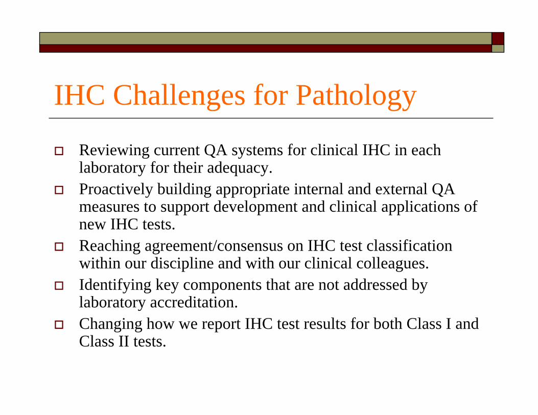
IHC Challenges for Pathology
Reviewing current QA systems for clinical IHC in each laboratory for their adequacy.
Proactively building appropriate internal and external QA measures to support development and clinical applications of new IHC tests.
Reaching agreement/consensus on IHC test classification within our discipline and with our clinical colleagues.
Identifying key components that are not addressed by laboratory accreditation.
Changing how we report IHC test results for both Class I and Class II tests.
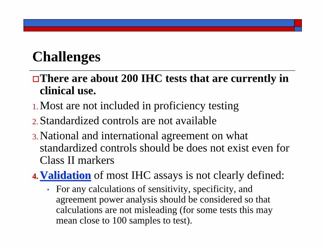
ChallengesThere are about 200 IHC tests that are currently in
clinical use.1. Most are not included in proficiency testing2. Standardized controls are not available3. National and international agreement on what
standardized controls should be does not exist even for Class II markers
4.4. ValidationValidation of most IHC assays is not clearly defined:• For any calculations of sensitivity, specificity, and
agreement power analysis should be considered so that calculations are not misleading (for some tests this may mean close to 100 samples to test).

Lack of Quality Control Samples and Standardization Lack of definitions and/or agreement what
samples should be used for either positive or negative controls
Lack of actual source of QC samples Lack of funds for appropriate generation of
QC samples Lack of knowledge dissemination in QC
including both laboratory physicians, technologists, managers, and users (oncologists, other…)

External Quality Assurance and Proficiency Testing (PT) in IHC Does not exist for many tests! Various programs that are providing PT do not clearly define
their targets: What are gold standards? What are reference values? Are assessments quantitative for quantitative IHC tests? Can participation in EQA be used for test validation? Are the EQA programs testing analytical or clinical
sensitivity and specificity or both (or neither)?

33% optimal33% optimal
33% good/suboptimal33% good/suboptimal
33% poor33% poor
NordiQC and CIQC Experience


CIQC Run2: HER2Average sensitivity: 91%, specificity: 98%Average sensitivity: 91%, specificity: 98%
Kendall’s coefficient of concordance = 0.96Kendall’s coefficient of concordance = 0.96

How to Define Discordant Results?
Outliers


Pilot Project Survey posted on www.cIQc.ca
Evaluation of stained slides:
Ki-67 CD117 CD3 CD20 CD34 CD61/F8-ra

CD3
EBMWG

CD3
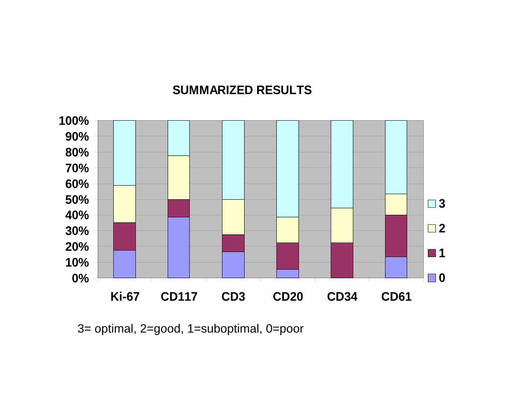
SUMMARIZED RESULTS
0%10%20%30%40%50%60%70%80%90%
100%
Ki-67 CD117 CD3 CD20 CD34 CD61
3
2
1
0
3= optimal, 2=good, 1=suboptimal, 0=poor

CenterCenter KiKi--6767 CD117CD117 CD3CD3 CD61CD61 CD34CD34 CD20CD20 Suboptimal/PoorSuboptimal/Poor TotalTotal11 11 11 33 11 11 11 55 66
22 11 00 22 22 11 22 66 66
33 11 33 33 33 33 33 11 66
44 33 11 33 33 22 33 11 66
55 22 22 22 22 33 22 00 66
66 22 33 22 33 33 33 00 66
77 00 00 00 .. 33 33 33 55
88 33 22 33 33 33 33 00 66
99 33 00 33 00 22 33 22 66
1010 33 22 00 11 33 33 22 66
1111 22 22 22 .. 33 33 00 55
1212 00 00 11 .. 22 22 33 55
1313 33 00 33 11 11 11 44 66
1414 00 33 33 00 33 33 22 66
1515 .. 00 00 11 11 00 55 55
1616 22 22 33 33 33 33 00 66
1717 33 33 11 33 33 11 22 66
1818 33 00 33 33 22 33 11 66
TotalTotal 35.50%35.50% 104104

Standardization of Bone Marrow Immunohistochemistry
International Council for Standardization in Haematology (ICSH) Working Party for
Standardization of Bone Marrow Immunohistochemistry
Emina Torlakovic, MD, PhD, FCAP – Canada (co-chair)Anna Porwit, MD, PhD -- Sweden (co-chair)Szu-Hee Lee, MBBChir, PhD, FRCPEdin, FRCPath, FRCPA --Australia Marciano Reis, MD, PhD, FRCPC – CanadaHans Kreipe, MD, PhD – GermanyKikkeri N. Naresh, MBBS, CCP, MD, FRCPath – United KingdomAlexander Tzankov, MD -- Switzerland Yoshito Sadahira, MD -- JapanElizabeth Hyjek, MD, PhD – USARussell K. Brynes, MD – USARobert McKenna, MD -- USA

Literature Review: Methods’ Description is Insufficient
Essential68Pretreatment time
Essential17Detection system
Essential61Pretreatment buffer
Essential48Pretreatment methods
Very important80Negative controls
Very important87Positive controls
Essential5272
DecalcificationDecalcification time
Essential2775
FixationFixaton time
Significance for IHC ResultsNot Described (%)

Formalin Modified Karnovski
CD3

High Expectations of Accuracy: To Treat or Not Treat?
Sensitivity and specificity of most tests is not defined
When possible to calculate sensitivity and specificity, standards are not set or not universally agreed upon
There is no tracking of clinical impact of reported IHC tests
There is no tracking of clinical impact of proficiency testing (PT) results of various programs that provide PT

Expression Level of ABC
OS
Targeted Therapy: Anti-ABC
PublishedReal?

Future
Patient safety is clearly identified as the no. 1 priority in the design and regulation of laboratory testing by all agencies and organizations
Standardization needs to address all parameters of the testing (pre-analytical, analytical, and post-analytical)
QA measures need to be tailored to the test type (Class I vs. Class II) as well as to make biological and statistical sense.

Future: Era of -OMICS In situ demonstration of protein expression -Omics studies at the moment are relatively
expensive and discovery-focused IHC required to confirm data obtained by other
methods including standard NB, WB, and SB Omics studies narrow our focus from large scale to
small scale (most important genes) Many “most important genes” are being detected by
immunohistochemistry

Paying Attention & Common Sense

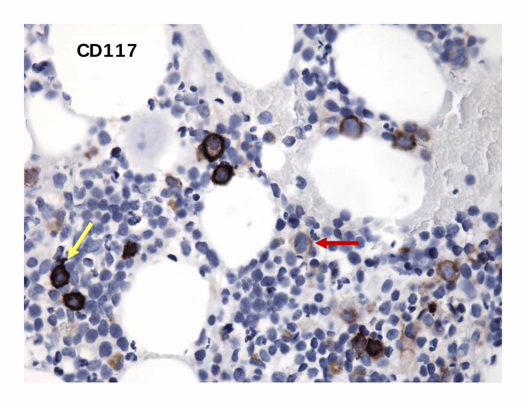
CD117

TDT: Negative
Cyclin D1: Positive

APAP--15 in 15 in Breast CA: Breast CA: Apocrine Apocrine PatternPattern

APAP--15 in 15 in Breast CA:Breast CA:IntracytoplasmicIntracytoplasmicLuminal Luminal PatternPattern

IS THIS POSITIVE GCDFP-15?
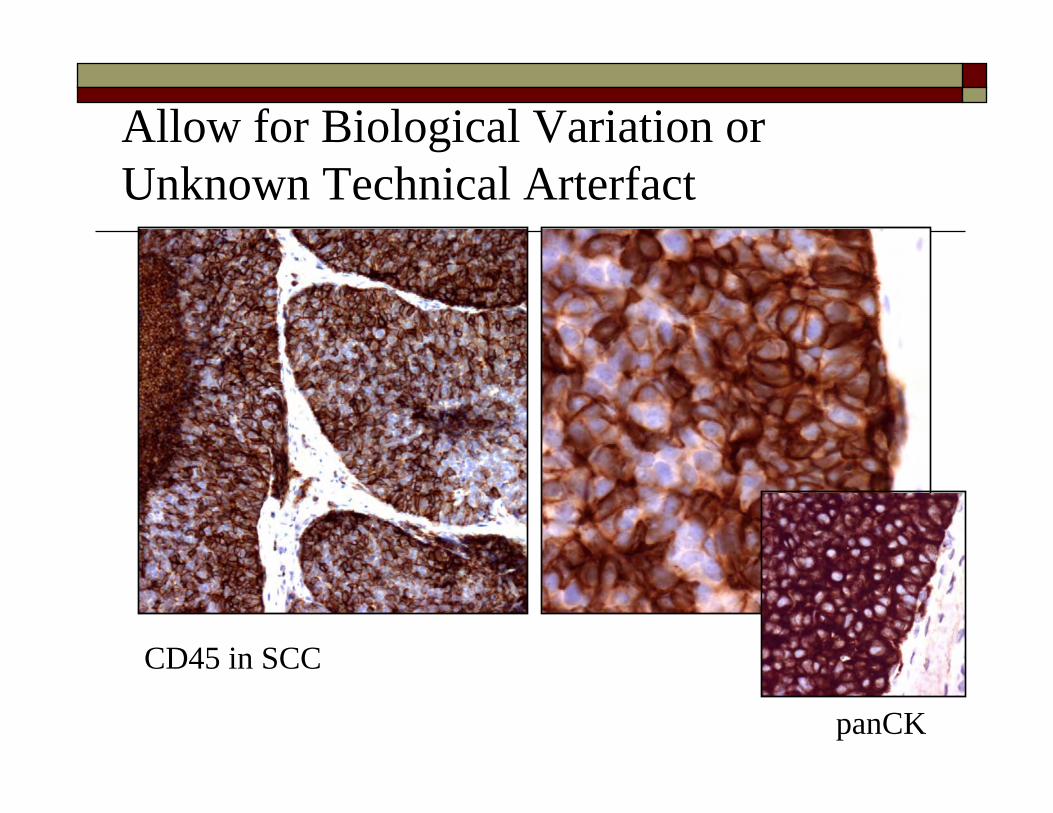
panCK
Allow for Biological Variation or Unknown Technical Arterfact
CD45 in SCC
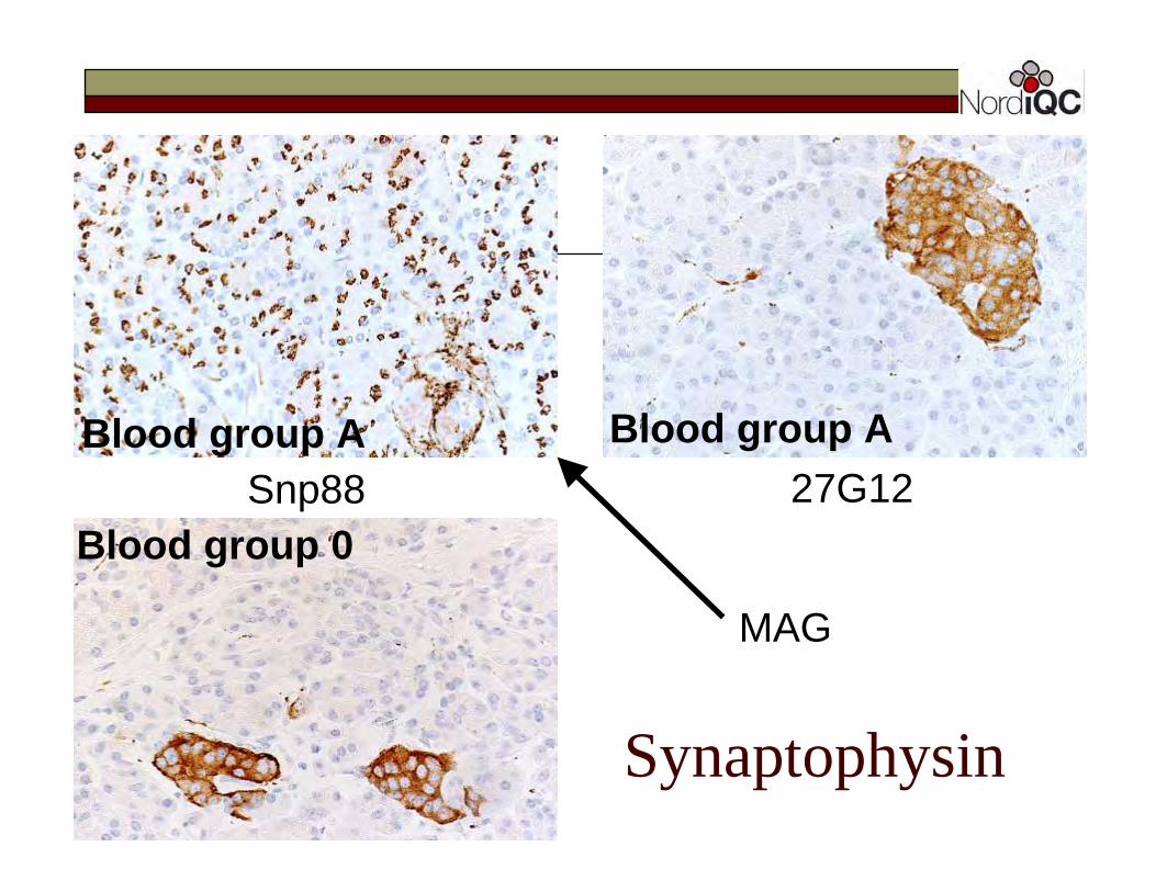
27G12Snp88
Synaptophysin
Blood group A Blood group A
Blood group 0
MAG

CMV (Kidney Bx):YES
CD30 in BM CD30 in BM aspirate: NOaspirate: NO
Single Cell Positivity: YES and NO

EBER in cHL: YES

Membranous vs. Cytoplasmic vs. Nuclear vs. Extracellular CD3: could be both, membranous and
cytoplasmic CK: cytoplasmic only CD30: Golgi,cytoplasmic,
membranous CD20: membranous only PSA: any pattern is good
CD3

Ber-EP4 CA-125
Membranous

Membranous: Cytokeratins

Cytokeratin: Dot-like & Filamentous Cytoplasmic

CK in Cortical Type CK in Cortical Type ThymomaThymoma: : DendriticDendritic

PSA

ALK-1 in ALCL with t(2;5)

Other Markers with Cytoplasmic + Nuclear Positivity
S-100 Calretinin Mutated NPM-1 CMV Hemoglobin A ER/PR and other typically nuclear markers
(sometimes) Other

Lymphoplasmacytic
lymphoma
Always Interpreted Together Kappa and lambda CD3, CD20 (and CD5) CD4 and CD8…


CD3 CD4 CD8
?

CD3 CD4 CD8

CD5 CD7

Distribution
Variation from cell to cell is more likely to be specific
Uniform positivity in all present cells should warrant special consideration to rule out false positive result

Monoclonal rabbit anti-CD79a

WHAT IS YOUR DIAGNOSIS?
Skin Bx:Blastic morphology, TdT+, Pax-5+,CD45-
Pax-5

WHAT MARKER AND WHAT TUMOR IS THIS?
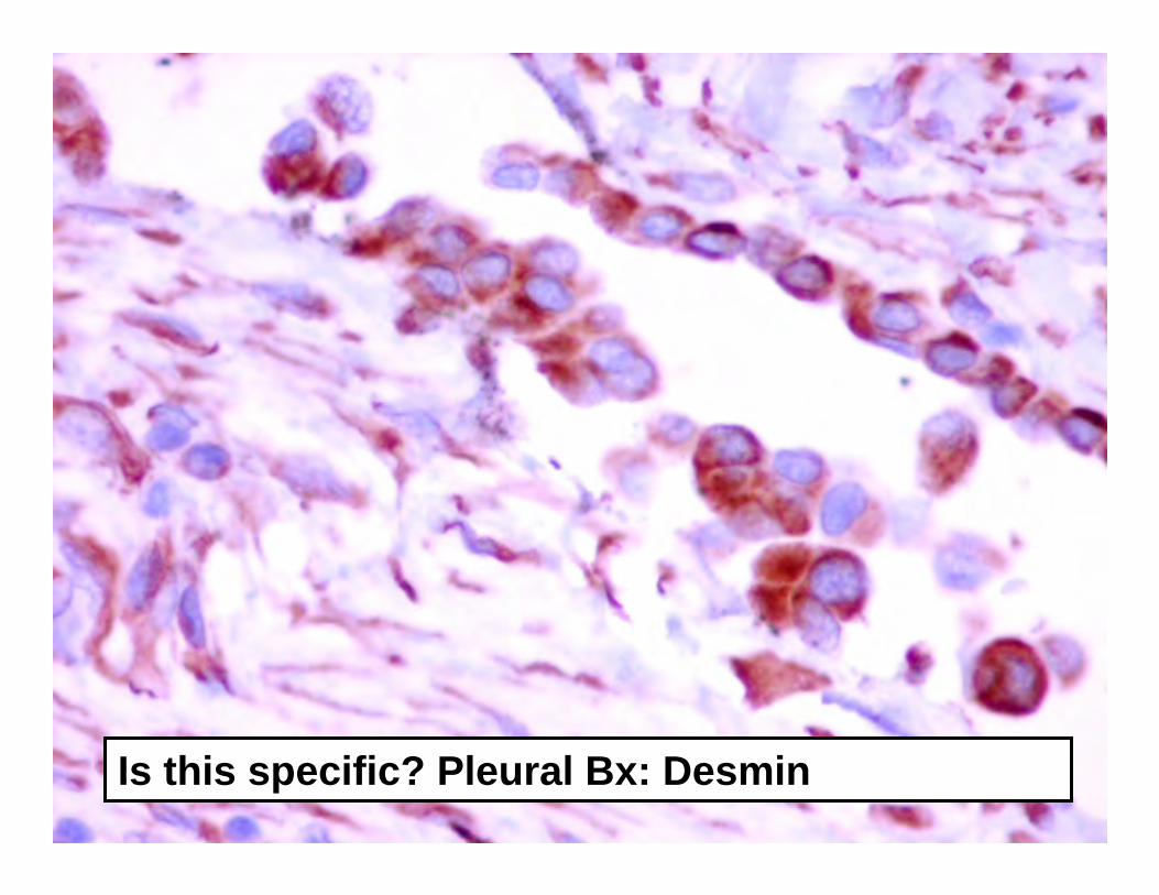
Is this specific? Pleural Bx: Desmin
![IHC PPT Ancillary Productsmy1hr-public.s3.amazonaws.com/documents/enroll/IHC PPT Ancillary Products[3].pdfAncillary Products From The IHC Group. The IHC Group Corporate Overview Ø](https://img.pdfslide.net/doc/110x75/5e38c9b5e1bb9a3e4e5b3bd8/ihc-ppt-ancillary-productsmy1hr-publics3-ppt-ancillary-products3pdf-ancillary.jpg)


![Color standardization for the …black [13 - 15] can be observed, as in Figures in this paper. To sum up, this paper presents the method of color standardization adjusted to the IHC](https://img.pdfslide.net/doc/110x75/5ed3158b18ac3f116443bbbd/color-standardization-for-the-black-13-15-can-be-observed-as-in-figures-in.jpg)