Embed Size (px)
Citation preview
HAL Id: hal-00875835https://hal-univ-rennes1.archives-ouvertes.fr/hal-00875835
Submitted on 22 Oct 2013
HAL is a multi-disciplinary open accessarchive for the deposit and dissemination of sci-entific research documents, whether they are pub-lished or not. The documents may come fromteaching and research institutions in France orabroad, or from public or private research centers.
L’archive ouverte pluridisciplinaire HAL, estdestinée au dépôt et à la diffusion de documentsscientifiques de niveau recherche, publiés ou non,émanant des établissements d’enseignement et derecherche français ou étrangers, des laboratoirespublics ou privés.
State of knowledge and concerns on cyanobacterialblooms and cyanotoxins.
Sylvain Merel, David Walker, Ruth Chicana, Shane Snyder, Estelle Baurès,Olivier Thomas
To cite this version:Sylvain Merel, David Walker, Ruth Chicana, Shane Snyder, Estelle Baurès, et al.. State of knowledgeand concerns on cyanobacterial blooms and cyanotoxins.. Environment International, Elsevier, 2013,59, pp.303-27. �10.1016/j.envint.2013.06.013�. �hal-00875835�

1
State of knowledge and concerns
on cyanobacterial blooms and cyanotoxins
Sylvain Merel1,2*, David Walker3, Ruth Chicana2, Shane Snyder1, Estelle Baurès4,
Olivier Thomas4
1 Department of Chemical and Environmental Engineering, University of Arizona, 1133
James E. Rogers Way, Tucson 85721, Arizona, USA
2 UMI 3157 CNRS, University of Arizona, 1133 James E. Rogers Way, Tucson 85721,
Arizona, USA
3 Environmental Research Laboratory, University of Arizona, 2601 E. Airport Drive, 85706
Tucson, Arizona, USA
4 Environment and Health Research Laboratory - IRSET, UMR 1085 INSERM, French School
of Public Health, Pr. Léon-Bernard Avenue – CS 74312, 35043 Rennes Cedex, France
* Corresponding author. E-mail: [email protected] or [email protected]
Tel: (+1) 520-621-6044
Fax: (+1) 520-621-6048
2
Abstract
Cyanobacteria are ubiquitous microorganisms considered as important contributors to the
formation of Earth’s atmosphere and nitrogen fixation. However, they are also frequently
associated with toxic blooms. Indeed, the wide range of hepatotoxins, neurotoxins and
dermatotoxins synthesized by these bacteria is a growing environmental and public health
concern. This paper provides a state of the art on the occurrence and management of harmful
cyanobacterial blooms in surface and drinking water, including economic impacts and
research needs. Cyanobacterial blooms usually occur according to a combination of
environmental factors e.g., nutrient concentration, water temperature, light intensity, salinity,
water movement, stagnation and residence time, as well as several other variables. These
environmental variables, in turn, have promoted the evolution and biosynthesis of strain-
specific, gene-controlled metabolites (cyanotoxins) that are often harmful to aquatic and
terrestrial life, including humans. Cyanotoxins are primarily produced intracellularly during
the exponential growth phase. Release of toxins into water can occur during cell death or
senescence but can also be due to evolutionary-derived or environmentally-mediated
circumstances such as allelopathy or relatively sudden nutrient limitation. Consequently,
when cyanobacterial blooms occur in drinking water resources, treatment has to remove both
cyanobacteria (avoiding cell lysis and subsequent toxin release) and aqueous cyanotoxins
previously released. Cells are usually removed with limited lysis by physical processes such
as clarification or membrane filtration. However, aqueous toxins are usually removed by both
physical retention, through adsorption on activated carbon or reverse osmosis, and chemical
oxidation, through ozonation or chlorination. While the efficient oxidation of the more
common cyanotoxins (microcystin, cylindrospermopsin, anatoxin and saxitoxin) has been
extensively reported, the chemical and toxicological characterization of their by-products
requires further investigation. In addition, future research should also investigate the removal

3
of poorly considered cyanotoxins (β-methylamino-alanine, lyngbyatoxin or aplysiatoxin) as
well as the economic impact of blooms.
Keywords: Cyanobacteria, Ecology, Bloom, Toxin, Drinking Water Treatment, Public Health,
Environmental Economy, Microcystin, Anatoxin, Cylindrospermopsin, Saxitoxin, BMAA
1. Introduction
Cyanobacteria were amongst the earliest organisms on Earth and the oxygen released
into the atmosphere through their photosynthesis may have been the precursor of the ozone
layer (Mur et al., 1999). Presently, while their importance in the evolutionary history of the
Earth should not be under-stated, these ubiquitous microorganisms are mostly associated with
eutrophic waters. Eutrophication of water resources is often considered as the primary cause
of water quality impairment on a world-wide basis. Eutrophication of drinking water
resources is primarily caused by excess nutrient loading and storage to lakes and reservoirs
due to human activities although climate change will likely play an increasing role in the
future (Heisler et al., 2008).
Cyanobacteria are known because of their ability to produce compounds (2-
methylisoborneol and geosmin) causing unpleasant tastes and odors in drinking water
(Falconer, 1999). However, over the last 2 decades, research priorities have progressively
focused on the harmful metabolites also potentially biosynthesised by these microorganisms.
Toxins of cyanobacteria (cyanotoxins) include hepatotoxins acting on the liver, neurotoxins
acting on the nervous system and dermatotoxins causing skin irritation. Since they have been
associated with numerous animal and human poisonings (Briand et al., 2003; Griffiths and
Saker, 2003; Kuiper-Goodman et al., 1999; Pouria et al., 1998), cyanotoxins are now a
growing environmental and public health concern.
4
Humans are potentially exposed to cyanotoxins through recreational activities such as
bathing in contaminated surface water and through consumption of unsuitably treated
drinking water produced from contaminated resources. Therefore, the efficient management
and protection of water resources from harmful cyanobacterial blooms is of critical
importance to protect human health. For that reason, this extensive review aims to provide a
better understanding of cyanobacterial blooms; from the causes of their occurrence and the
biosynthesis of toxins to the preventive and remedial options in surface water as well as
drinking water supplies. While giving the reader a list of key references for further reading,
this review also intends to underline the economic impacts of harmful cyanobacterial blooms
and identify major research needs.
2. Occurrence of harmful cyanobacterial blooms
There is no international definition or quantification for what a cyanobacterial bloom
is, however, this phenomenon is generally considered as a significant production of biomass
over a short period of time correlated with a diminution of phytoplankton diversity. In fact,
blooms of cyanobacteria are often mono-specific (or nearly so) and may form a dense layer of
cells at the surface of the water visible to the un-aided eye. The formation of cyanobacterial
blooms is controlled by environmental factors, and human poisonings are further conditioned
by the ability of the individual strains to perform the biosynthesis of cyanotoxins plus
subsequent exposure to these harmful metabolites.
2.1. Formation and monitoring of cyanobacterial blooms
Since cyanobacteria are primarily phototrophic microorganisms, groundwater
resources are not as vulnerable to bloom formation and associated problems as are surface
waters. Cyanobacteria, unlike several types of true algae, usually do not prefer flowing water

5
although some species have adapted to such conditions. Generally, cyanobacteria flourish in
more lentic aquatic ecosystems with relatively high concentrations of primary algal nutrients
(nitrogen, phosphorous, and carbon). In some lakes and reservoirs with long residence times,
nutrient accumulation and increasing trophic status favor blooms (sometimes nearly mono-
specific) of cyanobacteria. As lakes and reservoirs around the world continue to age due to
either natural or human causes, the resulting eutrophication will favor cyanobacterial bloom
formation over types of true algae. Therefore, the problem of cyanobacterial bloom formation
and subsequent risks to human health are an increasingly important and timely topic.
Given the many types of cyanobacteria and the diversity of habitats they have evolved
into, predicting all environmental variables required by an individual species to grow and
thrive is difficult if not impossible. Generally, the formation of cyanobacterial bloom is
regulated by a combination of three primary environmental factors (Fig. 1). The first one is
water temperature, several types of cyanobacteria preferring warmer water (25°C or above).
Consequently, global warming may increase the frequency and magnitude of cyanobacterial
blooms by favoring cyanobacteria among other phytoplankton species (Arheimer et al., 2005;
Dale et al., 2006; El-Shehawy et al., 2012; Jöhnk et al., 2008; O’Neil et al., 2012; Paerl and
Paul, 2012; Paul, 2008; Wiedner et al., 2007). The second environmental factor influencing
cyanobacterial bloom is light exposure. Although several species of cyanobacteria can be
considered hetero- or chemo-trophic, most species need a minimum of light availability for
photosynthesis to occur. The quality, intensity, and duration of light needed are species-
specific. Usually, pigmentation of cyanobacteria protects the cell from photoinhibition due to
high light intensities and also improves light harvesting through the absorption across a
broader region of the visible spectrum compared to other phytoplankton species (Mur et al.,
1999). Therefore, it would appear as if duration of light exposure is a more important growth
parameter than light intensity or quality. However, some species are extremely flexible in

6
their response to light exposure. Indeed, some cyanobacteria can persist in caves for months
with virtually no light and are viable and capable of growth immediately following light
exposure (Montechiaro and Giordano, 2006). The third factor leading to bloom formation is
the trophic status of the aquatic system. Cyanobacterial blooms mainly occur in eutrophic
reservoirs (El-Shehawy et al., 2012; Heisler et al., 2008) with N/P ratio ranging from 10 to 15
(Mur et al., 1999). However, another study considering 99 reservoirs indicates that the
occurrence of cyanobacterial blooms better correlates with the concentration of N total and P
total rather than N/P ratio (Downing et al., 2001).
The presence of cyanobacterial blooms is detected and monitored by different means.
A common approach is to measure chlorophyll a (Chl a), the primary photosynthetic pigment
contained in all phototrophic microorganisms. Chl a can be measured either in the field using
in situ sensors or samples can be collected for laboratory analyses. However, measurements
of Chl a do not discriminate cyanobacteria from algae, which pose a serious limitation on data
interpretation. Measuring specific cyanobacterial pigments such as phycocyanin may
overcome the problem (Brient et al., 2008; Gregor et al., 2007). In addition, the combined
analysis of Chl a and phycocyanin provides useful information with respect to the proportion
of cyanobacteria among other phytoplankton species.
Another common method of monitoring cyanobacterial blooms is the enumeration and
identification of cells by microscope. The advantage of doing so is that cyanobacteria species
can be accurately identified and the proportion of cyanobacteria compared to other species in
the phytoplankton assemblage. Microscope examination and enumeration has the highest
resolution of any other method. Nonetheless, it is time-consuming and requires a relatively
high level of taxonomic expertise. Like any other method, microscope identification of a
potentially-toxic species of cyanobacteria does not mean it is actively producing toxin.
However, enumeration and identification of cyanobacteria by microscope combined with
7
chemical identification of toxins in the water gives a good indication of the culprit species. In
addition to cell counting and identification, new techniques based on polymerase chain
reaction (PCR) of genes related to toxin synthesis have been proposed (Al-Tebrineh et al.,
2010; Barón-Sola et al., 2012; Baxa et al., 2010; dos Anjos et al., 2006; Hisbergues et al.,
2003; Kurmayer and Kutzenberger, 2003; Ostermaier and Kurmayer, 2010).
2.2. Origin of the toxicity
The toxicity of cyanobacteria is related to the biosynthesis of harmful metabolites
called cyanotoxins. However, cyanobacterial blooms are not necessarily associated with the
occurrence of toxins since not all the strains are toxic (Sarazin et al., 2002). Indeed,
cyanotoxins are produced only by the strains having the appropriate genes (Kurmayer and
Christiansen, 2009). In addition, even toxic strains do not automatically produce toxins since
several of them seem to have the capability to turn certain genes on or off depending upon
environmental conditions. Identification of strains having the appropriate genes, however, is
as-of-today the best method for determining whether a bloom is or may become toxic. Genes
known to have toxin-producing capabilities have been progressively identified in certain
strains (Kellmann et al., 2006; Kellmann et al., 2008; Moffitt and Neilan, 2004; Tillett et al.,
2000), allowing the development of PCR methods for the specific detection of potentially-
toxic cyanobacteria in environmental samples.
Although genetic identification is an excellent tool in determining the biosynthesis of
cyanotoxins, strain-specific environmental factors for toxin production exist. The iterative
nature of toxin production on a strain-specific level makes generalizations regarding toxin
production difficult. Environmental factors include but are not limited to light intensity and
exposure time, water movement and flow, allelopathic influences and competition for
resources, herbivory and grazing, nutrient concentrations and ratios, water temperature and
8
salinity, cell division and growth rate (Kosol et al., 2009; Orr and Jones, 1998; Sevilla et al.,
2008; Tonk et al., 2005). Indeed, the list of environmental factors and iterations seems almost
as large as the list of strains capable of toxin production. Although daunting, much more
empirical work needs to be done regarding environmental factors causing toxin production at
the individual strain level.
2.3. Literature available
The proliferation of Nodularia Spumigena described by Francis (1878) in lake
Alexandrina, Australia, is often referred to as the first report of toxic cyanobacterial bloom
even though no toxin was identified. Since then, the growing concerns associated with
cyanobacteria and their potentially toxic blooms have multiplied the publication of studies on
the topic. In fact, the annual amount of publications on cyanobacteria suddenly increased in
1991 and currently keeps growing (Fig.2). In December 2012, the Thomson Reuters Web of
Science database retrieved 18642 publications related to cyanobacteria, among which 15350
research articles and 1255 review articles. However, research on cyanobacteria is diverse and
articles related to blooms only represent 20% of the publications. In fact the literature
available on cyanobacteria covers more than 100 research areas including engineering and the
production of biofuels. However, most of the publications remain in the field of water biology
(22%), ecological science (18%) and microbiology (16%) while public health and
environmental health (0.3%) are sparsely considered.
3. Potential human exposure to cyanotoxins
Cyanotoxins have been associated with numerous animal poisonings worldwide, but
they are also a threat for human health. As presented in Fig. 1, human exposure to these
harmful metabolites can have 3 major origins. One route of exposure is the ingestion of
9
cyanobacteria-based food ingredients or shellfish which previously bioaccumulated toxins
through filtration of contaminated water (Ibelings and Chorus, 2007; Johnson et al., 2008;
Rellán et al., 2009; Saker et al., 2005). Another exposure route is possible through dermal
contact and accidental inhalation/ingestion during recreational activities in waters subjected to
a toxic bloom. The third route of exposure could be caused by the ingestion of drinking water
produced from a contaminated resource (Byth, 1980; Griffiths and Saker, 2003). Depending
upon the population served and type of treatment prior to delivery, this third exposure route
could affect a relatively large number of people. Some examples of intoxication and relevant
guidelines are provided with the chemical and biological properties of cyanotoxins.
4. Occurrence and properties of cyanotoxins
The word cyanotoxin refers to more than a hundred compounds that may strongly
differ in their chemical structure and toxicological property (Table 1). They are usually
arranged into 3 classes according to their target organ: hepatotoxins that induce liver injuries,
neurotoxins that alter the neuromuscular transmission and dermatotoxins that induce skin
irritation.
4.1. Cyanobacterial hepatotoxins
4.1.1. Microcystins
Microcystins (MCs) form the main family of cyanotoxins since they are the most
frequently studied and the most widespread. For example, their occurrence has been reported
in Asia, Europe, North Africa, North America and Scandinavian countries (Fristachi and
Sinclair, 2008). MCs were named according to Microcystis, the first genera of cyanobacteria
associated with their biosynthesis. However, MCs are also produced by Oscillatoria, Nostoc,

10
Anabaena and Anabaenopsis (Kaebernick and Neilan, 2001). As mentioned previously, toxin
synthesis is a complex process influenced by environmental conditions and depending on the
genetic properties of each cyanobacterial strain. In the last decade, the gene cluster mcyA-J
was identified as the origin of MCs biosynthesis. In fact, mcyA-J codes for a multienzyme
complex including peptide synthetase and poliketide synthase, allowing the components of
the toxin to be assembled non-ribosomally (Dittmann and Wiegand, 2006; Kaebernick and
Neilan, 2001).
As presented in Fig. 3, MCs are cyclic compounds enclosing 7 amino acids. Among
them, the unusual Adda amino acid is often associated with the toxicity of the molecule
because of its conjugated diene (Dawson, 1998). In addition, X and Z are usually referred to
as variable amino acids which multiple combinations make the difference between more than
70 variants of the toxin (Sivonen and Jones, 1999). Then, each variant is identified by the
initials of X and Z. For example, the common MC which has leucine (initial L) and arginine
(initial R) should be identified as MC-LR.
MCs are water-soluble and stable molecules (Sivonen and Jones, 1999). Once
absorbed by the organism, they are quickly concentrated in the liver (Fischer et al., 2000) and
bind to the protein phosphatase (Dawson, 1998; Gupta et al., 2003; Kuiper-Goodman et al.,
1999; MacKintosh et al., 1990). Depending upon dose and body weight, the inhibition of
protein phosphatase may lead successively to the accumulation of phosphorylated proteins in
the liver, cell necrosis, massive haemorrhage and death. For example, the lethal dose 50
(LD50) of MC-LR after intraperitoneal (i.p.) injection in mice ranges from 25 to 150 µg/kg
(Kuiper-Goodman et al., 1999). This value may differ according to the MC variant but MC-
LR is usually used as a reference. MCs are also considered to be potential tumor promoters
(Falconer, 1991; Nishiwaki-Matsushima et al., 1992).
11
Numerous animal and human intoxications by MCs have been reported (Hilborn et al.,
2007; Soares et al., 2006; Stewart et al., 2008). Most of the human poisonings were limited to
gastro-enteritis (Kuiper-Goodman et al., 1999; Teixeira et al., 1993) but, when water
containing the toxin was used for hemodialysis, MCs also caused the death of 60 patients at
the Brazilian dialysis centre of Caruaru in 1996 (Azevedo et al., 2002; Jochimsen et al., 1998;
Pouria et al., 1998; Yuan et al., 2006). Consequently, the World Health Organization
considered the MC-LR no observable adverse effect level (NOAEL) of 40 µg/kg/d obtained
after 13 weeks mice oral exposure (Fawell et al., 1999) and derived a guideline of 1 µg/L as a
maximum value for MC-LR in drinking water (WHO, 1998).
4.1.2. Nodularins
Nodularins (NODs) have been reported mainly in Australia, New Zealand and the
Baltic Sea (Sivonen and Jones, 1999; van Apeldoorn et al., 2007). Associated only with
Nodularia spumigena (Kaebernick and Neilan, 2001), their biosynthesis is regulated by genes
and performed non-ribosomally according to a mechanism similar to that involved in MC
production (Dittmann and Wiegand, 2006).
As shown in Fig. 3, NODs are cyclic pentapeptides structurally similar to MCs,
including the Adda moiety but only one variable amino acid Z. So far, 9 variants of this water
soluble and stable toxin have been identified (Codd et al., 2005), the most common being
NOD-R with Arginine as variable amino acid.
Like MCs, NODs are hepatotoxins acting through the inhibition of protein
phosphatase and are potential tumor promoters. According to the variant, the LD50 of NODs
in mice after i.p. injection ranges from 30 to 70 µg/kg (van Apeldoorn et al., 2007). However,
no human intoxication with NODs has been reported so far, and no guidelines have been
proposed for drinking water due to the lack of suitable toxicological data.
12
4.1.3. Cylindrospermopsin
Cylindrospermopsin (CYL) has been initially detected in Australia (Griffiths and
Saker, 2003; Saker et al., 1999), New Zealand (Stirling and Quilliam, 2001), and Thailand (Li
et al., 2001). Consequently, CYL was considered as a tropical toxin until its recent
characterization in temperate areas including Germany (Fastner et al., 2003; Fastner et al.,
2007; Rücker et al., 2007) and France (Brient et al., 2009). CYL was named according to
Cylindrospermopsis raciborskii, but other cyanobacteria like Aphanizomenon ovalisporum,
Raphidiopsis curvata and Umezakia natans can also perform the biosynthesis of the toxin
(Banker et al., 1997; Fristachi and Sinclair, 2008). Similarly to MCs, the synthesis of CYL
seems to be regulated by genes coding for polyketide synthase and peptide synthetase that
gather toxin’s components non-ribosomally (Schembri et al., 2001).
As shown in Fig. 3, CYL is a 415 Da tricyclic alkaloid enclosing a guanidine entity
along with a uracil moiety potentially responsible for the toxicity (Banker et al., 2001). So far,
the alteration of the hydroxyl group near the uracil moiety leads to the identification of 2 other
variants: 7-epicylindrospermopsin with a different OH orientation (Banker et al., 2000) and
the non-toxic deoxycylindrospermopsin without OH (Li et al., 2001; Norris et al., 1999).
CYL is highly water-soluble with a half-life greater than 10 days in high purity water
(Chiswell et al., 1999). After ingestion, the toxin mainly impacts the liver via the irreversible
inhibition of protein synthesis leading to cell death (Froscio et al., 2003; Froscio et al., 2008;
Metcalf et al., 2004). For example, CYL exhibits a 2100 µg/kg LD50 in mice, 24 hour after i.p.
injection (van Apeldoorn et al., 2007). However, CYL exposure can also lead to fetal toxicity
(Rogers et al., 2007), tumor initiation (Falconer and Humpage, 2001), micronucleus induction
and chromosome loss (Humpage et al., 2000).
13
The most famous case of human intoxication by CYL occurred in 1979 in Australia
and is often referred to as the Palm Island mystery disease (Bourke et al., 1983; Byth, 1980;
Griffiths and Saker, 2003). The application of an algaecide to eliminate a bloom of
cyanobacteria in the water supply resulted in CYL release and over 100 admissions of
children to the local hospital for gastroenteritis associated with the consumption of
contaminated drinking water. Therefore, based on the 30 µg/kg/d NOAEL observed on mice
orally exposed to CYL during 11 weeks, 1 µg/L was proposed as a guideline for maximum
concentration in drinking water (Humpage and Falconer, 2003).
4.2. Cyanobacterial neurotoxins
4.2.1. Anatoxin-a
The occurrence of anatoxin-a (ANTX-a) was reported in USA (Osswald et al., 2007),
Africa (Ballot et al., 2003; Krienitz et al., 2003), Asia (Namikoshi et al., 2003; Park et al.,
1993; Park et al., 1998) and Europe (Carrasco et al., 2007; Gugger et al., 2005; Viaggiu et al.,
2004). This toxin is mainly associated with 3 genera of cyanobacteria: Anabaena,
Aphanizomenon and Planktothrix (Osswald et al., 2007; van Apeldoorn et al., 2007). The
biosynthesis of ANTX-a has not been completely described yet, but the responsible genes
have been identified (Cadel-Six et al., 2009; Mejean et al., 2010).
As presented in Fig. 4, ANTX-a is a 165 Da alkaloid with a variant called
homoanatoxin-a resulting from the methylation of the carbon at the extremity of the ketone
function (van Apeldoorn et al., 2007). ANTX-a is highly water-soluble but unstable at pH
above 10 and transformed into a non-toxic form by sunlight exposure.
Once in the organism, ANTX-a induces paralysis by fixation on acetylcholine
receptors without being degraded by acetylcholinesterase (Osswald et al., 2007).
Consequently, death can occur by respiratory arrest when muscles involved in breathing
14
activity are affected. For example, the LD50 24 hours after i.p. injection in mice is 375 µg/kg
(van Apeldoorn et al., 2007).
ANTX-a has been responsible for various animal poisonings resulting in vomiting,
convulsion and respiratory arrest (Gugger et al., 2005; Henriksen et al., 1997; Krienitz et al.,
2003; Wood et al., 2007), but no human poisonings have yet been reported. So far, there is no
official guideline for ANTX-a in drinking water because of dissimilar results in subacute
toxicity studies (Kuiper-Goodman et al., 1999), but 3 µg/L has been suggested (van
Apeldoorn et al., 2007).
4.2.2. Anatoxin-a(s)
Anatoxin-a(s), known as ANTX-a(s), has been identified in restricted areas including
the United States, Scotland, Denmark and Brazil (Molica et al., 2005; Onodera et al., 1997;
Sivonen and Jones, 1999). The toxin has been associated only with Anabaena strains, but the
biosynthesis has not been completely explained yet.
As shown in Fig. 4, ANTX-a(s) is a 252 Da phosphate ester of a cyclic N-
hydroxyguanine (Sivonen and Jones, 1999; van Apeldoorn et al., 2007). Once absorbed in the
organism, ANTX-a(s) inhibits acetylcholinesterase (Molica et al., 2005) and induces muscular
paralysis with potential death by respiratory arrest. Very few toxicological studies have been
carried out and only the LD50 by i.p. injection into mice is available: 20-31 µg/kg (van
Apeldoorn et al., 2007). Consequently, no guideline has been proposed yet for ANTX-a(s) in
drinking water.
4.2.3. Saxitoxins
In freshwaters, saxitoxins (STXs) have been detected in Australia and USA (Kuiper-
Goodman et al., 1999). While Anabaena circinalis and Aphanizomenon flos-aquae seems to
15
be the main associated cyanobacteria, Lyngbya wollei and Cylindrospermopsis raciborskii
were also shown to perform the biosynthesis of the toxin (Nicholson et al., 2003). In sea
water, STXs are also produced by some dinoflagellates. Again, with the identification of a
relevant gene cluster, the knowledge of STXs biosynthesis has been improved (Kalaitzis et
al., 2010).
As presented in Fig. 4, STXs are tricyclic compounds ranging from 241 to 491 Da that
can be non-sulphated, singly sulphated or doubly sulphated (Nicholson et al., 2003; van
Apeldoorn et al., 2007). These water-soluble toxins can persist over 90 days in freshwater
(Jones and Negri, 1997), but they are altered by high temperatures and degraded into more
toxic variants (Sivonen and Jones, 1999).
STXs, also known as paralytic shellfish poisons, block sodium ion channels in nerve
axon membrane and induce nerve dysfunction, paralysis then death due to respiratory failure
(van Apeldoorn et al., 2007). For example, the LD50 of the most toxic variant in mice was
shown to be 10 µg/kg after i.p. injection.
Over the last century, STXs have been associated with numerous human intoxications
resulting in numbness, complete paralysis and even death (Kuiper-Goodman et al., 1999).
However, no intoxication through drinking water has been documented so far. While no
official guideline has been proposed for STXs in drinking water, Australia is considering a 3
µg STX eq/L to be used (van Apeldoorn et al., 2007).
4.2.4. β-N-methylamino-L-alanine
The cyanotoxin β-N-methylamino-L-alanine (BMAA) has been recently identified in
England (Metcalf et al., 2008), Peru (Johnson et al., 2008), South Africa (Esterhuizen and
Downing, 2008), China (Li et al., 2010) and Florida (Brand et al., 2010). BMAA has not been
extensively studied yet but a recent work indicates that this toxin may be produced by all
16
known groups of cyanobacteria (Cox et al., 2005). Indeed, cyanobacteria possess genes
coding for cysteine synthase-like enzyme and methyl transferase, both being involved in
BMAA biosynthesis (Aráoz et al., 2010a).
BMAA is a 118 Da non-protein amino acid shown in Fig. 4. It acts mostly on motor
neurons by fixation on glutamate receptors. In addition, BMAA could also cause
intraneuronal protein misfolding, the characteristic of neurodegeneration (Banack et al.,
2010a). In fact, there are assumptions that BMAA could be associated with various
neurodegenerative diseases such as the amyotrophic lateral sclerosis/parkinsonism dementia
complex in Guam or Alzheimer’s disease (Banack et al., 2010a; Murch et al., 2004; Pablo et
al., 2009). However, due to the lack of toxicological data such as LD50 or NOAEL, no
guideline has been proposed for BMAA in drinking water.
4.2.5. Other neurotoxins
In addition to the common freshwater toxins presented previously, a recent review also
mentioned the existence of 3 other marine cyanobacterial neurotoxins (Aráoz et al., 2010a).
Antillatoxin, kalkitoxin and jamaicamide are lipopeptides produced by Lyngbya majuscula
that induce neurotoxicity through the interaction with voltage-gated sodium channels.
However, due to the limited data available for these compounds, their chemical and biological
properties won’t be further developed in this paper.
4.3. Cyanobacterial dermatotoxins
Cyanobacterial dermatotoxins include aplysiatoxins (APTXs) and lynbyatoxins
(LTXs) mainly produced by Lyngbya majuscula (van Apeldoorn et al., 2007). So far, these
toxins have been detected only in marine water. After human exposure, characteristic
symptoms of poisoning include dermatitis as well as oral and gastrointestinal inflammation
17
resulting in diarrhea (Nagai et al., 1996; Osborne et al., 2007). In addition, APTXs and LTXs
are also potent tumor promoters through the activation of protein kinase C (van Apeldoorn et
al., 2007). However, due to the lack of data, their chemical and biological properties won’t be
further developed in this paper.
4.4. Literature available
MCs are probably the most common and the most well known toxic metabolites of
cyanobacteria but STX and ANTX were the first cyanotoxins to be studied in articles from the
early 1960s. Indeed, other toxins were only considered in studies published since the 1980s.
The annual amount of publications on cyanotoxins dramatically increased over the last
two decades (Fig. 2), mainly because of the constant progress in analytical science and the
growing awareness of the public health risk associated with these cell metabolites. In
December 2012, the Thomson Reuters Web of Science database retrieved 5293 publications
related to cyanotoxins, among which 4366 research articles, 467 proceedings and 198 review
articles. However, these publications are largely unevenly distributed among the different
toxins, as shown in Fig. 2. Indeed, with 2971 publications MCs account for more than 56% of
the overall literature. STXs come in second position with 27% of the literature but most of
these articles are actually associated to red tides rather than cyanobacteria. While NODs,
ANTX and CYL individually represent less than 10% of the literature available on
cyanotoxins, Fig. 2 reveals BMAA, LTX and APTX are largely understudied with less than
2% of the total amount of publications.
The literature available on cyanotoxins is also unevenly distributed between more than
100 research areas. Indeed, most of the studies focus on toxicology (24%), ecology (19%),
chemistry (18%) and pharmacology (17%). While water biology, biochemistry and molecular
biology still account for 13% of the literature, each of engineering and water resources only
18
represent 7%. With less than 5% of the overall amount of publications, other research areas
including economy, public health or epidemiology are even less considered.
5. Detection and quantification of cyanotoxins
A wide range of methods are available for the analysis of cyanotoxins (Table 2; Fig.
5). These include numerous techniques relying on biological and physico-chemical
approaches. However, according to the method employed and the kind of results expected,
samples often require specific preparation before analysis.
5.1. Sample preparation
After field sampling, samples should be stored at low temperature (4°C) and analyzed
as soon as possible in order to prevent any alteration of toxin distribution
(intracellular/extracellular). Sample preparation differs according to the kind of toxin
analyzed. As shown in Fig. 5, direct filtration of the sample only allows the detection of
extracellular toxins but an additional step inducing the lysis of cyanobacteria retained on the
filter would allow separate detection of intracellular toxins. However, cell lysis prior to
filtration allows the simultaneous detection of both extracellular and intracellular toxins
without determining their repartition. Cell lysis is often obtained by freezing-thawing
cyanobacteria or adding methanol in the sample (or onto the filter). Both methods directly
damage cell membranes and release intracellular toxins (Harada et al., 1999).
In addition, cyanotoxins in the filtrate may also undergo further purification and
concentration, usually through solid phase extraction (SPE). In this case at least 500 mL of
filtrate are poured through a cartridge containing a sorbent, usually a reversed phase C18.
Then toxins are eluted using methanol and water (usually 90% methanol). In order to further
increase the concentration of toxins, the cartridge eluate may be partially evaporated. This
19
practice can concentrate cyanotoxins by 3 orders of magnitude, which consequently improves
the detection limit of any subsequent analytical method.
5.2. Biological approach for the analysis of cyanotoxin
Cyanotoxins can be detected and quantified through a biological approach relying on
in vivo assays, immunological assays or biochemical assays, each with specific benefits and
limitations.
5.2.1. Toxin analysis by in vivo assays
Mouse bioassay is likely the most well known in vivo assay. In fact, it was the first
method developed to detect cyanotoxins in water even if it was actually designed to assess
their biological effects. The procedure consists in the i.p. injection of the sample in a
minimum of 3 mice followed by their necropsy after 24 hours (Falconer, 1993). The
observation of different symptoms reveals the presence of hepatotoxins or neurotoxins in the
matrix. For instance, while increasing the weight and the volume of the liver, the hepatotoxins
also induce the alteration of hepatic cells (Falconer, 1993). However, mouse bioassay does
not allow the exact identification of the toxin (MCs, NODs…) in the sample.
Despite its low sensitivity, mouse bioassay can be considered as a semi-quantitative
method when comparing the extent of the lesions to those observed on mice exposed to
different concentrations of a standard toxin. In this case, the results are often expressed as
equivalent of the standard toxin (usually MC-LR). Due to ethical issues with respect to animal
experiments and the development of new methods (faster, more sensitive and more specific)
for cyanotoxin measurement, the use of mouse bioassay is mostly limited to toxicological
research.
20
Alternative in vivo bioassays relying on less controversial organisms, like crustacean
larvae, have also been developed in order to quantify cyanotoxins (Kaushik and
Balasubramanian, 2012). Larvae of the selected organism (for example Artemia, Daphnia or
Thamnocephalus) are exposed to toxins through the incubation in a growth medium diluted in
a specific volume of the sample to be tested. However, while these assays can be performed in
a 96-well plate, they are still not specific and have a strong potential for interferences due to
matrix effects.
5.2.2. Toxin analysis by immunological assays
Cyanotoxins can also be detected through recognition and binding to specific
antibodies. For example, various ELISA (Enzyme-Linked ImmunoSorbent Assay) kits are
commercially available for the detection of MCs in water (Carmichael and An, 1999; Hilborn
et al., 2005; Lindner et al., 2004; Rapala et al., 2002). According to the antibody and the
procedure employed, these extremely sensitive methods can achieve a detection limit as low
as 4 ng/L with an upper quantitation limit (due to saturation) close to 2 µg/L for MC-LR
(Lindner et al., 2004). Therefore, while ELISA is successfully employed for the detection of
MCs, specific antibodies have also been designed to apply this method to the detection of
CYL and STXs (Bláhová et al., 2009; Campbell et al., 2009)
However, detection methods based on ELISA also have some limitations. For
instance, the different MCs variant cannot be distinguished and the results have to be
expressed as equivalent MC-LR/L. In addition, cross reactivity (even limited) with other
compounds in the sample may lead to overestimate the concentration of the toxins.

21
5.2.3. Toxin analysis by biochemical assays
Since MCs and NODs are potent inhibitors of protein phosphatase, these toxins can be
detected using a protein phosphatase inhibition assay known as PPIA (Almeida et al., 2006;
Bouaïcha et al., 2002; Heresztyn and Nicholson, 2001; Ortea et al., 2004; Rapala et al., 2002).
Before incubation with the relevant substrate, the enzyme is exposed to an aliquot of the
sample containing the toxin. Measuring the absorbance of the mixture at a specific
wavelength allows the detection of the substrate (or its transformation product) and the
assessment of the enzyme activity, which is inversely proportional to the concentration of the
toxin.
According to the method employed, PPIA can ensure toxin detection within a few
hours for a large number of samples. Such procedure allows the quantification of MC-LR
with a detection limit reaching 0.01 µg/L (Almeida et al., 2006). However, PPIA cannot
distinguish co-occurring variants of MCs and cannot distinguish MCs from NODs. Therefore,
results are often expressed as equivalent MC-LR/L. In addition, when analyzing bloom-
containing water, interferences with unknown compounds leading to overestimation or
underestimation of toxin concentration should be considered. Moreover, since PPIA detects
only MCs and NODs, further analysis should be undertaken to detect other cyanotoxins
potentially occurring in the sample.
5.3. Physico-chemical approach for the analysis of cyanotoxins
Cyanotoxins can also be analyzed through a physico-chemical approach often relying
on two steps, the separation of compounds present in the sample by chromatography followed
by their quantification with specific detectors. Depending on compatibility, a
chromatographic technique can be coupled to different detection technique and reciprocally.
22
5.3.1. Separation techniques
Separation techniques are commonly employed since they allow the discrimination of
several co-occurring toxins within a single analysis, even with a non-specific detector. Indeed,
toxins are then identified when pairing the separation profile of the sample with references
obtained from the analysis of standards or purified compounds in the same conditions.
Liquid chromatography (LC), usually with a reversed phase C18 or a HILIC column
and methanol/water or water/acetonitrile as a mobile phase, is likely the most common
separation method for cyanotoxins since it allows flexibility, rapidity and adaptability to a
wide range of detector relying on UV absorbance, fluorescence or mass spectrometry. Gas
chromatography (GC) is also used as a separation method for cyanotoxins (Kaushik and
Balasubramanian, 2012) but to a lower extent. Indeed, some cyanotoxins like MCs are large
molecules and not really volatile and GC separation requires more complex sample
preparation that may include derivatization. Capillary electrophoresis (CE) performing
separation of compounds according to their mass and charge can also be employed in the
analysis of cyanotoxins like ANTX, CYL and MCs (Vasas et al., 2004). However, even
though detection by mass spectrometry or fluorescence (after derivatization) could provide
sensitivity, a recent study indicates CE is not considered sufficiently robust yet to be used for
routine analysis (Kaushik and Balasubramanian, 2012).
5.3.2. Detection by UV absorbance and fluorescence
Monitoring UV absorbance was one of the first techniques to detect cyanotoxins after
LC separation. Indeed, MCs or CYL have specific UV spectra with a maximum absorbance at
240 nm and 262 nm respectively (Merel et al., 2009; Merel et al., 2010b). However, detection
by UV absorbance offers only limited sensitivity and low specificity. For instance, MCs have
similar UV spectra (Harada et al., 1999) and the identification of the variant depends only on
23
the retention time. Consequently, only 7 variants with analytical standards available can be
reliably identified and quantified by LC-UV. Those remaining have to be quantified and
reported as MC-LR equivalent. In addition, the lack of specificity further increases when
monitoring only the absorbance at selected wavelengths instead of the full UV spectrum and
potential interferences should be anticipated in complex matrices like bloom-containing
water.
Detection by fluorescence is also commonly used after LC separation. Therefore,
methods based on this technique have also been developed for the detection of cyanotoxins as
an alternative to UV absorbance (Harada et al., 1997; Kaushik and Balasubramanian, 2012).
Indeed, fluorescence detection usually significantly improves sensitivity. However,
cyanotoxins do not naturally fluoresce implying the addition of a derivatization process
during the sample preparation.
5.3.3. Detection by mass spectrometry
Mass spectrometry (MS) became increasingly common over the last decades due to its
high sensitivity in comparison to other detection methods and its availability for both LC and
GC. In addition, MS detects compounds based on their mass and charge, which therefore
limits the potential for interferences and improves selectivity. Moreover, the development of
tandem mass spectrometry (MS/MS) also enhanced specificity by further discriminating
compounds with similar mass and charge through their specific fragmentation pattern when
colliding with molecules of inert gas.
GC-MS methods have been developed and proven successful for the analysis of some
cyanotoxins like MCs but very complex procedures are required for sample preparation.
Indeed, published GC-MS methods for the analysis of MCs even require sample oxidation,
post-treatment to remove remaining reagents and derivatization. Consequently, cyanotoxins
24
are mostly detected by LC-MS or LC-MS/MS, allowing simultaneous detection of a larger
amount of toxins with a simpler sample preparation procedure (Kaushik and
Balasubramanian, 2012). Specific details about analytical conditions and methods can be
found in the relevant references provided in Table 2.
Cyanotoxins can also be detected by MS without preliminary chromatographic
separation, particularly with time of flight (TOF) mass spectrometers. For example MALDI-
TOF instruments can be used to perform toxin analysis in very small sample volume such as
cell colonies (Kaushik and Balasubramanian, 2012). Molecules enclosed in the dried and solid
sample are ionized by a laser beam and accurately identified through the high mass resolution
provided by the TOF instrument. However TOF mass spectrometers usually tend to be less
sensitive than other mass spectrometers from the same generation.
5.4. Challenges for the analysis of cyanotoxins
Cyanotoxins enclose a wide range of compounds with different properties and, despite
the major progress in analytical science over the last decades, their analysis still remains
challenging. The first challenge consists in developing new methods able to identify and
quantify simultaneously as many toxins as possible along with their different variants. Indeed,
there is no single method yet able to detect all the cyanotoxins potentially occurring in a water
sample. For instance, while biological methods are usually toxin specific, physical methods
like LC-MS also have limitations and quantifying several cyanotoxins might require several
sample analysis using different chromatographic conditions. The second challenge consists in
continuously improving method robustness and detection limit. For example, sample
preparation may result in a partial loss of analytes, biological methods are subject to matrix
interference called cross reaction and physical methods like LC-MS suffer matrix effect called
ion suppression. While cross reaction cannot be readily accounted for, spiking the sample a
25
stable isotope of the analyte can correct for eventual loss during sample preparation and ion
suppression. However, stable isotopes of cyanotoxins are not commercially available yet,
meaning that without such correction, concentration reported by LC-MS in bloom-containing
water might have been partially underestimated. Finally, another challenge consists in making
the analysis of cyanotoxins faster, cheaper and feasible in situ. For example sample
preparation is increasingly automated but also more commonly performed online, which
lower the volume of sample necessary and increase throughput. However, the current state of
the art does not yet allow sensitive mass spectrometric analysis in-situ, but serious progress
have been made on this topic with biological detection methods. Indeed, immunological
method have been adapted to develop strips for measurement of toxins in the field (Humpage
et al., 2012) but further research is still needed to achieve the same quantification capabilities
than laboratory based procedures.
6. Management of cyanobacterial blooms in surface water
The management of cyanobacterial blooms in surface water is a complex task which
aims to prevent, monitor, and treat such phenomenon while circumventing human health
issues. Different strategies can be applied to reach these objectives, but each potentially
contaminated site has to be carefully considered in order to select the more suitable approach.
6.1. Prevention of bloom occurrence
The concentration of nutrients (primarily species of C, N, and P) for cyanobacterial
and algal growth in water is a key factor in promoting bloom formation and overall
eutrophication of surface waters across the world. Decreasing the inputs of nutrients in
surface water is a primary, but long term strategy to prevent the occurrence of cyanobacterial
blooms (Downing et al., 2001; Paerl et al., 2011a; Paerl et al., 2011b). First effort to reduce
26
nutrient inputs was made in 1970s by increasing and improving wastewater treatment instead
of rejecting untreated effluents directly into surface waters. Within a few years, this approach
significantly decreased bloom occurrence in numerous surface waters in the US such as Lake
Washington, Lake Erie and the Potomac River (Heisler et al., 2008). Now, as most of the
largest cities in developed countries operate wastewater treatment facilities, further reduction
of nutrient inputs can be achieved by improving agricultural practices with particular
emphasis on the use of fertilizers and manure spreading (Chorus and Mur, 1999). However,
acting on such diffuse, non-point sources of nutrients is a long term strategy. In addition,
results may not be observed immediately since the high amount of nutrients already in lake
sediments and released through physico-chemical processes (autochthony) may still favor the
growth of cyanobacteria and overall eutrophication years into the future.
The management and control of cyanobacterial blooms is really the control and
management of nutrients from two sources, from outside of the lake or reservoir
(allochthonous) and from within the reservoir (autochthonous). Controlling non-point sources
of nutrients is difficult. Indeed some nutrients (i.e. nitrogen) are deposited atmospherically in
lakes and reservoirs from remote sources thousands of kilometers away. Also, almost every
human activity in the watershed affects nutrient loading to lakes and reservoirs. For instance,
urbanization in general is a huge source of nutrient delivery along with climate change and
wildland fires also resulting from anthropogenic activities.
To control non-point sources of pollution, the US Environmental Protection Agency
(EPA), under Section 303(d) of the Clean Water Act, has implemented a Total Maximum
Daily Load Program (TMDL) for surface waters of the US (US EPA 2012a). The TMDL
program is run on a state-by-state basis and attempts to:
27
1. Identify Quality Limited Waters. States must identify and prepare a list (US EPA
2012b) of waters that do not or are not expected to meet water quality standards after
applying existing required controls (e.g. minimum sewage treatment technology).
2. Establish Priority Waters/Watersheds. States must prioritize waters/watersheds and target
high priority waters/watersheds for TMDL development.
3. Develop TMDLs- For listed waters, States must develop TMDLs that will achieve water
quality standards, allowing for seasonal variations and an appropriate margin of safety. A
TMDL is a quantitative assessment of water quality problems, contributing sources, and
load reductions or control actions needed to restore and protect individual waterbodies.
The TMDL process attempts to control and reduce non-point sources of pollution
while taking into account the complexity involved with many different types of aquatic
ecosystems. It can be a lengthy process but it is relatively comprehensive in scope.
Although the TMDL process is a key tool for US regulation and control of non-point
sources of pollution delivered to surface waters of the US (allochthonous inputs), it is less
robust at controlling nutrient recycling (autochthonous) within lakes and reservoirs. To
address the problem of nutrient recycling and a reduction in cyanobacterial biomass requires
highly specialized techniques and expertise. Also, due to large differences in inherent
variability within aquatic systems, the “one-size-fits-all” approach almost never works. The
selection of what specific technique or set of techniques works the best in any given lake or
reservoir is site-specific. Lake and reservoir management should be on-going so that data can
be collected and analyzed in light of changing environmental conditions. Phytoplankton in
lakes and reservoirs is a highly dynamic assemblage of organisms and therefore, the
management of these aquatic resources should be equally as dynamic.
It is beyond the scope of this paper to address all the limnological concepts behind
lake and reservoir management for a reduction in trophic status and, therefore, in bloom
28
formation. The most important tool in reducing bloom formation is an ecological
understanding of what species are present and their requirements for growth. This knowledge
needs to be first and foremost otherwise, all management techniques aiming to reduce the
prevalence and magnitude of cyanobacterial blooms are hit-or-miss. Just a few lake and
reservoir management techniques known to control nutrient recycling are provided below:
- Aeration and mixing (including hypolimnetic aeration)
- Sediment dredging
- Sediment inactivation (usually by using aluminium sulfate)
- Dosing with aluminium sulfate to bind P and make it limiting for cyanobacterial growth.
- Algaecides to reduce cyanobacteria and algal biomass (and decrease bottom deposits).
Techniques should be carefully chosen and expert limnologists and lake managers
consulted. Results need to be data-driven rather than anecdotal and on-going management as
well as data collection should be implemented. Unfortunately, there are no quick fixes for
reducing trophic status and bloom formation so goals and objectives might take some time to
be achieved.
6.2. Eradication of occurring cyanobacterial blooms
The usual technique to eradicate a bloom of cyanobacteria consists in applying an
algaecide, usually copper sulfate (Hrudey et al., 1999). Indeed, cyanobacteria are among the
microorganisms most vulnerable to Cu2+ which affects electron transport in the photosystem
and the activity of fundamental enzymes (Hrudey et al., 1999; Le Jeune et al., 2006).
While algaecides efficiently eliminate occurring blooms, they also induce cell lysis
and subsequent release of intracellular toxins (Jones and Orr, 1994; Peterson et al., 1995). For
example, microscopic observation of Microcystis aeruginosa exposed to copper sulfate (650
29
µ/L for 24 hours) indicates massive cell membrane alteration correlated with an increasing
concentration of extracellular MC-LR (Kenefick et al., 1993). In addition, Cu2+ tends to
precipitate and accumulate in sediments, causing copper toxicity issue and allowing
cyanobacteria to bloom again after a few weeks.
Even though a recent study addresses the issue of copper toxicity by the application of
hydrogen peroxide instead of copper sulfate (Matthijs et al., 2012), algaecides should be
avoided has much as possible since it is barely a short term solution to blooms which further
leads to both ecological and public health risk through copper accumulation in sediment and
potentially significant toxin release (Griffiths and Saker, 2003). Thus, once a bloom occurs in
surface water there is no ideal curative measure, which strengthen the importance of
preventive measures described previously.
6.3. Bloom monitoring and prevention of health issues
Several countries besides the US, such as Germany, Finland, France or the Netherland,
have developed specialized monitoring procedures (Chorus, 2005; Ibelings, 2005; Rapala et
al., 2005). Differences in procedures exist yet but they all share some commonality. As
previously mentioned, phytoplankton counts and identification should be on-going and must
take into account seasonal variability in assemblage changes. Based upon total biomass, if
cyanobacteria are predominant, then toxins should be analyzed. If toxin concentrations are
low (different ranges of high, intermediate, and low by country) there may be no need for any
action other than public warning and awareness. On the contrary, intermediate toxin
concentration leads to public information with restriction of recreational activities while high
toxin concentration leads to prohibition of recreational activities, including fishing. Once
toxins are detected in surface water, their concentration has to be monitored until they become
non-detected again (biodegradation, etc.).
30
As it is impossible to constantly supervise every surface waters for toxin activity,
public information should be the primary means to prevent human intoxication.
7. Drinking water treatment and cyanobacteria
Until preventive measures (unique but long term solutions to blooms) discussed
previously successfully prevent the occurrence of cyanobacterial blooms in drinking water
resources, drinking water treatment should be able to protect consumer from exposure to
cyanotoxins. Consequently, drinking water treatment should remove cyanobacteria without
compromising cell integrity in order to simultaneously eliminate intracellular toxins, but it
should also remove potentially extracellular toxins previously released in raw water.
7.1. Overview of drinking water treatment
Drinking water treatment plants usually include a sequence of fundamental and
optional processes, as presented in Fig. 6. The succession of these processes strongly differs
according to the quality of the water resource but also according to the country and region.
For example, while the most basic treatment for a high quality surface water resource would
typically consist of coarse filtration followed by clarification to remove natural organic matter
(NOM) and disinfection to inactivate pathogens, the decreasing quality of the in-coming
surface water resource would require the application of additional processes to fulfill drinking
water quality standards.
Water treatment processes are usually divided into 2 categories: those based on the
retention of contaminants (clarification, adsorption, filtration…), and those based on the
degradation of contaminants (UV irradiation, ozonation, chlorination…). While retention-
based treatments generally require the regular application of cleaning procedure (backflush to
limit fouling) as well as the replacement of pseudo-consumables (activated carbon and
31
membranes), degradation-based treatments may lead to the formation of potentially harmful
known or unknown by-products such as trihalomethanes (THMs).
7.2. Impact of pre-treatment on cyanobacteria and cyanotoxins
Upon entering a water treatment plant, raw water is first coarsely filtered then
potentially pre-oxidized. Both of these steps are often referred to as pre-treatments, as
indicated in Fig. 6. Coarse filtration aims to remove macro-contaminants (leaves, plastic
bags…) that could either damage treatment facilities or disturb following treatment processes.
However, it does not greatly affect microcontaminants such as cyanobacteria and their toxins.
The optional pre-oxidation by chlorine or ozone aims to improve the efficiency of next
treatment steps, but it also damages the membrane of cyanobacteria (Miao and Tao, 2009). As
indicated in Table 3, pre-oxidation induces cell lysis and the release of intracellular toxins.
Also, the rapid consumption of chlorine and ozone by the high amount of dissolved organic
carbon (DOC) in water at this stage of the treatment is likely to prevent substantial toxin
oxidation. Consequently, while pre-oxidation is becoming less prevalent due to the production
of harmful by-products, it should also be avoided when a bloom occurs in drinking water
resources.
7.3. Impact of retention-based treatment on cyanobacteria and cyanotoxins
7.3.1. Coagulation-flocculation-sedimentation
The first steps in common drinking water treatment, coagulation-flocculation-
sedimentation, aims to remove colloidal material (negatively charged suspended particles) in
order to decrease turbidity. The addition of iron or aluminum salts neutralizes negative
charges of colloids and prevents electrostatic repulsion between particles. Consequently,
32
colloids tend to agglomerate and form bigger particles (flocs) subsequently removed by
sedimentation.
Cyanobacteria are microscopic microorganisms with negative charges on the
membrane that can be roughly considered as colloids and removed by coagulation-
flocculation-sedimentation. For example, up to 90% removal can be achieved on cultured
Microcystis (Hall et al., 2000), but the dose of coagulant has to be increased according to the
concentration of cyanobacteria in raw water and the organic matter content (Briley and
Knappe, 2002; Velzeboer et al., 1995). Indeed, the higher concentration of cyanobacteria the
more negative charges to be neutralized. In addition, the negative charges on the membranes
increase with the production of polysaccharides during exponential growth. Consequently, the
removal of cyanobacteria also depends on the age of the cells (Konno, 1993; Pieterse and
Cloot, 1997).
Although coagulation-flocculation-sedimentation is capable of removing
cyanobacteria, certain species containing gas vacuoles may disturb sedimentation by
preventing flocs to settle (Pieterse and Cloot, 1997). Therefore, some studies showed that
dissolved air flotation (DAF) could also efficiently remove cyanobacteria instead of
sedimentation (Teixeira and Rosa, 2006a; Teixeira et al., 2010). In this case, the air injected at
the bottom of the reactor carries the cells to the surface where they can be removed by
scrapping.
Both sedimentation and DAF efficiently remove intracellular toxins since various
studies have concluded that the elimination of cyanobacteria without damage to cell
membrane or toxin release can occur (Table 3). However, once transferred into the sludge
resulting from these processes, up to 90% of the cyanobacteria are lysed and released their
toxins within 24 hours (Drikas et al., 2001). Therefore, sludge should be quickly extracted in
order to avoid any back contamination of water by toxins diffusing to the aqueous phase.
33
On the contrary, coagulation-flocculation-sedimentation or DAF are not expected to
remove extracellular toxins since both of them are designed to remove particles. As indicated
in Table 3, this theory was confirmed by studies showing no difference in the concentration of
MCs after treatment. The impact of these processes on other cyanotoxins was not further
investigated.
7.3.2. Sand filtration
Slow sand filtration was shown to remove both cyanobacteria and their toxins during
water treatment (Grützmacher et al., 2002). For example, 85% to 99% removal of MCs could
be achieved when filtering water containing Planktothrix agardhii but the removal rate
drastically decreases at low water temperature. While the main process for the elimination of
MCs from a healthy cyanobacterial population was physical filtration of intracellular toxins
(Grützmacher et al., 2002), extracellular toxins were also shown to be biodegraded (Bourne et
al., 2006; Ho et al., 2006a; Ho et al., 2007). Indeed, the upper layer of sand filters potentially
allows the growth of microorganisms. Among them, some bacteria brought in from the raw
surface water resource could efficiently degrade MCs but a latency period might be required.
With the isolation of microorganisms able to assimilate MCs (Ho et al., 2007; Ho et al.,
2012a; Ho et al., 2012b) and the identification of the related genes (Bourne et al., 2001; Dziga
et al., 2012; Yan et al., 2012), biodegradation offers a promising alternative for the removal of
cyanotoxins in drinking water treatment. However, toxin-degrading microorganisms may not
be able to grow in each sand filter of each drinking water treatment plant and biodegradation
of cyanotoxins other than MCs have not been observed yet. Consequently, biodegradation of
cyanotoxins on slow sand filters should not yet be considered as a remedial treatment by itself
but as a link in the chain of a multi-barrier approach.

34
7.3.3. Membrane filtration
The term membrane filtration covers various processes characterized by the pore size
of the associated membrane: microfiltration (0.1-10 µm), ultrafiltration (1-100 nm),
nanofiltration (around 1 nm) and reverse osmosis (0.1 nm). These retention techniques have
received considerable attention for their potential to remove microcontaminants in drinking
water treatment. For example, according to the membrane employed, membrane filtration
processes can efficiently remove cyanobacteria and their toxins, as indicated in Table 3.
Microfiltration and ultrafiltration processes are particularly efficient to remove
cyanobacteria and intracellular toxins. For instance, both kinds of membranes were shown to
achieve 98% removal of Microcystis aeruginosa, a toxic cyanobacteria frequently detected in
drinking water resources (Chow et al., 1997). Although clogging and cell lysis are primary
concerns in any filtration technique, damage to cell membranes were shown to be non-
existent or limited during microfiltration and ultrafiltration, which prevents the increase of
extracellular toxins in the permeate (Table 3). On the contrary, extracellular toxins are not
expected to be removed by microfiltration membranes because of the pore size. Similarly,
even though ultrafiltration membranes were shown to remove extracellular MCs (Lee and
Walker, 2008), they may not be able to retain smaller toxins. On the other hand, both kind of
filtration techniques can be applied to remove extracellular toxins previously adsorbed on
powdered activated carbon (Campinas and Rosa, 2010a; Dixon et al., 2011a).
Theoretically, cyanobacteria should be efficiently removed by nanofiltration and
reverse osmosis (lower pore size compared to ultrafiltration), but cells are not supposed to
reach these processes. In fact, cyanobacteria are eliminated by previous treatments in order to
avoid immediate clogging of these membranes. However, both nanofiltration and reverse
osmosis are particularly efficient for the retention of extracellular toxins, as indicated in Table
3. For example, more than 95% removal could be observed for MC-LR and ANTX-a
35
(Teixeira and Rosa, 2006b) while 90-100% removal could be observed for CYL (Dixon et al.,
2010; Dixon et al., 2011b). In addition, reverse osmosis was also shown to remove NODs
(Vuori et al., 1997) but no published data are available concerning STXs, ANTX-a(s),
BMAA, APTX or LBTX.
Although membrane filtration seems to be a promising option to remove both
cyanobacteria and cyanotoxins during drinking water treatment, nanofiltration and reverse
osmosis are complex as well as expensive methods. Their high retention potential often
implies subsequent re-mineralization of the treated water. In addition, the cost associated with
the energy required by such processes makes them unaffordable for small drinking water
treatment units.
7.3.4. Activated carbon
In drinking water treatment, activated carbon is employed in two forms: powdered
(PAC) to perform adsorption simultaneously with clarification, or granulated (GAC) to
perform adsorption in percolation units. While activated carbon does not have any impact on
cyanobacteria and intracellular toxins, it can be successfully applied to remove extracellular
MCs, CYL, ANTX-a and STXs (Table 3).
The removal of cyanotoxins mostly depends on the kind of adsorbent employed
(Donati et al., 1994; Huang et al., 2007; Newcombe and Nicholson, 2004). Indeed, when
studying the adsorption of MC-LR on 8 activated carbons, adsorbents with the largest volume
of mesopores (pore diameter in the range 2-50 nm) were shown to be the most efficient
(Donati et al., 1994). However, other cyanotoxins may require other activated carbons. For
example, since STXs are smaller than MCs, using microporous instead of mesoporous carbon
is recommended for their removal (Newcombe and Nicholson, 2004).
36
Water quality also has a strong influence on the removal of cyanotoxins by activated
carbon since NOM can compete with contaminants and limit their adsorption (Donati et al.,
1994; Huang et al., 2007). This phenomenon clearly appears when comparing the adsorption
isotherms of MC-LR in ultrapure water treated with fresh versus preloaded adsorbent, or MC-
LR in ultrapure water versus surface water treated with fresh adsorbent (Lambert et al., 1996).
Indeed, the adsorption of the toxin significantly decreases in surface water and when using
preloaded activated carbon. Moreover, the isotherms obtained with surface water or
previously used activated carbon exhibit an alteration of the slope indicating much lower
adsorption capacity for toxin concentration below 0.15 µg/L. Therefore, although activated
carbon efficiently retains MC-LR, reaching lower concentration would require a high and
unusual amount of adsorbent for drinking water treatment (Lambert et al., 1996).
Activated carbon can also fix and grow specific microorganism and subsequently
eliminate cyanotoxins by biodegradation (Newcombe and Nicholson, 2004). Therefore, after
a latency period, the removal of cyanotoxins could progressively switch from adsorption to
biodegradation.
While activated carbon can efficiently retain cyanotoxins, their complete adsorption
would require a high amount of different adsorbent types, and their biodegradation on GAC
may not necessarily occur in each drinking water treatment plant. Consequently, activated
carbon should not be considered as an individual remediation measure but as a part of a multi-
barrier approach.
7.4. Impact of degradation-based treatment on cyanobacteria and cyanotoxins
7.4.1. UV irradiation and photocatalysis
UV irradiation is a potential process for drinking water disinfection since light in the
range 240-280 nm inactivates microorganisms by inducing DNA alteration. However,
37
increasing the UV dose can generate highly reactive hydroxyl radicals (OH•). Therefore, UV
irradiation can also be employed as an advanced oxidation process (AOP) in order to remove
organic contaminants. For this specific purpose, combining UV irradiation with ozone or
hydrogen peroxide usually enhances the efficacy of the treatment by increasing OH•
production. Additionally, photocatalysis of trace contaminants by titanium dioxide (TiO2) is
another UV-based AOP that could potentially be applied in drinking water treatment although
the formation of unknown and potentially toxic by-products remains an issue.
UV irradiation can potentially remove MCs, ANTX-a and CYL from drinking water
but its effect on other cyanotoxins has not been investigated (Afzal et al., 2010; He et al.,
2012; Kaya and Sano, 1998; Senogles et al., 2000a; Tsuji et al., 1995). The efficacy of such
treatment depends on the lamp type and design, the intensity of the irradiation, the UV
spectrum of each toxin and the turbidity of water. For instance, since MCs have a maximum
absorbance at 240 nm, they can be transformed by a germicidal lamp emitting at 254 nm.
When exposing MC-LR (10 mg/L in high purity water) to UV irradiation, toxin removal was
shown to increase from 60% within 30 minutes to 100% within 10 minutes while the
irradiation shifted from 147 µW/cm2 to 2550 µW/cm2 (Tsuji et al., 1995). As a result, 3 non-
toxic by-products have been identified: 2 geometrical isomers of MC-LR consisting in a
different conformation of the conjugated diene, plus another compound formed by addition
between the benzene ring and one of the double bonds of the conjugated diene (Kaya and
Sano, 1998). On the contrary, ANTX-a is not degraded using a low pressure disinfection UV
lamp (ANTX-a does not absorb at 254 nm) but only using a medium pressure UV lamp (Afzal
et al., 2010) with a broader UV emission spectrum.
For both MCs and ANTX-a, combining UV irradiation with the addition of hydrogen
peroxide enhances the degradation of the toxin (Afzal et al., 2010; He et al., 2012; Qiao et al.,
2005). While pH in the range of 7-8 was found to be optimal for MC-RR degradation,
38
increasing H2O2 over 1 mM and UV irradiation over 3.66 mW/cm2 would not further increase
the degradation rate (Qiao et al., 2005). Indeed, the production of OH• tends to increase with
the concentration of hydrogen peroxide but, at some point, H2O2 itself consumes hydroxyl
radicals and competes with water contaminants. Besides, toxin removal is also correlated with
water quality since OH• will also react with DOC (He et al., 2012). Similar results were
observed for ANTX-a, and the medium pressure UV lamp necessary for toxin elimination by
standalone UV irradiation can be replaced by a common low pressure UV lamp (Afzal et al.,
2010).
Adding a photocatalyst and increasing pH was shown to improve the efficiency of UV
irradiation to remove cyanotoxins (Senogles et al., 2001). For instance, the half-life of CYL
decreases from 14 min with UV irradiation alone to less than 2 min with UV irradiation in
presence of TiO2 (Senogles et al., 2001). In addition, photocatalysis by TiO2 also enhances the
transformation of NODs and MCs (Lawton et al., 1999; Liu et al., 2005) but, unlike CYL,
acidic conditions are preferable (Antoniou et al., 2008). While numerous by-products and
intermediates have been identified (Antoniou et al., 2008), it is considered that the
transformation of NODs and MCs mainly occurs through isomerization and subsequent attack
by OH• leading to substitution and cleavage of the Adda amino acid (Liu et al., 2005; Liu et
al., 2009), which is consistent with the lower toxicity of treated samples (Lawton et al., 1999;
Liu et al., 2005).
7.4.2. Ozonation
Ozone can potentially be used as a disinfection process to produce drinking water.
However, since this powerful oxidant is not persistent in water, it is mostly used to remove
trace organic contaminants by chemical degradation. For this purpose, ozone can also be used
39
along with H2O2 or Fe(II), which generates more OH• and usually enhances the degradation of
chemicals.
As indicated in Table 3, ozone reacts with all the common cyanotoxins but less
efficiently with STXs. For instance, at pH 7 and 20°C, 5 mg/L MC-LR in high purity water
can be completely removed by 2 mg/L O3 within 2 minutes (Al Momani and Jarrah, 2010).
Moreover, the reaction kinetic was shown to improve when decreasing pH as well as
increasing ozone dose with temperature (Al Momani et al., 2008; Al Momani and Jarrah,
2010; Shawwa and Smith, 2001). While the occurrence of NOM in the sample is known to
limit toxin removal by competing for O3, it is usually considered that the ozone dose
necessary to achieve a 0.05 mg/L residual ensures the complete removal of MCs (Brooke et
al., 2006; Newcombe and Nicholson, 2004). In fact, ozonation mainly alter MCs through
initial OH• attack on the conjugated diene of the molecule while further oxidation leads to the
cleavage of the Adda amino acid and the opening of the peptide ring (Al Momani and Jarrah,
2010; Miao et al., 2010). Such alteration of the toxin, particularly the Adda moiety, is
consistent with the decrease or total elimination of toxicity in ozonated samples analyzed
either by mouse bioassays or PPIA (Brooke et al., 2006; Miao et al., 2010).
Similar efficacy (with similar impact of temperature and DOC) has been observed
with CYL and ANTX-a but by-products and residual toxicity have not been considered (Al
Momani, 2007; Rodríguez et al., 2007a). However, while the removal of ANTX-a is
improved when pH increases (unlike MCs), STXs were shown to be poorly altered by
ozonation (Newcombe and Nicholson, 2004).
The use of ozone either with H2O2 or Fe(II) for the removal of cyanotoxins has not
been extensively studied. However, both O3/H2O2 and O3/Fe(II) where shown to enhance the
transformation of MCs and ANTX-a compare to O3 alone (Al Momani, 2007; Al Momani et
40
al., 2008). Further research could be undertaken to determine if O3/H2O2 and O3/Fe(II) may
also enhance the degradation of the recalcitrant STXs.
7.4.3. Chlorination and chloramination
Chlorination is frequently used to perform drinking water disinfection. Chlorine is
often preferred to ozone or UV irradiation since it is persistent and can prevent the
contamination of drinking water by pathogens in the distribution network. Similarly, in water
treatment facilities, chlorine can also be replaced by chloramine or chlorine dioxide.
However, chlorine and its substitutes are also oxidants that can react with water contaminants.
A recent review on the chlorination of cyanotoxins indicates that MCs and NODs as
well as CYL and STX are quickly transformed, but the kinetic was too slow for ANTX-a to
be altered during drinking water treatment (Merel et al., 2010a). Although MCs are highly
reactive with chlorine, the alteration of these toxins decreases when pH and DOC increase.
Indeed, increasing pH will progressively transform chlorine into ClO- (a weaker oxidant
prevailing at pH above 8) while DOC will compete with the toxin. It is usually considered
that MCs are efficiently transformed when pH is maintained below 8 and chlorine dose is
enough to ensure 0.5 mg/L residual after 30 minutes (Acero et al., 2005; Newcombe and
Nicholson, 2004; Nicholson et al., 1994). For instance, with chlorine 20 times in excess at pH
7, more than 99% of MC-LR in high purity water can be transformed within 5 minutes (Merel
et al., 2009). Similar efficacy is also expected in water treatment plants since for the worst
scenario (high toxin concentration with only chlorine residual, respectively 20 µg/L and 0.5
mg/L) chlorine would be at least 350 times in excess compare to MCs (Merel et al., 2009).
Additionally, chlorination of MCs leads to the formation of numerous by-products through
multiple hydroxylation or chlorine substitution on the initial toxin, including on the Adda
moiety (Merel et al., 2009; Tsuji et al., 1997). However, multiple studies based on bioassays
41
or PPIA have reported a decrease in toxicity of the mixture after chlorination (Merel et al.,
2010a; Nicholson et al., 1994; Rodríguez et al., 2008; Tsuji et al., 1997).
Various studies indicate that CYL is quickly transformed during chlorination (Banker
et al., 2001; Merel et al., 2010b; Newcombe and Nicholson, 2004). For instance, with chlorine
10 times in excess at pH 7 and 20°C, more than 98% of CYL can be transformed within 2
minutes (Merel et al., 2010b). While neutral pH was found to be optimum (unlike MCs), toxin
degradation was shown to occur twice faster when temperature increases from 10°C to 30°C
(Rodríguez et al., 2007b). Although only 3 by-products formed by chlorine addition followed
by cleavage of the uracil moiety have been identified, these were not shown to be toxic
(Banker et al., 2001; Merel et al., 2010b). Only one study reported liver injuries in 40% of
male mice when feeding the entire population with chlorinated cell free extract of C.
raciborskii (Senogles-Derham et al., 2003). However, the ratio chlorine/CYL was not
specified and chlorination is still usually considered to decrease the toxicity of the mixture.
Chlorination of STXs has not been extensively studied but some variants were shown
to be efficiently transformed under specific conditions (Nicholson et al., 2003). Unlike MCs
and CYL, toxin alteration improves from 20% to 98% when pH increases from 4 to 9.
However, toxin alteration also depends on the variant. In fact, the following vulnerability has
been established: GTX5 = dcSTX > STX > GTX3 = C2> C1> GTX2 (Nicholson et al., 2003).
Although several chlorination by-products remain unknown, chlorination is considered to
decrease the toxicity of the mixture since no acute toxicity could be observed by mouse
bioassay (Nicholson et al., 2003). However, further research should be undertaken to confirm
these results.
The use of ClO2 and monochloramine to remove cyanotoxins has not been extensively
studied. In fact, ClO2 also efficiently transforms MC-LR and decreases the toxicity of the
mixture but its efficiency with other cyanotoxins is limited or unknown. For example, CYL
42
has a 14.4 hour half-life when exposed to chlorine dioxide but a 1.7 minute half-life when
exposed to chlorine (Rodríguez et al., 2007a). Similarly, monochloramine is a weak oxidant
and reacts slowly with cyanotoxins. Indeed, even if monochloramine potentially reacts with
CYL (Banker et al., 2001), the kinetic constant is 2400-fold lower than with chlorine in
similar conditions (Merel et al., 2010a; Rodríguez et al., 2007b). Consequently,
chloramination is considered inappropriate to remove harmful metabolites from cyanobacteria
during drinking water treatment.
8. Regulation of cyanotoxins
The elaboration of a new regulation for cyanotoxins or any other chemical requires
extensive scientific background established from multiple research programs across several
disciplines. Indeed, as shown in Fig. 7, each regulating agency must consider multiple factors
in order to define meaningful but applicable values for regulation. While cyanotoxins include
multiple compounds, the first step of the regulation process consists in determining which
toxins must be considered, individually or as a mixture. After selecting a suitable analytical
method, the second step should estimate the occurrence of the selected toxins in the
environment in terms of frequency and concentration. Then, human exposure can be assessed
depending on the type of water and the maximum toxin concentration proposed from
toxicological studies. However, the limit or threshold value should remain economically
achievable with the technology currently available.
Toxic cyanobacterial blooms in surface water represent a growing public health
concern due to the multiple sources of human exposure to toxins. Therefore, more than a
decade after WHO’s guideline for MC-LR, several countries already regulated cyanotoxins
(Table 4) in drinking water and others like USA are also considering it. MCs are the most
regulated cyanotoxins and most of the countries consider the 1 µg/L guideline as the
43
maximum concentration. However, in most cases this value does not apply to MC-LR only
but to the sum of all MCs. Some countries also regulate CYL and STXs but while the
maximum concentration for STXs is identical at 3 µg/L, the maximum concentration for CYL
varies from 0.1 to 15 µg/L. New Zealand is the only country regulating MCs, NOD, CYL,
ANTX-a, homoANTX-a, ANTX-a(s) and STXs. However, emerging cyanotoxins like
BMAA, APTX or LTX are not considered, probably because the lack of data did not allow
the calculation of a guideline.
Cyanobacterial toxins are also considered by several countries in recreational waters
but only MCs are attributed specific values. As expected the maximum concentrations are
higher than those defined for drinking water since the exposure is lower in terms of frequency
(occasional for bathing but chronic for drinking water) and volume of water ingested.
Dermatotoxins to which exposure is expected to be significant in recreational water are not
considered, probably due to the lack of toxicological data available. In fact, the cell count
appears to be the dominant parameter monitored to assess the quality of recreational water.
Analyzing water for toxins is expensive and it cannot be done on a regular basis on
every water body. Therefore, most of the countries regulating cyanotoxins have developed
different risk management schemes with several alert levels based on cell counts. For
example, the World Health Organization has proposed 2 alert levels, 2 for 20,000 cells/ml and
3 for 100,000 cells/ml, this last leading to bathing prohibition (Chorus and Mur, 1999). The
density of cyanobacteria in water usually triggers the analysis of cyanotoxins which can lead
to administrative measures ranging from public information to restriction or interdiction of
recreational activities. However, more research and feedback from policy makers is needed to
improve regulation of cyanotoxins and protect human health without worsening the economic
impact of blooms.
44
9. Economic impact of cyanobacterial blooms
Cyanobacterial blooms in surface water may have a strong impact on local economies.
For example, the restriction or interdiction of recreational activities such as bathing or
shellfish harvesting in contaminated water can be detrimental to tourism. During the 1991
bloom of neurotoxic Anabaena which occurred in the Darling River, Central Australia, losses
to the tourism industry were estimated around $1.5 million (Steffensen, 2008). In the same
period, because of another bloom occurring in the Hawkesbury Nepean River, New South
Wales, Australia, revenues of tourism facilities were estimated to be $6.7 million lower
compared to the previous year (without a bloom). Considering that the bloom was not toxic,
the entire loss of revenues was only the consequence of a negative publicity (Steffensen,
2008). Multiple blooms over a wide range of latitudes can have a significant impact on the
tourism industry but the associated cost is not often calculated.
Preventive and remedial measures also contribute to the global economic impact of
cyanobacterial blooms. For example, preventing cyanobacterial growth by artificial water
circulation in a lake or reservoir requires the installation of various pumps at specific
locations and regular maintenance. Applying algaecides to eradicate an existing bloom is also
associated with an elevated cost. Indeed, in Australia, SA Water spends over $1 million a year
to apply algaecides and dispose of the copper contaminated water treatment sludge
(Steffensen, 2008). In order to prevent blooms by inhibiting photosynthesis, the same
company also covered three storage reservoirs for a total cost of over $7 million.
Once a bloom appears in surface water, monitoring the evolution of the phenomenon
implies the repetition of expensive analysis, sometimes over several months. For example,
toxicity tests may cost over $1,000 per sample and monitoring several sampling sites may be
necessary if a lake or reservoir is used for both recreational activities and drinking water
45
production. Therefore, in Australia, the cost estimation of bloom monitoring reaches over $8
million per year (Steffensen, 2008).
As indicated in Fig. 8, the global economic impact of cyanobacterial blooms should
also include costs related to water treatment, health consequences and research programs.
Indeed, while cyanobacterial blooms disturb drinking water production by increasing the
consumption of reactants, they also decrease the productivity through faster filter clogging.
Health consequences through human intoxication also represent a high expense, particularly
in the case of acute (liver failure) or long term (cancer or neurodegenerative diseases)
exposure. However, since moderate human intoxication is often limited to gastro-enteritis, a
symptom common to numerous pathologies, patients are treated without considering the
underlying cause and the relation with cyanotoxins is not or rarely established. Research
programs and conferences should also be held frequently so that the most current science
available can be used to alleviate the problem of cyanobacterial blooms in surface waters, but
these activities and events have an associated cost as well.
The overall impact of cyanobacterial blooms on the economy remains poorly
understood. Therefore, specific research is required on this topic with particular emphasis on
public health consequences and water treatment in the frame of global warming.
10. Research needs
The previous sections established a broad state of the art on harmful cyanobacterial
blooms, considered their occurrence, toxicity, management and economic impact. However,
several research needs still exist.
46
10.1. History of bloom occurrence and related environmental conditions
In most surface waters, long-term, quantitative data regarding phytoplankton and
cyanobacteria assemblage changes over time is non-existent. Paleolimnological techniques
can be applied to re-construct past histories of lakes and reservoirs by collecting and
analyzing sediment cores. Buried within the sediments of lakes and reservoirs is a repository
of proxy information that can be used to construct trophic state and environmental changes
over time. Information contained within sediment cores can also be used to predict past
climates, vegetation changes within the watershed, nutrient loading and recycling, etc.
Sediment cores can be age-dated using a variety of techniques including stable isotopes, varve
formation, etc. They can also be analyzed for algal and cyanobacterial pigments. By doing so,
it is possible to recreate past instances of bloom formation and the environmental
circumstances leading to their occurrence. Consequently, such historical data accumulated
over a period of time beyond human scale would greatly benefits the development of new and
more accurate predictive models for bloom occurrence.
10.2. Prediction of bloom occurrence
Predicting the occurrence of cyanobacterial blooms remains challenging.
Cyanobacteria and phytoplankton in general are very patchy assemblages that exhibit great
temporal and spatial variability. Many species of cyanobacteria exist and each has different
environmental requirements for survival and growth. For example, some species contain
heterocysts and are capable of “fixing” atmospheric nitrogen, some are filamentous and some
colonial (sometimes forming macroscopic colonies), some produce copious amounts of
exopolysaccharides, etc. Cyanobacteria are not a homogenous group of organisms and show
great diversity between species. This fact makes bloom prediction and management difficult.
While predictive models need to be established, refinement based upon predominant and
47
problematic species as well as individual ecosystems and environmental conditions can be
incorporated within a generalized framework.
Future research should also address the impact of climate change on bloom
occurrence. Climate change may increase the frequency and magnitude of blooms and alter
toxin distribution by cyanobacteria spreading to latitudes outside of their current range.
Increased runoff and altered hydrologic regimes may increase nutrient concentration in
surface waters, favoring the occurrence of cyanobacterial blooms and overall eutrophication.
Generalized and predictive models that address the overall spread or re-distribution of
cyanobacteria, and potential bloom formation and toxin formation, due to climate change are
needed.
10.3. Prediction of toxicity
The occurrence of cyanobacterial blooms in surface water does not necessarily imply
toxicity. Although it is clearly established that the ability of cyanobacteria to perform the
biosynthesis of harmful metabolites depends on their gene pool, the activation of these genes
by environmental factors remains poorly understood. Therefore, future research projects
should particularly focus on determining the environmental conditions needed for
biosynthesis of toxin on a species-by-species basis. Although this is a daunting task, the time
to begin building such a repository of information is now. If such information could be
incorporated into future or existing models, it would be a very valuable tool capable of
predicting blooms toxicity on a localized basis at least.
10.4. Bloom monitoring
Blooms of cyanobacteria occurring in surface waters need to be accurately monitored
due to their potential toxicity. Therefore, more efficient and affordable monitoring strategies
48
should be developed for both cyanobacteria (identification and enumeration) and their toxins
(identification and quantification). Particularly, further development of in situ monitoring
probes should be a priority. The automation and adaptation of molecular techniques for in situ
application (e.g., onsite PCR) would offer the capability for constant, real-time monitoring of
for potentially toxic cyanobacterial species. Similarly, automation and adaptation of ELISA
methods for onsite remote toxin analysis would be a major breakthrough in bloom
monitoring.
Besides in situ technique development, laboratory analysis of cyanotoxins also
requires further research in order to develop rapid and reliable methods for simultaneous
quantification of multiple compounds. Improvement of LC-MS or immunological methods
would significantly decrease the cost of analysis and improve bloom monitoring.
While blooms are carefully monitored in drinking water resources or surface waters
used for recreational activities, quantifying their occurrence often remains difficult. Indeed,
such phenomenons are often managed by local water authorities and are not reported into any
official national or international database. Consequently, significant amount of information
may remain inaccessible to scientists who have to rely on published literature, probably
describing a very low percentage of the blooms monitored worldwide. In addition, numerous
blooms occurring in water that are not submitted to specific control, such as private ponds, are
not even reported to local agencies. Therefore, both the occurrence of cyanobacterial blooms
and their potential health effects may be considerably underestimated. Consequently future
research should also be undertaken to address this concern.
10.5. Bloom and drinking water treatment
The present state of the art on the fate of cyanotoxins during drinking water treatment
clearly indicates that any individual toxin can be efficiently removed or transformed by at
49
least one process. However, no treatment process has been proven to simultaneously remove
or transform all the cyanotoxins. For instance, although chlorination can transform MCs, CYL
and STXs, the optimum pH is significantly different and chlorination of drinking water cannot
ensure the complete transformation of these toxins in a mixture. Consequently, the
elimination of cyanotoxins during drinking water treatment must be based on a multibarrier
approach. Therefore, future studies should focus on the removal of a mixture of toxins
(including less common ones such as ANTX-a(s) or BMAA for which no data are currently
available) through several treatment processes in series. However, while the concept of
multibarrier should not be difficult to implement for major water treatment facilities, it may
not be affordable for small scale units delivering water to a low amount of consumers. In this
context, some alternative measures, such as toxin removal by home filters, should also be
considered.
10.6. Regulation and economic impact
Cyanotoxins might be potential candidates for future regulation in drinking water. For
example, ANTX-a, CYL and MC-LR were on the third contaminant candidate list released by
the US EPA. Therefore, future research should investigate the suitability of regulating
cyanotoxins, considering occurrence and toxicological data but also analytical capabilities and
economic implications. Indeed, an inappropriate regulation of cyanotoxins in drinking water
could lead to significant expenses (modification of treatment plants, monitoring programs…)
sometimes unaffordable for small communities.
Blooms of cyanobacteria are often associated with a restriction of recreational
activities and direct consequences on tourism. While some examples have been studied in
Australia to assess the local economic loss, the overall economic impact of blooms remains
unknown. As presented in this review, future research should also estimate the cost of blooms

50
considering preventive and remedial measures, monitoring and research programs, water
treatment and health consequences.
11. Conclusion
Cyanobacteria are widespread microorganisms, naturally occurring in most of surface
water. It is well established that high concentration of nutrients is one of the most important
factor leading to their excessive development. According to the predominant strains, some
blooms are potentially harmful. Indeed, the gene pool of cyanobacteria determines their
ability to perform the biosynthesis of a wide range of toxins categorized into hepatotoxins,
neurotoxins and dermatotoxins. While the biosynthesis pathways have been identified for
most of them, the activation of the related genes by environmental factors and therefore the
conditions leading to toxin production remain unknown.
Humans are potentially exposed to harmful algal blooms through recreational
activities in contaminated water and through drinking water produced from contaminated
resources. Therefore, numerous strategies have been tested to prevent or eliminate blooms of
cyanobacteria. While water recirculation appears to be successful in the short term, a long
term and sustainable strategy consist in reducing the introduction of nutrients in surface
waters. Eradication of an occurring bloom by algaecides should be avoided because of the
subsequent release of toxins.
In order to protect consumer’s health, drinking water treatment has to remove both
intracellular and extracellular toxins. While the usual clarification and membrane filtration
efficiently removes cyanobacteria and intracellular toxins, combining ozonation and
chlorination should ensure the removal of the most common extracellular toxins. However,
although individual toxins can be efficiently removed or transformed by at least one treatment
step during the production of drinking water, no treatment has been proven to simultaneously
51
remove all the cyanotoxins in a mixture. Therefore, the efficient management of cyanotoxins
in drinking water treatment must be based on a multibarrier approach.
Even though cyanobacteria and their toxins have been extensively studied, further
research is required to address several gaps in the actual state of knowledge. For instance,
there is a major need for models predicting the occurrence of blooms but also their potential
toxicity. In addition, the impact of a changing climate on blooms also needs to be addressed.
Moreover, while the fate of common cyanotoxins during water treatment is already
documented, the fate of uncommon toxins such as BMAA, APTXs or LTXs remains
completely unknown. Besides, the occurrence of toxic cyanobacteria is also a major research
topic for economic sciences in order to assess the impact of blooms on local economies.
References
Acero JL, Rodriguez E, Meriluoto J, 2005. Kinetics of reactions between chlorine and the cyanobacterial toxins microcystins. Water Research 39, 1628-1638.
Acero JL, Rodríguez E, Majado ME, Sordo A, Meriluoto J, 2008. Oxidation of microcystin-LR with chlorine and permanganate during drinking water treatment. Journal of Water
Supply: Research and Technology-AQUA 57, 371-380. Afzal A, Oppenländer T, Bolton JR, El-Din MG, 2010. Anatoxin-a degradation by advanced
oxidation processes: vacuum-UV at 172 nm, photolysis using medium pressure UV
and UV/H2O2. Water Research 44, 278-286. Al-Tebrineh J, Mihali TK, Pomati F, Neilan BA, 2010. Detection of saxitoxin-producing
cyanobacteria and Anabaena circinalis in environmental water blooms by quantitative PCR. Applied and Environmental Microbiology 76, 7836-7842.
Al Momani F, 2007. Degradation of cyanobacteria anatoxin-a by advanced oxidation
processes. Separation and Purification Technology 57, 85-93. Al Momani F, Smith DW, Gamal El-Din M, 2008. Degradation of cyanobacteria toxin by
advanced oxidation processes. Journal of Hazardous Materials 150, 238-249. Al Momani FA, Jarrah N, 2010. Treatment and kinetic study of cyanobacterial toxin by
ozone. Journal of Environmental Science and Health Part A 45, 719-731.
Almeida VPS, Cogo K, Tsai SM, Moon DH, 2006. Colorimetric test for the monitoring of microcystins in cyanobacterial culture and environmental samples from southeast -
Brazil. Brazilian Journal of Microbiology 37, 192-198. An J, Carmichael WW, 1994. Use of a colorimetric protein phosphatase inhibition assay and
enzyme linked immunosorbent assay for the study of microcystins and nodularins.
Toxicon 32, 1495-1507.
52
Antoniou MG, Shoemaker JA, de la Cruz AA, Dionysiou DD, 2008. LC/MS/MS structure elucidation of reaction intermediates formed during the TiO2 photocatalysis of microcystin-LR. Toxicon 51, 1103-1118.
Aranda-Rodriguez R, Kubwabo C, Benoit FM, 2003. Extraction of 15 microcystins and nodularin using immunoaffinity columns. Toxicon 42, 587-599.
Aráoz R, Guérineau V, Rippka R, Palibroda N, Herdman M, Laprevote O, von Döhren H, Tandeau de Marsac N, Erhard M, 2008a. MALDI-TOF-MS detection of the low molecular weight neurotoxins anatoxin-a and homoanatoxin-a on lyophilized and fresh
filaments of axenic Oscillatoria strains. Toxicon 51, 1308-1315. Aráoz R, Herdman M, Rippka R, Ledreux A, Molgó J, Changeux JP, Tandeau de Marsac N,
Nghiêm HO, 2008b. A non-radioactive ligand-binding assay for detection of cyanobacterial anatoxins using Torpedo electrocyte membranes. Toxicon 52, 163-174.
Aráoz R, Molgó J, Tandeau de Marsac N, 2010a. Neurotoxic cyanobacterial toxins. Toxicon
56, 813-828. Aráoz R, Vilariño N, Botana LM, Molgó J, 2010b. Ligand-binding assays for cyanobacterial
neurotoxins targeting cholinergic receptors. Analytical and Bioanalytical Chemistry 397, 1695-1704.
Arheimer B, Andréasson J, Fogelberg S, Johnsson H, Pers CB, Persson K, 2005. Climate
change impact on water quality: model results from southern Sweden. AMBIO 34, 559-566.
Azevedo SMFO, Carmichael WW, Jochimsen EM, Rinehart KL, Lau S, Shaw GR, Eaglesham GK, 2002. Human intoxication by microcystins during renal dialysis treatment in Caruaru--Brazil. Toxicology 181-182, 441-446.
Azevedo J, Osswald J, Guilhermino L, Vasconcelos V, 2011. Development and validation of an SPE-HPLC-FL method for the determination of anatoxin-a in water and trout (Oncorhincus mykiss). Analytical Letters 44, 1431-1441.
Ballot A, Pflugmacher S, Wiegand C, Kotut K, Krienitz L, 2003. Cyanobacterial toxins in Lake Baringo, Kenya. Limnologica 33, 2-9.
Ballot A, Fastner J, Wiedner C, 2010. Paralytic shellfish poisoning toxin-producing cyanobacterium Aphanizomenon gracile in Northeast Germany. Applied And Environmental Microbiology 76, 1173-1180.
Banack SA, Caller TA, Stommel EW, 2010a. The cyanobacteria derived toxin β-n-methylamino- l-alanine and amyotrophic lateral sclerosis. Toxins 2, 2837-2850.
Banack SA, Downing TG, Spácil Z, Purdie EL, Metcalf JS, Downing S, Esterhuizen M, Codd GA, Cox PA, 2010b. Distinguishing the cyanobacterial neurotoxin β-n-methylamino-l-alanine (BMAA) from its structural isomer 2,4-diaminobutyric acid (2,4-DAB).
Toxicon 56, 868-879. Banker R, Carmeli S, Hadas O, Teltsch B, Poral R, Sukenik A, 1997. Identification of
cylindrospermopsin in Aphanizomenon ovalisporum (cyanophyceae) isolated form Lake Kinneret, Israel. Journal of Phycology 33, 613-616.
Banker R, Teltsch B, Sukenik A, Carmeli S, 2000. 7-epicylindrospermopsin, a toxic minor
metabolite of the cyanobacterium Aphanizomenon ovalisporum from Lake Kinneret, Israel. Journal of Natural Products 63, 387-389.
Banker R, Carmeli S, Werman M, Teltsch B, Porat R, Sukenik A, 2001. Uracil moiety is required for toxicity of the cyanobacterial hepatotoxin cylindrospermopsin. Journal of Toxicology and Environmental Health Part A 62, 281-288.
Barco M, Rivera J, Caixach J, 2002. Analysis of cyanobacterial hepatotoxins in water samples by microbore reversed-phase liquid chromatography-electrospray ionisation mass
spectrometry. Journal of Chromatography A 959, 103-111.

53
Barón-Sola Á, Ouahid Y, del Campo FF, 2012. Detection of potentially producing cylindrospermopsin and microcystin strains in mixed populations of cyanobacteria by simultaneous amplification of cylindrospermopsin and microcystin gene regions.
Ecotoxicology and Environmental Safety 75, 102-108. Bateman, K.P., Thibault, P., Douglas, D.J., White, R.L., 1995. Mass spectral analyses of
microcystins from toxic cyanobacteria using on-line chromatographic and electrophoretic separations. Journal of Chromatography A 712, 253-268.
Baxa DV, Kurobe T, Ger KA, Lehman PW, Teh SJ, 2010. Estimating the abundance of toxic
Microcystis in the San Francisco Estuary using quantitative real-time PCR. Harmful Algae 9, 342-349.
Ben-Gigirey B, Rodríguez-Velasco ML, Otero A, Vieites JM, Cabado AG, 2012. A comparative study for PSP toxins quantification by using MBA and HPLC official methods in shellfish. Toxicon 60, 864-873.
Bláhová L, Oravec M, Marsálek B, Sejnohová L, Simek Z, Bláha L, 2009. The first occurrence of the cyanobacterial alkaloid toxin cylindrospermopsin in the Czech
Republic as determined by immunochemical and LC/MS methods. Toxicon 53, 519-524.
Blay P, Hui J, Chang J, Melanson J, 2011. Screening for multiple classes of marine biotoxins
by liquid chromatography–high-resolution mass spectrometry. Analytical and Bioanalytical Chemistry 400, 577-585.
Bogialli S, Bruno M, Curini R, Di Corcia A, Laganà A, 2006. Simple and rapid determination of anatoxin-a in lake water and fish muscle tissue by liquid-chromatography-tandem mass spectrometry. Journal of Chromatography A 1122, 180-185.
Bouaïcha N, Maatouk I, Vincent G, Levi Y, 2002. A colorimetric and fluorometric microplate assay for the detection of microcystin-LR in drinking water without preconcentration. Food and Chemical Toxicology 40, 1677-1683.
Bourke ATC, Hawes RB, Neilson A, Stallman ND, 1983. An outbreak of hepato-enteritis (the Palm Island mystery disease) possibly caused by algal intoxication. Toxicon 21, 45-
48. Bourne DG, Riddles P, Jones GJ, Smith W, Blakeley RL, 2001. Characterisation of a gene
cluster involved in bacterial degradation of the cyanobacterial toxin microcystin LR.
Environmental Toxicology 16, 523-534. Bourne DG, Blakeley RL, Riddles P, Jones GJ, 2006. Biodegradation of the cyanobacterial
toxin microcystin LR in natural water and biologically active slow sand filters. Water Research 40, 1294-1302.
Brand LE, Pablo J, Compton A, Hammerschlag N, Mash DC, 2010. Cyanobacterial blooms
and the occurrence of the neurotoxin, beta-n-methylamino-l-alanine (BMAA), in south Florida aquatic food webs. Harmful Algae 9, 620-635.
Briand JF, Jacquet S, Bernard C, Humbert JF, 2003. Health hazards for terrestrial vertebrates from toxic cyanobacteria in surface water ecosystems. Veterinary Research 34, 361-377.
Brient L, Lengronne M, Bertrand E, Rolland D, Sipel A, Steinmann D, Baudin I, Legeas M, Le Rouzic B, Bormans M, 2008. A phycocyanin probe as a tool for monitoring
cyanobacteria in freshwater bodies. Journal of Environmental Monitoring 10, 248-255. Brient L, Lengronne M, Bormans M, Fastner J, 2009. First occurrence of cylindrospermopsin
in freshwater in France. Environmental Toxicology 24, 415-420.
Briley DS, Knappe DRU, 2002. Optimizing ferric sulfate coagulation of algae with streaming current measurements. Journal of the American Water Works Association 94, 80-90.
Brooke S, Newcombe G, Nicholson B, Klass G, 2006. Decrease in toxicity of microcystins LA and LR in drinking water by ozonation. Toxicon 48, 1054-1059.

54
Byth S, 1980. Palm Island mystery disease. Medical Journal of Australia 2, 40-42. Cadel-Six S, Iteman I, Peyraud-Thomas C, Mann S, Ploux O, Mejean A, 2009. Identification
of a polyketide synthase coding sequence specific for anatoxin-a-producing
Oscillatoria cyanobacteria. Applied and Environmental Microbiology 75, 4909-4912. Campbell K, Huet AC, Charlier C, Higgins C, Delahaut P, Elliott CT, 2009. Comparison of
ELISA and SPR biosensor technology for the detection of paralytic shellfish poisoning toxins. Journal of Chromatography B 877, 4079-4089.
Campinas M, Rosa MJ, 2006. The ionic strength effect on microcystin and natural organic
matter surrogate adsorption onto PAC. Journal of Colloid and Interface Science 299, 520-529.
Campinas M, Rosa MJ, 2010a. Removal of microcystins by PAC/UF. Separation and Purification Technology 71, 114-120.
Campinas M, Rosa MJ, 2010b. Evaluation of cyanobacterial cells removal and lysis by
ultrafiltration. Separation and Purification Technology 70, 345-353. Carmichael WW, An J, 1999. Using an enzyme linked immunosorbent assay (ELISA) and a
protein phosphatase inhibition assay (PPIA) for the detection of microcystins and nodularins. Natural Toxins 7, 377-385.
Carrasco D, Moreno E, Paniagua T, Hoyos Cd, Wormer L, Sanchis D, Cirés S, Martín-del-
Pozo D, Codd GA, Quesada A, 2007. Anatoxin-a occurrence and potential cyanobacterial anatoxin-a producers in spanish reservoirs. Journal of Phycology 43,
1120-1125. Cervantes Cianca RC, Baptista MS, da Silva LP, Lopes VR, Vasconcelos VM, 2012.
Reversed-phase HPLC/FD method for the quantitative analysis of the neurotoxin
BMAA (β-N-methylamino- l-alanine) in cyanobacteria. Toxicon 59, 379-384. Chen J, Yan T, Xu J, He S, Zhao P, Yan X, 2012. Simultaneous determination of toxins in
algae and water samples by high-performance liquid chromatography with triple
quadrupole mass spectrometry. Journal of Separation Science 35, 1094-1101. Chiswell RK, Shaw GR, Eaglesham G, Smith MJ, Norris RL, Seawright AA, Moore MR,
1999. Stability of cylindrospermopsin, the toxin from the cyanobacterium, cylindrospermopsis raciborskii: effect of pH, temperature, and sunlight on decomposition. Environmental Toxicology 14, 155-161.
Chorus I, Mur LR, 1999. Preventative measures, in: Chorus I, Bartram J (Eds.), Toxic cyanobacteria in water: a guide to their public health consequences, monitoring and
management. E & FN Spon, London, UK, pp. 235-273. Chorus I, 2005. Germany: approaches to assessing and managing the cyanotoxin risk, in:
Chorus I (Ed.), Current approaches to cyanotoxin risk assessment, risk management
and regulations in different countries. Umweltbundesamt, Berlin, Germany, pp. 59-67. Chorus I, 2012. Current approaches to Cyanotoxin risk assessment, risk management and
regulations in different countries. Federal Environment Agency, Dessau-Roßlau, Germany.
Chow CWK, Panglisch S, House J, Drikas M, Burch MD, Gimbel R, 1997. A study of
membrane filtration for the removal of cyanobacterial cells. Journal of Water Supply: Research and Technology-Aqua 46, 324-334.
Chow CWK, House J, Velzeboer RMA, Drikas M, Burch MD, Steffensen DA, 1998. The effect of ferric chloride flocculation on cyanobacterial cells. Water Research 32, 808-814.
Chow CWK, Drikas M, House J, Burch MD, Velzeboer RMA, 1999. The impact of conventional water treatment processes on cells of the cyanobacterium Microcystis
aeruginosa. Water Research 33, 3253-3262.
55
Codd GA, lindsay J, Young FM, Morrison LF, Metcalf JS, 2005. Harmful cyanobacteria: from mass mortalities to management measures, in: Huisman J, Matthijs HCP, Visser PM (Eds.), Harmful cyanobacteria. Springer, Dordrecht, Netherlands, pp. 1-23.
Cohen SA, 2012. Analytical techniques for the detection of α-amino-β-methylaminopropionic acid. Analyst 137, 1991-2005.
Cong L, Huang B, Chen Q, Lu B, Zhang J, Ren Y, 2006. Determination of trace amount of microcystins in water samples using liquid chromatography coupled with triple quadrupole mass spectrometry. Analytica Chimica Acta 569, 157-168.
Cox PA, Banack SA, Murch SJ, Rasmussen U, Tien G, Bidigare RR, Metcalf JS, Morrison LF, Codd GA, Bergman B, 2005. Diverse taxa of cyanobacteria produce beta-n-
methylamino-l-alanine, a neurotoxic amino acid. Proceedings of the National Academy of Sciences of the United States of America 102, 5074-5078.
Dale B, Edwards M, Reid PC, 2006. Climate change and harmful algal blooms, in: Granéli E,
Turner JT (Eds.), Ecology of harmful algae. Springer, New York, USA, pp. 367-378. Daly RI, Ho L, Brookes JD, 2007. Effect of chlorination on Microcystis aeruginosa cell
integrity and subsequent microcystin release and degradation. Environmental Science and Technology 41, 4447-4453.
Dawson RM, 1998. The toxicology of microcystins. Toxicon 36, 953-962.
DeGrasse SL, van de Riet J, Hatfield R, Turner A, 2011. Pre- versus post-column oxidation liquid chromatography fluorescence detection of paralytic shellfish toxins. Toxicon
57, 619-624. del Campo FF, Ouahid Y, 2010. Identification of microcystins from three collection strains of
Microcystis aeruginosa. Environmental Pollution 158, 2906-2914.
Delgado LF, Charles P, Glucina K, Morlay C, 2012. The removal of endocrine disrupting compounds, pharmaceutically activated compounds and cyanobacterial toxins during drinking water preparation using activated carbon-A review. Science of the Total
Environment 435, 509-525. Dell'Aversano C, Eaglesham GK, Quilliam MA, 2004. Analysis of cyanobacterial toxins by
hydrophilic interaction liquid chromatography-mass spectrometry. Journal of Chromatography A 1028, 155-164.
Dell’Aversano, C., Hess, P., Quilliam, M.A., 2005. Hydrophilic interaction liquid
chromatography–mass spectrometry for the analysis of paralytic shellfish poisoning (PSP) toxins. Journal of Chromatography A 1081, 190-201.
Devic, E., Li, D.H., Dauta, A., Henriksen, P., Codd, G.A., Marty, J.L., Fournier, D., 2002. Detection of anatoxin-a(s) in environmental samples of cyanobacteria by using a biosensor with engineered acetylcholinesterases. Applied And Environmental
Microbiology 68, 4102-4106. Diehnelt CW, Peterman SM, Budde WL, 2005. Liquid chromatography-tandem mass
spectrometry and accurate m/z measurements of cyclic peptide cyanobacteria toxins. TrAC Trends in Analytical Chemistry 24, 622-634.
Dimitrakopoulos IK, Kaloudis TS, Hiskia AE, Thomaidis NS, Koupparis MA, 2010.
Development of a fast and selective method for the sensitive determination of anatoxin-a in lake waters using liquid chromatography-tandem mass spectrometry and
phenylalanine-d(5) as internal standard. Analytical and Bioanalytical Chemistry 397, 2245-2252.
Dittmann E, Wiegand C, 2006. Cyanobacterial toxins - occurrence, biosynthesis and impact
on human affairs. Molecular Nutrition and Food Research 50, 7-17. Dixon MB, Falconet C, Ho L, Chow CWK, O'Neill BK, Newcombe G, 2010. Nanofiltration
for the removal of algal metabolites and the effects of fouling. Water Science and Technology 61, 1189-1199.

56
Dixon MB, Richard Y, Ho L, Chow CWK, O'Neill BK, Newcombe G, 2011a. A coagulation-powdered activated carbon-ultrafiltration - multiple barrier approach for removing toxins from two australian cyanobacterial blooms. Journal of Hazardous Materials
186, 1553-1559. Dixon MB, Falconet C, Ho L, Chow CWK, O'Neill BK, Newcombe G, 2011b. Removal of
cyanobacterial metabolites by nanofiltration from two treated waters. Journal of Hazardous Materials 188, 288-295.
Donati C, Drikas M, Hayes R, Newcombe G, 1994. Microcystin-LR adsorption by powdered
activated carbon. Water Research 28, 1735-1742. Dörr, F.A., Rodríguez, V., Molica, R., Henriksen, P., Krock, B., Pinto, E., 2010. Methods for
detection of anatoxin-a(s) by liquid chromatography coupled to electrospray ionization-tandem mass spectrometry. Toxicon 55, 92-99.
dos Anjos FM, Bittencourt-Oliveira MdC, Zajac MP, Hiller S, Christian B, Erler K, Luckas
B, Pinto E, 2006. Detection of harmful cyanobacteria and their toxins by both PCR amplification and LC-MS during a bloom event. Toxicon 48, 239-245.
Downing JA, Watson SB, McCauley E, 2001. Predicting cyanobacteria dominance in lakes. Canadian Journal of Fisheries and Aquatic Sciences 58, 1905-1908.
Drikas M, Chow CWK, House J, Burch MD, 2001. Using coagulation, flocculation, and
settling to remove toxic cyanobacteria. Journal of the American Water Works Association 93, 100-111.
Dziga D, Wladyka B, Zielińska G, Meriluoto J, Wasylewski M, 2012. Heterologous expression and characterisation of microcystinase. Toxicon 59, 578-586.
Eaglesham GK, Norris RL, Shaw GR, Smith MJ, Chiswell RK, Davis BC, Neville GN,
Seawright AA, Moore MR, 1999. Use of HPLC-MS/MS to monitor cylindrospermopsin, a blue-green algal toxin, for public health purposes. Environmental Toxicology 14, 151-154.
El-Shehawy R, Gorokhova E, Fernández-Piñas F, del Campo FF, 2012. Global warming and hepatotoxin production by cyanobacteria: what can we learn from experiments? Water
Research 46, 1420-1429. Esterhuizen M, Downing TG, 2008. β-n-methylamino-l-alanine (BMAA) in novel South
African cyanobacterial isolates. Ecotoxicology and Environmental Safety 71, 309-313.
Faassen EJ, Gillissen F, Lürling M, 2012. A comparative study on three analytical methods for the determination of the neurotoxin BMAA in cyanobacteria. Plos One 7, e36667.
Faassen EJ, Gillissen F, Zweers HAJ, Lurling M, 2009. Determination of the neurotoxins BMAA (beta-N-methylamino-L-alanine) and DAB (alpha-,gamma-diaminobutyric acid) by LC-MSMS in Dutch urban waters with cyanobacterial blooms. Amyotrophic
Lateral Sclerosis 10, 79-84. Falconer IR, 1991. Tumor promotion and liver injury caused by oral consumption of
cyanobacteria. Environmental Toxicology and Water Quality 6, 177-184. Falconer IR, 1993. Measurement of toxins from blue-green algae in water and foodstuffs, in:
Falconer IR (Ed.), Algal toxins in seafood and drinking water. Academic Press,
London, UK, pp. 165-175. Falconer IR, 1999. An overview of problems caused by toxic blue-green algae
(cyanobacteria) in drinking and recreational water. Environmental Toxicology 14, 5-12.
Falconer IR, Humpage AR, 2001. Preliminary evidence for in vivo tumour initiation by oral
administration of extracts of the blue-green alga Cylindrospermopsis raciborskii containing the toxin cylindrospermopsin. Environmental Toxicology 16, 192-195.
57
Fang, X., Fan, X., Tang, Y., Chen, J., Lu, J., 2004. Liquid chromatography/quadrupole time-of-flight mass spectrometry for determination of saxitoxin and decarbamoylsaxitoxin in shellfish. Journal of Chromatography A 1036, 233-237.
Fastner J, Heinze R, Humpage AR, Mischke U, Eaglesham GK, Chorus I, 2003. Cylindrospermopsin occurrence in two german lakes and preliminary assessment of
toxicity and toxin production of Cylindrospermopsis raciborskii (cyanobacteria) isolates. Toxicon 42, 313-321.
Fastner J, Rücker J, Stüken A, Preubel K, Nixdorf B, Chorus I, Köhler A, Wiedner C, 2007.
Occurrence of the cyanobacterial toxin cylindrospermopsin in northeast Germany. Environmental Toxicology 22, 26-32.
Fawell JK, Mitchell RE, Everett DJ, Hill RE, 1999. The toxicity of cyanobacterial toxins in the mouse: I microcystin-LR. Human and Experimental Toxicology 18, 162-167.
Ferrão-Filho AdS, Soares MCS, Magalhães VF, Azevedo SMFO, 2009. Biomonitoring of
cyanotoxins in two tropical reservoirs by cladoceran toxicity bioassays. Ecotoxicology and Environmental Safety 72, 479-489.
Ferrão-Filho AdS, Soares MC, Magalhães VF, Azevedo SMFO, 2010. A rapid bioassay for detecting saxitoxins using a Daphnia acute toxicity test. Environmental Pollution 158, 2084-2093.
Fischer WJ, Hitzfeld BC, Tencalla F, Eriksson JE, Mikhailov A, Dietrich DR, 2000. Microcystin-LR toxicodynamics, induced pathology, and immunohistochemical
localization in livers of blue-green algae exposed rainbow trout (Oncorhynchus mykiss). Toxicological Sciences 54, 365-373.
Francis G, 1878. Poisonous Australian Lake. Nature 18, 11-12.
Frias HV, Mendes MA, Cardozo KHM, Carvalho VM, Tomazela D, Colepicolo P, Pinto E, 2006. Use of electrospray tandem mass spectrometry for identification of microcystins during a cyanobacterial bloom event. Biochemical and Biophysical Research
Communications 344, 741-746. Fristachi A, Sinclair JL, 2008. Occurrence of cyanobacterial harmful algal blooms workgroup
report, in: Hudnell KH (Ed.), Cyanobacterial harmful algal blooms: state of the science and research needs. Springer, New York, USA, pp. 45-103.
Froscio SM, Humpage AR, Burcham PC, Falconer IR, 2003. Cylindrospermopsin-induced
protein synthesis inhibition and its dissociation from acute toxicity in mouse hepatocytes. Environmental Toxicology 18, 243-251.
Froscio SM, Humpage AR, Wickramasinghe W, Shaw G, Falconer IR, 2008. Interaction of the cyanobacterial toxin cylindrospermopsin with the eukaryotic protein synthesis system. Toxicon 51, 191-198.
Furey A, Crowley J, Lehane M, James KJ, 2003. Liquid chromatography with electro spray ion-trap mass spectrometry for the determination of anatoxins in cyanobacteria and
drinking water. Rapid Communications in Mass Spectrometry 17, 583-588. Gijsbertsen-Abrahamse AJ, Schmidt W, Chorus I, Heijman SGJ, 2006. Removal of
cyanotoxins by ultrafiltration and nanofiltration. Journal of Membrane Science 276,
252-259. Graham D, Kisch H, Lawton LA, Robertson PKJ, 2010. The degradation of microcystin-LR
using doped visible light absorbing photocatalysts. Chemosphere 78, 1182-1185. Gregor J, Marsálek B, Sípková H, 2007. Detection and estimation of potentially toxic
cyanobacteria in raw water at the drinking water treatment plant by in vivo
fluorescence method. Water Research 41, 228-234. Griffiths DJ, Saker ML, 2003. The Palm Island mystery disease 20 years on: a review of
research on the cyanotoxin cylindrospermopsin. Environmental Toxicology 18, 78-93.
58
Grützmacher G, Böttcher G, Chorus I, Bartel H, 2002. Removal of microcystins by slow sand filtration. Environmental Toxicology 17, 386-394.
Gugger M, Lenoir S, Berger C, Ledreux A, Druart JC, Humbert JF, Guette C, Bernard C,
2005. First report in a river in France of the benthic cyanobacterium Phormidium favosum producing anatoxin-a associated with dog neurotoxicosis. Toxicon 45, 919-
928. Gupta N, Pant SC, Vijayaraghavan R, Rao PVL, 2003. Comparative toxicity evaluation of
cyanobacterial cyclic peptide toxin microcystin variants (LR, RR, YR) in mice.
Toxicology 188, 285-296. Gurbuz F, Metcalf JS, Karahan AG, Codd GA, 2009. Analysis of dissolved microcystins in
surface water samples from Kovada Lake, Turkey. Science of the Total Environment 407, 4038-4046.
Hall T, Hart J, Croll B, Gregory R, 2000. Laboratory-scale investigations of algal toxin
removal by water treatment. Journal of the Chartered Institution of Water and Environmental Management 14, 143-149.
Halme M, Rapinoja M-L, Karjalainen M, Vanninen P, 2012. Verification and quantification of saxitoxin from algal samples using fast and validated hydrophilic interaction liquid chromatography–tandem mass spectrometry method. Journal of Chromatography B
880, 50-57. Harada, K.-i., Oshikata, M., Shimada, T., Nagata, A., Ishikawa, N., Suzuki, M., Kondo, F.,
Shimizu, M., Yamada, S., 1997. High-performance liquid chromatographic separation of microcystins derivatized with a highly fluorescent dienophile. Natural Toxins 5, 201-207.
Harada K-i, Kondo F, Lawton L, 1999. Laboratory analysis of cyanotoxins, in: Chorus I, Bartram J (Eds.), Toxic cyanobacteria in water: a guide to their public health consequences, monitoring and management. E & FN Spon, London, UK, pp. 369-405.
He X, Pelaez M, Westrick JA, O’Shea KE, Hiskia A, Triantis T, Kaloudis T, Stefan MI, de la Cruz AA, Dionysiou DD, 2012. Efficient removal of microcystin-LR by UV-C/H2O2
in synthetic and natural water samples. Water Research 46, 1501-1510. Heisler J, Glibert PM, Burkholder JM, Anderson DM, Cochlan W, Dennison WC, Dortch Q,
Gobler CJ, Heil CA, Humphries E, Lewitus A, Magnien R, Marshall HG, Sellner K,
Stockwell DA, Stoecker DK, Suddleson M, 2008. Eutrophication and harmful algal blooms: a scientific consensus. Harmful Algae 8, 3-13.
Henriksen P, Carmichael WW, An J, Moestrup Ø, 1997. Detection of an anatoxin-a(s)-like anticholinesterase in natural blooms and cultures of cyanobacteria/blue-green algae from danish lakes and in the stomach contents of poisoned birds. Toxicon 35, 901-913.
Heresztyn T, Nicholson BC, 2001. Determination of cyanobacterial hepatotoxins directly in water using a protein phosphatase inhibition assay. Water Research 35, 3049-3056.
Herranz S, Marazuela MD, Moreno-Bondi MC, 2012. Automated portable array biosensor for multisample microcystin analysis in freshwater samples. Biosensors and Bioelectronics 33, 50-55.
Herrmann A, Rosén J, Jansson D, Hellenäs K-E, 2012. Evaluation of a generic multi-analyte method for detection of >100 representative compounds correlated to emergency
events in 19 food types by ultrahigh-pressure liquid chromatography–tandem mass spectrometry. Journal of Chromatography A 1235, 115-124.
Hilborn ED, Carmichael WW, Yuan M, Azevedo SMFO, 2005. A simple colorimetric
method to detect biological evidence of human exposure to microcystins. Toxicon 46, 218-221.
59
Hilborn ED, Carmichael WW, Soares RM, Yuan M, Servaites JC, Barton HA, Azevedo SMFO, 2007. Serologic evaluation of human microcystin exposure. Environmental Toxicology 22, 459-463.
Himberg K, Keijola AM, Hiisvirta L, Pyysalo H, Sivonen K, 1989. The effect of water treatment processes on the removal of hepatotoxins from Microcystis and Oscillatoria
cyanobacteria: a laboratory study. Water Research 23, 979-984. Hisbergues M, Christiansen G, Rouhiainen L, Sivonen K, Börner T, 2003. PCR-based
identification of microcystin-producing genotypes of different cyanobacterial genera.
Archives of Microbiology 180, 402-410. Ho L, Meyn T, Keegan A, Hoefel D, Brookes J, Saint CP, Newcombe G, 2006a. Bacterial
degradation of microcystin toxins within a biologically active sand filter. Water Research 40, 768-774.
Ho L, Onstad G, von Gunten U, Rinck-Pfeiffer S, Craig K, Newcombe G, 2006b. Differences
in the chlorine reactivity of four microcystin analogues. Water Research 40, 1200-1209.
Ho L, Hoefel D, Saint CP, Newcombe G, 2007. Isolation and identification of a novel microcystin-degrading bacterium from a biological sand filter. Water Research 41, 4685-4695.
Ho L, Slyman N, Kaeding U, Newcombe G, 2008. Optimizing PAC and chlorination practices for cylindrospermopsin removal. Journal of American Water Works
Association 100, 88-96. Ho L, Lambling P, Bustamante H, Duker P, Newcombe G, 2011. Application of powdered
activated carbon for the adsorption of cylindrospermopsin and microcystin toxins from
drinking water supplies. Water Research 45, 2954-2964. Ho L, Sawade E, Newcombe G, 2012a. Biological treatment options for cyanobacteria
metabolite removal – A review. Water Research 46, 1536-1548.
Ho L, Tang T, Monis PT, Hoefel D, 2012b. Biodegradation of multiple cyanobacterial metabolites in drinking water supplies. Chemosphere 87, 1149-1154.
Ho L, Dreyfus J, Boyer J, Lowe T, Bustamante H, Duker P, Meli T, Newcombe G, 2012c. Fate of cyanobacteria and their metabolites during water treatment sludge management processes. Science of The Total Environment 424, 232-238.
Hoeger SJ, Dietrich DR, Hitzfeld BC, 2002. Effect of ozonation on the removal of cyanobacterial toxins during drinking water treatment. Environmental Health
Perspectives 110, 1127-1132. Hrudey S, Burch M, Drikas M, Gregory R, 1999. Remedial measures, in: Chorus I, Bartram J
(Eds.), Toxic cyanobacteria in water: a guide to their public health consequences,
monitoring and management. E & FN Spon, London, UK, pp. 275-312. Huang WJ, Cheng BL, Cheng YL, 2007. Adsorption of microcystin-LR by three types of
activated carbon. Journal of Hazardous Materials 141, 115-122. Huang TL, Zhao JW, Chai BB, 2008. Mechanism studies on chlorine and potassium
permanganate degradation of microcystin-LR in water using high-performance liquid
chromatography tandem mass spectrometry. Water Science and Technology 58, 1079-1084.
Humpage AR, Fenech M, Thomas P, Falconer IR, 2000. Micronucleus induction and chromosome loss in transformed human white cells indicate clastogenic and aneugenic action of the cyanobacterial toxin, cylindrospermopsin. Mutation Research 472, 155-
161. Humpage AR, Falconer IR, 2003. Oral toxicity of the cyanobacterial toxin
cylindrospermopsin in male swiss albino mice: determination of no observed adverse
60
effect level for deriving a drinking water guideline value. Environmental Toxicology 18, 94-103.
Humpage AR, Magalhaes VF, Froscio SM, 2010. Comparison of analytical tools and
biological assays for detection of paralytic shellfish poisoning toxins. Analytical and Bioanalytical Chemistry 397, 1655-1671.
Humpage AR, Froscio SM, Lau HM, Murphy D, Blackbeard J, 2012. Evaluation of the Abraxis Strip Test for Microcystins™ for use with wastewater effluent and reservoir water. Water Research 46, 1556-1565.
Ibelings BW, 2005. Risks of toxic cyanobacterial blooms in recreational waters: guidelines in the netherlands, in: Chorus I (Ed.), Current approaches to cyanotoxin risk assessment,
risk management and regulations in different countries. Umweltbundesamt, Berlin, Germany, pp. 85-91.
Ibelings BW, Chorus I, 2007. Accumulation of cyanobacterial toxins in freshwater "seafood"
and its consequences for public health: a review. Environmental Pollution 150, 177-192.
James KJ, Sherlock IR, Stack MA, 1997. Anatoxin-a in irish freshwater and cyanobacteria, determined using a new fluorimetric liquid chromatographic method. Toxicon 35, 963-971.
James KJ, Furey A, Sherlock IR, Stack MA, Twohig M, Caudwell FB, Skulberg OM, 1998. Sensitive determination of anatoxin-a, homoanatoxin-a and their degradation products
by liquid chromatography with fluorimetric detection. Journal of Chromatography A 798, 147-157.
James, K.J., Crowley, J., Hamilton, B., Lehane, M., Skulberg, O., Furey, A., 2005. Anatoxins
and degradation products, determined using hybrid quadrupole time-of-flight and quadrupole ion-trap mass spectrometry: forensic investigations of cyanobacterial neurotoxin poisoning. Rapid Communications in Mass Spectrometry 19, 1167-1175.
Ji Y, Huang J-L, Fu J, Wu MS, Cui CW, 2008. Degradation of microcystin-RR in water by chlorine dioxide. Journal of China University of Mining and Technology 18, 623-628.
Jochimsen EM, Carmichael WW, An JS, Cardo DM, Cookson ST, Holmes CEM, Antunes MBD, de Melo DA, Lyra TM, Barreto VST, Azevedo S, Jarvis WR, 1998. Liver failure and death after exposure to microcystins at a hemodialysis center in Brazil.
New England Journal of Medicine 338, 873-878. Jöhnk KD, Huisman J, Sharples J, Sommeijer B, Visser PM, Stroom JM, 2008. Summer
heatwaves promote blooms of harmful cyanobacteria. Global Change Biology 14, 495-512.
Johnson HE, King SR, Banack SA, Webster C, Callanaupa WJ, Cox PA, 2008. Cyanobacteria
(Nostoc commune) used as a dietary item in the Peruvian highlands produce the neurotoxic amino acid BMAA. Journal of Ethnopharmacology 118, 159-165.
Jones GJ, Orr PT, 1994. Release and degradation of microcystin following algicide treatment of a Microcystis aeruginosa bloom in a recreational lake, as determined by HPLC and protein phosphatase inhibition assay. Water Research 28, 871-876.
Jones GJ, Negri AP, 1997. Persistence and degradation of cyanobacterial paralytic shellfish poisons (PSPs) in freshwaters. Water Research 31, 525-533.
Kaebernick M, Neilan BA, 2001. Ecological and molecular investigations of cyanotoxin production. FEMS Microbiology Ecology 35, 1-9.
Kalaitzis JA, Chau R, Kohli GS, Murray SA, Neilan BA, 2010. Biosynthesis of toxic
naturally-occurring seafood contaminants. Toxicon 56, 244-258. Kaushik R, Balasubramanian R, 2012. Methods and Approaches Used for Detection of
Cyanotoxins in Environmental Samples: a Review. Critical Reviews in Environmental Science and Technology, DOI 10.1080/10643389.2011.644224 (in press).
61
Kaya K, Sano T, 1998. A photodetoxification mechanism of the cyanobacterial hepatotoxin microcystin-LR by ultraviolet irradiation. Chemical Research in Toxicology 11, 159-163.
Kaya, K., Sano, T., 1999. Total microcystin determination using erythro-2-methyl-3-(methoxy-d3)-4-phenylbutyric acid (MMPB-d3) as the internal standard. Analytica
Chimica Acta 386, 107-112. Kaya, K., Sano, T., Inoue, H., Takagi, H., 2001. Selective determination of total normal
microcystin by colorimetry, LC/UV detection and/or LC/MS. Analytica Chimica Acta
450, 73-80. Kellmann R, Mills T, Neilan BA, 2006. Functional modeling and phylogenetic distribution of
putative cylindrospermopsin biosynthesis enzymes. Journal of Molecular Evolution 62, 267-280.
Kellmann R, Mihali TK, Jeon YJ, Pickford R, Pomati F, Neilan BA, 2008. Biosynthetic
intermediate analysis and functional homology reveal a saxitoxin gene cluster in cyanobacteria. Applied and Environmental Microbiology 74, 4044-4053.
Kenefick SL, Hrudey SE, Peterson HG, Prepas EE, 1993. Toxin release from Microcystis aeruginosa after chemical treatment. Water Science and Technology 27, 433-440.
Khreich N, Lamourette P, Lagoutte B, Ronco C, Franck X, Creminon C, Volland H, 2010. A
fluorescent immunochromatographic test using immunoliposomes for detecting microcystins and nodularins. Analytical and Bioanalytical Chemistry 397, 1733-1742.
Kikuchi, S., Kubo, T., Kaya, K., 2007. Cylindrospermopsin determination using 2-[4-(2-hydroxyethyl)-1-piperazinyl]ethanesulfonic acid (HEPES) as the internal standard. Analytica Chimica Acta 583, 124-127.
Konno H, 1993. Settling ang coagulation of slender type diatoms. Water Science and Technology 27, 231-240.
Kosol S, Schmidt J, Kurmayer R, 2009. Variation in peptide net production and growth
among strains of the toxic cyanobacterium Planktothrix spp. European Journal of Phycology 44, 49-62.
Krienitz L, Ballot A, Kotut K, Wiegand C, Pütz S, Metcalf JS, Codd GA, Pflugmacher S, 2003. Contribution of hot spring cyanobacteria to the mysterious deaths of lesser flamingos at Lake Bogoria, Kenya. FEMS Microbiology Ecology 43, 141-148.
Krüger T, Mönch B, Oppenhäuser S, Luckas B, 2002. LC-MS/MS determination of the isomeric neurotoxins BMAA (β-n-methylamino-l-alanine) and DAB (2,4-
diaminobutyric acid) in cyanobacteria and seeds of Cycas revoluta and Lathyrus latifolius. Toxicon 55, 547-557.
Krüger T, Christian B, Luckas B, 2009. Development of an analytical method for the
unambiguous structure elucidation of cyclic peptides with special appliance for hepatotoxic desmethylated microcystins. Toxicon 54, 302-312.
Kubo T, Kato N, Hosoya K, Kaya K, 2008. Effective determination method for a cyanobacterial neurotoxin, b-n-methylamino- l-alanine. Toxicon 51, 1264-1268.
Kuiper-Goodman T, Falconer I, Fitzgerald J, 1999. Human health aspects, in: Chorus I,
Bartram J (Eds.), Toxic cyanobacteria in water: a guide to their public health consequences, monitoring and management. E & FN Spon, London, UK, pp. 113-153.
Kull TPJ, Backlund PH, Karlsson KM, Meriluoto JAO, 2004. Oxidation of the cyanobacterial hepatotoxin microcystin-LR by chlorine dioxide: reaction kinetics, characterization, and toxicity of reaction products. Environmental Science and Technology 38, 6025-
6031. Kull TPJ, Sjövall OT, Tammenkoski MK, Backlund PH, Meriluoto JAO, 2006. Oxidation of
the cyanobacterial hepatotoxin microcystin-LR by chlorine dioxide: influence of natural organic matter. Environmental Science and Technology 40, 1504-1510.
62
Kurmayer R, Kutzenberger T, 2003. Application of real-time PCR for quantification of microcystin genotypes in a population of the toxic cyanobacterium Microcystis sp. Applied and Environmental Microbiology 69, 6723-6730.
Kurmayer R, Christiansen G, 2009. The genetic basis of toxin production in cyanobacteria. Freshwater Reviews 2, 31-50.
Lajeunesse A, Segura PA, Gélinas M, Hudon C, Thomas K, Quilliam MA, Gagnon C, 2012. Detection and confirmation of saxitoxin analogues in freshwater benthic Lyngbya wollei algae collected in the St. Lawrence River (Canada) by liquid chromatography–
tandem mass spectrometry. Journal of Chromatography A 1219, 93-103. Lam AK, Prepas EE, Spink D, Hrudey SE, 1995. Chemical control of hepatotoxic
phytoplankton blooms: implications for human health. Water Research 29, 1845-1854. Lambert TW, Holmes CFB, Hrudey SE, 1996. Adsorption of microcystin-LR by activated
carbon and removal in full scale water treatment. Water Research 30, 1411-1422.
Lawrence, J.F., Niedzwiadek, B., 2001. Quantitative determination of paralytic shellfish poisoning toxins in shellfish by using prechromatographic oxidation and liquid
chromatography with fluorescence detection. Journal Of AOAC International 84, 1099-1108.
Lawrence, J.F., Niedzwiadek, B., Menard, C., Lau, B.P.Y., Lewis, D., Kuper-Goodman, T.,
Carbone, S., Holmes, C., 2001. Comparison of liquid chromatography/mass spectrometry, ELISA, and phosphatase assay for the determination of microcystins in
blue-green algae products. Journal Of AOAC International 84, 1035-1044. Lawrence, J.F., Niedzwiadek, B., Menard, C., 2004. Quantitative determination of paralytic
shellfish poisoning toxins in shellfish using prechromatographic oxidation and liquid
chromatography with fluorescence detection: Interlaboratory study. Journal Of AOAC International 87, 83-100.
Lawrence, J.F., Niedzwiadek, B., Menard, C., 2005. Quantitative determination of paralytic
shellfish poisoning toxins in shellfish using prechromatographic oxidation and liquid chromatography with fluorescence detection: Collaborative study. Journal Of AOAC
International 88, 1714-1732. Lawton, L.A., Edwards, C., Codd, G.A., 1994. Extraction and high-performance liquid
chromatographic method for the determination of microcystins in raw and treated
waters. Analyst 119, 1525-1530. Lawton LA, Robertson PKJ, 1999. Physico-chemical treatment methods for the removal of
microcystins (cyanobacterial hepatotoxins) from potable waters. Chemical Society Reviews 28, 217-224.
Lawton LA, Robertson PKJ, Cornish BJPA, Jaspars M, 1999. Detoxification of microcystins
(cyanobacterial hepatotoxins) using TiO2 photocatalytic oxidation. Environmental Science and Technology 33, 771-775.
Lawton LA, Edwards C, 2008. Conventional laboratory methods for cyanotoxins, in: Hudnell KH (Ed.), Cyanobacterial harmful algal blooms: state of the science and research needs. Springer, New York, USA, pp. 513-537.
Lawton LA, Chambers H, Edwards C, Nwaopara AA, Healy M, 2010. Rapid detection of microcystins in cells and water. Toxicon 55, 973-978.
Laycock MV, Donovan MA, Easy DJ, 2010. Sensitivity of lateral flow tests to mixtures of saxitoxins and applications to shellfish and phytoplankton monitoring. Toxicon 55, 597-605.
Laycock MV, Anderson DM, Naar J, Goodman A, Easy DJ, Donovan MA, Li A, Quilliam MA, Al Jamali E, Alshihi R, 2012. Laboratory desalination experiments with some
algal toxins. Desalination 293, 1-6.
63
Le Jeune AH, Charpin M, Deluchat V, Briand JF, Lenain JF, Baudu M, Amblard C, 2006. Effect of copper sulphate treatment on natural phytoplanktonic communities. Aquatic Toxicology 80, 267-280.
Lee J, Walker HW, 2008. Mechanisms and factors influencing the removal of microcystin-LR by ultrafiltration membranes. Journal of Membrane Science 320, 240-247.
Li R, Carmichael WW, Brittain S, Eaglesham GK, Shaw GR, Mahakhant A, Noparatnaraporn N, Yongmanitchai W, Kaya K, Watanabe MM, 2001. Isolation and identification of the cyanotoxin cylindrospermopsin and deoxy-cylindrospermopsin from a Thailand
strain of Cylindrospermopsis raciborskii (cyanobacteria). Toxicon 39, 973-980. Li A, Tian Z, Li J, Yu R, Banack SA, Wang Z, 2010. Detection of the neurotoxin BMAA
within cyanobacteria isolated from freshwater in China. Toxicon 55, 947-953. Lindner P, Molz R, Yacoub-George E, Dürkop A, Wolf H, 2004. Development of a highly
sensitive inhibition immunoassay for microcystin-LR. Analytica Chimica Acta 521,
37-44. Liu I, Lawton LA, Robertson PKJ, 2003. Mechanistic studies of the photocatalytic oxidation
of microcystin-LR: an investigation of byproducts of the decomposition process. Environmental Science and Technology 37, 3214-3219.
Liu I, Lawton LA, Bahnemann D, Robertson PKJ, 2005. The photocatalytic destruction of the
cyanotoxin, nodularin using TiO2. Applied Catalysis B: Environmental 60, 245-252. Liu I, Lawton LA, Bahnemann DW, Liu L, Proft B, Robertson PKJ, 2009. The photocatalytic
decomposition of microcystin-LR using selected titanium dioxide materials. Chemosphere 76, 549-553.
Locke, S.J., Thibault, P., 1994. Improvement in detection limits for the determination of
paralytic shellfish poisoning toxins in shellfish tissues using capillary electrophoresis/electrospray mass spectrometry and discontinuous buffer systems. Analytical Chemistry 66, 3436-3446.
Ma M, Liu R, Liu H, Qu J, 2012. Chlorination of Microcystis aeruginosa suspension: cell lysis, toxin release and degradation. Journal of Hazardous Materials 217–218, 279-
285. MacKintosh C, Beattie KA, Klumpp S, Cohen P, Codd GA, 1990. Cyanobacterial
microcystin-LR is a potent and specific inhibitor of protein phosphatases 1 and 2a
from both mammals and higher plants. FEBS Letters 264, 187-192. Mathys W, Surholt B, 2004. Analysis of microcystins in freshwater samples using high
performance liquid chromatography and an enzyme-linked immunosorbent assay. International Journal of Hygiene and Environmental Health 207, 601-605.
Matthijs HCP, Visser PM, Reeze B, Meeuse J, Slot PC, Wijn G, Talens R, Huisman J, 2012.
Selective suppression of harmful cyanobacteria in an entire lake with hydrogen peroxide. Water Research 46, 1460-1472.
McElhiney J, Lawton LA, 2005. Detection of the cyanobacterial hepatotoxins microcystins. Toxicology and Applied Pharmacology 203, 219-230.
Mejean A, Mazmouz R, Mann S, Calteau A, Medigue C, Ploux O, 2010. The genome
sequence of the cyanobacterium Oscillatoria sp. Pcc 6506 reveals several gene clusters responsible for the biosynthesis of toxins and secondary metabolites. Journal
of Bacteriology 192, 5264-5265. Merel S, LeBot B, Clément M, Seux R, Thomas O, 2009. MS identification of microcystin-
LR chlorination by-products. Chemosphere 74, 832-839.
Merel S, Clément M, Thomas O, 2010a. State of the art on cyanotoxins in water and their behaviour towards chlorine. Toxicon 55, 677-691.
Merel S, Clément M, Mourot A, Fessard V, Thomas O, 2010b. Characterization of cylindrospermopsin chlorination. Science of the Total Environment 408, 3433-3442.
64
Meriluoto, J., 1997. Chromatography of microcystins. Analytica Chimica Acta 352, 277-298. Metcalf JS, Bell SG, Codd GA, 2000. Production of novel polyclonal antibodies against the
cyanobacterial toxin microcystin-LR and their application for the detection and
quantification of microcystins and nodularin. Water Research 34, 2761-2769. Metcalf JS, Barakate A, Codd GA, 2004. Inhibition of plant protein synthesis by the
cyanobacterial hepatotoxin, cylindrospermopsin. FEMS Microbiology Letters 235, 125-129.
Metcalf JS, Banack SA, Lindsay J, Morrison LF, Cox PA, Codd GA, 2008. Co-occurrence of
β-n-methylamino-l-alanine, a neurotoxic amino acid with other cyanobacterial toxins in british waterbodies, 1990-2004. Environmental Microbiology 10, 702-708.
Miao H, Tao W, 2009. The mechanisms of ozonation on cyanobacteria and its toxins removal. Separation and Purification Technology 66, 187-193.
Miao H-F, Qin F, Tao G-J, Tao W-Y, Ruan W-Q, 2010. Detoxification and degradation of
microcystin-LR and -RR by ozonation. Chemosphere 79, 355-361. Moffitt MC, Neilan BA, 2004. Characterization of the nodularin synthetase gene cluster and
proposed theory of the evolution of cyanobacterial hepatotoxins. Applied and Environmental Microbiology 70, 6353-6362.
Molica RJR, Oliveira EJA, Carvalho PVVC, Costa ANSF, Cunha MCC, Melo GL, Azevedo
SMFO, 2005. Occurrence of saxitoxins and an anatoxin-a(s)-like anticholinesterase in a brazilian drinking water supply. Harmful Algae 4, 743-753.
Montechiaro F, Giordano, 2006. Effect of prolonged dark incubation on pigments and photosynthesis of the cave-dwelling cyanobacterium Phormidium autumnale (Oscillatoriales, Cyanobacteria). Phycologia 45, 704-710.
Msagati TAM, Siame BA, Shushu DD, 2006. Evaluation of methods for the isolation, detection and quantification of cyanobacterial hepatotoxins. Aquatic Toxicology 78, 382-397.
Mur LR, Skulberg MO, Utkilen H, 1999. Cyanobacteria in the environment, in: Chorus I, Bartram J (Eds.), Toxic cyanobacteria in water: a guide to their public health
consequences, monitoring and management. E & FN Spon, London, UK, pp. 15-40. Murch SJ, Cox PA, Banack SA, Steele JC, Sacks OW, 2004. Occurrence of β-methylamino-l-
alanine (BMAA) in ALS/PDC patients from Guam. Acta Neurologica Scandinavica
110, 267-269. Nagai H, Yasumoto T, Hokama Y, 1996. Aplysiatoxin and debromoaplysiatoxin as the
causative agents of a red alga Gracilaria coronopifolia poisoning in Hawaii. Toxicon 34, 753-761.
Namikoshi M, Murakami T, Watanabe MF, Oda T, Yamada J, Tsujimura S, Nagai H, Oishi S,
2003. Simultaneous production of homoanatoxin-a, anatoxin-a, and a new non-toxic 4-hydroxyhomoanatoxin-a by the cyanobacterium Raphidiopsis mediterranea Skuja.
Toxicon 42, 533-538. Neffling MR, Spoof L, Meriluoto J, 2009. Rapid LC-MS detection of cyanobacterial
hepatotoxins microcystins and nodularins--comparison of columns. Analytica Chimica
Acta 653, 234-241. Neumann U, Weckesser J, 1998. Elimination of microcystin peptide toxins from water by
reverse osmosis. Environmental Toxicology and Water Quality 13, 143-148. Newcombe G, Cook D, Brooke S, Ho L, Slyman N, 2003. Treatment options for microcystin
toxins: similarities and differences between variants. Environmental Technology 24,
299-308. Newcombe G, Nicholson B, 2004. Water treatment options for dissolved cyanotoxins. Journal
of Water Supply: Research and Technology-AQUA 53, 227-239.
65
Nicholson BC, Rositano J, Burch MD, 1994. Destruction of cyanobacterial peptide hepatotoxins by chlorine and chloramine. Water Research 28, 1297-1303.
Nicholson BC, Shaw GR, Morrall J, Senogles PJ, Woods TA, Papageorgiou J, Kapralos C,
Wickramasinghe W, Davis BC, Eaglesham GK, Moore MR, 2003. Chlorination for degrading saxitoxins (paralytic shellfish poisons) in water. Environmental Technology
24, 1341-1348. Nishiwaki-Matsushima R, Ohta T, Nishiwaki S, Suganuma M, Kohyama K, Ishikawa T,
Carmichael WW, Fujiki H, 1992. Liver tumor promotion by the cyanobacterial cyclic
peptide toxin microcystin-LR. Journal of Cancer Research and Clinical Oncology 118, 420-424.
Norris RL, Eaglesham GK, Pierens G, Shaw GR, Smith MJ, Chiswell RK, Seawright AA, Moore MR, 1999. Deoxycylindrospermopsin, an analog of cylindrospermopsin from Cylindrospermopsis raciborskii. Environmental Toxicology 14, 163-165.
O’Neil JM, Davis TW, Burford MA, Gobler CJ, 2012. The rise of harmful cyanobacteria blooms: the potential roles of eutrophication and climate change. Harmful Algae 14,
313-334. Oehrle SA, Southwell B, Westrick J, 2010. Detection of various freshwater cyanobacterial
toxins using ultra-performance liquid chromatography tandem mass spectrometry.
Toxicon 55, 965-972. Onodera H, Oshima Y, Henriksen P, Yasumoto T, 1997. Confirmation of anatoxin-a(s), in the
cyanobacterium Anabaena lemmermannii, as the cause of bird kills in danish lakes. Toxicon 35, 1645-1648.
Onstad GD, Strauch S, Meriluoto J, Codd GA, von Gunten U, 2007. Selective oxidation of
key functional groups in cyanotoxins during drinking water ozonation. Environmental Science and Technology 41, 4397-4404.
Orr PT, Jones GJ, 1998. Relationship between microcystin production and cell division rates
in nitrogen-limited microcystis aeruginosa cultures. Limnology and Oceanography 43, 1604-1614.
Orr PT, Jones GJ, Hamilton GR, 2004. Removal of saxitoxins from drinking water by granular activated carbon, ozone and hydrogen peroxide--implications for compliance with the australian drinking water guidelines. Water Research 38, 4455-4461.
Ortea PM, Allis O, Healy BM, Lehane M, Ní Shuilleabháin A, Furey A, James KJ, 2004. Determination of toxic cyclic heptapeptides by liquid chromatography with detection
using ultra-violet, protein phosphatase assay and tandem mass spectrometry. Chemosphere 55, 1395-1402.
Osborne NJ, Shaw GR, Webb PM, 2007. Health effects of recreational exposure to Moreton
Bay, Australia waters during a Lyngbya majuscula bloom. Environment International 33, 309-314.
Osswald J, Rellán S, Gago A, Vasconcelos V, 2007. Toxicology and detection methods of the alkaloid neurotoxin produced by cyanobacteria, anatoxin-a. Environment International 33, 1070-1089.
Ostermaier V, Kurmayer R, 2010. Application of real-time PCR to estimate toxin production by the cyanobacterium Planktothrix sp. Applied and Environmental Microbiology 76,
3495-3502. Pablo J, Banack SA, Cox PA, Johnson TE, Papapetropoulos S, Bradley WG, Buck A, Mash
DC, 2009. Cyanobacterial neurotoxin BMAA in ALS and Alzheimer's disease. Acta
Neurologica Scandinavica 120, 216-225. Paerl HW, Hall NS, Calandrino ES, 2011a. Controlling harmful cyanobacterial blooms in a
world experiencing anthropogenic and climatic-induced change. Science of the Total Environment 409, 1739-1745.
66
Paerl HW, Xu H, McCarthy MJ, Zhu G, Qin B, Li Y, Gardner WS, 2011b. Controlling harmful cyanobacterial blooms in a hyper-eutrophic lake (Lake Taihu, China): the need for a dual nutrient (N & P) management strategy. Water Research 45, 1973-1983.
Paerl HW, Paul VJ, 2012. Climate change: links to global expansion of harmful cyanobacteria. Water Research 46, 1349-1363.
Pan M, Mabry TJ, Cao P, Moini M, 1997. Identification of nonprotein amino acids from cycad seeds as N-ethoxycarbonyl ethyl ester derivatives by positive chemical-ionization gas chromatography-mass spectrometry. Journal of Chromatography A 787,
288-294. Pantelić D, Svirčev Z, Simeunović J, Vidović M, Trajković I, 2013. Cyanotoxins:
Characteristics, production and degradation routes in drinking water treatment with reference to the situation in Serbia. Chemosphere 91, 421-441.
Park H-D, Watanabe MF, Harada K-I, Nagai H, Suzuki M, Watanabe M, Hayashi H, 1993.
Hepatotoxin (microcystin) and neurotoxin (anatoxin-a) contained in natural blooms and strains of cyanobacteria from japanese freshwaters. Natural Toxins 1, 353-360.
Park H-D, Kim B, Kim E, Okino T, 1998. Hepatotoxic microcystins and neurotoxic anatoxin-a in cyanobacterial blooms from korean lakes. Environmental Toxicology and Water Quality 13, 225-234.
Paul VJ, 2008. Global warming and cyanobacterial harmful algal blooms, in: Hudnell KH (Ed.), Cyanobacterial harmful algal blooms: state of the science and research needs.
Springer, New York, USA, pp. 239-257. Pelaez M, Falaras P, Likodimos V, Kontos AG, de la Cruz AA, O'shea K, Dionysiou DD,
2010. Synthesis, structural characterization and evaluation of sol-gel-based NF-TiO2
films with visible light-photoactivation for the removal of microcystin-LR. Applied Catalysis B: Environmental 99, 378-387.
Pelaez M, Nolan NT, Pillai SC, Seery MK, Falaras P, Kontos AG, Dunlop PSM, Hamilton
JWJ, Byrne JA, O'Shea K, Entezari MH, Dionysiou DD, 2012a. A review on the visible light active titanium dioxide photocatalysts for environmental applications.
Applied Catalysis B: Environmental 125, 331-349. Pelaez M, Falaras P, Kontos AG, de la Cruz AA, O'Shea K, Dunlop PSM, Byrne JA,
Dionysiou DD, 2012b. A comparative study on the removal of cylindrospermopsin
and microcystins from water with NF-TiO2-P25 composite films with visible and UV–vis light photocatalytic activity. Applied Catalysis B: Environmental 121–122,
30-39. Pelander, A., Ojanperä, I., Sivonen, K., Himberg, K., Waris, M., Niinivaara, K., Vuori, E.,
1996. Screening for cyanobacterial toxins in bloom and strain samples by thin layer
chromatography. Water Research 30, 1464-1470. Pelander, A., Ojanperä, I., Lahti, K., Niinivaara, K., Vuori, E., 2000. Visual detection of
cyanobacterial hepatotoxins by thin-layer chromatography and application to water analysis. Water Research 34, 2643-2652.
Pendleton P, Schumann R, Wong SH, 2001. Microcystin-LR adsorption by activated carbon.
Journal of Colloid and Interface Science 240, 1-8. Peterson HG, Hrudey SE, Cantin IA, Perley TR, Kenefick SL, 1995. Physiological toxicity,
cell membrane damage and the release of dissolved organic carbon and geosmin by Aphanizomenon flos-aquae after exposure to water treatment chemicals. Water Research 29, 1515-1523.
Pieterse AJH, Cloot A, 1997. Algal cells and coagulation, flocculation and sedimentation processes. Water Science and Technology 36, 111-118.
Pietsch J, Bornmann K, Schimdt W, 2002. Relevance of intra- and extracellular cyanotoxins for drinking water treatment. Acta Hydrochimica Hydrobiologica 30, 7-15.
67
Pouria S, de Andrade A, Barbosa J, Cavalcanti RL, Barreto VTS, Ward CJ, Preiser W, Poon GK, Neild GH, Codd GA, 1998. Fatal microcystin intoxication in haemodialysis unit in Caruaru, Brazil. The Lancet 352, 21-26.
Qiao RP, Li N, Qi XH, Wang QS, Zhuang YY, 2005. Degradation of microcystin-RR by UV radiation in the presence of hydrogen peroxide. Toxicon 45, 745-752.
Rapala J, Erkomaa K, Kukkonen J, Sivonen K, Lahti K, 2002. Detection of microcystins with protein phosphatase inhibition assay, high-performance liquid chromatography-UV detection and enzyme-linked immunosorbent assay: comparison of methods. Analytica
Chimica Acta 466, 213-231. Rapala J, Issakainen J, Salmela J, Hoppu K, Lahti K, Lepistö L, 2005. Finland: The network
of monitoring cyanobacteria and their toxins (1998-2004), in: Chorus I (Ed.), Current approaches to cyanotoxin risk assessment, risk management and regulations in different countries. Umweltbundesamt, Berlin, Germany, pp. 47-53.
Rawn, D.F.K., Lau, B.P.Y., Niedzwiadek, B., Lawrence, J.F., 2005. Improved method for the determination of anatoxin-a and two of its metabolites in blue-green algae using liquid
chromatography with fluorescence detection. Journal Of AOAC International 88, 1741-1747.
Rellán S, Osswald J, Saker M, Gago-Martinez A, Vasconcelos V, 2009. First detection of
anatoxin-a in human and animal dietary supplements containing cyanobacteria. Food and Chemical Toxicology 47, 2189-2195.
Robertson PKJ, Robertson JMC, Bahnemann DW, 2012. Removal of microorganisms and their chemical metabolites from water using semiconductor photocatalysis. Journal of Hazardous Materials 211–212, 161-171.
Robillot C, Hennion MC, 2004. Issues arising when interpreting the results of the protein phosphatase 2a inhibition assay for the monitoring of microcystins. Analytica Chimica Acta 512, 339-346.
Rodríguez E, Onstad GD, Kull TPJ, Metcalf JS, Acero JL, von Gunten U, 2007a. Oxidative elimination of cyanotoxins: comparison of ozone, chlorine, chlorine dioxide and
permanganate. Water Research 41, 3381-3393. Rodríguez E, Sordo A, Metcalf JS, Acero JL, 2007b. Kinetics of the oxidation of
cylindrospermopsin and anatoxin-a with chlorine, monochloramine and permanganate.
Water Research 41, 2048-2056. Rodríguez EM, Acero JL, Spoof L, Meriluoto J, 2008. Oxidation of MC-LR and -RR with
chlorine and potassium permanganate: toxicity of the reaction products. Water Research 42, 1744-1752.
Rodríguez P, Alfonso A, Botana AM, Vieytes MR, Botana LM, 2010. Comparative analysis
of pre- and post-column oxidation methods for detection of paralytic shellfish toxins. Toxicon 56, 448-457.
Rogers EH, Zehr RD, Gage MI, Humpage AR, Falconer IR, Marr M, Chernoff N, 2007. The cyanobacterial toxin, cylindrospermopsin, induces fetal toxicity in the mouse after exposure late in gestation. Toxicon 49, 855-864.
Rositano J, Nicholson BC, Pieronne P, 1998. Destruction of cyanobacterial toxins by ozone. Ozone Science and Engineering 20, 223-238.
Rositano J, Newcombe G, Nicholson B, Sztajnbok P, 2001. Ozonation of NOM and algal toxins in four treated waters. Water Research 35, 23-32.
Rücker J, Stüken A, Nixdorf B, Fastner J, Chorus I, Wiedner C, 2007. Concentrations of
particulate and dissolved cylindrospermopsin in 21 Aphanizomenon-dominated temperate lakes. Toxicon 50, 800-809.
68
Saker ML, Thomas AD, Norton JH, 1999. Cattle mortality attributed to the toxic cyanobacterium Cylindrospermopsis raciborskii in an outback region of north Queensland. Environmental Toxicology 14, 179-182.
Saker ML, Jungblut AD, Neilan BA, Rawn DFK, Vasconcelos VM, 2005. Detection of microcystin synthetase genes in health food supplements containing the freshwater
cyanobacterium Aphanizomenon flos-aquae. Toxicon 46, 555-562. Sano, T., Nohara, K., Shiraishi, F., Kaya, K., 1992. A method for micro-determination of total
microcystin content in waterblooms of cyanobacteria (blue-green algae). International
Journal of Environmental Analytical Chemistry 49, 163-170. Sarazin G, Quiblier-Llobéras C, Bertru G, Brient L, Vezie C, Bernard C, Couté A, Hennion
MC, Robillot C, Tandeau de Marsac N, 2002. First assessment of the toxical risk associated with fresh water cyanobacteria in france: The EFFLOCYA research program. Journal of Water Science 15, 315-326.
Schembri MA, Neilan BA, Saint CP, 2001. Identification of genes implicated in toxin production in the cyanobacterium Cylindrospermopsis raciborskii. Environmental
Toxicology 16, 413-421. Senogles P, Scott A, Shaw G, 2000a. Effciency of UV treatment with and without titanium
dioxide for the degradation of the cyanotoxin cylindrospermopsin. Resource and
Environmental Biotechnology 3, 71-85. Senogles P, Shaw G, Smith M, Norris R, Chiswell R, Mueller J, Sadler R, Eaglesham G,
2000b. Degradation of the cyanobacterial toxin cylindrospermopsin, from cylindrospermopsis raciborskii, by chlorination. Toxicon 38, 1203-1213.
Senogles P-J, Scott JA, Shaw G, Stratton H, 2001. Photocatalytic degradation of the
cyanotoxin cylindrospermopsin, using titanium dioxide and UV irradiation. Water Research 35, 1245-1255.
Senogles-Derham P-J, Seawright A, Shaw G, Wickramisingh W, Shahin M, 2003.
Toxicological aspects of treatment to remove cyanobacterial toxins from drinking water determined using the heterozygous p53 transgenic mouse model. Toxicon 41,
979-988. Sevilla E, Martin-Luna B, Vela L, Bes MT, Fillat MF, Peleato ML, 2008. Iron availability
affects mcyD expression and microcystin-LR synthesis in Microcystis aeruginosa
PCC7806. Environmental Microbiology 10, 2476-2483. Shan Y, Shi X, Dou A, Zou C, He H, Yang Q, Zhao S, Lu X, Xu G, 2011. A fully automated
system with on-line micro solid-phase extraction combined with capillary liquid chromatography-tandem mass spectrometry for high throughput analysis of microcystins and nodularin-R in tap water and lake water. Journal of Chromatography
A 1218, 1743-1748. Sharma VK, Triantis TM, Antoniou MG, He X, Pelaez M, Han C, Song W, O’Shea KE, de la
Cruz AA, Kaloudis T, Hiskia A, Dionysiou DD, 2012. Destruction of microcystins by conventional and advanced oxidation processes: a review. Separation and Purification Technology 91, 3-17.
Shawwa AR, Smith DW, 2001. Kinetics of microcystin-LR oxidation by ozone. Ozone-Science and Engineering 23, 161-170.
Shephard GS, Stockenström S, De Villiers D, Engelbrecht WJ, Sydenham EW, Wessels GFS, 1998. Photocatalytic degradation of cyanobacterial microcystin toxins in water. Toxicon 36, 1895-1901.
Shephard GS, Stockenström S, de Villiers D, Engelbrecht WJ, Wessels GFS, 2002. Degradation of microcystin toxins in a falling film photocatalytic reactor with
immobilized titanium dioxide catalyst. Water Research 36, 140-146.
69
Shi H-X, Qu J-H, Wang A-M, Ge J-T, 2005. Degradation of microcystins in aqueous solution with in situ electrogenerated active chlorine. Chemosphere 60, 326-333.
Shi H, Ding J, Timmons T, Adams C, 2012. pH effects on the adsorption of saxitoxin by
powdered activated carbon. Harmful Algae 19, 61-67. Sivonen K, Jones G, 1999. Cyanobacterial toxins, in: Chorus I, Bartram J (Eds.), Toxic
cyanobacteria in water: a guide to their public health consequences, monitoring and management. E & FN Spon, London, UK, pp. 41-111.
Soares RM, Yuan M, Servaites JC, Delgado A, Magalhães VF, Hilborn ED, Carmichael WW,
Azevedo SMFO, 2006. Sublethal exposure from microcystins to renal insufficiency patients in Rio de Janeiro, Brazil. Environmental.Toxicology 21, 95-103.
Sorlini S, Collivignarelli C, 2011. Microcystin-LR removal from drinking water supplies by chemical oxidation and activated carbon adsorption. Journal of Water Supply Research and Technology-Aqua 60, 403-411.
Spoof L, Karlsson K, Meriluoto J, 2001. High-performance liquid chromatographic separation of microcystins and nodularin, cyanobacterial peptide toxins, on C18 and amide C16
sorbents. Journal of Chromatography A 909, 225-236. Spoof L, Meriluoto J, 2002. Rapid separation of microcystins and nodularin using a
monolithic silica C18 column. Journal of Chromatography A 947, 237-245.
Spoof L, Vesterkvist P, Lindholm T, Meriluoto J, 2003. Screening for cyanobacterial hepatotoxins, microcystins and nodularin in environmental water samples by reversed-
phase liquid chromatography-electrospray ionisation mass spectrometry. Journal of Chromatography A 1020, 105-119.
Spoof L, Neffling MR, Meriluoto J, 2009. Separation of microcystins and nodularins by ultra-
performance liquid chromatography. Journal of Chromatography B 877, 3822-3830. Spoof L, Neffling MR, Meriluoto J, 2010. Fast separation of microcystins and nodularins on
narrow-bore reversed-phase columns coupled to a conventional HPLC system.
Toxicon 55, 954-964. Steffensen DA, 2008. Economic cost of cyanobacterial blooms, in: Hudnell KH (Ed.),
Cyanobacterial harmful algal blooms: state of the science and research needs. Springer, New York, USA, pp. 855-865.
Stewart I, Seawright AA, Shaw GR, 2008. Cyanobacterial poisoning in livestock, wild
mammals and birds - an overview, in: Hudnell KH (Ed.), Cyanobacterial harmful algal blooms: state of the science and research needs. Springer, New York, USA, pp. 613-
637. Stirling DJ, Quilliam MA, 2001. First report of the cyanobacterial toxin cylindrospermopsin
in New Zealand. Toxicon 39, 1219-1222.
Sun F, Pei H-Y, Hu W-R, Ma C-X, 2012. The lysis of Microcystis aeruginosa in AlCl3 coagulation and sedimentation processes. Chemical Engineering Journal 193–194,
196-202. Teixeira MdG, Costa MdC, de Carvalho VL, Pereira MdS, Hage E, 1993. Gastroenteritis
epidemic in the area of the Itaparica Dam, Bahia, Brazil. Bulletin of the Pan American
Health Organization 27, 244-253. Teixeira MR, Rosa MJ, 2005. Microcystins removal by nanofiltration membranes. Separation
and Purification Technology 46, 192-201. Teixeira MR, Rosa MJ, 2006a. Comparing dissolved air flotation and conventional
sedimentation to remove cyanobacterial cells of Microcystis aeruginosa: part I: the
key operating conditions. Separation and Purification Technology 52, 84-94. Teixeira MR, Rosa MJ, 2006b. Neurotoxic and hepatotoxic cyanotoxins removal by
nanofiltration. Water Research 40, 2837-2846.

70
Teixeira MR, Rosa MJ, 2006c. Integration of dissolved gas flotation and nanofiltration for M. Aeruginosa and associated microcystins removal. Water Research 40, 3612-3620.
Teixeira MR, Sousa V, Rosa MJ, 2010. Investigating dissolved air flotation performance with
cyanobacterial cells and filaments. Water Research 44, 3337-3344. Tillett D, Dittmann E, Erhard M, von Dohren H, Borner T, Neilan BA, 2000. Structural
organization of microcystin biosynthesis in Microcystis aeruginosa PCC7806: an integrated peptide-polyketide synthetase system. Chemistry and Biology 7, 753-764.
Tillmanns AR, Pick FR, Aranda-Rodriguez R, 2007. Sampling and analysis of microcystins:
implications for the development of standardized methods. Environmental Toxicology 22, 132-143.
Tong P, Zhang L, He Y, Tang S, Cheng J, Chen G, 2010. Analysis of microcystins by capillary zone electrophoresis coupling with electrospray ionization mass spectrometry. Talanta 82, 1101-1106.
Tonk L, Visser PM, Christiansen G, Dittmann E, Snelder E, Wiedner C, Mur LR, Huisman J, 2005. The microcystin composition of the cyanobacterium Planktothrix agardhii
changes toward a more toxic variant with increasing light intensity. Applied and Environmental Microbiology 71, 5177-5181.
Törökné, A., Asztalos, M., Bánkiné, M., Bickel, H., Borbély, G., Carmeli, S., Codd, G.A.,
Fastner, J., Huang, Q., Humpage, A., Metcalf, J.S., Rábai, E., Sukenik, A., Surányi, G., Vasas, G., Weiszfeiler, V., 2004. Interlaboratory comparison trial on
cylindrospermopsin measurement. Analytical Biochemistry 332, 280-284. Triantis T, Tsimeli K, Kaloudis T, Thanassoulias N, Lytras E, Hiskia A, 2010. Development
of an integrated laboratory system for the monitoring of cyanotoxins in surface and
drinking waters. Toxicon 55, 979-989. Tsuji K, Watanuki T, Kondo F, Watanabe MF, Suzuki S, Nakazawa H, Suzuki M, Uchida H,
Harada Ki, 1995. Stability of microcystins from cyanobacteria--ii. Effect of UV light
on decomposition and isomerization. Toxicon 33, 1619-1631. Tsuji K, Watanuki T, Kondo F, Watanabe MF, Nakazawa H, Suzuki M, Uchida H, Harada
Ki, 1997. Stability of microcystins from cyanobacteria--iv. Effect of chlorination on decomposition. Toxicon 35, 1033-1041.
Turner A, Hatfield R, Rapkova M, Higman W, Algoet M, Suarez-Isla B, Cordova M, Caceres
C, van de Riet J, Gibbs R, Thomas K, Quilliam M, Lees D, 2011. Comparison of AOAC 2005.06 LC official method with other methodologies for the quantitation of
paralytic shellfish poisoning toxins in UK shellfish species. Analytical and Bioanalytical Chemistry 399, 1257-1270.
Twist H, Codd GA, 1997. Degradation of the cyanobacterial hepatotoxin, nodularin, under
light and dark conditions. FEMS Microbiology Letters 151, 83-88. US EPA, 2012a. Impaired waters and total maximum daily loads,
http://water.epa.gov/lawsregs/lawsguidance/cwa/tmdl/index.cfm (updated March 6th, 2012; visited May 2nd, 2012).
US EPA, 2012b. Clean Water Act § 303(d) List of Impaired Waters
http://yosemite.epa.gov/r10/water.nsf/91c5465a73ff369c882564f4007d6db0/250e807b6bf513f1882567dd0061e347!OpenDocument (updated May 2nd, 2012; visited May
2nd, 2012). van Apeldoorn ME, van Egmond HP, Speijers GJA, Bakker GJI, 2007. Toxins of
cyanobacteria. Molecular Nutrition and Food Research 51, 7-60.
Vasas G, Gáspár A, Páger C, Surányi G, Máthé C, Hamvas MM, Borbely G, 2004. Analysis of cyanobacterial toxins (anatoxin-a, cylindrospermopsin, microcystin-LR) by
capillary electrophoresis. Electrophoresis 25, 108-115.
71
Velzeboer R, Drikas M, Donati C, Burch M, Steffensen D, 1995. Release of geosmin by Anabaena circinalis following treatment with aluminium sulphate. Water Science and Technology 31, 187-194.
Viaggiu E, Melchiorre S, Volpi F, Di Corcia A, Mancini R, Garibaldi L, Crichigno G, Bruno M, 2004. Anatoxin-a toxin in the cyanobacterium Planktothrix rubescens from a
fishing pond in northern Italy. Environmental Toxicology 19, 191-197. Villatte, F., Schulze, H., Schmid, R.D., Bachmann, T.T., 2002. A disposable
acetylcholinesterase-based electrode biosensor to detect anatoxin-a(s) in water.
Analytical And Bioanalytical Chemistry 372, 322-326. Vuori E, Pelander A, Himberg K, Waris M, Niinivaara K, 1997. Removal of nodularin from
brackish water with reverse osmosis or vacuum distillation. Water Research 31, 2922-2924.
Wang H, Ho L, Lewis DM, Brookes JD, Newcombe G, 2007a. Discriminating and assessing
adsorption and biodegradation removal mechanisms during granular activated carbon filtration of microcystin toxins. Water Research 41, 4262-4270.
Wang J, Pang X, Ge F, Ma Z, 2007b. An ultra-performance liquid chromatography-tandem mass spectrometry method for determination of microcystins occurrence in surface water in Zhejiang province, China. Toxicon 49, 1120-1128.
Ward, C.J., Beattie, K.A., Lee, E.Y.C., Codd, G.A., 1997. Colorimetric protein phosphatase inhibition assay of laboratory strains and natural blooms of cyanobacteria:
comparisons with high-performance liquid chromatographic analysis for microcystins. FEMS Microbiology Letters 153, 465-473.
Warhurst AM, Raggett SL, McConnachie GL, Pollard SJ, Chipofya V, Codd GA, 1997.
Adsorption of the cyanobacterial hepatotoxin microcystin-LR by a low-cost activated carbon from the seed husks of the pan-tropical tree, Moringa oleifera. Science of the Total Environment 207, 207-211.
Welker M, Bickel H, Fastner J, 2002. HPLC-PDA detection of cylindrospermopsin--opportunities and limits. Water Research 36, 4659-4663.
Westrick JA, Szlag DC, Southwell BJ, Sinclair J, 2010. A review of cyanobacteria and cyanotoxins removal/inactivation in drinking water treatment. Analytical Bioanalytical Chemistry 397, 1705-1714.
WHO, 1998. Guidelines for drinking-water quality. Addendum to volume 2, health criteria and other supporting information, second ed. World Health Organization, Geneva.
Wiedner C, Rücker J, Brüggemann R, Nixdorf B, 2007. Climate change affects timing and size of populations of an invasive cyanobacterium in temperate regions. Oecologia 152, 473-484.
Wirsing, B., Flury, T., Wiedner, C., Neumann, U., Weckesser, J., 1999. Estimation of the microcystin content in cyanobacterial field samples from German lakes using the
colorimetric protein–phosphatase inhibition assay and RP-HPLC. Environmental Toxicology 14, 23-29.
Wong CK, Hung P, Ng EAL, Lee KLH, Wong GTC, Kam KM, 2010. Operational application
of a rapid antibody-based detection assay for first line screening of paralytic shellfish toxins in shellfish. Harmful Algae 9, 636-646.
Wood SA, Selwood AI, Rueckert A, Holland PT, Milne JR, Smith KF, Smits B, Watts LF, Cary CS, 2007. First report of homoanatoxin-a and associated dog neurotoxicosis in New Zealand. Toxicon 50, 292-301.
Xagoraraki I, Harrington GW, Zulliger K, Zeier B, Krick W, Karner DA, Standridge JH, Westrick JA, 2006. Inactivation kinetics of the cyanobacterial toxin microcystin-LR
by free chlorine. Journal of Environmental Engineering 132, 818-823.
72
Xia Y, Deng J, Jiang L, 2010. Simple and highly sensitive detection of hepatotoxin microcystin-LR via colorimetric variation based on polydiacetylene vesicles. Sensors and Actuators B: Chemical 145, 713-719.
Yan H, Wang J, Chen J, Wei W, Wang H, Wang H, 2012. Characterization of the first step involved in enzymatic pathway for microcystin-RR biodegraded by Sphingopyxis sp.
USTB-05. Chemosphere 87, 12-18. Yang J, Chen D-X, Deng A-P, Huang Y-P, Chen C-C, 2011. Visible-light-driven
photocatalytic degradation of microcystin-LR by Bi-doped TiO2. Research on
Chemical Intermediates 37, 47-60. Yuan M, Namikoshi M, Otsuki A, Watanabe MF, Rinehart KL, 1999. Electrospray ionization
mass spectrometric analysis of microcystins, cyclic heptapeptide hepatotoxins: modulation of charge states and [M + H]+ to [M + Na]+ ratio. Journal of the American Society for Mass Spectrometry 10, 1138-1151.
Yuan M, Carmichael WW, 2004. Detection and analysis of the cyanobacterial peptide hepatotoxins microcystin and nodularin using SELDI-TOF mass spectrometry.
Toxicon 44, 561-570. Yuan M, Carmichael WW, Hilborn ED, 2006. Microcystin analysis in human sera and liver
from human fatalities in Caruaru, Brazil 1996. Toxicon 48, 627-640.
Zamyadi A, Ho L, Newcombe G, Daly RI, Burch M, Baker P, Prévost M, 2010. Release and oxidation of cell-bound saxitoxins during chlorination of Anabaena circinalis cells.
Environmental Science and Technology 44, 9055-9061. Zamyadi A, Ho L, Newcombe G, Bustamante H, Prévost M, 2012. Fate of toxic
cyanobacterial cells and disinfection by-products formation after chlorination. Water
Research 46, 1524-1535. Zamyadi A, Fan Y, Daly RI, Prévost M, 2013. Chlorination of Microcystis aeruginosa: Toxin
release and oxidation, cellular chlorine demand and disinfection by-products
formation. Water Research 47, 1080-1090. Zhang, L., Ping, X., Yang, Z., 2004. Determination of microcystin-LR in surface water using
high-performance liquid chromatography/tandem electrospray ionization mass detector. Talanta 62, 191-198.
Zhou X, Meng Y, Ma H, Tao G, 2011. Method for determination of microcystin-leucine-
arginine in water samples based on the quenching of the fluorescence of bioconjugates between CdSe/CdS quantum dots and microcystin-leucine-arginine antibody.
Microchimica Acta 173, 259-266. Zong W, Sun F, Sun X, 2013. Evaluation on the generative mechanism and biological toxicity
of microcystin-LR disinfection by-products formed by chlorination. Journal of
Hazardous Materials 252–253, 293-299. Zotou, A., Jefferies, T.M., Brough, P.A., Gallagher, T., 1993. Determination of anatoxin-a
and homoanatoxin in blue-gree-algal extracts by high-performence liquid-chromatography and gas-chromatography mass-spectrometry. Analyst 118, 753-758.
73
Figure Captions
Fig. 1: Origin of toxic cyanobacterial blooms and human exposure.
Fig. 2: Overview of literature available on cyanobacteria and cyanotoxins.
Fig. 3: Structure of cyanobacterial hepatotoxins.
Fig. 4: Structure of cyanobacterial neurotoxins.
Fig. 5: Overview of sample preparation and analytical methods for the detection of
cyanotoxins.
Fig. 6: Overview of drinking water treatment and the overall impact on cyanobacteria and
cyanotoxins.
Fig. 7: Parameters to consider when building a new regulation for cyanotoxins in water.
Fig. 8: Aspects to consider when assessing the economic impact of blooms.
Table Captions
Table 1: Toxicological characteristics of cyanotoxins.
Table 2: Literature survey of analytical methods for the detection of cyanotoxins.
Table 3: Literature survey of common water treatment processes and the related impact on
cyanobacteria and their toxins.
Table 4: Overview of existing regulation on cyanotoxins in drinking water and recreational
water (Chorus, 2012).
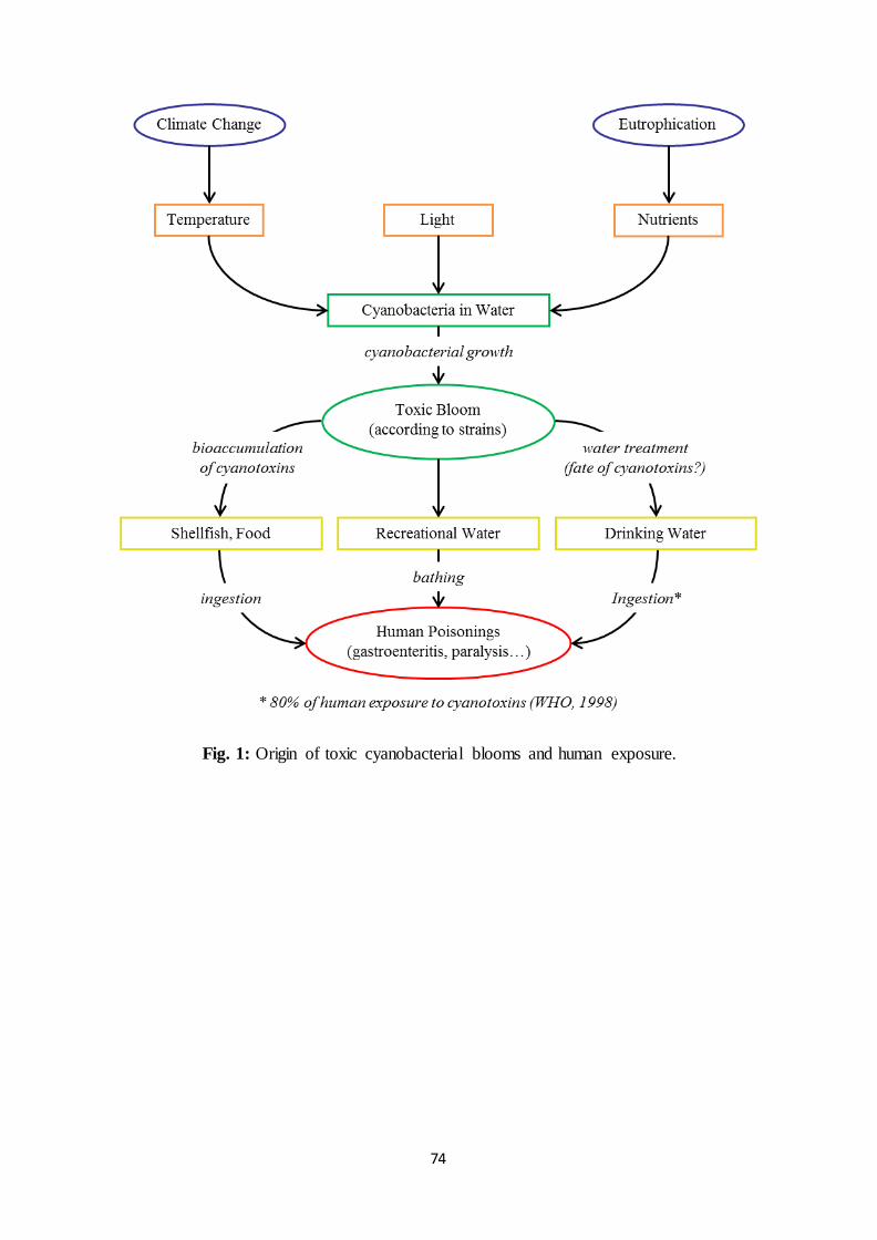
74
Fig. 1: Origin of toxic cyanobacterial blooms and human exposure.
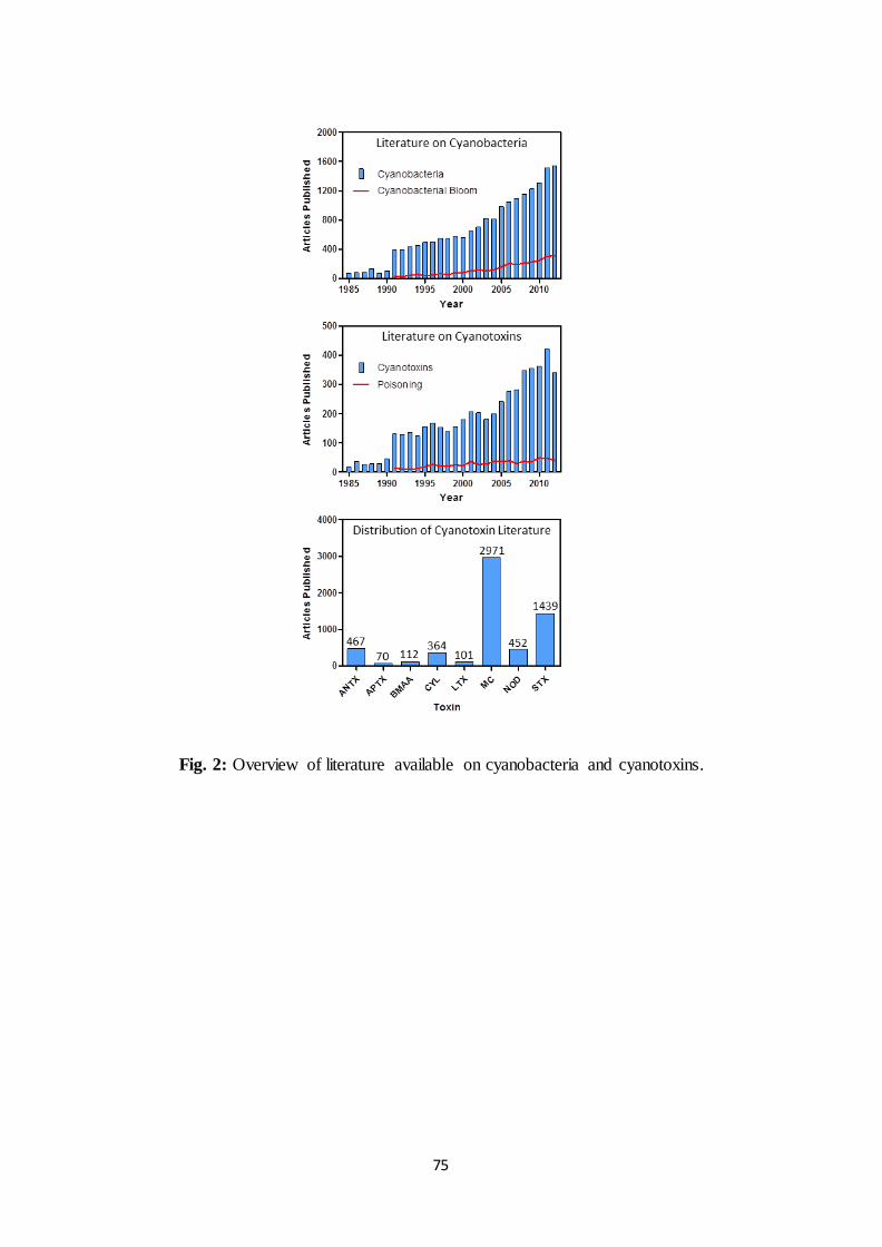
75
Fig. 2: Overview of literature available on cyanobacteria and cyanotoxins.
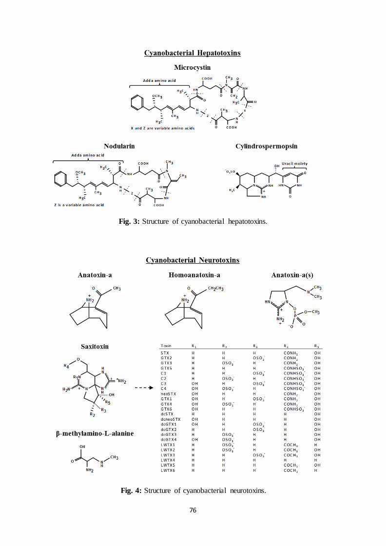
76
Fig. 3: Structure of cyanobacterial hepatotoxins.
Fig. 4: Structure of cyanobacterial neurotoxins.
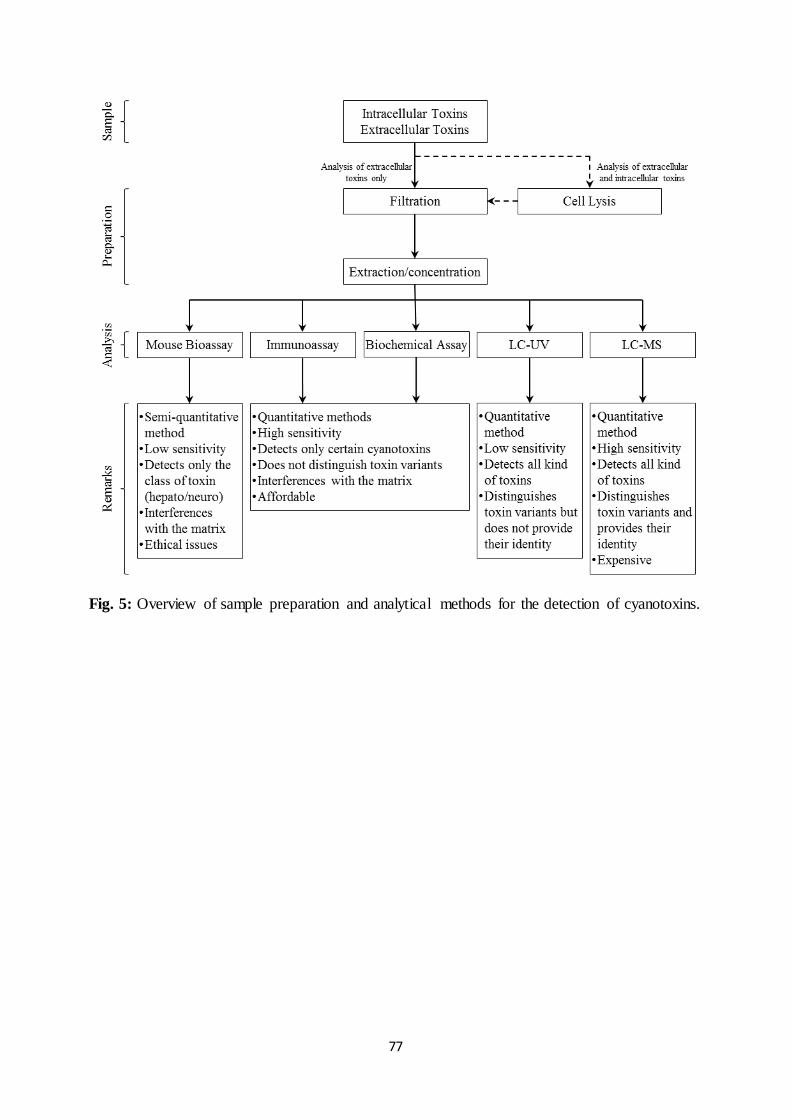
77
Fig. 5: Overview of sample preparation and analytical methods for the detection of cyanotoxins.
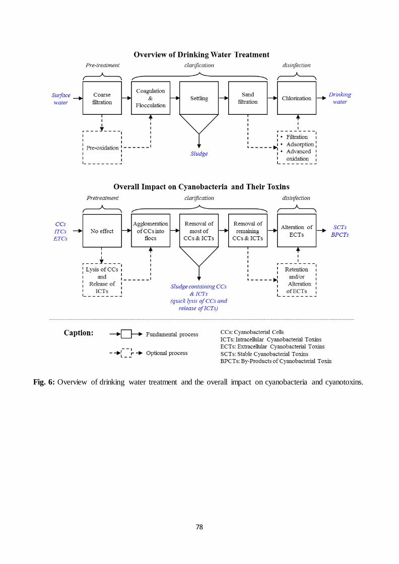
78
Fig. 6: Overview of drinking water treatment and the overall impact on cyanobacteria and cyanotoxins.
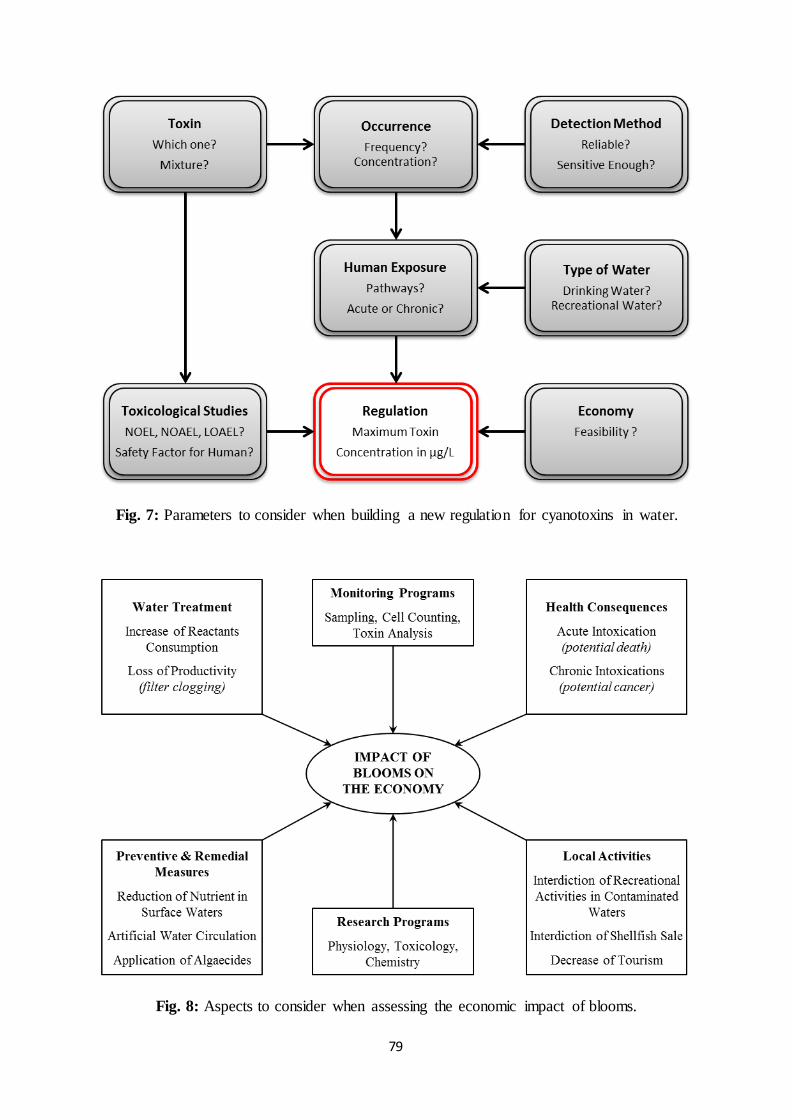
79
Fig. 7: Parameters to consider when building a new regulation for cyanotoxins in water.
Fig. 8: Aspects to consider when assessing the economic impact of blooms.
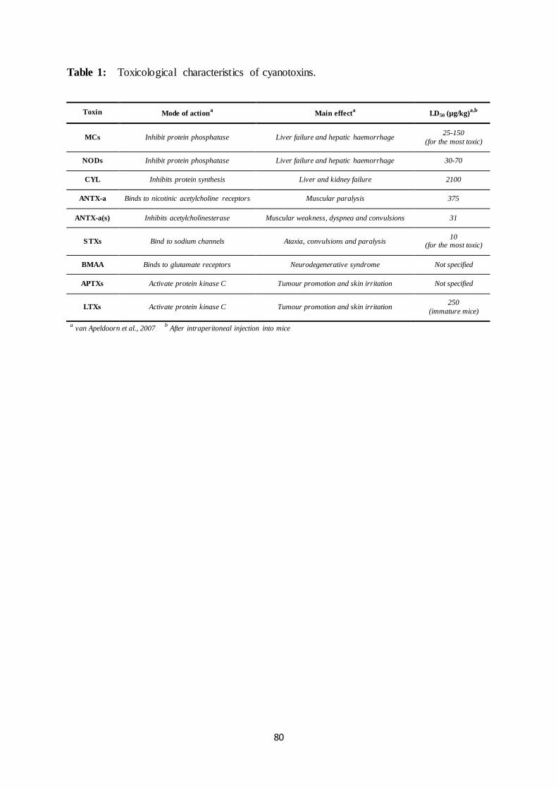
80
Table 1: Toxicological characteristics of cyanotoxins.
Toxin Mode of actiona Main effect
a LD50 (µg/kg)
a,b
MCs Inhibit protein phosphatase Liver failure and hepatic haemorrhage 25-150
(for the most toxic)
NODs Inhibit protein phosphatase Liver failure and hepatic haemorrhage 30-70
CYL Inhibits protein synthesis Liver and kidney failure 2100
ANTX-a Binds to nicotinic acetylcholine receptors Muscular paralysis 375
ANTX-a(s) Inhibits acetylcholinesterase Muscular weakness, dyspnea and convulsions 31
STXs Bind to sodium channels Ataxia, convulsions and paralysis 10
(for the most toxic)
BMAA Binds to glutamate receptors Neurodegenerative syndrome Not specified
APTXs Activate protein kinase C Tumour promotion and skin irritation Not specified
LTXs Activate protein kinase C Tumour promotion and skin irritation 250
(immature mice)
a van Apeldoorn et al., 2007
b After intraperitoneal injection into mice
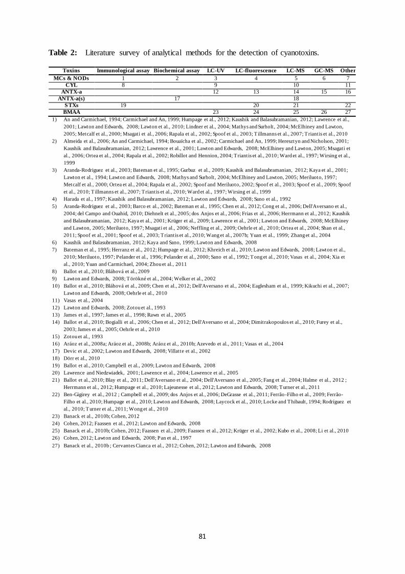
81
Table 2: Literature survey of analytical methods for the detection of cyanotoxins.
Toxins Immunological assay Biochemical assay LC-UV LC-fluorescence LC-MS GC-MS Other
MCs & NODs 1 2 3 4 5 6 7
CYL 8 9 10 11
ANTX-a 12 13 14 15 16
ANTX-a(s) 17 18
STXs 19 20 21 22
BMAA 23 24 25 26 27
1) An and Carmichael, 1994; Carmichael and An, 1999; Humpage et al., 2012; Kaushik and Balasubramanian, 2012; Lawrence et al.,
2001; Lawton and Edwards, 2008; Lawton et al., 2010; Lindner et al., 2004; Mathys and Surholt, 2004; McElhiney and Lawton,
2005; Metcalf et al., 2000; Msagati et al., 2006; Rapala et al., 2002; Spoof et al., 2003; T illmanns et al., 2007; Triantis et al., 2010
2) Almeida et al., 2006; An and Carmichael, 1994; Bouaïcha et al., 2002; Carmichael and An, 1999; Heresztyn and Nicholson, 2001;
Kaushik and Balasubramanian, 2012; Lawrence et al., 2001; Lawton and Edwards, 2008; McElhiney and Lawton, 2005; Msagati et
al., 2006; Ortea et al., 2004; Rapala et al., 2002; Robillot and Hennion, 2004; Triantis et al., 2010; Ward et al., 1997; Wirsing et al.,
1999
3) Aranda-Rodriguez et al., 2003; Bateman et al., 1995; Gurbuz et al., 2009; Kaushik and Balasubramanian, 2012; Kaya et al., 2001;
Lawton et al., 1994; Lawton and Edwards, 2008; Mathys and Surholt, 2004; McElhiney and Lawton, 2005; Meriluoto, 1997;
Metcalf et al., 2000; Ortea et al., 2004; Rapala et al., 2002; Spoof and Meriluoto, 2002; Spoof et al., 2003; Spoof et al., 2009; Spoof
et al., 2010; T illmanns et al., 2007; Triantis et al., 2010; Ward et al., 1997; Wirsing et al., 1999
4) Harada et al., 1997; Kaushik and Balasubramanian, 2012; Lawton and Edwards, 2008; Sano et al., 1992
5) Aranda-Rodriguez et al., 2003; Barco et al., 2002; Bateman et al., 1995; Chen et al., 2012; Cong et al., 2006; Dell'Aversano et al.,
2004; del Campo and Ouahid, 2010; Diehnelt et al., 2005; dos Anjos et al., 2006; Frias et al., 2006; Herrmann et al., 2012; Kaushik
and Balasubramanian, 2012; Kaya et al., 2001; Krüger et al., 2009; Lawrence et al., 2001; Lawton and Edwards, 2008; McElhiney
and Lawton, 2005; Meriluoto, 1997; Msagati et al., 2006; Neffling et al., 2009; Oehrle et al., 2010; Ortea et al., 2004; Shan et al.,
2011; Spoof et al., 2001; Spoof et al., 2003; Triantis et al., 2010; Wang et al., 2007b; Yuan et al., 1999; Zhang et al., 2004
6) Kaushik and Balasubramanian, 2012; Kaya and Sano, 1999; Lawton and Edwards, 2008
7) Bateman et al., 1995; Herranz et al., 2012; Humpage et al., 2012; Khreich et al., 2010; Lawton and Edwards, 2008; Lawton et al.,
2010; Meriluoto, 1997; Pelander et al., 1996; Pelander et al., 2000; Sano et al., 1992; Tong et al., 2010; Vasas et al., 2004; Xia et
al., 2010; Yuan and Carmichael, 2004; Zhou et al., 2011
8) Ballot et al., 2010; Bláhová et al., 2009
9) Lawton and Edwards, 2008; Törökné et al., 2004; Welker et al., 2002
10) Ballot et al., 2010; Bláhová et al., 2009; Chen et al., 2012; Dell'Aversano et al., 2004; Eaglesham et al., 1999; Kikuchi et al., 2007;
Lawton and Edwards, 2008; Oehrle et al., 2010
11) Vasas et al., 2004
12) Lawton and Edwards, 2008; Zotou et al., 1993
13) James et al., 1997; James et al., 1998; Rawn et al., 2005
14) Ballot et al., 2010; Bogialli et al., 2006; Chen et al., 2012; Dell'Aversano et al., 2004; Dimitrakopoulos et al., 2010; Furey et al.,
2003; James et al., 2005; Oehrle et al., 2010
15) Zotou et al., 1993
16) Aráoz et al., 2008a; Aráoz et al., 2008b; Aráoz et al., 2010b; Azevedo et al., 2011; Vasas et al., 2004
17) Devic et al., 2002; Lawton and Edwards, 2008; Villatte et al., 2002
18) Dörr et al., 2010
19) Ballot et al., 2010; Campbell et al., 2009; Lawton and Edwards, 2008
20) Lawrence and Niedzwiadek, 2001; Lawrence et al., 2004; Lawrence et al., 2005
21) Ballot et al., 2010; Blay et al., 2011; Dell'Aversano et al., 2004; Dell'Aversano et al., 2005; Fang et al., 2004; Halme et al., 2012 ;
Herrmann et al., 2012; Humpage et al., 2010; Lajeunesse et al., 2012; Lawton and Edwards, 2008; Turner et al., 2011
22) Ben-Gigirey et al., 2012 ; Campbell et al., 2009; dos Anjos et al., 2006; DeGrasse et al., 2011; Ferrão-Filho et al., 2009; Ferrão-
Filho et al., 2010; Humpage et al., 2010; Lawton and Edwards, 2008; Laycock et al., 2010; Locke and Thibault, 1994; Rodríguez et
al., 2010; Turner et al., 2011; Wong et al., 2010
23) Banack et al., 2010b; Cohen, 2012
24) Cohen, 2012; Faassen et al., 2012; Lawton and Edwards, 2008
25) Banack et al., 2010b; Cohen, 2012; Faassen et al., 2009; Faassen et al., 2012; Krüger et al., 2002; Kubo et al., 2008; Li et al., 2010
26) Cohen, 2012; Lawton and Edwards, 2008; Pan et al., 1997
27) Banack et al., 2010b ; Cervantes Cianca et al., 2012; Cohen, 2012; Lawton and Edwards, 2008
82
Table 3: Literature survey of common water treatment processes and the related impact on
cyanobacteria and their toxins.
P
reo
xid
ati
on
C
lari
fica
tio
n
Mem
bra
ne f
iltr
ati
on
A
dso
rpti
on
P
ho
to(c
ata
)ly
sis
Ozo
na
tio
n
Ch
lori
na
tio
n
Cy
an
ob
act
eri
a
Cel
l ly
sis1
To
xin
rel
ease
1
Cel
l re
mo
va
l2
No
to
xin
rel
ease
in
wa
ter3
To
xin
rel
ease
in
slu
dg
e4
Cel
l re
mo
va
l5
Lo
w c
ell
lysi
s5
N.A
.*
N.A
.*
N.A
.*
N.A
.*
MC
P
ote
nti
al
toxi
n a
lter
ati
on
**
No
eff
ect6
T
oxi
n r
emo
va
l7
To
xin
rem
ova
l8
To
xin
alt
era
tio
n9
To
xin
alt
era
tio
n1
0
To
xin
alt
era
tio
n1
1
NO
D
Po
ten
tia
l to
xin
alt
era
tio
n*
*
No
da
ta
To
xin
rem
ova
l12
No
da
ta
To
xin
alt
era
tio
n1
3
To
xin
alt
era
tio
n1
4
To
xin
alt
era
tio
n1
5
CY
L
Po
ten
tia
l to
xin
alt
era
tio
n*
*
No
da
ta
To
xin
rem
ova
l16
To
xin
rem
ova
l17
To
xin
alt
era
tio
n1
8
To
xin
alt
era
tio
n1
9
To
xin
alt
era
tio
n2
0
ST
X
Po
ten
tia
l to
xin
alt
era
tio
n*
*
No
da
ta
To
xin
rem
ova
l21
To
xin
rem
ova
l22
No
da
ta
Po
or
toxi
n a
lter
ati
on
23
To
xin
alt
era
tio
n2
4
AN
TX
-a
Po
ten
tia
l to
xin
alt
era
tio
n*
*
No
da
ta
To
xin
rem
ova
l25
To
xin
rem
ova
l26
To
xin
alt
era
tio
n2
7
To
xin
alt
era
tio
n2
8
No
eff
ect2
9
Oth
er
tox
ins
No
da
ta
No
da
ta
No
da
ta
No
da
ta
No
da
ta
No
da
ta
No
da
ta
1
) D
aly
et
al.,
20
07
; H
all et
al.
, 200
0; H
oeg
er e
t al
., 2
00
2;
Lam
et
al.,
19
95
; M
a et
al.
, 2
01
2;
Mia
o a
nd
Tao
, 2
00
9;
Pet
erso
n e
t al
., 1
99
5;
Zam
yad
i et
al.
, 2
01
0;
Zam
yad
i et
al.
, 2
01
3
2)
Ch
ow
et
al.,
19
99
; D
rik
as e
t al
., 2
001
; H
all et
al.
, 200
0; H
o e
t al
., 2
012
c;
Lam
et
al.,
19
95
; M
a et
al.
, 2
01
2; P
iets
ch e
t al
., 2
00
2; Sun
et
al.,
20
12; T
eixei
ra a
nd R
osa
, 200
6a;
Tei
xei
ra e
t al
., 2
01
0;
Vel
zebo
er
et a
l.,
19
95
3)
Ch
ow
et
al.,
19
98
; C
how
et
al.,
1999
; D
rikas
et al
., 2
00
1; H
all et
al.
, 200
0; P
eter
son e
t al
., 1
995
; Sun
et
al.,
20
12
4)
Dri
kas
et
al.,
20
01
; H
o e
t al
., 2
01
2c;
Sun
et
al.,
20
12
5)
Cam
pin
as a
nd R
osa
, 2
01
0b;
Ch
ow
et
al.,
19
97
; G
ijsb
erts
en-A
bra
ham
se e
t al
., 2
00
6
6)
Dri
kas
et
al.,
20
01
; H
all
et a
l.,
20
00
; H
imber
g e
t al
., 1
98
9
7)
Cam
pin
as a
nd R
osa
, 20
10a;
Dix
on e
t al
., 2
010
; D
ixo
n e
t al
., 2
01
1a;
Dix
on
et
al.,
20
11
b;
Gij
sber
tsen
-
Abra
ham
se e
t al
., 2
00
6;
Hal
l et
al.
, 2
00
0;
Law
ton
an
d R
ober
tso
n,
19
99
; L
ee a
nd W
alk
er,
20
08
; N
eum
ann
an
d W
eckes
ser,
199
8; P
ante
lić
et a
l.,
20
13
; Tei
xei
ra a
nd R
osa
, 2
00
5;
Tei
xei
ra a
nd R
osa
, 2
00
6b;
Tei
xei
ra a
nd R
osa
, 2
00
6c;
Wes
tric
k e
t al
., 2
01
0
8)
Cam
pin
as a
nd R
osa
, 20
06; C
amp
inas
an
d R
osa
, 2010
a; D
elgad
o e
t al
., 2
01
2; D
onat
i et
al.
, 1
99
4;
Hal
l et
al.
, 20
00; H
imber
g e
t al
., 1
98
9; H
o e
t al
., 2
01
1; H
uan
g e
t al
., 2
00
7; L
amber
t et
al.
, 199
6; L
awto
n a
nd
Ro
ber
tso
n, 1
999
; N
ewco
mbe
et a
l.,
20
03
; N
ewco
mbe
and N
ich
ols
on
, 2
00
4;
Pan
teli
ć et
al.
, 2
01
3;
Pen
dle
ton
et al
., 2
001
; So
rlin
i an
d C
oll
ivig
nar
elli, 2
01
1;
Wan
g e
t al
., 2
00
7a;
War
hurs
t et
al.,
19
97
; W
estr
ick
et
al.,
20
10
9)
An
ton
iou e
t al
., 2
00
8;
Gra
ham
et
al.,
20
10
; H
e et
al.
, 2
01
2;
Kay
a an
d S
ano
, 1
99
8;
Law
ton
an
d
Ro
ber
tso
n, 1
999
; L
awto
n e
t al
., 1
99
9; L
iu e
t al
., 2
003; L
iu e
t al
., 2
00
9; P
ante
lić
et a
l.,
20
13
; Pel
aez
et
al.,
20
10
; P
elae
z et
al.
, 2012
a; P
elae
z et
al.
, 201
2b; Q
iao
et al
., 2
005
; R
ober
tso
n e
t al
., 2
012; Sh
arm
a et
al
., 2
01
2;
Sh
eph
ard e
t al
., 1
99
8;
Sh
eph
ard e
t al
., 2
00
2;
Tsu
ji e
t al
., 1
99
5;
Yan
g e
t al
., 2
01
1
10
) A
l M
om
ani et
al.
, 2008
; A
l M
om
ani an
d J
arra
h, 2
010; B
rook
e et
al.
, 20
06; H
all et
al.
, 20
00;
Him
ber
g
et a
l., 1
98
9; H
oeg
er e
t al
., 2
002
; L
awto
n a
nd R
ober
tso
n,
19
99
; M
iao
an
d T
ao,
20
09
; M
iao
et
al.,
2
01
0; N
ewco
mbe
et a
l., 2
003
; N
ewco
mbe
and N
icho
lson
, 20
04; O
nst
ad e
t al
., 2
00
7;
Pan
teli
ć et
al.
,
20
13
; Ro
drí
guez
et
al.,
20
07
a; R
osi
tan
o e
t al
., 1
99
8;
Ro
sita
no
et
al.,
20
01
; Sh
arm
a et
al.
, 2
01
2;
Sh
aww
a an
d S
mit
h,
20
01
; W
estr
ick
et
al.,
20
10
11
) A
cero
et
al.,
200
5; A
cero
et al
., 2
00
8; D
aly e
t al
., 2
007; H
all et
al.
, 20
00
; Ho
et
al.,
20
06
b;
Huan
g e
t al
., 2
00
8; Ji
et
al.,
200
8; K
ull
et
al.,
2004
; K
ull
et
al.,
20
06; L
awto
n a
nd R
ober
tso
n, 1
999
; M
erel
et al
., 2
00
9; M
erel
et
al.,
2010
a; N
ewco
mbe
and N
ich
ols
on, 2
004
; N
ich
ols
on
et
al.,
19
94
; P
ante
lić
et a
l.,
20
13
; Ro
drí
guez
et
al.,
200
7a;
Ro
drí
guez
et al
., 2
00
8; Sen
ogle
s-D
erham
et
al.,
20
03
; Sh
arm
a et
al.
, 2
01
2; Sh
i et
al.
, 2
00
5;
So
rlin
i an
d C
oll
ivig
nar
elli
, 2
01
1;
Tri
anti
s et
al.
, 2
01
2;
Tsu
ji e
t al
., 1
99
7;
Wes
tric
k e
t al
., 2
010
; X
agora
raki et
al.
, 20
06; Z
amyad
i et
al.
, 201
2; Z
amyad
i et
al.
, 2013
; Z
ong e
t al
.,
20
13
12
) L
awto
n a
nd R
ober
tso
n,
19
99
; V
uo
ri e
t al
., 1
99
7
13
) L
iu e
t al
., 2
00
5;
Ro
ber
tso
n e
t al
., 2
01
2;
Tw
ist
and C
odd,
19
97
14
) R
osi
tan
o e
t al
., 1
99
8
15
) M
erel
et
al.,
20
10
a;
Nic
ho
lso
n e
t al
., 1
99
4;
Pan
teli
ć et
al.
, 2
01
3
16
) D
ixo
n e
t al
., 2
01
0;
Dix
on
et
al.,
20
11
b
17
) D
elgad
o e
t al
., 2
01
2; H
o e
t al
., 2
00
8; H
o e
t al
., 2
011
; N
ewco
mbe
and N
icho
lson
, 20
04; P
ante
lić
et a
l.,
20
13
; Wes
tric
k e
t al
., 2
01
0
18
) P
elae
z et
al.
, 2
01
2b;
Ro
ber
tso
n e
t al
., 2
01
2;
Sen
ogle
s et
al.
, 2
00
0a;
Sen
ogle
s et
al.
, 2
00
1
19
) N
ewco
mbe
and N
ich
ols
on, 2
00
4; O
nst
ad e
t al
., 2
00
7;
Pan
teli
ć et
al.
, 2
01
3; R
odrí
guez
et
al.,
2
00
7a ;
Wes
tric
k e
t al
., 2
01
0
20
) B
ank
er e
t al
., 2
00
1; H
o e
t al
., 2
008
; M
erel
et
al.,
20
10
a; M
erel
et
al.,
20
10
b;
New
com
be
and
Nic
ho
lso
n,
20
04
; P
ante
lić
et a
l.,
20
13
; Ro
drí
guez
et
al.,
20
07
a; R
odrí
guez
et
al.,
20
07
b;
Sen
ogle
s et
al.
, 20
00
b; Sen
ogle
s-D
erham
et
al.,
20
03
; W
estr
ick
et
al.,
20
10
; Z
amy
adi
et a
l.,
20
12
21
) L
ayco
ck e
t al
., 2
01
2;
Wes
tric
k e
t al
., 2
01
0
22
) D
elgad
o e
t al
., 2
01
2;
New
com
be
and N
ich
ols
on
, 2
00
4;
Orr
et
al.,
20
04
; Sh
i et
al.
, 2
01
2;
Wes
tric
k e
t al
., 2
01
0
23
) N
ewco
mbe
and N
ich
ols
on
, 2
00
4;
Orr
et
al.,
20
04
; R
osi
tan
o e
t al
., 2
00
1
24
) M
erel
et
al.,
20
10
a; N
ewco
mbe
and N
ich
ols
on, 2
00
4;
Nic
ho
lso
n e
t al
., 2
00
3;
Pan
teli
ć et
al.
, 2
01
3; S
eno
gle
s-D
erham
et al
., 2
003; W
estr
ick e
t al
., 2
010; Z
amyad
i et
al.
, 201
0; Z
amy
adi et
al.,
2
01
2
25
) G
ijsb
erts
en-A
bra
ham
se e
t al
., 2
00
6;
Tei
xei
ra a
nd R
osa
, 2
00
6b
; W
estr
ick
et
al.,
20
10
26
) H
all
et a
l.,
20
00
; N
ewco
mbe
and N
ich
ols
on
, 2
00
4
27
) A
fzal
et
al.,
20
10
28
) A
l M
om
ani,
20
07;
Hal
l et
al.
, 2
00
0;
New
com
be
and N
ich
ols
on
, 2
00
4;
On
stad
et
al.,
20
07
; P
ante
lić
et a
l.,
20
13
; Ro
sita
no
et
al.,
19
98
; R
osi
tan
o e
t al
., 2
00
1;
Ro
drí
guez
et
al.,
20
07
a;
Wes
tric
k e
t al
., 2
01
0
29
) H
all et
al.
, 20
00; M
erel
et al
., 2
010
a; N
ewco
mbe
and N
ich
ols
on, 2
00
4; R
odrí
guez
et al
., 2
00
7a;
R
odrí
guez
et
al.,
20
07
b;
Wes
tric
k e
t al
., 2
01
0
*
Cy
ano
bac
teri
a ar
e n
ot li
kel
y to
rea
ch t
hes
e tr
eatm
ents
sin
ce t
hey
are
pre
vio
usl
y r
emo
ved
by
cl
arif
icat
ion
**
A
s a
fun
ctio
n o
f: r
aw
wat
er q
ual
ity, t
he
amo
unt o
f o
xid
izin
g a
gen
t, t
he
reac
tiv
ity
of
the
tox
in
wit
h c
hlo
rin
e o
r o
zon
e
83
Table 4: Overview of existing regulation on cyanotoxins in drinking water and recreational
water (Chorus, 2012).
M
Cs
NO
D
CY
L
AN
TX
-a
Hom
oA
NT
X-a
A
NT
X-a
(s)
ST
X
WH
O
1µ
g/L
-
- -
- -
-
Au
stra
lia
1.3
µg/L
(1
0µ
g/L
) -
1µ
g/L
-
- -
3µ
g/L
Bra
zil
1µ
g/L
-
15µ
g/L
-
- -
3µ
g/L
Can
ada
1.5
µg/L
* (2
0µ
g/L
) -
- 3.7
µg/L
-
- -
Czech
Repu
bli
c 1µ
g/L
*
- -
- -
- -
Fra
nce
1µ
g/L
(2
5µ
g/L
) -
- -
- -
-
Germ
an
y
1µ
g/L
(1
0µ
g/L
) -
0.1
µg/L
-
- -
-
Hu
ngary
-
(
20µ
g/L
) -
- -
- -
-
Italy
-
(
25µ
g/L
) -
- -
- -
-
Neth
erl
an
d
1µ
g/L
(2
0µ
g/L
)
New
Zeala
nd
1µ
g/L
(1
2µ
g/L
) 1µ
g/L
1µ
g/L
6µ
g/L
2µ
g/L
1µ
g/L
3µ
g/L
Sin
gapore
1µ
g/L
*
- -
- -
- -
Spain
1µ
g/L
-
- -
- -
-
Tu
rkey
1µ
g/L
(2
5µ
g/L
) -
- -
- -
-
Uru
gu
ay
1µ
g/L
-
- -
- -
-
Sou
th A
fric
a
1µ
g/L
-
- -
- -
-
US
A
- -
- -
- -
-
* T
he
valu
e applies
to M
C-L
R o
nly
( )
The
valu
e bet
wee
n p
are
nth
esis
ref
er to r
ecre
ational
wate
r