Embed Size (px)
Citation preview

Stathmin Overexpression Inhibits Megakaryocyte Maturation and Platelet
Production
David C Gajzer1, M.D. Camelia Iancu-Rubin2, Ph.D.
1Cardiovascular Research Center, Department of Medicine and 2Division of Hematology and Medical Oncology, Department of Medicine and The Tisch Cancer Institute, Mount Sinai School of Medicine, New York
Original research article was published as “Stathmin downregulation is required for megakaryocyte maturation and platelet production” - Blood, 117(17):4580-9 (2011)
Iancu-Rubin C1, Gajzer D1, Tripodi J1, Najfeld V1,2, Gordon RE2, Hoffman R1,3 and Atweh GF4
1 Division of Hematology and Medical Oncology, Department of Medicine and The Tisch Cancer Institute, Mount Sinai School of Medicine, New York, NY 2 Department of Pathology, Mount Sinai School of Medicine, New York, NY 3 Department of Gene and Cell Medicine, Mount Sinai School of Medicine, New York, NY4 Division of Hematology/Oncology, Department of Medicine and UC Cancer Institute, University of Cincinnati College of Medicine, Cincinnati, OH

Background and Rationale
MegakaryocytopoiesisThe maturation process of the hematopoietic progenitor cells that go on to form blood platelets
Microtubules in megakaryocytopoiesisMicrotubules help maintain cell structure, provide platforms for intracellular transport and form the spindle during mitosis. In megakaryocytes, they make up the multipolar mitotic spindle and thus enable endomitosis (a form of abortive mitosis, leading to >4N DNA content) (Nagata et al., 1997, Vitrat et al. 1998)
Proplatelet extension and platelet formation In megakaryocytes, microtubules provide the structural scaffold required for extension of proplatelets and ensure the transport of cytoplasmic organelles and specific granules into nascent platelets (Italiano JE et al., 1999; Richardson JL et al., 2005)

Stathmin
a/b tubulinheterodimers
Promotes microtubule
depolymerization
Sequesters tubulin heterodimers
and prevents polymerization
• Stathmin is a microtubule-regulatory protein that destabilizes microtubules and regulates microtubule dynamics. It is important for cellular proliferation and cell motility. Interfering with its expression results in abnormal mitotic spindles and aberrant exit from mitosis
• All cell types express stathmin in their proliferative stages but, with the exception of neurons, they do not express stathmin in the mature stages
• Stathmin is overexpressed in a wide variety of solid tumors and hematological malignancies

Stathmin Expression During Megakaryocyte Maturation
• In murine MK and in human leukemic cell lines with MK-like features (K562 and HEL), stathmin is downregulated during differentiation and its level of expression correlates inversely with the level of ploidy (Chang et al. 2001, Iancu-Rubin et al. 2003)
• Alterations of stathmin expression in these cell lines are associated with abnormal mitotic spindles and changes in their propensity to become polyploid (Iancu-Rubin et al. 2003, 2005)
• Stathmin is expressed at very low levels in normal bone marrow megakaryocytes and is not found in platelets (Brattsand G. 1993, Rowlands DC, 1995, Iancu-Rubin et al. 2003)

Is Stathmin Important For Megakaryocytopoiesis and Platelet
Production?
HypothesisHypothesis
Stathmin may be important in primary megakaryocytopoiesis where its expression may be necessary for proliferation of megakaryocyte progenitors in the early stages while its suppression may be required for megakaryocyte maturation in the later stages.
In our report, we assessed the influence of stathmin expression on human primary MK development and identified a role for stathmin in MK maturation and platelet formation.
Methods
• Primary megakaryocyte culturesTwo-step liquid culture system:Human peripheral blood (PB)-derived CD34+ hematopoietic stem cells were cultured in a two-step liquid culture system for 14 days. hSCF and hTPO were added to the culture medium for the first six days, then cells were cultured in the absence of hSCF for an additional 7-10 days.Semi-solid collagen-based cultures:PB-derived CD34+ cells were cultured in semi-solid collagen-based cultures in MegaCult medium supplemented with hTPO, hIL-3, hIL-6 for 16 days until microscopic analysis.
• FIV-based vectors and lentiviral transductionsSTMN-WT or STMN-4A stathmin cDNA was excised from pTRE2-STMN plasmids and cloned into FIV-based pCDF1-MCS2-EF1-copGFP plasmid backbone. STMN-4A cDNA was generated by site-directed mutagenesis which replaced serine for alanine residues at the four phosphorylation sites (4A). An empty control vector expressing only copGFP was used as a control. The transfer vectors were co-transfected with pCPR-ΔEnv and pCI-VSV plasmids into the 293T packaging cell line using lipofectamine 2000. Viral supernatants were harvested on day 3 and viral titers determined by flow cytometric analysis. Successful packaging and viral functionality was confirmed by transduction of HEK293 cells and subsequent gene and protein expression analysis.
• Gene expression analysisTotal RNA was purified using the RNeasy purification kit (Qiagen, MD). 1 μg total RNA was used for cDNA synthesis with the Omniscript kit (Qiagen, MD). One tenth of the cDNA was used for quantitative real-time PCR on a RealPlex MasterCycler (Eppendorf, NY). Primers used were specific for human stathmin, GATA-1 and platelet factor 4 (SABiosciences, MD). cDNA templates were mixed with 1X IQ SYBR Green Supermix (Bio-Rad Laboratories, CA) and 0.2 mM of each primer in a total volume of 50 μl in PCR plates (Fisher Scientific, PA) in duplicates.

Methods
•Protein expression analysisCells were lysed (50 mM Tris-Cl, 15 mM NaCl, 1% Triton-X, 40 mg/ml protease inhibitor cocktail, Roche Molecular Biochemicals, IN), then 50 μg of protein were separated on 12.5% SDS-PAGE gels and transferred to a polyvinylidene difluoride (PVDF) membrane. Polyclonal anti-stathmin antibodies (Calbiochem, CA) and goat anti-rabbit IgG-horseradish peroxidase conjugated (HRP) secondary antibodies (Pierce, IL) were used to detect stathmin. Monoclonal anti-actin antibodies (Oncogene Research Products, MA) and goat anti-mouse IgM-HRP secondary antibodies (Calbiochem, CA) were used to detect actin. The proteins were visualized by enhanced chemiluminescence detection (ECL, Amersham Pharmacia Biotech, NJ).
•Microscopic analysisCells were placed on slides using a Shandon centrifuge (Life Sciences International, England), fixed in methanol for 5 minutes and stained with Wright-Giemsa (Sigma, MO) for 20 minutes. MegaCult cultures were fixed and stained using the manufacturer’s staining kit (StemCell Technologies, Canada). Immunofluorescence labeling of cells fixed in 4% paraformaldehyde was performed using phycoerythrin (PE)-conjugated anti-CD41 antibodies (Becton-Dickinson, CA) and 1.5 μM Hoechst 33342 (Sigma, MO) for nuclear staining. The slides were mounted with Vectashield mounting solution (Vector Laboratories, CA) and analyzed using a Zeiss Axiophot 2 fluorescence microscope using a 63X/1.25 oil objective. Image acquisition was performed using a Hammamatsu Orca CCD camera and OpenLab software.
•Flow cytometric analysisCultured cells were labeled with PE-conjugated anti-CD41 antibodies and allophycocyanin (APC)-conjugated anti-CD42b antibodies (Becton-Dickinson, CA ) for 45 minutes then incubated with 5 μl 7-aminoactinomycin (7-AAD) for 15 additional minutes. The data was acquired and analyzed using a FACSCanto II flow cytometer and FACS Diva software (Becton-Dickinson, CA).

Methods
•Analysis of culture-derived plateletsMK cultures were harvested and centrifuged at 800 rpm for 5 minutes and supernatants were collected and further centrifuged at 3500 rpm for 10 minutes. The pellet containing culture-derived platelets was re-suspended in phosphate buffered saline (PBS) with 0.1% fetal bovine serum (FBS) and labeled with CD41-PE and CD42-APC antibodies for 30 minutes at room temperature. Reticulated platelets were identified following incubation with 2 μg/ml thiazole orange (TO) (Sigma, MO) for additional 15 minutes at room temperature in the dark. The cells were then washed, re-suspended in PBS with 0.1% FBS and analyzed by flow cytometry. PB-derived human platelets were immunolabeled in the same manner and used as control.
•Fluorescence in situ hybridization (FISH)Uninfected and lentivirus-infected MK were fixed then labeled with two different DNA probes, the alpha satellite sequence of the centromeric region of chromosome X (Xp11.1-q11.1), and the satellite III sequence of chromosome Y (Yq12). These DNA Probes were obtained from Abbott Molecular (Des Plaines, IL) and labeled with SpectrumOrange and SpectrumGreen, respectively. FISH probe hybridization signals were visualized using an Axioplan 2 fluorescence microscope (Zeiss, Germany) at 100X magnification and imaged with CytoVision software (Genetix Corp., CA).
•Statistical analysisData are expressed as means +SD and analyzed by using student’s unpaired t-test. P values of < 0.05 were considered statistically significant.
Results
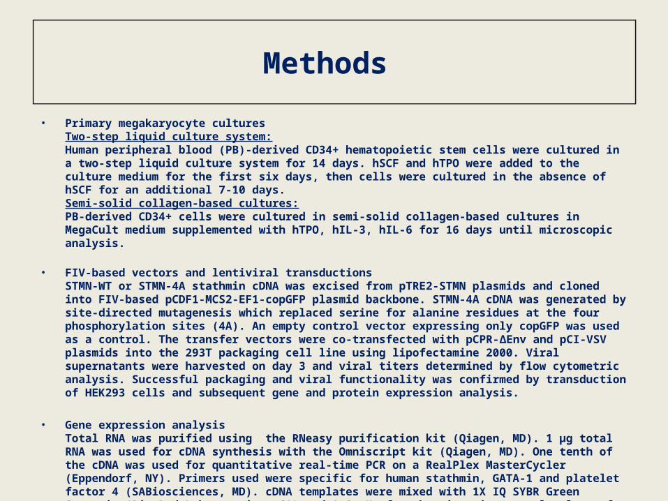
• Characterization of primary MK culturesBy the end of the maturation step (day 14 in liquid culture), 80-90% of cells were MK, out of which 40-50% were mature MK.
MK
PTL
CD42
bCD
42b
MK
PTL
GPIIb/IIIa Staining
Wright-Giemsa Staining
Flow Cytometric Analysis of Cultured Cells

Results
• Stathmin expression is downregulated during ex vivo megakaryocytopoiesis

Results
• Lentiviral-mediated forced stathmin expression prevents physiological stathmin downregulation in maturing megakaryocytes
After confirming physiological downregulation of stathmin during megakaryocytopoiesis, cells cultured in our two-step liquid culture system were transduced with either FIV-GFP (empty control), FIV-STMN or FIV-STMN4A. FIV-STMN4A, expressing mutant non-phosphorylatable stathmin, was used because it was shown previously that ectopically-expressed wild-type stathmin activity is often lost due to phosphorylation by cellular kinases. Use of the mutant form of stathmin allowed for analysis of the effects of a constitutively active stathmin protein on microtubules.

ResultsSS
C
GFP
CD42
b-AP
C
CD41-PE
Uninfected cells were used as negative controls to set a gate for detection of infected cells based on GFP fluorescence. Subsequently, the fraction of CD41+/CD42b+ cells within the GFP+ population was quantified.
Expression of wild-type stathmin did not significantly alter CD41/CD42b express-ion while expression of the constitutively active form of stathmin resulted in over 50% reduction in the fraction of mature MK.
• Sustained stathmin expression inhibits megakaryocyte maturation
Results
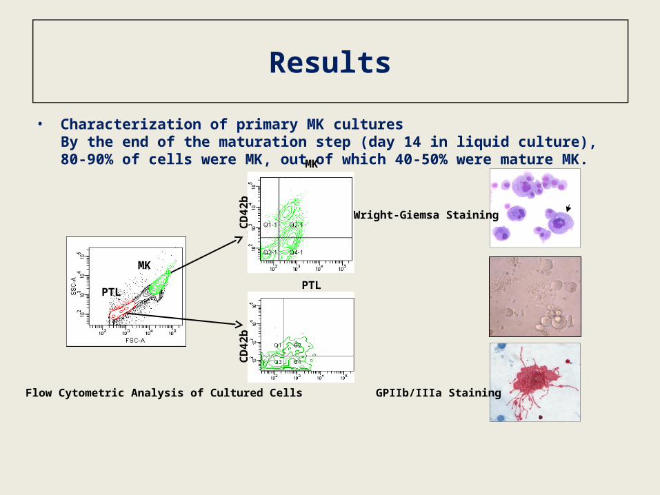
• Sustained stathmin expression is associated with low GATA-1 and PF-4 expression
PF4 expression was barely detectable in CD34+ cells whereas it was markedly up-regulated in mature MK infected with the control lentiviral vector. A small but not statistically significant increase in PF4 expression was observed in MK that were transduced with the wild-type lentiviral vector. However, up-regulation of PF4 was not observed in MK transduced with the lentivirus that encodes the constitutively active form of stathmin.
Increased GATA-1 levels were detected in mature MK transduced with control lentiviruses compared to primary CD34+ cells. MK transduced with lentiviruses expressing wild-type stathmin were also characterized by up-regulation of GATA-1 expression. Up-regulation of GATA- 1 was not observed in MK transduced with lentiviruses expressing the constitutively active form of stathmin.

Results
2N 32N16N8N4N
• Sustained stathmin expression inhibits polyploidization
A typical human cell with 2N DNA content (i.e. diploid) has one copy of each chromosome X and Y (or two copies of an X-chromosome if the cell originated from a female), a cell with 4N DNA content (i.e. tetraploid) has two copies of each chromosome, a cell with 8N DNA has four copies of each chromosome and so on. Representative examples of MK with ploidy levels ranging from 2N to 32N are presented. An increase in ploidy levels correlates with the nuclear complexity detected by DAPI staining (blue fluorescence) and with nuclear size. In order to assess the effects of stathmin on the generation of MK with high ploidy levels, we analyzed by FISH cells with ≥8N DNA and categorized them in three ploidy classes based on the number of X and Y chromosome copies. Sustained stathmin expression was associated with a decrease in the fraction of MK with 16N and 32N DNA in both wild type- and constitutively active stathmin-expressing cultures. This was accompanied by an accumulation of MK with lower ploidy (i.e. 8N DNA content). These results indicate that sustained expression of stathmin impaired the ability of MK to achieve high ploidy levels.

Results
• Sustained stathmin expression interferes with platelet production by primary MK
Following transduction with control or stathmin-expressing lentiviruses, MK cultures were inspected by microscopy to visualize proplatelet formation. By day 14, control cultures contained large MK displaying long cytoplasmic extensions resembling proplatelets. The frequency of proplatelet-bearing MK was much lower in the cultures infected with stathmin expressing lentiviruses. Because the number of platelets derived in culture is directly proportional to the number of proplatelets formed, we collected and labeled culture-derived platelets from MK transduced with control or stathmin-expressing lentiviruses and quantified them by flow cytometry. Platelets derived from uninfected MK cultures were used as negative control for GFP expression. As illustrated above, platelet-sized particles derived from lentivirus infected cultures are GFP+, indicating that they were derived from the GFP positive MK. From the platelet-sized GFP+ events, we quantified only those expressing CD41. The results presented in the bar graph show a two-fold reduction in the fraction of platelets derived in vitro from MK expressing wild-type or phosphorylation-deficient stathmin as compared to those produced by control MK. This finding indicates that sustained stathmin expression has a negative effect on in vitro platelet production, suggesting that alterations of expression of an MT-regulatory protein can interfere with platelet production by primary MK.
PTLFS
C
GFP CD41
CD42
b
Conclusions
• Downregulation of stathmin is required for normal megakaryocytopoiesis
• Sustained levels of active stathmin inhibit megakaryocyte maturation, the ability to achieve high ploidy and the capacity to generate platelets in vitro
• The results of this study validate the importance of microtubules for megakaryocytopoiesis
• Dysregulation of stathmin might contribute to alterations of the megakaryocyte lineage in hematological malignancies such as leukemia and myelodysplastic syndrome which express very high levels of stathmin: deficient platelet production and bleeding from low platelet counts is one of the most common causes of death in these patients
• This study supports the development of small stathmin-like molecules capable of targeting megakaryocyte-specific b1-tubulin to limit excessive thrombocytosis. As it accounts for more than 90% of platelet microtubules and its expression is restricted to the MK lineage, the β1-isoform of tubulin may be an excellent therapeutic target for disrupting microtubule function exclusively in megakaryocytes


![How Does Stathmin Destabilize Microtubules? · shrink continually in the living cells qua the harmony between these processes is vital for normal cell function [1-5]. Microtubules](https://img.pdfslide.net/doc/110x75/5ad8d6847f8b9a32618e1199/how-does-stathmin-destabilize-microtubules-continually-in-the-living-cells-qua.jpg)