Embed Size (px)
Citation preview
Nuclear Instruments and Methods in Physics Research A 649 (2011) 15–18
Contents lists available at ScienceDirect
Nuclear Instruments and Methods inPhysics Research A
0168-90
doi:10.1
n Corr
E-m
journal homepage: www.elsevier.com/locate/nima
Status of the Center for Advanced Microstructures and Devices(CAMD)—2010
Amitava Roy n, Eizi Morikawa, Henry Bellamy, Challa Kumar, Jost Goettert, Victor Suller,Kevin Morris, Richard Kurtz, John Scott
J. Bennett Johnston, Sr., Center for Advanced Microstructures and Devices, Louisiana State University, Baton Rouge, LA 70806, USA
a r t i c l e i n f o
Available online 30 December 2010
Keywords:
Synchrotron radiation
Instrumentation
Accelerator
02/$ - see front matter & 2010 Elsevier B.V. A
016/j.nima.2010.11.132
esponding author.
ail address: [email protected] (A. Roy).
a b s t r a c t
The J. Bennett Johnston, Sr., Center for Advanced Microstructures and Devices (CAMD) is a 1.3 GeV
synchrotron-radiation facility owned and operated by the State of Louisiana. Fifteen beamlines provide
radiation for CAMD users and cover the spectral range from the far IR to X-rays of ca. 40 keV. Eleven of
them receive radiation from bending magnets and four from a 7 T wavelength shifter. A wide range of
basic and applied scientific experiments as well as microfabrication are performed at these beamlines.
The nanomaterial synthesis and characterization laboratory at CAMD continues to add new instru-
ments such as SQUID magnetometer (Quantum Deign MPMS XL5) and high precision microfluidic-
based nanomaterials synthesis equipment complementing already available facilities. We have recently
received NSF MRI funding for a multipole 7.5 T wiggler that will become operational in 2012. Generous
equipment donations from the University of California at Riverside (Professor Jory Yarmoff) and the
University of Bonn (ELSA facility) will provide users with two additional VUV beamlines in the near
future.
& 2010 Elsevier B.V. All rights reserved.
1. Introduction
The J. Bennett Johnston, Sr., Center for Advanced Microstructuresand Devices (CAMD) /http://www.camd.lsu.eduS has been operatingas a synchrotron-light source since 1992. CAMD provides infrastruc-ture for research and education in synchrotron-based science andtechnology, principally focusing on materials science, microfabrica-tion, nano-fabrication and environmental and biological sciences.Researchers from Louisiana university as well as from national andinternational institutions use the beamlines and the laboratories atCAMD. The facility is open to all users and is free of charge for non-proprietary research. CAMD is owned by the State of Louisiana; theoperational budget is provided by Louisiana State University (LSU).CAMD has developed into a strong regional center for the use ofsynchrotron radiation. In 2009 CAMD had 275 users from 23 statesand many other countries.
2. Accelerator operation and recent upgrades
The CAMD light source was conceived as a second generationfacility of moderate brightness, principally for X-ray-lithographic
ll rights reserved.
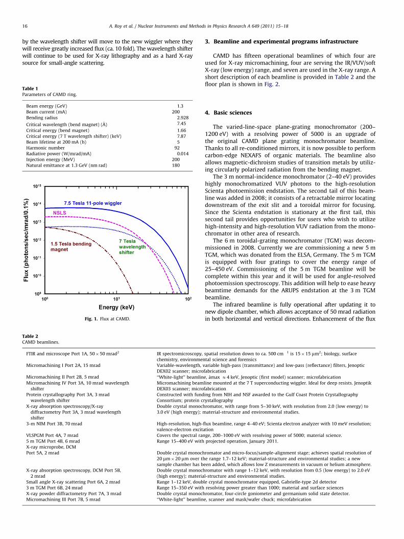
applications. The characteristics of the ring are shown in Table 1and the flux is shown in Fig. 1. Since 2003 CAMD has operated at1.3 GeV although energies up to 1.5 GeV are possible. A 7 Twavelength-shifter was installed in 1998 in one of the twoavailable straight sections. Due to budget constraints the ringpresently operates weekdays, 14 h per day except for two days ofmaintenance and studies every two weeks. The beam lifetime ispresently 12 h at 100 mA. In 2009 the machine was operated for2200 h with 95% reliability. The integrated beam that wasdelivered to users was 348 A h at an average current of 154 mA.
The brightness of the CAMD light source has been increased by afactor of ca. 10 by careful optimization of the ring focusing magnetsover the past few years. Some experiments benefit from increasedflux density even without an increase in beam brightness. Thistechnique has been applied since 2002 to the protein crystallographybeamline, which uses the wavelength shifter. It increases the fluxdensity at the wavelength shifter source point by a factor 5, shrinkingthe vertical beam size through the use of a special operating mode,minibeta. The electron-beam source position is stabilized continu-ously and achieves typically 750 mm variation over the life of a user-beam fill.
In 2009 CAMD was awarded an NSF Major Research Instrumenta-tion (MRI) grant to purchase an 11 pole 7.5 T superconductingwiggler, which we plan to install in an unused straight section inearly 2012. The protein crystallography, X-ray absorption spectro-scopy and X-ray diffraction, and tomography beamlines now served
A. Roy et al. / Nuclear Instruments and Methods in Physics Research A 649 (2011) 15–1816
by the wavelength shifter will move to the new wiggler where theywill receive greatly increased flux (ca. 10 fold). The wavelength shifterwill continue to be used for X-ray lithography and as a hard X-raysource for small-angle scattering.
Table 1Parameters of CAMD ring.
Beam energy (GeV) 1.3
Beam current (mA) 200
Bending radius 2.928
Critical wavelength (bend magnet) (A) 7.45
Critical energy (bend magnet) 1.66
Critical energy (7 T wavelength shifter) (keV) 7.87
Beam lifetime at 200 mA (h) 5
Harmonic number 92
Radiative power (W/mrad/mA) 0.014
Injection energy (MeV) 200
Natural emittance at 1.3 GeV (nm rad) 180
Fig. 1. Flux at CAMD.
Table 2CAMD beamlines.
FTIR and microscope Port 1A, 50�50 mrad2 IR spectromicroscopy,
chemistry, environmen
Micromachining I Port 2A, 15 mrad Variable-wavelength, v
DEX02 scanner; micro
Micromachining II Port 2B, 5 mrad ‘‘White-light’’ beamlin
Micromachining IV Port 3A, 10 mrad wavelength
shifter
Micromachining beam
DEX03 scanner; micro
Protein crystallography Port 3A, 3 mrad
wavelength shifter
Constructed with fund
Consortium; protein cr
X-ray absorption spectroscopy/X-ray
diffractometry Port 3A, 3 mrad wavelength
shifter
Double crystal monoch
3.0 eV (high energy); m
3-m NIM Port 3B, 70 mrad High-resolution, high-fl
valence-electron excita
VLSPGM Port 4A, 7 mrad Covers the spectral ran
5 m TGM Port 4B, 6 mrad Range 15–400 eV with
X-ray microprobe, DCM
Port 5A, 2 mrad Double crystal monoch
20 mm�20 mm over th
sample chamber has b
X-ray absorption spectroscopy, DCM Port 5B,
2 mrad
Double crystal monoch
(high energy); materia
Small angle X-ray scattering Port 6A, 2 mrad Range 1–12 keV, doub
3 m TGM Port 6B, 24 mrad Range 15–350 eV with
X-ray powder diffractometry Port 7A, 3 mrad Double crystal monoch
Micromachining III Port 7B, 5 mrad ‘‘White-light’’ beamlin
3. Beamline and experimental programs infrastructure
CAMD has fifteen operational beamlines of which four areused for X-ray micromachining, four are serving the IR/VUV/softX-ray (low energy) range, and seven are used in the X-ray range. Ashort description of each beamline is provided in Table 2 and thefloor plan is shown in Fig. 2.
4. Basic sciences
The varied-line-space plane-grating monochromator (200–1200 eV) with a resolving power of 5000 is an upgrade ofthe original CAMD plane grating monochromator beamline.Thanks to all re-conditioned mirrors, it is now possible to performcarbon-edge NEXAFS of organic materials. The beamline alsoallows magnetic-dichroism studies of transition metals by utiliz-ing circularly polarized radiation from the bending magnet.
The 3 m normal-incidence monochromator (2–40 eV) provideshighly monochromatized VUV photons to the high-resolutionScienta photoemission endstation. The second tail of this beam-line was added in 2008; it consists of a retractable mirror locatingdownstream of the exit slit and a toroidal mirror for focusing.Since the Scienta endstation is stationary at the first tail, thissecond tail provides opportunities for users who wish to utilizehigh-intensity and high-resolution VUV radiation from the mono-chromator in other area of research.
The 6 m toroidal-grating monochromator (TGM) was decom-missioned in 2008. Currently we are commissioning a new 5 mTGM, which was donated from the ELSA, Germany. The 5 m TGMis equipped with four gratings to cover the energy range of25–450 eV. Commissioning of the 5 m TGM beamline will becomplete within this year and it will be used for angle-resolvedphotoemission spectroscopy. This addition will help to ease heavybeamtime demands for the ARUPS endstation at the 3 m TGMbeamline.
The infrared beamline is fully operational after updating it tonew dipole chamber, which allows acceptance of 50 mrad radiationin both horizontal and vertical directions. Enhancement of the flux
spatial resolution down to ca. 500 cm�1 is 15�15 mm2; biology, surface
tal science and forensics
ariable high-pass (transmittance) and low-pass (reflectance) filters, Jenoptic
fabrication
e, amax E4 keV, Jenoptic (first model) scanner; microfabrication
line mounted at the 7 T superconducting wiggler. Ideal for deep resists. Jenoptik
fabrication
ing from NIH and NSF awarded to the Gulf Coast Protein Crystallography
ystallography
romator, with range from 5–30 keV, with resolution from 2.0 (low energy) to
aterial-structure and environmental studies.
ux beamline, range 4–40 eV; Scienta electron analyzer with 10 meV resolution;
tion
ge, 200–1000 eV with resolving power of 5000; material science.
projected operation, January 2011.
romator and micro-focus/sample-alignment stage; achieves spatial resolution of
e range 1.7–12 keV; material-structure and environmental studies; a new
een added, which allows low Z measurements in vacuum or helium atmosphere.
romator with range 1–12 keV, with resolution from 0.5 (low energy) to 2.0 eV
l-structure and environmental studies.
le crystal monochromator equipped, Gabrielle-type 2d detector
resolving power greater than 1000; material and surface sciences
romator, four-circle goniometer and germanium solid state detector.
e, scanner and mask/wafer chuck; microfabrication
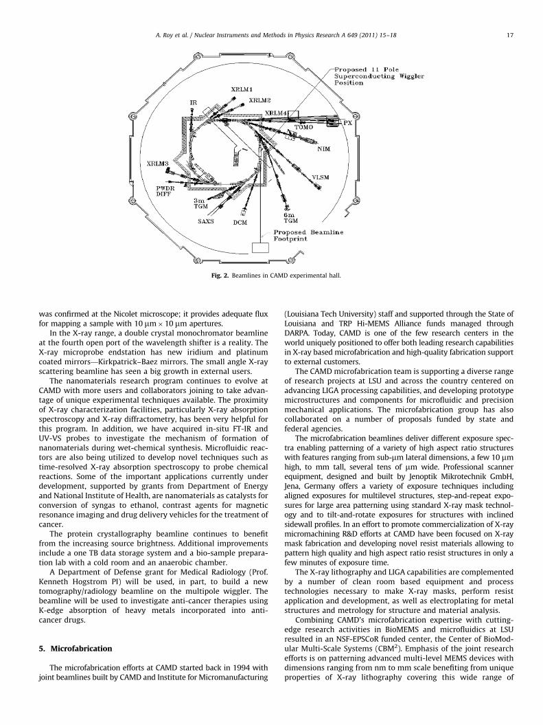
Fig. 2. Beamlines in CAMD experimental hall.
A. Roy et al. / Nuclear Instruments and Methods in Physics Research A 649 (2011) 15–18 17
was confirmed at the Nicolet microscope; it provides adequate fluxfor mapping a sample with 10 mm�10 mm apertures.
In the X-ray range, a double crystal monochromator beamlineat the fourth open port of the wavelength shifter is a reality. TheX-ray microprobe endstation has new iridium and platinumcoated mirrors—Kirkpatrick–Baez mirrors. The small angle X-rayscattering beamline has seen a big growth in external users.
The nanomaterials research program continues to evolve atCAMD with more users and collaborators joining to take advan-tage of unique experimental techniques available. The proximityof X-ray characterization facilities, particularly X-ray absorptionspectroscopy and X-ray diffractometry, has been very helpful forthis program. In addition, we have acquired in-situ FT-IR andUV-VS probes to investigate the mechanism of formation ofnanomaterials during wet-chemical synthesis. Microfluidic reac-tors are also being utilized to develop novel techniques such astime-resolved X-ray absorption spectroscopy to probe chemicalreactions. Some of the important applications currently underdevelopment, supported by grants from Department of Energyand National Institute of Health, are nanomaterials as catalysts forconversion of syngas to ethanol, contrast agents for magneticresonance imaging and drug delivery vehicles for the treatment ofcancer.
The protein crystallography beamline continues to benefitfrom the increasing source brightness. Additional improvementsinclude a one TB data storage system and a bio-sample prepara-tion lab with a cold room and an anaerobic chamber.
A Department of Defense grant for Medical Radiology (Prof.Kenneth Hogstrom PI) will be used, in part, to build a newtomography/radiology beamline on the multipole wiggler. Thebeamline will be used to investigate anti-cancer therapies usingK-edge absorption of heavy metals incorporated into anti-cancer drugs.
5. Microfabrication
The microfabrication efforts at CAMD started back in 1994 withjoint beamlines built by CAMD and Institute for Micromanufacturing
(Louisiana Tech University) staff and supported through the State ofLouisiana and TRP Hi-MEMS Alliance funds managed throughDARPA. Today, CAMD is one of the few research centers in theworld uniquely positioned to offer both leading research capabilitiesin X-ray based microfabrication and high-quality fabrication supportto external customers.
The CAMD microfabrication team is supporting a diverse rangeof research projects at LSU and across the country centered onadvancing LIGA processing capabilities, and developing prototypemicrostructures and components for microfluidic and precisionmechanical applications. The microfabrication group has alsocollaborated on a number of proposals funded by state andfederal agencies.
The microfabrication beamlines deliver different exposure spec-tra enabling patterning of a variety of high aspect ratio structureswith features ranging from sub-mm lateral dimensions, a few 10 mmhigh, to mm tall, several tens of mm wide. Professional scannerequipment, designed and built by Jenoptik Mikrotechnik GmbH,Jena, Germany offers a variety of exposure techniques includingaligned exposures for multilevel structures, step-and-repeat expo-sures for large area patterning using standard X-ray mask technol-ogy and to tilt-and-rotate exposures for structures with inclinedsidewall profiles. In an effort to promote commercialization of X-raymicromachining R&D efforts at CAMD have been focused on X-raymask fabrication and developing novel resist materials allowing topattern high quality and high aspect ratio resist structures in only afew minutes of exposure time.
The X-ray lithography and LIGA capabilities are complementedby a number of clean room based equipment and processtechnologies necessary to make X-ray masks, perform resistapplication and development, as well as electroplating for metalstructures and metrology for structure and material analysis.
Combining CAMD’s microfabrication expertise with cutting-edge research activities in BioMEMS and microfluidics at LSUresulted in an NSF-EPSCoR funded center, the Center of BioMod-ular Multi-Scale Systems (CBM2). Emphasis of the joint researchefforts is on patterning advanced multi-level MEMS devices withdimensions ranging from nm to mm scale benefiting from uniqueproperties of X-ray lithography covering this wide range of

A. Roy et al. / Nuclear Instruments and Methods in Physics Research A 649 (2011) 15–1818
dimensions with unprecedented accuracy and tightly controlledtolerances. Ultimately complex chip designs are transferred intomold inserts and replicated conveniently using hot embossing orother replication techniques. The overall design modular designand fabrication concept allows the fabrication of easily reconfi-gurable fluidic devices utilizing standardized fluidic, electrical andoptical interfaces accelerating the fast development of newsolutions in the area of BioMEMS.
6. Concluding remarks
Though CAMD is primarily a soft X-ray ring, more options arebecoming available for hard X-ray research. It is hoped that thewavelength shifter will stay operational while the 11-pole wiggler isadded. The wiggler is optimized for X-rays in the range of 4–20 keV.The idea that another short multipole wiggler can be added in ashort straight section for VUV applications is also being entertained.