Embed Size (px)
Citation preview
REVIEW Open Access
Stem cell therapies and benefaction ofsomatic cell nuclear transfer cloning inCOVID-19 eraBirbal Singh1, Gorakh Mal1, Vinod Verma2, Ruchi Tiwari3, Muhammad Imran Khan4, Ranjan K. Mohapatra5,Saikat Mitra6, Salem A. Alyami7, Talha Bin Emran8*, Kuldeep Dhama9* and Mohammad Ali Moni10*
Abstract
Background: The global health emergency of COVID-19 has necessitated the development of multiple therapeuticmodalities including vaccinations, antivirals, anti-inflammatory, and cytoimmunotherapies, etc. COVID-19 patientssuffer from damage to various organs and vascular structures, so they present multiple health crises. Mesenchymalstem cells (MSCs) are of interest to treat acute respiratory distress syndrome (ARDS) caused by SARS-CoV-2 infection.
Main body: Stem cell-based therapies have been verified for prospective benefits in copious preclinical and clinicalstudies. MSCs confer potential benefits to develop various cell types and organoids for studying virus-humaninteraction, drug testing, regenerative medicine, and immunomodulatory effects in COVID-19 patients. Apart frompaving the ways to augment stem cell research and therapies, somatic cell nuclear transfer (SCNT) holds uniqueability for a wide range of health applications such as patient-specific or isogenic cells for regenerative medicineand breeding transgenic animals for biomedical applications. Being a potent cell genome-reprogramming tool, theSCNT has increased prominence of recombinant therapeutics and cellular medicine in the current era of COVID-19.As SCNT is used to generate patient-specific stem cells, it avoids dependence on embryos to obtain stem cells.
Conclusions: The nuclear transfer cloning, being an ideal tool to generate cloned embryos, and the embryonicstem cells will boost drug testing and cellular medicine in COVID-19.
Keywords: SCNT, Genome reprogramming, Stem cells, SARS-CoV-2, COVID-19, Regenerative medicine, Bio-pharming
Highlights
� MSCs serve as potential regenerative medicine anddrug-testing tools in COVID-19.
� SCNT cloning is an alternative means of obtainingnuclear-transfer embryonic stem cells (NT-ESCs)which negate dependence on embryos to generateESCs.
� Nuclear transfer cloning produces cloned transgenicanimals for clinical investigations and recombinanttherapeutics for health applications.
IntroductionCoronavirus disease 2019 (COVID-19), as a pandemic,has affected badly several areas of human life. Especiallycountries with weak economies, limited resources, and
© The Author(s). 2021 Open Access This article is licensed under a Creative Commons Attribution 4.0 International License,which permits use, sharing, adaptation, distribution and reproduction in any medium or format, as long as you giveappropriate credit to the original author(s) and the source, provide a link to the Creative Commons licence, and indicate ifchanges were made. The images or other third party material in this article are included in the article's Creative Commonslicence, unless indicated otherwise in a credit line to the material. If material is not included in the article's Creative Commonslicence and your intended use is not permitted by statutory regulation or exceeds the permitted use, you will need to obtainpermission directly from the copyright holder. To view a copy of this licence, visit http://creativecommons.org/licenses/by/4.0/.The Creative Commons Public Domain Dedication waiver (http://creativecommons.org/publicdomain/zero/1.0/) applies to thedata made available in this article, unless otherwise stated in a credit line to the data.
* Correspondence: [email protected]; [email protected];[email protected] of Pharmacy, BGC Trust University Bangladesh, Chittagong4381, Bangladesh9Division of Pathology, ICAR-Indian Veterinary Research Institute, Izatnagar,Bareilly, Uttar Pradesh 243 122, India10WHO Collaborating Centre on eHealth, UNSW Digital Health, Faculty ofMedicine, School of Public Health and Community Medicine, UNSW Sydney,Sydney, NSW 2052, AustraliaFull list of author information is available at the end of the article
Singh et al. Stem Cell Research & Therapy (2021) 12:283 https://doi.org/10.1186/s13287-021-02334-5
poorly developed health system are in big trouble [1–3].However, the prevalence rates of infection in developedcountries like the USA and several other countries arevery high [4, 5]. But, these countries have ample health-care facilities for patients to compensate for their tech-nology and research and provide better protection totheir frontline medical practitioners. According to theunpublished report in certain underdeveloped countries,due to the limitation of available resources, a significantnumber of healthcare workers have lost their lives dueto COVID-19. Whatever be the circumstances, everyoneis working up to the potential within available resourcesto fight against COVID-19. Several therapies, like cellu-lar therapies, plasma therapies, antivirals, certain inhibi-tors, etc., are being applied to treat patients [6]. Plasmatherapy is an adjuvant preventive medication for critic-ally sick COVID-19 patients before long-term clinicaltrial treatment alternatives are available [7]. Plasma fromrecovering patients, mainly after serious infection, canproduce large amounts of polyclonal, pathogen-specificantibodies [8]. These antibodies confer purposeful pro-tective immunity to recipients with similar infectionconditions [9]. Convalescent plasma is assumed to per-form primarily by counteracting viral components inviral pathogenic diseases [10]. Additionally, some avail-able antiviral agents like remdesivir, favipiravir, ribavirin,etc., have been established with promising activitiesagainst SARS-CoV-2 [11].The rodents (mice, rats, and rabbits); non-human pri-
mates (NHPs), mainly the macaques (Macaca mulatta),Cynomolgus monkeys (Macaca fascicularis), and Africangreen monkey (Chlorocebus sabaeus); and some farm an-imals, such as pigs, cats, and dogs and the large domesticanimals [12], are on the leading edge as model species tostudy pharmacology, immunology, nutrition, and drugtesting and comprehend and cure the human diseases.Microbiome-targeted therapies [13–15], phage therapy[16, 17], antibody-antibiotic conjugates, stem cell-basedtherapies [18, 19], and nanomedicine [20–22] haveemerged as prospective unconventional therapies to curehuman diseases. Cellular medicine uses living cells, tis-sues, or biological processes to repair and regenerate thebody exclusive of dependence on major surgery. Inhumans, cell therapy is used to treat and cure diseasessuch as cancer by injecting living cells into patients. Ex-amples of the diseases treated with stem cells and regen-erative medicine include spinal cord injuries (SCI) [23],tendon and ligament injury and musculoskeletal healing[24], diabetes [25, 26], cancers [27–29], and cognitivedysfunctions [30, 31].Allogenic, autologous, xenogenic, embryonic stem cells
(ESCs); MSCs and neural stem cells; and hematopoieticstem cell therapies are expansively used in humans. Inanimals, stem cells are used to treat tendon, ligament,
and cartilage injuries in sports animals [32–34]. Matureor differentiated cell transplantation and de-differentiated cells genetically reprogrammed or inducedpluripotent stem cells (iPSCs) are used to treat a rangeof human diseases and pathologies [18, 35]. Several clin-ical and preclinical trials have confirmed the potency ofstem cell therapy. Also, 88 trials for COVID-19 patientpopulations were reported as exploring the protectionand effectiveness of stem cell transplantation therapy orstem cell-derived exosomes [36]. Investigated symptomsinvolve COVID-19 with severe pneumonia, respiratorydistress, acute respiratory distress syndrome (ARDS),and pulmonary fibrosis. The bulk of trials were recordedfor “COVID-19” (19 out of 88) and “severe pneumonia”patients (37 out of 88). As per a meta-analysis of 50,466hospital-admitted COVID-19 patients, 14.8% of COVID-19 patients developed ARDS [37]. Interestingly, only oneof 88 cellular therapy-based trials ascertained the adverseeffects, including severe kidney injury. Otherwise, mostof them had favorable benefits with stem cell therapy[38].This review presents the potential of stem cell therap-
ies, as well as the support of SCNT cloning in cellularmedicine in the COVID-19 era. The concepts and ideasemanating from this study can be considered to enhancedrug testing and discovery for prevention againstCOVID-19.
COVID-19 and cellular medicineThe SARS-CoV-2 infections including its variants arenew and deadlier than other ssRNA coronaviruses,which are relatively innocuous. Ever since its emergencein mid of December 2019 in Wuhan city, China, SARS-CoV-2 has spread across continents and countries, in-fecting more than 128 million people while killing nearly3 million as of March 30, 2021 (COVID-19 CoronavirusPandemic, World meters, 2021).It posed high global health concerns due to its ongoing
pandemic and crippled the human life socio-economically. The infected people are under enormousstress due to fear of fatal outcomes. SARS-CoV-2 causesmultiple health problems [39], such as inflammation,hypoxia, oxidative stress, mitochondrial dysfunction [40],myocarditis due to intense immune response [41], pro-pensity of clotting in micro- and large blood vessels [42],direct infection of cardiomyocytes and cardiogenic shock[43], neurological and neuropsychiatric illnesses [44],kidney damage [45], and immunopathological conditionsincluding cytokine storm [39, 46].A cytokine storm, also referred to as hypercytokine-
mia, is a physiological response in people and other liv-ing organisms that triggers an unregulated and excessiverelease of pro-inflammatory signaling molecules calledcytokines [47]. Cytokine storm as a hallmark of COVID-
Singh et al. Stem Cell Research & Therapy (2021) 12:283 Page 2 of 16
19 is characterized by the presence of various immunecells. Elicited immune response by these cells releasesinterleukin (IL) IL-2, IL-6, IL-7, interferon gamma, tumornecrosis factor, granulocyte colony-stimulating factor, in-ducible protein-10, and macrophage chemoattractantprotein-1, which suggests a well-determined effort by theimmune system, but is misconceived [48, 49]. As theMSCs possess regenerative, immunomodulatory, and anti-inflammatory properties stemmed from their stemness,these cells can play an essential role as therapies and pa-tient health management in COVID-19 [50, 51].COVID-19-associated cytokine storm resulting from
displaced immune system activation drives unbalancedimmune response, inflammation, and intravascular coagu-lation and induces vascular damage in patients. Thesecomplications result in vascular cardiopulmonary collapseand confer a poor prognosis [52, 53]. Besides these com-plications, ACE2 receptors are widely distributed in tissuesof various organs such as kidneys, cardiac muscles, andsmooth and endothelial muscles of multiple organs, whichexplicates that SARS-CoV-2 can generate systemic dis-eases and damage various organs [51]. Under these threat-ening conditions, in addition to an appropriatelyoptimized immune response from the system, repair ofdamaged endothelial is a crucial question in recovery andgetting rid of COVID-19. Given these preliminary scenar-ios, endothelial progenitor cells (EPCs) may play a key rolein inhibiting or reversing this damage in these patients[54]. Current evidence indicates EPCs of bone marroworigin as potential candidates for vascular repair and con-siderably promising therapeutics [55, 56]. In critical pa-tients and severe cases of COVID-19, EPCs can play anessential role in maintaining vascular endothelial func-tions, hence contributing to the stem cell therapies inCOVID-19 patients [54].Rapid research advances have paved the ways to de-
velop potential therapeutic interventions, vaccines,drugs, therapies, and immunomodulators to tackleCOVID-19, few of which are in the last phases of clinicaltrials. Success has been achieved to develop anti-SARS-CoV-2 vaccines, and vaccination is currently in progress[57–64]. Immunological interventions, immune-mediated, gene-editing-based therapeutics, and cell-based therapies [65–72] utilizing natural killer (NK) cells[73, 74], T cells [75, 76], convalescent plasma, neutraliz-ing antibodies, monoclonal antibodies [62, 77, 78], cyto-kines, interferons [79, 80], toll-like receptors (TLRs) [81,82], and stem cell-based therapies [7, 83] have also beenexploited to curb the SARS-CoV-2.
Stem cell-based COVID-19 therapyIndividual humans’ immune system plays a fundamentalrole in reclamation and protection against infections.Therefore, evading the detrimental cytokine storm
elicited in response to virus invasion is very important incritically affected COVID-19 patients. Immunomodula-tory and regenerative properties of MSCs make thempromising candidates to cure or lessen the sufferingfrom COVID-19. Intravenously administered MSCs mi-grate and cumulate in pulmonary microvasculature,where they secrete various paracrine factors which pro-tect and rejuvenate pulmonary epithelial cells and im-prove lung functioning [84]. In addition, theadministered MSCs due to their ability to home and acton injured organs such as the heart, liver, and kidney areuseful as cellular medicine [85].Stem cell-based therapies can be stated as any treat-
ment strategies for severe health issues or medical con-ditions that implicate the usage of any sort of feasiblehumans’ stem cells, such as induced pluripotent stemcells (iPSCs), ESCs, and adult stem cells for allogeneicand autologous therapies. Stem cells may confer the po-tential remedy if tissue and organ transplantation areneeded through their capacity to distinguish between thespecific cell types used to repair diseased tissues [86, 87].Currently, stem cell-based therapies are being evaluatedagainst SARS-CoV-2 and for their potential in treatingCOVID-19 patients [7, 83, 88–90]. MSCs, also recog-nized as mesenchymal stromal cells or medicinal signal-ing cells, are multipotent stromal cells that can set apartinto a choice of cell sorts, including bone cells (osteo-blasts), cartilage cells (chondrocytes), muscle cells (myo-cytes), and adipocytes which can differentiate into othercell lineages [91–93]. MSCs with their immunomodula-tory and regenerative properties have been put forwardas promising therapeutic regimens for severe SARS-CoV-2 and can impact the management of COVID-19patients [94–103]. MSCs can be harvested from differentadult tissues with their regenerative and multipotentcapacities [104]. Leng et al. investigated the effect of asingle dose of transfused MSCs in seven COVID-19 pa-tients and reported a decrease in cytokine levels and pa-tient’s improvement within 2 days. Authors furtherreported that natural killer and T cells responsible forcytokine storm also disappeared after 6 days with no sideeffects [105]. O’Driscoll mentioned a case study inBaoshan, China, which also showed a similar outcome[106]. Another case study of MSC infusion showed sig-nificant patient improvement after 24 h without consid-erable side effects [107]. MSCs communicate viaparacrine mechanisms such as excretion of extracellularvesicles (EVs), including exosomes and macrovesicles[108–110]. The EVs (exosomes and ectosomes) affectimmune cells by induction of anti-inflammatory macro-phages, regulatory dendritic cells, regulatory T and Bcells, and inactivation of T cells; halt cytokine storm andpro-angiogenic action; and secrete a variety of bioactiveand immunomodulatory factors. These properties make
Singh et al. Stem Cell Research & Therapy (2021) 12:283 Page 3 of 16
them suitable for treatments of severe cases of COVID-19 [83, 111–119]. Published reports suggest that manybenefits of MSCs can be achieved by exosomes and ecto-somes while avoiding cellular transfusion and associatedretaliation [120–122]. So, MSC-derived EVs of varioussources like the umbilical cord, peripheral blood, amni-otic fluid, etc., can also give almost all MSC therapeuticbenefits and are being evaluated for the same [106, 123].Promising therapeutic aspects of EVs of MSCs includenasal administration or inhalation and non-replicativenature that are void of side effects like uncontrolled celldivision associated with cell therapy [106]. Otherwise,MSCs inhibit tissue fibrosis and cell death, releasing im-munomodulatory factors that suppress the cytokinestorms in COVID-19 patients [118]. Intravenous infu-sion of perinatal, i.e., umbilical cord and Wharton’s jelly,MSCs improved pulmonary functions and symptoms inCOVID-19 pneumonia. The treated patients had im-proved CD3+, CD4+, and CD8+ cell count and decreasedIL-6, TNF-α, and C-reactive proteins [124].Several trials have been conducted to evaluate the clin-
ical avenues and therapeutic potentials of MSCs in treat-ing COVID-19 patients. Few of them have proved thesafety and efficacy of MSCs, suggesting their potent clin-ical applications and prospects to fight against COVID-19 [86, 94, 98, 101, 125–136]. Thus, MSCs might consti-tute potentially life-saving treatment options for critic-ally ill COVID-19 patients, could reduce severity andlongevity of ARDS in such patients, and reduce the asso-ciated mortality rates during the pandemic. However, in-depth studies and large-scale trials for confirming andvalidating the safety, potency, and efficacy of MSCs, sup-ported with reliable and sufficient scientific evidence,would aid in optimizing the use of MSCs and MSC-derived products, cell-free therapies, and stem cell trans-plantation therapy to alleviate and treat critically severeCOVID-19 pneumonia and illness. Explorative researchis warranted to resolve prominent challenges such as thefate of administered MSCs, validating their safety and ef-ficacy, homing capacity, resistance to the disease micro-environment, and stem cell quality management. Also,due attention is needed for adopting strict ethical, regu-latory permissions and clinical guidelines for developingsafer and effective MSC-based therapies for patients withCOVID-19 [128, 137–140].Two clinical studies were undertaken to consider the
potential improvements of MSCs in COVID-19 patientswith high fever, dyspnea (difficulty in breathing), andpneumonia. A total of 10 individuals were selected assubjects, and the analysis was performed on sevenSARS-CoV-2-positive patients, four of whom had severesymptoms, two exhibiting common sorts of the syn-drome, and one of whom was seriously ill. Theremaining three patients were recruited in placebo
monitoring for acute symptoms. Clinical-grade humanMSCs were injected intravenously into all seven patients.The patients were administered with 1 × 10⁶ MSCs perkilogram of body weight while their situation was ser-iously deteriorating and studied for a cumulative dur-ation of 14 days. Before the administration of MSCs,patients were recorded with elevated fever (body temper-atures varying from 38.5 to 39 °C), lowered oxygen satur-ation, dyspnea, and pneumonia. This study ascertainedthat the symptoms, which had appeared in patients be-fore infusions, receded within 2–4 days after administrat-ing the MSCs. The oxygen saturation at rest, with orwithout oxygen uptake, increased to around 95% [50,141].Intravenously administered MSCs were found to travel
straight into the lungs where several factors secreted byMSCs played a major role in immunomodulation, pro-tecting alveolar epithelial cells, combating pulmonary fi-brosis, along with providing improved lung functions,which make greater benefits for treating severe pulmon-ary conditions in COVID-19 [124]. Another researchconducted by Liang et al. [142] included a severeventilator-ridden COVID-19 patient treated with a hu-man umbilical cord MSC (hUCMSC). The patient wasmonitored at intervals every 3 days with three infusionsof 5 × 10⁷ hUCMSC and was capable of walking only 4days after the second MSC infusion. The patient did notdisplay detectable side effects, and the critical parame-ters such as T-cell counts were returned to the normalrange [142]. Another study was carried out to observethe efficacy of intravenous infusion of bone marrow-derived MSCs in ARDS [143]. The research was under-taken in the first step with the intravenous injection ofstromal MSCs in 9 ARDS patients. The MSC infusionwas found to be effective and showed no adverse effects[143]. Phase 2 of the study indicated that a sheep modelwith bacterial pneumonia was less severely impaired byacute lung damage in therapy with hMSCs [144].Following an intravenous infusion, significant popula-
tions of the MSCs were found to migrate and gather inthe lungs where they protected alveolar epithelial cells,re-established pulmonary microenvironment, and curedlung dysfunction besides preventing the pulmonary fi-brosis [145]. Systemic infusion of 5 × 10⁷ human umbil-ical cord MSCs (UC-MSCs) through intravenousinjection every 3 days for 3 three times improved healthoutcomes in the patients and hence could be consideredas a prospective alternate therapy to treat COVID-19 ingeneral and elderly patients [146].The patients having acute SARS-CoV-2-induced
ARDS accompanied by severe hypoxemia were givenintravenous infusions of 200 × 106 UC-MSCs daily (6cases) or placental MSCs (5 cases) [147]. The treatmentdid not cause any adverse effects after 24–48 h post cell
Singh et al. Stem Cell Research & Therapy (2021) 12:283 Page 4 of 16
infusion. The study concludes that multiple infusions ofhigh doses of allogenic UC- and placental MSCs canswiftly improve COVID-19-induced ARDS in some pa-tients. However, MSC therapy should be avoided in ex-ceptional patients who are prone to develop sepsis ormulti-organ failure [147]. Nevertheless, studiesemphasize on the need for randomized multicenter clin-ical trials with long-term follow-up of the stem celltreatments [83, 147, 148]. Indeed, the use of MSCsagainst COVID-19 is still at the initial stages. As stemcell treatments have already revealed positive outcomesin patients with pulmonary pathologies, there are pros-pects of hopeful therapies using assorted types of MSCsin immunomodulation, regenerative medicine, cell or tis-sue engineering, and anti-inflammatory treatments [89].A parallel assigned controlled, non-randomized, phase
1 clinical trial using intravenous infusion of 3 × 10⁷ cellsper fusion on days 0, 3, and 6 was conducted to evaluatethe safety and efficacy of UC-MSCs to treat severeCOVID-19 pulmonary pathology [148]. Despite diminu-tive initial side effects viz., transient facial flushing, slightfever, and transient hypoxia at 12 h post-infusion, thepatients recovered soon and were discharged from theclinics. The study infers that intravenous UC-MSC infu-sion is safe and tolerable in moderate and severeCOVID-19 patients [148]. UC-MSCs have been sug-gested for compassionate applications in critically illCOVID-19 patients to decrease mortality and morbid-ities [83]. In countries like China where modalities toprevent COVID-19 were limited, the patients weretreated with stem cells such as UC-MSCs [83].Indeed, MSCs are very useful in the treatment of
COVID-19 for the reason that they mitigate or decreasethe cytokine storm through their immunomodulatorycapacity via inhibition of T and B cell proliferation andthrough effective regulation of pro-inflammatory cyto-kines to improve the microenvironment for endogenousrepair [149, 150]. With regard to COVID-19, Leng et al.[50] used BM-derived MSCs intravenously in 7 patientswith COVID-19 (1 with critically severe illness, 4 withsevere illnesses, and 2 with common illnesses). Mass cy-tometry and cytokine analysis of the patients’ peripheralblood also showed loss of over-activated T cells and NKcells, an increase in anti-inflammatory cytokines like IL-10, and a decrease in pro-inflammatory cytokines suchas TNF-alpha. Hence, it shows that MSC has a potentialto decrease cytokine storm.Cell- and tissue-based remedies such as stem cell ther-
apy, NK therapy, T-cell therapy, chimeric antigen recep-tor, stem cell-exosomes, extracellular vesicles, and tissueproducts are of considerable therapeutic interest againstinfectious diseases [151, 152]. Despite being at the initialstages, the use of MSCs against COVID-19 has attractedsubstantial attention as regenerative medicine. As stem
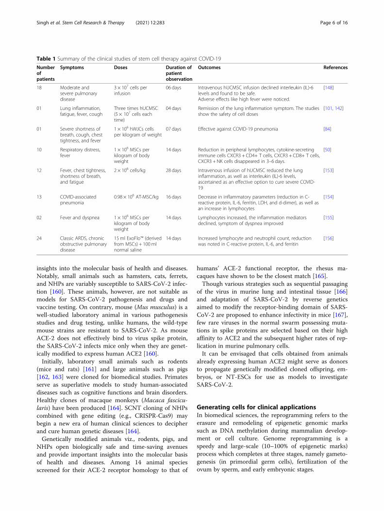
cell treatments have already revealed positive outcomesin patients with pulmonary pathologies, there are pros-pects of hopeful therapies using assorted types of MSCsin immunomodulation, regenerative medicine, cell or tis-sue engineering, and anti-inflammatory treatments [89].The clinical studies which ascertained the efficacy ofstem cell therapy have been highlighted in Table 1.In view of the importance of stem cell therapies in
COVID-19, there is a need to generate patient-specificclinical-grade immunocompatible cells, ESCs and MSCs.We have emphasized SCNT cloning as a futuristic toolto generate stem cells from fetal, neonates, or adult hu-man donors. The stem cells generated by SCNT and in-duced pluripotency are promising tools for personalizedrequirements of regenerative medicine, transplantation,and disease modeling [157].
SCNT cloning for biomedical and regenerativemedicineSCNT initially introduced in 1952 to discover embryodevelopment in frogs came into broad publicity in 1997when Prof. Ian Wilmut and his team succeeded to re-program the sheep fibroblasts and produced “Dolly,” thefirst cloned mammalian species [158]. The technique(Fig. 1) has been modified and adapted to clone severalmammalian species, including laboratory animals, live-stock, and endangered wild mammals including non-human primates (NHPs) [159]. The underlying principleof SCNT is that the body cells in an individual possessan identical genome despite being different in pheno-types, niches, and functions. SCNT organizes the repro-gramming of the genome of donor nuclei andaccordingly provides the means of transforming a ma-tured cell to a totipotency state comparable to an em-bryo. Hence, SCNT cloning circumvents the processesthat generally ensue during gametogenesis andfertilization and enable the embryo to undergo normaldevelopment.Transgenic animal models are important bioresources
for developmental biology and biomedical research.Highly exigent need of unraveling, preventing, and con-trolling the SARS-CoV-2 necessitates the search for op-timal animal models. The paucity of a befitting COVID-19 preclinical model animal is one of the major impedi-ments. The researchers are in race to understand themolecular mechanisms of SARS-CoV-2 infection to re-purpose the currently available drugs to develop animalmodels and antiviral therapies against the pandemic. NTcloning is used to generate stem cells and progeniescharacteristically identical to the already borne live ordeceased organism.Coronaviruses affect a broad range of mammalian spe-
cies. Wild-type or genetically modified rodents, pigs, andNHPs have been of paramount interest and provide key
Singh et al. Stem Cell Research & Therapy (2021) 12:283 Page 5 of 16
insights into the molecular basis of health and diseases.Notably, small animals such as hamsters, cats, ferrets,and NHPs are variably susceptible to SARS-CoV-2 infec-tion [160]. These animals, however, are not suitable asmodels for SARS-CoV-2 pathogenesis and drugs andvaccine testing. On contrary, mouse (Mus musculus) is awell-studied laboratory animal in various pathogenesisstudies and drug testing, unlike humans, the wild-typemouse strains are resistant to SARS-CoV-2. As mouseACE-2 does not effectively bind to virus spike protein,the SARS-CoV-2 infects mice only when they are genet-ically modified to express human ACE2 [160].Initially, laboratory small animals such as rodents
(mice and rats) [161] and large animals such as pigs[162, 163] were cloned for biomedical studies. Primatesserve as superlative models to study human-associateddiseases such as cognitive functions and brain disorders.Healthy clones of macaque monkeys (Macaca fascicu-laris) have been produced [164]. SCNT cloning of NHPscombined with gene editing (e.g., CRISPR-Cas9) maybegin a new era of human clinical sciences to decipherand cure human genetic diseases [164].Genetically modified animals viz., rodents, pigs, and
NHPs open biologically safe and time-saving avenuesand provide important insights into the molecular basisof health and diseases. Among 14 animal speciesscreened for their ACE-2 receptor homology to that of
humans’ ACE-2 functional receptor, the rhesus ma-caques have shown to be the closest match [165].Though various strategies such as sequential passaging
of the virus in murine lung and intestinal tissue [166]and adaptation of SARS-CoV-2 by reverse geneticsaimed to modify the receptor-binding domain of SARS-CoV-2 are proposed to enhance infectivity in mice [167],few rare viruses in the normal swarm possessing muta-tions in spike proteins are selected based on their highaffinity to ACE2 and the subsequent higher rates of rep-lication in murine pulmonary cells.It can be envisaged that cells obtained from animals
already expressing human ACE2 might serve as donorsto propagate genetically modified cloned offspring, em-bryos, or NT-ESCs for use as models to investigateSARS-CoV-2.
Generating cells for clinical applicationsIn biomedical sciences, the reprogramming refers to theerasure and remodeling of epigenetic genomic markssuch as DNA methylation during mammalian develop-ment or cell culture. Genome reprogramming is aspeedy and large-scale (10–100% of epigenetic marks)process which completes at three stages, namely gameto-genesis (in primordial germ cells), fertilization of theovum by sperm, and early embryonic stages.
Table 1 Summary of the clinical studies of stem cell therapy against COVID-19
Numberofpatients
Symptoms Doses Duration ofpatientobservation
Outcomes References
18 Moderate andsevere pulmonarydisease
3 × 10⁷ cells perinfusion
06 days Intravenous hUCMSC infusion declined interleukin (IL)-6levels and found to be safe.Adverse effects like high fever were noticed.
[148]
01 Lung inflammation,fatigue, fever, cough
Three times hUCMSC(5 × 10⁷ cells eachtime)
04 days Remission of the lung inflammation symptom. The studiesshow the safety of cell doses
[101, 142]
01 Severe shortness ofbreath, cough, chesttightness, and fever
1 × 10⁶ hWJCs cellsper kilogram of weight
07 days Effective against COVID-19 pneumonia [84]
10 Respiratory distress,fever
1 × 10⁶ MSCs perkilogram of bodyweight
14 days Reduction in peripheral lymphocytes, cytokine-secretingimmune cells CXCR3 + CD4+ T cells, CXCR3 + CD8+ T cells,CXCR3 + NK cells disappeared in 3–6 days.
[50]
12 Fever, chest tightness,shortness of breath,and fatigue
2 × 10⁶ cells/kg 28 days Intravenous infusion of hUCMSC reduced the lunginflammation, as well as interleukin (IL)-6 levels,ascertained as an effective option to cure severe COVID-19
[153]
13 COVID-associatedpneumonia
0.98 × 10⁶ AT-MSC/kg 16 days Decrease in inflammatory parameters (reduction in C-reactive protein, IL-6, ferritin, LDH, and d-dimer), as well asan increase in lymphocytes
[154]
02 Fever and dyspnea 1 × 10⁶ MSCs perkilogram of bodyweight
14 days Lymphocytes increased, the inflammation mediatorsdeclined, symptom of dyspnea improved
[155]
24 Classic ARDS, chronicobstructive pulmonarydisease
15ml ExoFlo™ (derivedfrom MSCs) + 100mlnormal saline
14 days Increased lymphocyte and neutrophil count, reductionwas noted in C-reactive protein, IL-6, and ferritin
[156]
Singh et al. Stem Cell Research & Therapy (2021) 12:283 Page 6 of 16
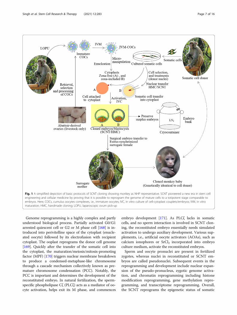
Genome reprogramming is a highly complex and partlyunderstood biological process. Partially activated G0/G1arrested quiescent cell or G2 or M phase cell [168] is in-troduced into perivitelline space of the cytoplast (enucle-ated oocyte) followed by its electrofusion with recipientcytoplast. The ooplast reprograms the donor cell genome[169]. Quickly after the transfer of the somatic cell intothe cytoplast, the maturation/meiosis/mitosis-promotingfactor (MPF) [170] triggers nuclear membrane breakdownto produce a condensed-metaphase-like chromosomethrough a cascade mechanism collectively known as pre-mature chromosome condensation (PCC). Notably, thePCC is important and determines the development of thereconstituted embryo. In natural fertilization, the sperm-specific phospholipase Cζ (PLCζ) acts as a mediator of oo-cyte activation, helps exit its M phase, and commences
embryo development [171]. As PLCζ lacks in somaticcells, and no sperm interaction is involved in SCNT clon-ing, the reconstituted embryo essentially needs simulatedactivation to undergo auxiliary development. Various sup-plements, i.e., artificial oocyte activators (AOAs), such ascalcium ionophores or SrCl2 incorporated into embryoculture medium, activate the reconstituted embryos.Sperm and oocyte pronuclei are present in fertilized
zygotes, whereas nuclei in reconstituted or SCNT em-bryos are called pseudonuclei. Subsequent events in thereprogramming and development include nuclear expan-sion of the pseudo-pronucleus, zygotic genome activa-tion, and chromatin reprogramming including histonemodification reprogramming, gene methylation repro-gramming, and transcriptome reprogramming. Overall,the SCNT reprograms the epigenetic status of somatic
Fig. 1 A simplified depiction of basic protocols of SCNT cloning showing monkey as NHP representative. SCNT pioneered a new era in stem cellengineering and cellular medicine by proving that it is possible to reprogram the genome of mature cells to a totipotent stage comparable toembryos. Here, COCs, cumulus oocytes complexes, i.e., immature oocytes; IVC, in vitro culture of cell-cytoplast couplets/embryos; IVM, in vitromaturation; HMC, handmade cloning; LOPU, laparoscopic ovum pick-up
Singh et al. Stem Cell Research & Therapy (2021) 12:283 Page 7 of 16
cells used as donor cells within a brief period thoughfew regions are not reprogrammed, or they resist repro-gramming [169].
Regenerative medicineInner cell mass (ICM) cells of cloned embryos serve as asource of NT-ESCs. In humans, nuclear transfer cloningis used to generate patient-specific NT-ESCs which areisogenic and immunocompatible. This process is knownas therapeutic cloning. Handmade cloning (HMC) [172,173], a modified procedure of nuclear transfer cloning, issimpler as it circumvents dependency on expensive mi-cromanipulation to produce cytoplast from IVM oocytesand transfer of donor cell across zona pellucida of thecytoplast.NT-ESCs can be propagated, preserved, and de-
differentiated into other cell types. Notably, the mamma-lian cells can also be reprogrammed by ectopic or in-duced expression of exogenous genetic factors Oct5,Sox2, Klf4, and cMyc (OSKM), also known as Yamanakafactors [174]; non-genetic elements and small molecules[175]; microRNAs [176]; synthesized transcription fac-tors [177]; combinations of chemical compounds [178,179]; and cell fusion [180]. The phenomenon ofintestine-specific caudal-related homeobox (CDX), espe-cially the CDX1-induced SALL4 and KLF5-mediated in-testinal epithelial cell reprogramming into tissue stem-like progenitor cells [181], has led to the concept ofusing beneficial microorganisms such as lactic acid bac-teria to reprogram the somatic cells [182, 183]. In nu-clear transfer cloning, the cytoplast possessesmitochondria and other factors that support the metab-olism competency, cope with metabolic oxidative stress,and assist rejuvenation of donor cells [184].
Organoids technology and COVID-19Organoids are tiny, self-organized 3D tissue derivedfrom adult cells, ESCs, or reprogrammed stem cells, i.e.,iPSCs, which recapitulate much of the complexity, se-lected properties, and genetic signatures of original tis-sues and organ [185, 186]. Organoids bridge thepreclinical and clinical science and have resolved variousresearch anomalies and therapeutic challenges. Like dif-ferent types of the organs, the organoids are also of dif-ferent types and are analyzed by various sequencingmethods, molecular imaging, and spectrometry. Multipleorgan stem cell-derived organoids have been developedfor disease modeling, host-pathogen interactions, drugdiscovery, regenerative medicine, and studyingorganogenesis.Human organoids generated from patient biopsies or
stem cells are used for drug screening and study bio-medical complications, genetic disorders, infectious dis-eases, and disease modeling with high precision. Human
iPSC-derived monolayer brain cells and region-specificbrain organoids have revealed that compared to choroidplexus epithelial cells, the neurons and astrocytes aresparsely infected with SARS-CoV-2 [187]. Human stemcell lung organoids were found to be susceptible toSARS-CoV-2 infection exhibiting vigorous induction ofchemokines [188]. Analysis of human airway organoidsshows that SARS-CoV-2 has multi-basic cleavage sites inits spike protein which increase its infectivity towardsairway cells, and compared to other coronaviruses, theSARS-CoV-2 enters more rapidly into airway cells [189].High-throughput screening of interaction of the humanlung organoid model with some FDA-approved drugsagainst COVID-19 has shown that imatinib, mycopheno-lic acid, and quinacrine dihydrochloride could signifi-cantly inhibit the SARS-CoV-2 [188]. Hence, organ-specific stem cell-derived organoids provide valuabletools to identify therapeutics and drugs against COVID-19. Stem cells generated by induced pluripotency orSCNT reprogramming might be a valuable resource togenerate multiple types of organoids to screen efficacyand safety of drugs, basic virology, and pathogenesis ofSARS-CoV-2.
SCNT vis-à-vis bio-pharmingThe transgenic cloned animals have several applicationsin research, medicine and agriculture. Recombinant pro-teins produced through transgenic animals have post-translational maturity and stability. Compared to cul-tured animal cells, transgenic mammalian species serveas an excellent platform to produce monoclonal anti-bodies (mAbs) in milk. An improved version of cetuxi-mab, a mAb against epidermal growth factor receptor, isproduced at a larger scale in transgenic goats [190].Gene-edited animals that serve as donors of clinical-
grade stem cells are produced by SCNT [191–193]. Aline of transgenic goats has been designed to express hu-man lysozyme in their mammary glands [194]. Targetedchanges in the animal genome are likely to initiate a newera of bio-pharming [195]. Healthcare applications in-clude production of target-specific stem cells and thera-peutic proteins [196], mAbs [190], released into the milkof animals, to the use of genetically modified (GM) ani-mals to produce organs for xenotransplantation areenvisaged.The use of cloned mammalian livestock (goats, pigs,
and cattle) for commercial production of recombinanthuman proteins and nutraceuticals has been reviewedelsewhere [197–199]. Currently, information is lackingon the synthesis of recombinant SARS-CoV-2 S proteinusing cloned transgenic mice, rabbits, pigs, or milch ani-mals such as goats, sheep, or cattle. More recently, re-combinant SARS-CoV-2 S protein has been producedusing baculovirus-silkworm expression system. S
Singh et al. Stem Cell Research & Therapy (2021) 12:283 Page 8 of 16
proteins secreted into silkworm serum have been puri-fied and would be used for the development of immuno-detection, immunoglobulin, and vaccine developmentagainst the virus [200]. Delay in development of clonedtransgenic animals as model animals or source of recom-binant SARS-CoV-2 proteins is probably due to low effi-ciency of SCNT cloning to produce embryos, longgestation periods, more age of achieving maturity, andexpression of recombinant proteins in mammary tissueand excretion into milk.
Development of model animals and therapeuticcellsThere is an urgent need of animal models to screen andevaluate vaccines and drugs to treat COVID-19. SCNT isat present the most reliable method to produce clonedand transgenic animals including livestock. However,SARS-CoV-2 pathogenicity is studied using macaques,
ferrets, cats, and hamsters. NHPs are instrumental for thepreclinical evaluation of vaccines against COVID-19 [104].Transgenic mice expressing human ACE2 are the cur-rently in vivo systems to discover SARS-CoV-2 [201, 202].Though some vaccines are already underway, there is
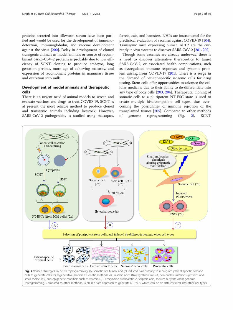
a need to discover alternative therapeutics to targetSARS-CoV-2, or associated health complications, suchas dysregulated immune responses and systemic prob-lem arising from COVID-19 [201]. There is a surge inthe demand of patient-specific isogenic cells for drugtesting. Stem cells offer opportunities to advance the cel-lular medicine due to their ability to de-differentiate intoany type of body cells [203, 204]. Therapeutic cloning ofsomatic cells to a pluripotent NT-ESC state is used tocreate multiple histocompatible cell types, thus over-coming the possibilities of immune rejection of thetransplanted tissues [205]. Compared to other methodsof genome reprogramming (Fig. 2), SCNT
Fig. 2 Various strategies: (a) SCNT reprogramming, (b) somatic cell fusion, and (c) induced pluripotency to reprogram patient-specific somaticcells to generate cells for regenerative medicine. Genetic methods viz., nucleic acids (NA), synthetic mRNA, non-nucleic methods (proteins andsmall molecules), and epigenetic modifiers such as vitamin C, 5-azacytidine, trichostatin A, valproic acid, sodium butyrate assist genomereprogramming. Compared to other methods, SCNT is a safe approach to generate NT-ESCs, which can be de-differentiated into other cell types
Singh et al. Stem Cell Research & Therapy (2021) 12:283 Page 9 of 16
reprogramming of cells is anticipated to generatepatient-specific therapeutic grade cells. The iPSCs gener-ated by OSKM have repulsive concerns that restrict theiruse in biomedical applications. The use of retrovirusesmay cause cancer or tumor formation. In addition, retro-viruses insert their DNA into the host genome and trig-ger the expression of cancer-causing genes. C-Myc (oneof the genes used in reprogramming) is a known onco-gene whose overexpression can induce teratoma. More-over, all the pluripotency factors are not equallyexpressed, and reprogramming of non-dividing cellssuch as peripheral blood mononuclear cells (PBMC) andaged skin fibroblasts is very low. Nonetheless, the retro-viral vectors, epiosmal vectors, and Sendai viruses had acomparable reprogramming efficiency and did not affectgene expression in fibroblast-derived human iPSCs[206].Patient-specific NT-ESCs serve as valuable in vitro dis-
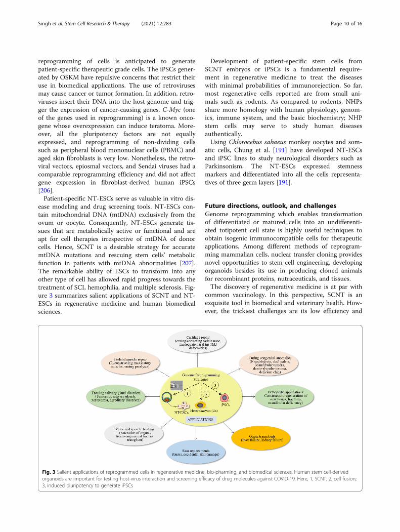
ease modeling and drug screening tools. NT-ESCs con-tain mitochondrial DNA (mtDNA) exclusively from theovum or oocyte. Consequently, NT-ESCs generate tis-sues that are metabolically active or functional and areapt for cell therapies irrespective of mtDNA of donorcells. Hence, SCNT is a desirable strategy for accuratemtDNA mutations and rescuing stem cells’ metabolicfunction in patients with mtDNA abnormalities [207].The remarkable ability of ESCs to transform into anyother type of cell has allowed rapid progress towards thetreatment of SCI, hemophilia, and multiple sclerosis. Fig-ure 3 summarizes salient applications of SCNT and NT-ESCs in regenerative medicine and human biomedicalsciences.
Development of patient-specific stem cells fromSCNT embryos or iPSCs is a fundamental require-ment in regenerative medicine to treat the diseaseswith minimal probabilities of immunorejection. So far,most regenerative cells reported are from small ani-mals such as rodents. As compared to rodents, NHPsshare more homology with human physiology, genom-ics, immune system, and the basic biochemistry; NHPstem cells may serve to study human diseasesauthentically.Using Chlorocebus sabaeus monkey oocytes and som-
atic cells, Chung et al. [191] have developed NT-ESCsand iPSC lines to study neurological disorders such asParkinsonism. The NT-ESCs expressed stemnessmarkers and differentiated into all the cells representa-tives of three germ layers [191].
Future directions, outlook, and challengesGenome reprogramming which enables transformationof differentiated or matured cells into an undifferenti-ated totipotent cell state is highly useful techniques toobtain isogenic immunocompatible cells for therapeuticapplications. Among different methods of reprogram-ming mammalian cells, nuclear transfer cloning providesnovel opportunities to stem cell engineering, developingorganoids besides its use in producing cloned animalsfor recombinant proteins, nutraceuticals, and tissues.The discovery of regenerative medicine is at par with
common vaccinology. In this perspective, SCNT is anexquisite tool in biomedical and veterinary health. How-ever, the trickiest challenges are its low efficiency and
Fig. 3 Salient applications of reprogrammed cells in regenerative medicine, bio-pharming, and biomedical sciences. Human stem cell-derivedorganoids are important for testing host-virus interaction and screening efficacy of drug molecules against COVID-19. Here, 1, SCNT; 2, cell fusion;3, induced pluripotency to generate iPSCs
Singh et al. Stem Cell Research & Therapy (2021) 12:283 Page 10 of 16
developmental abnormalities resulting due to imperfectepigenetic reprogramming.Poor-quality pre-implantation embryos or blastocysts
resulting due to incomplete reprogramming or aberrantdevelopment such as delayed development, low number ofblastomeres or embryo cells, and genome instability im-pede yield of ESCs. Another issue crucial for the successand implementation of SCNT in humans and primates isthe availability of oocytes for research purposes. It evi-dently involves financial and ethical implications. It is en-visaged that research on applications of SCNT and theiPSCs to produce sperm and oocytes is of high biomedicalimportance and will continue in near future [208].The fundamental hurdles related to animal cloning
need to be resolved by analyzing underlying cellular andmolecular mechanisms. Mitochondrial proteins (Mfn2and Bcnl3L) deregulation or dysfunction in cloned em-bryos hampers their development [209]; hence, multiplestrategies should be developed to overcome aberrationsin mitochondrial protein expression.Nuclear transfer cloning is a persuasive research tool
to generate distinctive cell type or 3D tissue models(organoids) for disease pathogenesis and regenerativemedicine. In particular, patient-specific NT-ESC-derivedlung organoids can serve as a disease model to studySARS-CoV-2 infection and drug screening tools to iden-tify candidate COVID-19 therapeutics. There is a needto develop an analysis of brain-, lung- and kidney cell-derived organoids and study them to determine differen-tial organ-specific SARS-CoV-2 tropisms. This will helpto determine infection susceptibility of different cells,mechanisms of SARS-CoV-2-induced cell dysfunction,and treatments apart from reducing the need for animalexperiments.Concomitant use of human iPSCs and CRISPR/Cas9
has novel prospects and opportunities to drug screeningand developing biotherapeutics to prevent infectious andnon-infectious diseases. SCNT technology could add tomodern medicine when pooled with CRISPR/Cas9-me-diated genome editing. As potent genome altering tools,iPSCs and CRISPR/Cas9 have advanced basic and trans-lational research and allow deep insights into develop-mental biology and pharmaceutical research [210, 211].One distinctive feature of SCNT is that it enables the
direct generation of embryo and organisms from singlecultured or genetically modified donor cell. Further, NTcloning is a desirable method of generating stem cellswithout tumorigenic factors such as cMyc as genome-reprogramming factor, and early embryo developmentare elicited by ooplast components, electric impulse, andchemicals as supplements in pre-implantation stages ofembryo culture media. This feature allows opportunitiesof rapid and efficient generation of cloned CRISPR/Cas9genome-edited experimental animal models.
Xenotransplantation of organs is a promising strategyto alleviate the shortage of organs for humans. Addition-ally, in combination with gene editors such as CRISPR-Cas9, SCNT can rapidly produce gene-edited cloned ani-mals such as pigs and livestock with desirable traits suchas fast growth rate, resistance to biotic and abiotic stress,and short breeding interval. It is need of the hour toproduce an animal model expressing human ACE2 tostudy the pathogenicity of SARS-CoV-2 and gene-editedanimals to fulfill the demand for therapeutic proteins,immunocompatible cells, tissues, and organs for humantransplantation [212, 213].Moreover, MSCs, NT-ESCs, cells and tissues, and
organoids will advance the understanding of the patho-physiology of COVID-19, can contribute to study SARS-CoV-2, and adapted to assess potential drugs or vac-cines. Hence, advancements in SCNT will further benefithuman regenerative therapies by propagating transgenicand genetically modified animals to study SARS-CoV-2and its interaction with host.In conclusion, COVID-19 caused by SARS-CoV-2 and
its emerging variants is a serious pandemic and necessi-tates multiple level interventions to stop health prob-lems. Therapeutic cloning in its present embodiment isa viable alternative to develop ESCs, cells, and tissuesfrom an individual of any age. SCNT is a widely usedtechnique to clone early-stage embryos and obtain stemcells, stem cell-derived gametes, organoids, exosomes,and transgenic animal models for biomedical applica-tions. The efficiency of SCNT needs improvements toexploit its copious potential in biomedical sciences.
AbbreviationsSCNT: Somatic cell nuclear transfer; MSCs: Mesenchymal stem cells;ARDS: Acute respiratory distress syndrome; ESCs: Embryonic stem cells; NT-ESCs: Nuclear-transfer embryonic stem cells; COVID-19: Coronavirus disease2019; SARS-CoV-2: Severe acute respiratory syndrome coronavirus 2;SCI: Spinal cord injuries; EPCs: Endothelial progenitor cells; iPSCs: Inducedpluripotent stem cells; NHPs: Non-human primates; IL: Interleukin; NK: Naturalkiller; TLRs: Toll-like receptors; TNF: Tumor necrosis factor; hUCMSC: Humanumbilical cord MSC; EVs: Extracellular vesicles; MPF: Mitosis-promoting factor;PCC: Premature chromosome condensation; HMC: Handmade cloning;mAbs: Monoclonal antibodies; GM: Genetically modified; NA: Nucleic acids;mtDNA: Mitochondrial DNA; PBMC: Peripheral blood mononuclear cells
AcknowledgementsAll the authors acknowledge and thank their respective institutes anduniversities.
Authors’ contributionsAll authors contributed to the conception and the main idea of the work. B.S, G. M, V. V, R. T, M.I.K, R.K.M, S. M, S.A.A, T.B.E, and K. D drafted the maintext, figures, and tables. K. D and M.A.M supervised the work and providedthe comments and additional scientific information. R. T, K. D, T.B.E, andM.A.M also reviewed and revised the text. All authors read and approved thefinal version of the work to be published.
FundingThis compilation is a review article written, compiled and designed by itsauthors and required no substantial funding to be stated. This research was
Singh et al. Stem Cell Research & Therapy (2021) 12:283 Page 11 of 16
supported by the Deanship of Scientific Research, Imam MohammadIbn Saud Islamic University (IMSIU), Saudi Arabia, Grant No. (21-13-18-029).
Availability of data and materialsNot applicable.
Declarations
Ethics approval and consent to participateNot applicable.
Consent for publicationNot applicable.
Competing interestsThe authors declare that they have no competing interests.
Author details1ICAR-Indian Veterinary Research Institute Regional Station, Palampur,Himachal Pradesh, India. 2Stem Cell Research Centre, Department ofHematology, Sanjay Gandhi Post-Graduate Institute of Medical Sciences,Lucknow, India. 3Department of Veterinary Microbiology and Immunology,College of Veterinary Sciences, Uttar Pradesh Pandit Deen Dayal UpadhyayaPashu Chikitsa Vigyan Vishwavidyalaya Evam Go Anusandhan Sansthan(DUVASU), Mathura 281001, India. 4Hefei National Lab for Physical Sciences atthe Microscale and the Centers for Biomedical Engineering, University ofScience and Technology of China, Hefei, China. 5Department of Chemistry,Government College of Engineering, Keonjhar, Odisha, India. 6Department ofPharmacy, Faculty of Pharmacy, University of Dhaka, Dhaka 1000, Bangladesh.7Department of Mathematics and Statistics, Imam Mohammad Ibn SaudIslamic University, Riyadh 11432, Saudi Arabia. 8Department of Pharmacy,BGC Trust University Bangladesh, Chittagong 4381, Bangladesh. 9Division ofPathology, ICAR-Indian Veterinary Research Institute, Izatnagar, Bareilly, UttarPradesh 243 122, India. 10WHO Collaborating Centre on eHealth, UNSWDigital Health, Faculty of Medicine, School of Public Health and CommunityMedicine, UNSW Sydney, Sydney, NSW 2052, Australia.
Received: 3 February 2021 Accepted: 12 April 2021
References1. Bilal M, Khan MI, Nazir MS, Ahmed I, Iqbal HMN. Coronaviruses and COVID-
19 – complications and lessons learned for the future. J Pure ApplMicrobiol. 2020;14:725–31.
2. Hussain N, Ahmed A, Khan MI, Zhu W, Nadeem Z, Bilal M. A real-timeupdated portrayal of covid-19 diagnosis and therapeutic options. J Exp BiolAgric Sci. 2020;8:S21–33.
3. Iqbal MS, Sardar N, Akmal W, Qadri AM, Nawaz R, Miraj A, et al. Severe acuterespiratory syndrome coronaviruses and 21st century pandemic: anoverview of functional receptors and challenge of therapeutic success. J ExpBiol Agric Sci. 2020;8:S87–102.
4. Shah STA, Iftikhar A, Khan MI, Mansoor M, Mirza AF, Bilal M. Predictingcovid-19 infections prevalence using linear regression tool. J Exp Biol AgricSci. 2020;8:S01–8.
5. Ali Shah ST, Mansoor M, Mirza AF, Dilshad M, Khan MI, Farwa R, et al.Predicting COVID-19 spread in Pakistan using the siR model. J Pure ApplMicrobiol. 2020;14:1423–30.
6. Shih HI, Wu CJ, Tu YF, Chi CY. Fighting COVID-19: a quick review ofdiagnoses, therapies, and vaccines. Biom J. 2020;43:341–54.
7. Li Z, Niu S, Guo B, Gao T, Wang L, Wang Y, et al. Stem cell therapy forCOVID-19, ARDS and pulmonary fibrosis. Cell Prolif. 2020;53(12):e12939.https://doi.org/10.1111/cpr.12939.
8. Marano G, Vaglio S, Pupella S, Facco G, Catalano L, Liumbruno GM, et al.Convalescent plasma: new evidence for an old therapeutic tool? BloodTransfus. 2016;14:152–7.
9. Keller MA, Stiehm ER. Passive immunity in prevention and treatment ofinfectious diseases. Clin Microbiol Rev. 2000;13:602–14.
10. Rojas M, Rodríguez Y, Monsalve DM, Acosta-Ampudia Y, Camacho B, GalloJE, et al. Convalescent plasma in Covid-19: possible mechanisms of action.Autoimmun Rev. 2020;19:102554. https://doi.org/10.1016/j.autrev.2020.102554.
11. Frediansyah A, Tiwari R, Sharun K, Dhama K, Harapan H. Antivirals forCOVID-19: a critical review. Clin Epidemiol Glob Heal. 2021;9:90–8.
12. Singh B, Mal G, Kues WA, Yadav PS. The domesticated buffalo - anemerging model for experimental and therapeutic use of extraembryonictissues. Theriogenology. 2020;151:95–102.
13. Wang L, Xia T, Guo T, Ru Y, Jiang Y, Cui W, et al. Recombinant Lactobacilluscasei expressing capsid protein vp60 can serve as vaccine against rabbithemorrhagic disease virus in rabbits. Vaccines. 2019;7(4):172. https://doi.org/10.3390/vaccines7040172.
14. Malard F, Dore J, Gaugler B, Mohty M. Introduction to host microbiomesymbiosis in health and disease. Mucosal Immunol. 2021;14:547–54. https://doi.org/10.1038/s41385-020-00365-4.
15. Singh B, Mal G, Marotta F. Designer probiotics: paving the way to livingtherapeutics. Trends Biotechnol. 2017;35:679–82.
16. Tiwari R, Chakraborty S, Dhama K, Wani MY, Kumar A, Kapoor S. Wonderworld of phages: potential biocontrol agents safeguarding biosphere andhealth of animals and humans - current scenario and perspectives. PakistanJ Biol Sci. 2014;17:316–28.
17. Lenneman BR, Fernbach J, Loessner MJ, Lu TK, Kilcher S. Enhancing phagetherapy through synthetic biology and genome engineering. Curr OpinBiotechnol. 2021;68:151–9.
18. Shi Y, Inoue H, Wu JC, Yamanaka S. Induced pluripotent stem celltechnology: a decade of progress. Nat Rev Drug Discov. 2017;16:115–30.
19. Suman S, Domingues A, Ratajczak J, Ratajczak MZ. Potential clinical applicationsof stem cells in regenerative medicine. Adv Exp Med Biol. 2019;1201:1–22.
20. Prasad M, Lambe UP, Brar B, Shah I, JM, Ranjan K, et al. Nanotherapeutics: aninsight into healthcare and multi-dimensional applications in medical sectorof the modern world. Biomed Pharmacother. 2018;97:1521–37.
21. Tabassum N, Verma V, Kumar M, Kumar A, Singh B. Nanomedicine in cancerstem cell therapy: from fringe to forefront. Cell Tissue Res. 2018;374:427–38.
22. Contera S, De La Serna JB, Tetley TD. Biotechnology, nanotechnology andmedicine. Emerg Top Life Sci. 2021;4:551–4.
23. Gazdic M, Volarevic V, Harrell CR, Fellabaum C, Jovicic N, Arsenijevic N et al.Stem cells therapy for spinal cord injury. Int J Mol Sci. 2018;19(4):1039.https://doi.org/10.3390/ijms19041039.
24. Andia I, Maffulli N. Biological therapies in regenerative sports medicine.Sport Med. 2017;47:807–28.
25. Kalra K, Chandrabose ST, Ramasamy TS, Kasim NHBA. Advances in thegeneration of functional β-cells from induced pluripotent stem cells as acure for diabetes mellitus. Curr Drug Targets. 2018;19:1463–77.
26. Kondo Y, Toyoda T, Inagaki N, Osafune K. iPSC technology-basedregenerative therapy for diabetes. J Diabetes Investig. 2018;9:234–43.
27. Kim J, Zaret KS. Reprogramming of human cancer cells to pluripotency formodels of cancer progression. EMBO J. 2015;34:739–47.
28. Zhang CL, Huang T, Wu BL, He WX, Liu D. Stem cells in cancer therapy:opportunities and challenges. Oncotarget. 2017;8:75756–66.
29. Honda T, Ando M, Ando J, Ishii M, Sakiyama Y, Ohara K, et al. Sustainabletumor-suppressive effect of iPSC-derived rejuvenated T cells targetingcervical cancers. Mol Ther. 2020;28:2394–405.
30. Doi D, Magotani H, Kikuchi T, Ikeda M, Hiramatsu S, Yoshida K, et al. Pre-clinical study of induced pluripotent stem cell-derived dopaminergicprogenitor cells for Parkinson’s disease. Nat Commun. 2020;11:3369. https://doi.org/10.1038/s41467-020-17165-w.
31. Liu Q, Zhang L, Zhang J. Induced pluripotent stem cell-derived neuralprogenitor cell transplantation promotes regeneration and functionalrecovery after post-traumatic stress disorder in rats. Biomed Pharmacother.2021;133:110981. https://doi.org/10.1016/j.biopha.2020.110981.
32. Yadav PS, Singh RK, Singh B. Fetal stem cells in farm animals: applications inhealth and production. Agric Res. 2012;1:67–77.
33. Mann A, Yadav RP, Singh J, Kumar D, Singh B, Yadav PS. Culture,characterization and differentiation of cells from buffalo (Bubalus bubalis)amnion. Cytotechnology. 2013;65:23–30.
34. Singh B, Mal G, Gautam SK, Mukesh M, et al. Stem cells and cellularreprogramming to advance livestock industry. In: Singh B, et al. editors.Advances in Animal Biotechnology. Switzerland: Springer Nature; 2019. pp.215–26. ISBN: 978-3-030-21309-1.
35. Lanza R, Russell DW, Nagy A. Engineering universal cells that evade immunedetection. Nat Rev Immunol. 2019;19:723–33.
36. Gupta A, Kashte S, Gupta M, Rodriguez HC, Gautam SS, Kadam S.Mesenchymal stem cells and exosome therapy for COVID-19: current statusand future perspective. Hum Cell. 2020;33:907–18.
Singh et al. Stem Cell Research & Therapy (2021) 12:283 Page 12 of 16
37. Liu Y, Sun W, Li J, Chen L, Wang Y, Zhang L, et al. Clinical features andprogression of acute respiratory distress syndrome in coronavirus disease2019. medRxiv. 2020. https://doi.org/10.1101/2020.02.17.20024166.
38. Sun P, Qie S, Liu Z, Ren J, Li K, Xi J. Clinical characteristics of hospitalizedpatients with SARS-CoV-2 infection: a single arm meta-analysis. J Med Virol.2020;92:612–7.
39. Dhama K, Patel SK, Pathak M, Yatoo MI, Tiwari R, Malik YS et al. An updateon SARS-CoV-2/COVID-19 with particular reference to its clinical pathology,pathogenesis, immunopathology and mitigation strategies. Travel MedInfect Dis. 2020;37:101755. https://doi.org/10.1016/j.tmaid.2020.101755.
40. Saleh J, Peyssonnaux C, Singh KK, Edeas M. Mitochondria and microbiotadysfunction in COVID-19 pathogenesis. Mitochondrion. 2020;54:1–7.
41. Puntmann VO, Carerj ML, Wieters I, Fahim M, Arendt C, Hoffmann J, et al.Outcomes of cardiovascular magnetic resonance imaging in patientsrecently recovered from coronavirus disease 2019 (COVID-19). JAMA Cardiol.2020;5:1265–73.
42. Topol EJ. COVID-19 can affect the heart. Science. 2020;370:408–9.43. Xiong TY, Redwood S, Prendergast B, Chen M. Coronaviruses and the cardiovascular
system: acute and long-term implications. Eur Heart J. 2020;41:1798–800.44. Paterson RW, Brown RL, Benjamin L, Nortley R, Wiethoff S, Bharucha T, et al.
The emerging spectrum of COVID-19 neurology: clinical, radiological andlaboratory findings. Brain. 2020;143:3104–20.
45. Patel SK, Singh R, Rana J, Tiwari R, Natesan S, Harapan H, et al. The kidneyand COVID-19 patients – important considerations. Travel Med Infect Dis.2020;37:101831. https://doi.org/10.1016/j.tmaid.2020.101831.
46. Keam S, Megawati D, Patel SK, Tiwari R, Dhama K, Harapan H.Immunopathology and immunotherapeutic strategies in severe acuterespiratory syndrome coronavirus 2 infection. Rev Med Virol. 2020;30:(5):e2123. https://doi.org/10.1002/rmv.2123.
47. Farsalinos K, Niaura R, Le Houezec J, Barbouni A, Tsatsakis A, Kouretas D,et al. Editorial: nicotine and SARS-CoV-2: COVID-19 may be a disease of thenicotinic cholinergic system. Toxicol Reports. 2020;7:658–63.
48. MD PCH, MD YW, MD PXL, PhD PLR, MD PJZ, MD YH, et al. Clinical featuresof patients infected with 2019 novel coronavirus in Wuhan, China. Lancet.2020;1–10. Available from: https://doi.org/10.1016/S0140-6736(20)30183-5%0Apapers3://publication/doi/10.1016/S0140-6736(20)30183-5
49. Mehta P, McAuley DF, Brown M, Sanchez E, Tattersall RS, Manson JJ. COVID-19: consider cytokine storm syndromes and immunosuppression. Lancet.2020;395:1033–4.
50. Leng Z, Zhu R, Hou W, Feng Y, Yang Y, Han Q, et al. Transplantation ofACE2- mesenchymal stem cells improves the outcome of patients withcovid-19 pneumonia. Aging Dis. 2020;11:216–28.
51. Muraca M, Pessina A, Pozzobon M, Dominici M, Galderisi U, Lazzari L, et al.Mesenchymal stromal cells and their secreted extracellular vesicles as therapeutictools for COVID-19 pneumonia? J Control Release. 2020;325:135–40.
52. Varga Z, Flammer AJ, Steiger P, Haberecker M, Andermatt R, Zinkernagel AS, et al.Endothelial cell infection and endotheliitis in COVID-19. Lancet. 2020;395:1417–8.
53. Guzik TJ, Mohiddin SA, Dimarco A, Patel V, Savvatis K, Marelli-Berg FM, et al.COVID-19 and the cardiovascular system: implications for risk assessment,diagnosis, and treatment options. Cardiovasc Res. 2020;116:1666–87.
54. Karaahmet F, Kocaman SA. Endothelial progenitor cells and mesenchymalstem cells to overcome vascular deterioration and cytokine storm in criticalpatients with COVID-19. Med Hypotheses. 2020;144:109973. https://doi.org/10.1016/j.mehy.2020.109973.
55. Kocaman SA, Yalçın MR, Yağcı M, Sahinarslan A, Türkoğlu S, Arslan U et al.Endothelial progenitor cells (CD34+KDR+) and monocytes may provide thedevelopment of good coronary collaterals despite the vascular risk factorsand extensive atherosclerosis. Anadolu Kardiyol Derg. 2011;11:290–9. https://doi.org/10.5152/akd.2011.078.
56. Bianconi V, Sahebkar A, Kovanen P, Bagaglia F, Ricciuti B, Calabrò P, et al.Endothelial and cardiac progenitor cells for cardiovascular repair: acontroversial paradigm in cell therapy. Pharmacol Ther. 2018;181:156–68.
57. Darvish M, Shahverdi M. Therapeutic measures for the novel coronavirus: areview of current status and future perspective. Curr Mol Med. 2020. Inpress. https://doi.org/10.2174/1566524020666201203170230.
58. Burrage DR, Koushesh S, Sofat N. Immunomodulatory drugs in themanagement of SARS-CoV-2. Front Immunol. 2020;11:1844. https://doi.org/10.3389/fimmu.2020.01844. eCollection 2020.
59. Hussman JP. Cellular and molecular pathways of COVID-19 and potentialpoints of therapeutic intervention. Front Pharmacol. 2020;11:1169. https://doi.org/10.3389/fphar.2020.01169. eCollection 2020.
60. Rabaan AA, Al-Ahmed SH, Sah R, Tiwari R, Yatoo MI, Patel SK, et al. SARS-CoV-2/COVID-19 and advances in developing potential therapeutics and vaccines tocounter this emerging pandemic. Ann Clin Microbiol Antimicrob. 2020;19:40. Inpress. https://doi.org/10.1186/s12941-020-00384-w.
61. Saha RP, Sharma AR, Singh MK, Samanta S, Bhakta S, Mandal S, et al.Repurposing drugs, ongoing vaccine, and new therapeutic developmentinitiatives against COVID-19. Front Pharmacol. 2020;11:1258. https://doi.org/10.3389/fphar.2020.01258. eCollection 2020.
62. Sharun K, Tiwari R, Iqbal Yatoo M, Patel SK, Natesan S, Dhama J, et al. Antibody-based immunotherapeutics and use of convalescent plasma to counterCOVID-19: advances and prospects. Expert Opin Biol Ther. 2020;20:1033–46.
63. Vellingiri B, Jayaramayya K, Iyer M, Narayanasamy A, Govindasamy V, GiridharanB, et al. COVID-19: a promising cure for the global panic. Sci Total Environ.2020;725:138277. https://doi.org/10.1016/j.scitotenv.2020.138277.
64. Iqbal Yatoo M, Hamid Z, Parray OR, Wani AH, Ul Haq A, Saxena A, et al.COVID-19 - recent advancements in identifying novel vaccine candidatesand current status of upcoming SARS-CoV-2 vaccines. Hum VaccinesImmunother. 2020;16:2891–904.
65. Florindo HF, Kleiner R, Vaskovich-Koubi D, Acúrcio RC, Carreira B, Yeini E,et al. Immune-mediated approaches against COVID-19. Nat Nanotechnol.2020;15:630–45.
66. Alijotas-Reig J, Esteve-Valverde E, Belizna C, Selva-O’Callaghan A, Pardos-GeaJ, Quintana A, et al. Immunomodulatory therapy for the management ofsevere COVID-19. Beyond the anti-viral therapy: a comprehensive review.Autoimmun Rev. 2020;19
67. Felsenstein S, Herbert JA, McNamara PS, Hedrich CM. COVID-19:immunology and treatment options. Clin Immunol. 2020;215:108448.https://doi.org/10.1016/j.clim.2020.108448.
68. Golchin A. Cell-based therapy for severe COVID-19 patients: clinical trialsand cost-utility. Stem Cell Rev Rep. 2021;17:56–62. https://doi.org/10.1007/s12015-020-10046-1.
69. Khoury M, Cuenca J, Cruz FF, Figueroa FE, Rocco PRM, Weiss DJ. Currentstatus of cell-based therapies for respiratory virus infections: applicability toCOVID-19. Eur Respir J. 2020;55(6):2000858. https://doi.org/10.1183/13993003.00858-2020.
70. Rada G, Corbalán J, Rojas P, COVID-19 L·OVE Working Group. Cell-basedtherapies for COVID-19: a living systematic review. Medwave. 2020;20(11):e8079. https://doi.org/10.5867/medwave.2020.11.8078.
71. Razmi M, Hashemi F, Gheytanchi E, Dehghan Manshadi M, Ghods R, MadjdZ. Immunomodulatory-based therapy as a potential promising treatmentstrategy against severe COVID-19 patients: a systematic review. IntImmunopharmacol. 2020;88:106942. https://doi.org/10.1016/j.intimp.2020.106942.
72. Ramezankhani R, Solhi R, Memarnejadian A, Nami F, Hashemian SMR, TricotT, et al. Therapeutic modalities and novel approaches in regenerativemedicine for COVID-19. Int J Antimicrob Agents. 2020;56(6):106208. https://doi.org/10.1016/j.ijantimicag.2020.106208.
73. Market M, Angka L, Martel AB, Bastin D, Olanubi O, Tennakoon G, et al.Flattening the COVID-19 curve with natural killer cell basedimmunotherapies. Front Immunol. 2020;11:1512. https://doi.org/10.3389/fimmu.2020.01512. eCollection 2020.
74. van Eeden C, Khan L, Osman MS, Tervaert JWC. Natural killer celldysfunction and its role in covid-19. Int J Mol Sci. 2020;21:1–17.
75. Hu Y, Tan Su Yin E, Yang Y, Wu H, Wei G, Su J, et al. CAR T-cell treatmentduring the COVID-19 pandemic: management strategies and challenges.Curr Res Transl Med. 2020;68:111–8.
76. Stephen-Victor E, Das M, Karnam A, Pitard B, Gautier JF, Bayry J. Potential ofregulatory T-cell-based therapies in the management of severe COVID-19.Eur Respir J. 2020;56
77. Dassarma B, Tripathy S, Matsabisa M. Emergence of ancient convalescentplasma (CP) therapy: to manage COVID-19 pandemic. Transfus Clin Biol.2021;28:123–7. https://doi.org/10.1016/j.tracli.2020.11.004.
78. Jahanshahlu L, Rezaei N. Monoclonal antibody as a potential anti-COVID-19.Biomed Pharmacother. 2020;129:110337. https://doi.org/10.1016/j.biopha.2020.110337.
79. Moosavi R, Mohammad Amin M. Cytokine-targeted therapy in severely illCOVID-19 patients: options and cautions. Eurasian J Med Oncol. 2020;4:179–81. https://doi.org/10.14744/ejmo.2020.72142.
80. Nile SH, Nile A, Qiu J, Li L, Jia X, Kai G. COVID-19: pathogenesis, cytokinestorm and therapeutic potential of interferons. Cytokine Growth Factor Rev.2020;53:66–70.
Singh et al. Stem Cell Research & Therapy (2021) 12:283 Page 13 of 16
81. Angelopoulou A, Alexandris N, Konstantinou E, Mesiakaris K, Zanidis C,Farsalinos K, et al. Imiquimod - a toll like receptor 7 agonist - is an idealoption for management of COVID 19. Environ Res. 2020;188:109858. https://doi.org/10.1016/j.envres.2020.109858.
82. Patra R, Chandra Das N, Mukherjee S. Targeting human TLRs to combatCOVID-19: a solution? J Med Virol. 2021;93:615–7.
83. Atluri S, Manchikanti L, Hirsch JA. Expanded umbilical cord mesenchymal stemcells (UC-MSCs) as a therapeutic strategy in managing critically ill COVID-19patients: the case for compassionate use. Pain Physician. 2020;23:E71–84.
84. Lee RH, Pulin AA, Seo MJ, Kota DJ, Ylostalo J, Larson BL, et al. IntravenoushMSCs improve myocardial infarction in mice because cells embolized inlung are activated to secrete the anti-inflammatory protein TSG-6. Cell StemCell. 2009;5:54–63.
85. Monsel A, Zhu YG, Gennai S, Hao Q, Liu J, Lee JW. Cell-based therapy foracute organ injury: preclinical evidence and ongoing clinical trials usingmesenchymal stem cells. Anesthesiology. 2014;121:1099–121.
86. Zhang FQ, Jiang JL, Zhang JT, Niu H, Fu XQ, Zeng LL. Current status andfuture prospects of stem cell therapy in Alzheimer’s disease. Neural RegenRes. 2020;15:242–50.
87. Aly RM. Current state of stem cell-based therapies: an overview. Stem CellInvestig. 2020;7
88. Abbaspanah B, Abroun S, Zarrabi M, Mozdgir A, Mollanouri M. Stem celltherapy: a promising approach in treatment of COVID 19. Curr Stem CellRes Ther. 2020. In press. https://doi.org/10.2174/1574888X15666201012165700. Online ahead of print.
89. Coelho A, Alvites RD, Branquinho MV, Guerreiro SG, Maurício AC.Mesenchymal stem cells (MSCs) as a potential therapeutic strategy inCOVID-19 patients: literature research. Front Cell Dev Biol. 2020;8:602647.https://doi.org/10.3389/fcell.2020.602647. eCollection 2020.
90. Desai D, Shende P. Nanoconjugates-based stem cell therapy for themanagement of COVID-19. Stem Cell Rev Reports. 2021;17(1):231–40.https://doi.org/10.1007/s12015-020-10079-6. Epub 2020 Nov 7.
91. Ankrum JA, Ong JF, Karp JM. Mesenchymal stem cells: immune evasive, notimmune privileged. Nat Biotechnol. 2014;32:252–60.
92. Mahla RS. Stem cells applications in regenerative medicine and disease therapeutics.Int J Cell Biol. 2016;2016:6940283. https://doi.org/10.1155/2016/6940283.
93. Caplan AI. Mesenchymal stem cells: time to change the name! Stem CellsTransl Med. 2017;6:1445–51.
94. Feng Y, Huang J, Wu J, Xu Y, Chen B, Jiang L, et al. Safety and feasibility ofumbilical cord mesenchymal stem cells in patients with COVID-19pneumonia: a pilot study. Cell Prolif. 2020;53
95. Harrell CR, Jovicic BP, Djonov V, Volarevic V. Therapeutic potential ofmesenchymal stem cells and their secretome in the treatment of SARS-CoV-2-induced acute respiratory distress syndrome. Anal Cell Pathol (Amst).2020;2020:1939768. https://doi.org/10.1155/2020/1939768. eCollection 2020.
96. Ibrahim C, Semaan H, El-Sabban M, Najjar F, Hamade A. Addressing theimportance of stem cell-based therapy: a perspective in the treatment ofCOVID-19. Curr Mol Med. 2020;20
97. Khorshidi M, Zarezadeh M, Emami M, Olang B, Moradi MO. Promisingimpacts of mesenchymal stem cell therapy in treatment of SARS-CoV-2(COVID-19). Hear Lung. 2020;49:745–8.
98. Li J, Wang X, Li N, Jiang Y, Huang H, Wang T, et al. Feasibility ofmesenchymal stem cell therapy for COVID-19: a mini review. Curr GeneTher. 2020;20:285–8.
99. Mazzeo A, Santos EJC. Mesenchymal stem cells in the treatment of coronavirus-induced pneumonia (COVID-19). Einstein (Sao Paulo). 2020;18:eCE5802.
100. Rocha JLM, de Oliveira WCF, Noronha NC, dos Santos NCD, Covas DT,Picanço-Castro V, et al. Mesenchymal stromal cells in viral infections:implications for COVID-19. Stem Cell Rev Reports. 2020;17:71–93. https://doi.org/10.1007/s12015-020-10032-7.
101. Shetty R, Murugeswari P, Chakrabarty K, Jayadev C, Matalia H, Ghosh A, et al.Stem cell therapy in coronavirus disease 2019: current evidence and futurepotential. Cytotherapy. 2020;9:S1465-3249(20)30932-4. https://doi.org/10.1016/j.jcyt.2020.11.001.
102. Yilmaz R, Adas G, Cukurova Z, Kart Yasar K, Isiksacan N, Oztel ON, et al.Mesenchymal stem cells treatment in COVID-19 patient with multi-organinvolvement. Bratislava Med J. 2020;121:847–52.
103. Zhu Y, Geng S, Li Q, Jiang H. Transplantation of mesenchymal stem cells: apotential adjuvant therapy for COVID-19. Front Bioeng Biotechnol. 2020;8:557652. https://doi.org/10.3389/fbioe.2020.557652. eCollection 2020.
104. Di JR, Liu MQ, Chen Y, Shan C, Zhou YW, Shen XR, et al. Pathogenesis ofSARS-CoV-2 in transgenic mice expressing human angiotensin-convertingenzyme 2. Cell. 2020;182:50–58.e8.
105. Leng Z, Zhu R, Hou W, Feng Y, Yang Y, Han Q, et al. Transplantation ofACE2- mesenchymal stem cells improves the outcome of patients withCOVID-19 pneumonia. Aging Dis. 2020;11(2):216–28. https://doi.org/10.14336/AD.2020.0228. eCollection 2020.
106. O’Driscoll L. Extracellular vesicles from mesenchymal stem cells as a Covid-19 treatment. Drug Discov Today. 2020;25:1124–5.
107. Soler Rich R, Rius Tarruella J, Melgosa Camarero MT. Expandedmesenchymal stem cells: a novel therapeutic approach for SARS-CoV-2pneumonia (COVID-19). Concepts regarding a first case in Spain. MedClínica. 2020;155:318–9.
108. Doorn J, Moll G, Le Blanc K, Van Blitterswijk C, De Boer J. Therapeuticapplications of mesenchymal stromal cells: paracrine effects and potentialimprovements. Tissue Eng - Part B Rev. 2012;18:101–15.
109. Silini AR, Magatti M, Cargnoni A, Parolini O. Is immune modulation themechanism underlying the beneficial effects of amniotic cells and theirderivatives in regenerative medicine? Cell Transplant. 2017;26:531–9.
110. Li J, Huang S, Wu Y, Gu C, Gao D, Feng C, et al. Paracrine factors frommesenchymal stem cells: a proposed therapeutic tool for acute lung injuryand acute respiratory distress syndrome. Int Wound J. 2014;11:114–21.
111. Al-Khawaga S, Abdelalim EM. Potential application of mesenchymal stemcells and their exosomes in lung injury: an emerging therapeutic option forCOVID-19 patients. Stem Cell Res Ther. 2020;11(1):437.https://doi.org/10.1186/s13287-020-01963-6.
112. Alzahrani FA, Saadeldin IM, Ahmad A, Kumar D, Azhar EI, Siddiqui AJ, et al. Thepotential use of mesenchymal stem cells and their derived exosomes asimmunomodulatory agents for COVID-19 patients. Stem Cells Int. 2020;2020:8835986. https://doi.org/10.1155/2020/8835986. eCollection 2020.
113. Gardin C, Ferroni L, Chachques JC, Zavan B. Could mesenchymal stem cell-derived exosomes be a therapeutic option for critically ill COVID-19patients? J Clin Med. 2020;9:2762.
114. Gugjoo MB, Hussain S, Amarpal SRA, Dhama K. Mesenchymal stem cell-mediated immuno-modulatory and anti-inflammatory mechanisms inimmune and allergic disorders. Recent Patents Inflamm Allergy Drug Discov.2020;14:3–14.
115. Tsuchiya A, Takeuchi S, Iwasawa T, Kumagai M, Sato T, Motegi S, et al.Therapeutic potential of mesenchymal stem cells and their exosomes insevere novel coronavirus disease 2019 (COVID-19) cases. Inflamm Regen.2020;40:14. https://doi.org/10.1186/s41232-020-00121-y. eCollection 2020.
116. Jeyaraman M, John A, Koshy S, Ranjan R, Anudeep TC, Jain R, et al.Fostering mesenchymal stem cell therapy to halt cytokine storm in COVID-19. Biochim Biophys Acta - Mol Basis Dis. 2021;1867(2):166014. https://doi.org/10.1016/j.bbadis.2020.166014.
117. Rao V, Thakur S, Rao J, Arakeri G, Brennan PA, Jadhav S et al. Mesenchymalstem cells-bridge catalyst between innate and adaptive immunity in COVID 19.Med Hypotheses. 2020;143:109845. https://doi.org/10.1016/j.mehy.2020.109845.
118. Sadeghi S, Soudi S, Shafiee A, Hashemi SM. Mesenchymal stem celltherapies for COVID-19: current status and mechanism of action. Life Sci.2020;262:118493. https://doi.org/10.1016/j.lfs.2020.118493.
119. Xiao K, Hou F, Huang X, Li B, Qian ZR, Xie L. Mesenchymal stem cells:current clinical progress in ARDS and COVID-19. Stem Cell Res Ther. 2020;11(1):305.https://doi.org/10.1186/s13287-020-01804-6.
120. Mahida RY, Matsumoto S, Matthay MA. Extracellular vesicles: a new frontierfor research in acute respiratory distress syndrome. Am J Respir Cell MolBiol. 2020;63:15–24.
121. O’Driscoll L. Expanding on exosomes and ectosomes in cancer. N Engl JMed. 2015;372:2359–62.
122. Worthington EN, Hagood JS. Therapeutic use of extracellular vesicles foracute and chronic lung disease. Int J Mol Sci. 2020;21(7):2318. https://doi.org/10.3390/ijms21072318.
123. Tsiapalis D, O’Driscoll L. Mesenchymal stem cell derived extracellular vesiclesfor tissue engineering and regenerative medicine applications. Cells. 2020;9(4):991. https://doi.org/10.3390/cells9040991.
124. Zhang Y, Ding J, Ren S, Wang W, Yang Y, Li S, et al. Intravenous infusion ofhuman umbilical cord Wharton’s jelly-derived mesenchymal stem cells as apotential treatment for patients with COVID-19 pneumonia. Stem Cell ResTher. 2020;11(1):207. https://doi.org/10.1186/s13287-020-01725-4.
125. Qin H, Zhao A. Mesenchymal stem cell therapy for acute respiratory distresssyndrome: from basic to clinics. Protein Cell. 2020;11:707–22.
Singh et al. Stem Cell Research & Therapy (2021) 12:283 Page 14 of 16
126. Rezakhani L, Kelishadrokhi AF, Soleimanizadeh A, Rahmati S. Mesenchymalstem cell (MSC)-derived exosomes as a cell-free therapy for patients infectedwith COVID-19: real opportunities and range of promises. Chem Phys Lipids.2021;234:105009. https://doi.org/10.1016/j.chemphyslip.2020.105009.
127. Tao J, Nie Y, Wu H, Cheng L, Qiu Y, Fu J, et al. Umbilical cord blood-derivedmesenchymal stem cells in treating a critically ill COVID-19 patient. J InfectDev Ctries. 2020;14:1138–45.
128. Li C, Zhao H, Wang B. Challenges for mesenchymal stem cell-based therapyfor COVID-19. Drug Des Devel Ther. 2020;14:3995–4001.
129. Yao D, Ye H, Huo Z, Wu L, Wei S. Mesenchymal stem cell research progressfor the treatment of COVID-19. J Int Med Res. 2020;48:300060520955063.https://doi.org/10.1177/0300060520955063.
130. Zengin R, Beyaz O, Koc ES, Akinci IO, Kocagoz S, Sagcan G, et al.Mesenchymal stem cell treatment in a critically ill COVID-19 patient: a casereport. Stem Cell Investig. 2020;7:17. https://doi.org/10.21037/sci-2020-024.eCollection 2020.
131. Canham MA, Campbell JDM, Mountford JC. The use of mesenchymalstromal cells in the treatment of coronavirus disease 2019. J Transl Med.2020;18:359. https://doi.org/10.1186/s12967-020-02532-4.
132. Cao Y, Wu H, Zhai W, Wang Y, Li M, Li M, et al. A safety consideration ofmesenchymal stem cell therapy on COVID-19. Stem Cell Res. 2020;49:102066. https://doi.org/10.1016/j.scr.2020.102066.
133. Gentile P, Sterodimas A, Pizzicannella J, Calabrese C, Garcovich S. Researchprogress on mesenchymal stem cells (MSCs), adipose-derived mesenchymalstem cells (AD-MSCs), drugs, and vaccines in inhibiting COVID-19 disease.Aging Dis. 2020;11:1191–201.
134. Gorman E, Millar J, McAuley D, O’Kane C. Mesenchymal stromal cells foracute respiratory distress syndrome (ARDS), sepsis, and COVID-19 infection:optimizing the therapeutic potential. Expert Rev Respir Med. 2021;15(3):301–24. https://doi.org/10.1080/17476348.2021.1848555.
135. Irmak DK, Darıcı H, Karaöz E. Stem cell based therapy option in COVID-19: isit really promising? Aging Dis. 2020;11:1174–91.
136. Klimczak A. Perspectives on mesenchymal stem/progenitor cells and theirderivates as potential therapies for lung damage caused by COVID-19.World J Stem Cells. 2020;12:1013–22.
137. Chen Y, Zhang Q, Peng W, Liu D, You Y, Liu X, et al. Efficacy and safety ofmesenchymal stem cells for the treatment of patients infected with COVID-19: a systematic review and meta-analysis protocol. BMJ Open. 2020;10(12):e042085. https://doi.org/10.1136/bmjopen-2020-042085.
138. khoury maroun, Ikonomou L, Dominici massimo, Le Blanc K, Levine BL,Weiss DJ. The coronavirus pandemic: a pitfall or a fast-track for validatingcell therapy products? Stem Cells Dev. 2021;30:119–27. https://doi.org/10.1089/scd.2020.0122.
139. Wang J, Zou W, Liu J. Mesenchymal stem cells in the treatment of COVID-19 - progress and challenges. Shengwu Gongcheng Xuebao/Chinese JBiotechnol. 2020;36:1970–8.
140. Tufan AC. Mesenchymal stem cells as a treatment strategy for coronavirusdisease 2019 (COVID-19): need for authority regulations and clinicalguidelines. Curr Stem Cell Res Ther. 2020. In press.https://doi.org/10.2174/1574888X16999201124224816.
141. Ray S, Rajarshi K, Chatterjee A. Combating COVID-19 with mesenchymalstem cell therapy. Biotechnol Rep (Amst). 2020;26:e00467. https://doi.org/10.1016/j.btre.2020.e00467. eCollection 2020 Jun.
142. Liang B, Chen J, Li T, Wu H, Yang W, Li Y, et al. Clinical remission of acritically ill COVID-19 patient treated by human umbilical cordmesenchymal stem cells: a case report. Medicine. 2020;99:e21429.
143. Wilson J, Liu K, Zhuo N, Caballero L, McMillan M, Fang X, et al.Mesenchymal stem (stromal) cells for treatment of ARDS: a phase 1 clinicaltrial. Lancet Respir Med. 2015;3(1):24–32. https://doi.org/10.1016/S2213-2600(14)70291-7.
144. Asmussen S, Ito H, Traber DL, Lee JW, Cox RA, Hawkins HK, et al. Humanmesenchymal stem cells reduce the severity of acute lung injury in a sheepmodel of bacterial pneumonia. Thorax. 2014;69:819–25.
145. Golchin A, Seyedjafari E, Ardeshirylajimi A. Mesenchymal stem cell therapyfor COVID-19: present or future. Stem Cell Rev Reports. 2020;16:427–33.
146. Chen H, Zhang L, He Z, Wang D, Liu L, Zhang W, et al. Systemic administrationof human umbilical cord-derived mesenchymal stem cells effectivelyameliorates the outcomes of a critically ill elderly patient with COVID-19 withmultiple comorbidities: a case report. World Acad Sci J. 2020;2:1–1.
147. Hashemian SMR, Aliannejad R, Zarrabi M, Soleimani M, Vosough M, HosseiniSE, et al. Mesenchymal stem cells derived from perinatal tissues for
treatment of critically ill COVID-19-induced ARDS patients: a case series.Stem Cell Res Ther. 2021;12(1):91. https://doi.org/10.1186/s13287-021-02165-4.
148. Meng F, Xu R, Wang S, Xu Z, Zhang C, Li Y, et al. Human umbilical cord-derived mesenchymal stem cell therapy in patients with COVID-19: a phase1 clinical trial. Signal Transduct Target Ther. 2020;5(1):172. https://doi.org/10.1038/s41392-020-00286-5.
149. Majolo F, da Silva GL, Vieira L, Timmers LFSM, Laufer S, Goettert MI. Review oftrials currently testing stem cells for treatment of respiratory diseases: facts knownto date and possible applications to COVID-19. Stem Cell Rev Reports. 2021;17(1):44–55. https://doi.org/10.1007/s12015-020-10033-6.
150. Xiong J, Bao L, Qi H, Feng Z, Shi Y. Mesenchymal stem cell-based therapyfor COVID-19: possibility and potential. Curr Stem Cell Res Ther. 2020;16:105–8.
151. Basiri A, Pazhouhnia Z, Beheshtizadeh N, Hoseinpour M, Saghazadeh A,Rezaei N. Regenerative medicine in COVID-19 treatment: real opportunitiesand range of promises. Stem Cell Rev Rep. 2021;17:163–75.
152. Pironti G, Andersson DC, Lund LH. Mechanistic and therapeutic implicationsof extracellular vesicles as a potential link between Covid-19 andcardiovascular disease manifestations. Front Cell Dev Biol. 2021;9:640723.https://doi.org/10.3389/fcell.2021.640723. eCollection 2021.
153. Shu L, Niu C, Li R, Huang T, Wang Y, Ji N, et al. Treatment of severe COVID-19with human umbilical cord mesenchymal stem cells. Stem Cell Res Ther. 2020;11(1):361. https://doi.org/10.1186/s13287-020-01875-5.
154. Sánchez-Guijo F, García-Arranz M, López-Parra M, Monedero P, Mata-MartínezC, Santos A, et al. Adipose-derived mesenchymal stromal cells for thetreatment of patients with severe SARS-CoV-2 pneumonia requiringmechanical ventilation. A proof of concept study. EClinicalMedicine. 2020;25:100454. https://doi.org/10.1016/j.eclinm.2020.100454.
155. Tang L, Jiang Y, Zhu M, Chen L, Zhou X, Zhou C, et al. Clinical study usingmesenchymal stem cells for the treatment of patients with severe COVID-19. Front Med. 2020;14:664–73.
156. Sengupta V, Sengupta S, Lazo A, Woods P, Nolan A, Bremer N. Exosomesderived from bone marrow mesenchymal stem cells as treatment for severeCOVID-19. Stem Cells Dev. 2020;29:747–54.
157. Zhang ZP, Zhang JT, Huang SC, He XY, Deng LX. Double sperm cloning(DSC) is a promising strategy in mammalian genetic engineering and stemcell research. Stem Cell Res Ther. 2020;11(1):388. https://doi.org/10.1186/s13287-020-01907-0.
158. Wilmut I, Schnieke AE, Mcwhir J, Kind AJ, Campbell KHS. Viable offspringderived from fetal and adult mammalian cells. Cloning Stem Cells. 2007;9:3–7.
159. Singh B, Chauhan MS, Singla SK, Gautam SK, Verma V, Manik RS, et al.Reproductive biotechniques in buffaloes (Bubalus bubalis): status, prospectsand challenges. Reprod Fertil Dev. 2009;21:499–510.
160. Gurumurthy CB, Quadros RM, Richardson GP, Poluektova LY, Mansour SL,Ohtsuka M. Genetically modified mouse models to help fight COVID-19. NatProtoc. 2020;15:3777–87.
161. Kaminuma O, Katayama K, Inoue K, Saeki M, Nishimura T, Kitamura N, et al.Hyper-reactive cloned mice generated by direct nuclear transfer of antigen-specific CD 4 + T cells. EMBO Rep. 2017;18:885–93.
162. Polejaeva IA, Chen SH, Vaught TD, Page RL, Mullins J, Ball S, et al. Cloned pigsproduced by nuclear transfer from adult somatic cells. Nature. 2000;407:86–90.
163. Lee SE, Hyun H, Park MR, Choi Y, Son YJ, Park YG, et al. Production oftransgenic pig as an Alzheimer’s disease model using a multi-cistronicvector system. PLoS One. 2017;12(6):e0177933. https://doi.org/10.1371/journal.pone.0177933. eCollection 2017.
164. Liu Z, Cai Y, Wang Y, Nie Y, Zhang C, Xu Y, et al. Cloning of macaquemonkeys by somatic cell nuclear transfer. Cell. 2018;172:881-887.e7. https://doi.org/10.1016/j.cell.2018.01.020 Epub 2018 Feb 1.
165. Zhao X, Chen D, Szabla R, Zheng M, Li G, Du P, et al. Broad and differentialanimal angiotensin-converting enzyme 2 receptor usage by SARS-CoV-2. JVirol. 2020;94(18):e00940-20. https://doi.org/10.1128/JVI.00940-20.
166. Gu H, Chen Q, Yang G, He L, Fan H, Deng YQ, et al. Adaptation of SARS-CoV-2 in BALB/c mice for testing vaccine efficacy. Science. 2020;369(6511):1603-1607. https://doi.org/10.1126/science.abc4730.
167. Dinnon KH 3rd, Leist SR, Schäfer A, Edwards CE, Martinez DR, MontgomerySA, et al. A mouse-adapted model of SARS-CoV-2 to test COVID-19countermeasures. Nature. 2020;586(7830):560–6. https://doi.org/10.1038/s41586-020-2708-8.
168. Ono Y, Shimozawa N, Ito M, Kono T. Cloned mice from fetal fibroblast cellsarrested at metaphase by a serial nuclear transfer. Biol Reprod. 2001;64:44–50.
Singh et al. Stem Cell Research & Therapy (2021) 12:283 Page 15 of 16
169. Matoba S, Zhang Y. Somatic cell nuclear transfer reprogramming:mechanisms and applications. Cell Stem Cell. 2018;23:471–85.
170. Campbell K. Cell cycle co-ordination in embryo cloning by nuclear transfer.Rev Reprod. 1996;1:40–6.
171. Saunders CM, Larman MG, Parrington J, Cox LJ, Royse J, Blayney LM, et al.PLCζ: a sperm-specific trigger of Ca2+ oscillations in eggs and embryodevelopment. Development. 2002;129:3533–44.
172. Vajta G, Lewis IM, Hyttel P, Thouas GA, Trounson AO. Somatic cell cloningwithout micromanipulators. Cloning. 2001;3:89–95.
173. Vajta G, Maddox-Hyttel P, Skou CT, Tecirlioglu RT, Peura TT, Lai L, et al.Highly efficient and reliable chemically assisted enucleation method forhandmade cloning in cattle. Reprod Fertil Dev. 2005;17:791–7.
174. Yamanaka S, Takahashi K. Induction of pluripotent stem cells from mouseembryonic and adult fibroblast cultures by defined factors. Cell. 2006;126:663–76. Available from: http://linkinghub.elsevier.com/retrieve/pii/S0092867406009767%5Cnfile:///Files/42/42506fb6-567f-4e3f-9f0b-053408c9ceab.pdf%5Cnpapers3://publication/doi/10.1016/j.cell.2006.07.024
175. Li X, Xu J, Deng H. Small molecule-induced cellular fate reprogramming:promising road leading to Rome. Curr Opin Genet Dev. 2018;52:29–35.
176. Dang J, Rana TM. Enhancing induced pluripotent stem cell generation byMicroRNA. Methods Mol Biol. 2016;1357:71–84.
177. Black JB, Gersbach CA. Synthetic transcription factors for cell fatereprogramming. Curr Opin Genet Dev. 2018;52:13–21.
178. Li X, Liu D, Ma Y, Du X, Jing J, Wang L, et al. Direct reprogramming offibroblasts via a chemically induced XEN-like state. Cell Stem Cell. 2017;21:264–273.e7.
179. Takeda Y, Harada Y, Yoshikawa T, Dai P. Chemical compound-based directreprogramming for future clinical applications. Biosci Rep. 2018;38(3):BSR20171650. https://doi.org/10.1042/BSR20171650.
180. Fang Y, Guo J, Wu S, Li X, Zhao J, Li Y, et al. Cellular reprogramming bysingle-cell fusion with mouse embryonic stem cells in pig. J Cell Physiol.2020;235:3558–68.
181. Fujii Y, Yoshihashi K, Suzuki H, Tsutsumi S, Mutoh H, Maeda S, et al. CDX1confers intestinal phenotype on gastric epithelial cells via induction ofstemness-associated reprogramming factors SALL4 and KLF5. Proc NatlAcad Sci U S A. 2012;109:20584–9.
182. Ohta K, Kawano R, Ito N. Lactic acid bacteria convert human fibroblasts tomultipotent cells. PLoS One. 2012;7:e51866.
183. Ito N, Ohta K. Reprogramming of human somatic cells by bacteria. DevelopGrowth Differ. 2015;57:305–12.
184. Wu DY, Zhang X, Miao YL. Reprogramming of aged cells into pluripotentstem cells by nuclear transfer. Methods Mol Biol. 2019;2045:271–81.
185. Kim J, Koo BK, Knoblich JA. Human organoids: model systems for humanbiology and medicine. Nat Rev Mol Cell Biol. 2020;21:571–84.
186. Dutta D, Heo I, Clevers H. Disease modeling in stem cell-derived 3Dorganoid systems. Trends Mol Med. 2017;23:393–410.
187. Jacob F, Pather SR, Huang WK, Zhang F, Wong SZH, Zhou H, et al. Humanpluripotent stem cell-derived neural cells and brain organoids reveal SARS-CoV-2 neurotropism predominates in choroid plexus epithelium. Cell StemCell. 2020;27:937–950.e9.
188. Han Y, Duan X, Yang L, Nilsson-Payant BE, Wang P, Duan F, et al.Identification of SARS-CoV-2 inhibitors using lung and colonic organoids.Nature. 2021;589:270–5.
189. Mykytyn AZ, Breugem TI, Riesebosch S, Schipper D, van den Doel PB, RottierRJ, et al. SARS-CoV-2 entry into human airway organoids is serine protease-mediated and facilitated by the multibasic cleavage site. Elife. 2021;10:e64508. https://doi.org/10.7554/eLife.64508.
190. Laible G, Cole S, Brophy B, Maclean P, How Chen L, Pollock DP, Cavacini L,Fournier N, De Romeuf C, Masiello NC, Gavin WG, Wells DN, Meade HM.Transgenic goats producing an improved version of cetuximab in milk.FASEB Bioadv. 2020;2(11):638–52. https://doi.org/10.1096/fba.2020-00059.eCollection 2020.
191. Chung YG, Seay M, Elsworth JD, Eugene RD. Generation of pluripotent stemcells using somatic cell nuclear transfer and induced pluripotent somaticcells from African green monkeys. Stem Cells Dev. 2020;29:1294–307.
192. Zhu X, Wei Y, Zhan Q, Yan A, Feng J, Liu L, et al. CRISPR/Cas9-mediatedbiallelic knockout of IRX3 reduces the production and survival of somaticcell-cloned bama minipigs. Animals (Basel). 2020;10(3):501. https://doi.org/10.3390/ani10030501.
193. Huang J, Wang A, Huang C, Sun Y, Song B, Zhou R, et al. Generation ofmarker-free pbd-2 knock-in pigs using the crispr/cas9 and cre/loxp systems.Genes (Basel). 2020;11:1–14.
194. Maga EA, Shoemaker CF, Rowe JD, BonDurant RH, Anderson GB, Murray JD.Production and processing of milk from transgenic goats expressing humanlysozyme in the mammary gland. J Dairy Sci. 2006;89:518–24.
195. Sheets TP, Park CH, Park KE, Powell A, Donovan DM, Telugu BP. Somatic cellnuclear transfer followed by CRIPSR/Cas9 microinjection results in highlyefficient genome editing in cloned pigs. Int J Mol Sci. 2016;17(12):2031.https://doi.org/10.3390/ijms17122031.
196. Li L, Meng H, Zou Q, Zhang J, Cai L, Yang B, et al. Establishment of gene-edited pigs expressing human blood-coagulation factor VII and albumin forbioartificial liver use. J Gastroenterol Hepatol. 2019;34:1851–9.
197. Houdebine LM. Production of pharmaceutical proteins by transgenicanimals. Rev Sci Tech. 2018;37:131–9.
198. Singh B, Mal G, Gautam SK, Mukesh M. Transgenesis and geneticallyengineered livestock as live bioreactors. In: Singh B, et al. Editors. Advancesin Animal Biotechnology . Switzerland: Springer Nature; 2019. pp. 249–64.ISBN: 978-3-030-21309-1.
199. Monzani PS, Adona PR, Ohashi OM, Meirelles FV, Wheeler MB. Transgenic bovineas bioreactors: challenges and perspectives. Bioengineered. 2016;7:123–31.
200. Fujita R, Hino M, Ebihara T, Nagasato T, Masuda A, Lee JM, et al. Efficientproduction of recombinant SARS-CoV-2 spike protein using the baculovirus-silkworm system. Biochem Biophys Res Commun. 2020;529:257–62.
201. Cleary SJ, Pitchford SC, Amison RT, Carrington R, Robaina Cabrera CL,Magnen M, et al. Animal models of mechanisms of SARS-CoV-2 infectionand COVID-19 pathology. Br J Pharmacol. 2020;177:4851–65.
202. Bao L, Deng W, Huang B, Gao H, Liu J, Ren L, et al. The pathogenicity ofSARS-CoV-2 in hACE2 transgenic mice. Nature. 2020;583:830–3.
203. Kimbrel EA, Lanza R. Current status of pluripotent stem cells: moving thefirst therapies to the clinic. Nat Rev Drug Discov. 2015;14:681–92.
204. Kimbrel EA, Lanza R. Next-generation stem cells — ushering in a new era ofcell-based therapies. Nat Rev Drug Discov. 2020;19:463–79.
205. Lanza RP, Chung HY, Yoo JJ, Wettstein PJ, Blackwell C, Borson N, et al.Generation of histocompatible tissues using nuclear transplantation. NatBiotechnol. 2002;20:689–96.
206. Trevisan M, Desole G, Costanzi G, Lavezzo E, Palù G, Barzon L.Reprogramming methods do not affect gene expression profile of humaninduced pluripotent stem cells. Int J Mol Sci. 2017;18(1):206. https://doi.org/10.3390/ijms18010206.
207. Tachibana M, Amato P, Sparman M, Gutierrez NM, Tippner-Hedges R, Ma H,et al. Human embryonic stem cells derived by somatic cell nuclear transfer.Cell. 2013;154:465–6.
208. Gouveia C, Huyser C, Egli D, Pepper MS. Lessons learned from somatic cellnuclear transfer. Int J Mol Sci. 2020;21(7):2314. https://doi.org/10.3390/ijms21072314.
209. Czernik M, Toschi P, Zacchini F, Iuso D, Ptak GE. Deregulated expression ofmitochondrial proteins Mfn2 and Bcnl3L in placentae from sheep somaticcell nuclear transfer (SCNT) conceptuses. PLoS One. 2017;12(1):e0169579.https://doi.org/10.1371/journal.pone.0169579. eCollection 2017.
210. Li XF, Zhou YW, Cai PF, Fu WC, Wang JH, Chen JY, et al. CRISPR/Cas9facilitates genomic editing for large-scale functional studies in pluripotentstem cell cultures. Hum Genet. 2019;138:1217–25.
211. De Masi C, Spitalieri P, Murdocca M, Novelli G, Sangiuolo F. Application ofCRISPR/Cas9 to human-induced pluripotent stem cells: from gene editing todrug discovery. Hum Genomics. 2020;14(1):25. https://doi.org/10.1186/s40246-020-00276-2.
212. Niu D, Wei HJ, Lin L, George H, Wang T, Lee IH, et al. Inactivation of porcineendogenous retrovirus in pigs using CRISPR-Cas9. Science. 2017;357:1303–7.
213. Wang X, Qu J, Li J, He H, Liu Z, Huan Y. Epigenetic reprogramming during somaticcell nuclear transfer: recent progress and future directions. Front Genet. 2020;11:205.https://doi.org/10.3389/fgene.2020.00205. eCollection 2020.
Publisher’s NoteSpringer Nature remains neutral with regard to jurisdictional claims inpublished maps and institutional affiliations.
Singh et al. Stem Cell Research & Therapy (2021) 12:283 Page 16 of 16