Embed Size (px)
Citation preview

Undergraduate Journal of Experimental Microbiology and Immunology (UJEMI) Vol. 24:1-16 Copyright © September 2019, M&I UBC
September 2019 Vol. 24:1-16 Undergraduate Research Article https://jemi.microbiology.ubc.ca/ 1
Steps toward a luciferase assay system for investigating gene expression Adam Mesa, Ru Lan Xu, Annie Yip, Ada Zhang Department of Microbiology and Immunology, University of British Columbia, Vancouver, British Columbia, Canada SUMMARY The luciferase reporter assay is a powerful method of studying gene expression at the transcriptional level because of its high sensitivity and convenience. It detects the activity of luciferase, a light-producing enzyme encoded by the luxCDABE operon. By cloning promoters of interest upstream of the luxCDABE operon in the pCS26 vector, light production can be correlated to promoter activity. In this study, we developed steps toward a luciferase assay system that can be used to investigate gene expression in a variety of biological models by attempting to establish positive and negative controls within the context of the previously proposed AcrS repression of the acrAB and acrEF operons. To assess this repression, luciferase activity can be compared in the Escherichia coli wild-type BW25113 and ΔacrS JW3232-1 strains transformed with pCS26 vectors fused with promoters. Attempts at cloning the promoters of acrAB and acrEF into pCS26 through Gibson cloning failed, likely as a result of inefficient enzyme activity. We assessed the suitability of ydcWp-pCS26 as a positive control, which contains the promoter of ydcW, an aldehyde dehydrogenase gene, fused upstream of the luxCDABE operon. Through luciferase assay measurements, we determined that ydcWp-pCS26 acts as an appropriate positive control for studying AcrS repression which produces light at consistent levels regardless of the presence of AcrS. ydcWp-pCS26 can be similarly evaluated in future experiments as a potential positive control due to its constitutive expression. We also attempted to create a negative control containing a non-promoter insert upstream of the luxCDABE operon in pCS26 which should not produce light in any condition. Cloning the negative control by inverse PCR was unsuccessful but should be continued along with construction of the acrABp- and acrEFp-pCS26 constructs in future experiments to create a complete luciferase assay system functional for investigating regulation of acrAB and acrEF and various other biological systems.
INTRODUCTION
he luciferase reporter assay is an adaptable tool for studying gene expression at the transcriptional level with high versatility and reproducibility. It is a desirable
alternative to traditional assays because of its sensitivity, dynamic range, and convenience (1, 2). The assay measures the activity of luciferase, a class of oxidative enzymes encoded by the luxCDABE operon originally from Photorhabdus luminescens that release energy in the form of light (1). The resulting bioluminescence signal is practical in studying complex biological systems, as it can generate 10 to 1000-fold greater assay sensitivity than common fluorescent reporters such as GFP (1). This vastly improves accuracy, allowing even subtle changes in transcription to deliver quantifiable signals within an elaborate biological environment (1, 3). This is possible because of the lack of natural bioluminescence in most host cells, such as Escherichia coli, limiting the sensitivity of the luciferase reporter assay only by the low background noise of the detector (4). Thus, the luciferase assay has improved sensitivity and detection range over conventional assays (1). Furthermore, the luciferase assay is cell-based, allowing experiments to be conducted in conditions which are representative of physiological contexts, leading to increased validity in results (4).
When the luxCDABE operon is expressed, the LuxA-LuxB heterodimer forms luciferase, while LuxC, LuxD and LuxE catalyze the production of the substrate for the reaction (3). The spontaneous light production from the luciferase reaction allows the luciferase assay to be performed immediately on bacterial cells or lysed eukaryotic cells (4).
T
Published Online: 18 September 2019
Citation: Mesa A, Xu RL, Yip A, Zhang A. 2019. Steps toward a luciferase assay system for investigating gene expression. UJEMI 24:1-16
Editor: Julia Huggins, University of British Columbia
Copyright: © 2019 Undergraduate Journal of Experimental Microbiology and Immunology. All Rights Reserved.
Address correspondence to: https://jemi.microbiology.ubc.ca/

UJEMI Mesa et al.
September 2019 Volume 24: 1-16 Undergraduate Research Article https://jemi.microbiology.ubc.ca/ 2
Several inexpensive commercial luciferase assay kits exist for eukaryotic cells, providing expression vectors and enhancing reagents for the reaction (1). Using these kits, the assay yields linear results over eight orders of magnitude within minutes and can detect less than 10-20 moles of luciferase (1, 4). The light output resulting from luciferase expression can be detected spontaneously in bacterial cells, allowing the luciferase assay to be performed without commercial kits. The ease of conducting a luciferase assay on samples simultaneously in 96-well plates confers advantages over traditional labor-intensive methods such as reverse transcriptase-quantitative PCR (RT-qPCR). Luciferase activity is rapid and does not require post-translational modifications, making the assay simple and convenient to apply to various biological systems (4). The continual improvements of luciferase-based reporter vectors in sensitivity and versatility make the luciferase assay a promising tool for investigating gene expression in a wide variety of biological models (4).
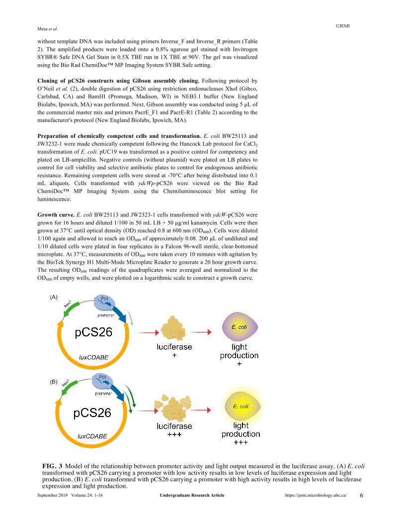
The method behind the luciferase assay involves cloning the promoter of a gene or operon of interest upstream of a promoterless luxCDABE operon in an expression vector such as pCS26 (1). pCS26 is a low copy-number bacterial expression vector which contains a kanamycin resistance gene cassette and promoter cloning sites in addition to a promoterless luxCDABE operon. The promoter of interest drives the expression of luciferase, which emits light that can then be measured by a luminometer (4). Promoter activity is correlated to the light output that results from luciferase expression under the control of the promoter of interest (4). Decreased promoter activity results in reduced expression of luciferase from the luxCDABE operon and thus reduced light, while increased promoter activity results in increased light production (Figure 3).
AcrS is a putative repressor encoded upstream of the acrEF operon, as seen in Figure 2A (2). We hypothesize that it represses the acrAB and acrEF operons (2), which encode the AcrAB and AcrEF multidrug efflux pumps (Figure 1). These pumps actively export a wide range of antibiotics, providing the bacterium with intrinsic drug resistance (2). Since AcrS can decrease antibiotic resistance by repressing the expression of the acrAB and acrEF operons, its activity has clinical applications. In this study, we proposed positive and negative controls that can be applied to a variety of biological models using AcrS repression as an example.
In the luciferase assay, a positive control produces consistent levels of light, while a negative control does not produce light regardless of the condition, such as the presence of AcrS. Our main aim was to evaluate controls within the context of AcrS repression by comparing luciferase activity resulting from promoter activity in the E. coli wild-type BW25113 and ΔacrS JW3232-1 strains from the Keio collection (5). To assess repression of the acrAB and acrEF operons, we also attempted to create pCS26 constructs with the promoters of acrAB (acrABp) and acrEF (acrEFp) fused upstream of the luxCDABE operon. We expect decreased light production from both constructs in the wild-type strain compared to the ΔacrS strain due to AcrS repression. We evaluated the suitability of
FIG. 1 Representation of the orientation of the AcrAB and AcrEF efflux pumps in the membrane of E. coli (15).

UJEMI Mesa et al.
September 2019 Volume 24: 1-16 Undergraduate Research Article https://jemi.microbiology.ubc.ca/ 3
ydcWp-pCS26 as a positive control, which ensures AcrS is not causing global repression. ydcWp-pCS26 contains the promoter of ydcW, an aminobutyraldehyde dehydrogenase gene, fused upstream of the luxCDABE operon in pCS26 (6). We propose that ydcW is a potential positive control that is unaffected by AcrS and thus produces consistent levels of light in the luciferase assay in the wild-type and ΔacrS strains (7). We attempted to create a negative control by cloning a random non-promoter fragment upstream of the luxCDABE operon in pCS26. Since it is a promoterless vector, it should not produce light in either strain and act as a negative control. We hypothesize that ydcWp-pCS26 and promoterless pCS26 are controls that can be used in future luciferase assays studying gene regulation in a variety of biological systems. METHODS AND MATERIALS
Bacterial strains, plasmids, and growth conditions. Refer to Table 1 for the strains used. All E. coli strains were grown in Luria-Bertani (LB) broth made following the Hancock Lab recipe on a shaker at 200 rpm at 37°C for 16 hours. LB was supplemented with the appropriate antibiotic (ampicillin at 100 µg/mL and kanamycin at 50 µg/mL) for the cells transformed with vectors. pCS26 vector (Table 1) is a 9.3 kb low copy-number pZS derivative with strong transcriptional terminators and unique promoter cloning sites (XhoI and BamHI) for insertion upstream of the luxCDABE operon (2). ydcWp-pCS26 (Table 1) contains the promoter of ydcW from a Salmonella enterica serovar Typhimurium 14028 reporter library (8). pUC19 plasmid (Table 1) is a 2.7 kb high copy-number empty backbone cloning vector used as a positive control during transformation (9).
Plasmid preparation of pUC19 and pCS26 from host strains. Plasmid host strains were grown in 10 mL of LB with the appropriate antibiotic for 16 hours at 37°C with shaking (200 rpm). 10 mL of cell culture was used for plasmid isolation using the Bio Basic EZ-10 Spin Column Plasmid DNA Miniprep Kit (Bio Basic #BS654) as per manufacturer’s instructions. To optimize the yield of plasmid DNA, 10 mL of overnight culture was separated into two 5 mL parts during the addition of Solutions I, II and III. The supernatants were then concentrated into one spin column for subsequent steps. The resulting plasmid yield and concentration were assessed using the Thermo Scientific NanoDrop 2000 before storage at -20°C.
Genotypic confirmation of Keio strains. Following Hay et al. protocol (10), genomic PCR amplification was performed on E. coli BW25113 and JW3232-1 using primers flanking acrS (Table 2). PCR amplicons were loaded onto a 1% agarose gel stained with SYBR® Safe DNA Gel Stain in 0.5X TBE (Invitrogen) run at 100V. Amplicons were visualized using the Bio Rad ChemiDoc™ MP Imaging System.
FIG. 2 Proposed model of AcrS repression of the acrAB and acrEF operons and structure of pCS26 constructs. (A) AcrS is encoded upstream of the acrEF operon and hypothesized to repress acrEF and acrAB (2). (B) Adapted sequence map of pCS26 (Addgene plasmid # 47640) with a promoter insertion site upstream of the luxCDABE operon (2, 16).

UJEMI Mesa et al.
September 2019 Volume 24: 1-16 Undergraduate Research Article https://jemi.microbiology.ubc.ca/ 4
Colony PCR and sequencing of the pCS26 cloning site. Colony PCR was performed using pCS26 FWD and REV primers amplifying a 250 bp (Table 2) region flanking the XhoI and BamHI promoter insertion region in pCS26. PCR was performed in 25 µL reactions using the Platinum® Taq DNA Polymerase (Invitrogen) as per manufacturer instructions. Addition of template DNA was performed by touching an individual colony with a micropipette tip and inoculating the reaction mix contained in a PCR tube. A negative control without DNA and positive control using E. coli DH5⍺ carrying pUC19 with universal primers were also performed (Table 2). A lysis step of 3 min at 98°C was performed on the mixtures in the thermocycler. Thermocycler conditions were set according to the manufacturer recommendations with the appropriate annealing temperature for the primers (Table 2) to perform 40 cycles of PCR amplification. PCR products were then loaded onto a 1% agarose gel stained with SYBR® Safe DNA Gel Stain in 0.5X TBE (Invitrogen) run at 100V. Amplicons were visualized using the Bio Rad ChemiDoc™ MP Imaging System. Plasmid constructs were then prepared according to GENEWIZ Sanger Sequencing requirements for sequencing. The results of sequencing were identified using NCBI Nucleotide BLAST.
Inverse PCR of pCS26 to generate promoterless pCS26 as a negative control for the luciferase assay. Inverse PCR was performed using primers (Inverse_F and Inverse_R in Table 2) which amplify the entire ydcWp-pCS26 vector or gel purified XhoI and BamHI-digested pCS26 vector. The primers introduced a short, random non-promoter fragment in the region between the XhoI and BamHI promoter insertion sites and were designed to insert EcoRI restriction enzyme digestion sites at the 5’ ends for subsequent circularization of the amplicons. PCR was performed in 20 µL volumes following manufacturer instructions for Phusion High-Fidelity DNA Polymerase (Thermo Fisher Scientific) with 0% or 3% DMSO addition. Recommended thermocycler settings were used for reverse touchdown PCR with an initial 5 cycles at an annealing temperature of 57°C, followed by 35 cycles with a gradient of annealing temperatures from 60°C to 75°C. All extensions were extended to 6 minutes to ensure sufficient time for extension. ydcWp-pCS26 vector with pCS26 FWD and REV primers (Table 2) was used as a positive control. A negative control

UJEMI Mesa et al.
September 2019 Volume 24: 1-16 Undergraduate Research Article https://jemi.microbiology.ubc.ca/ 5 without template DNA was included using primers Inverse_F and Inverse_R primers (Table
UJEMI Mesa et al.
September 2019 Volume 24: 1-16 Undergraduate Research Article https://jemi.microbiology.ubc.ca/ 6
without template DNA was included using primers Inverse_F and Inverse_R primers (Table 2). The amplified products were loaded onto a 0.8% agarose gel stained with Invitrogen SYBR® Safe DNA Gel Stain in 0.5X TBE run in 1X TBE at 90V. The gel was visualized using the Bio Rad ChemiDoc™ MP Imaging System SYBR Safe setting.
Cloning of pCS26 constructs using Gibson assembly cloning. Following protocol by O’Neil et al. (2), double digestion of pCS26 using restriction endonucleases XhoI (Gibco, Carlsbad, CA) and BamHI (Promega, Madison, WI) in NEB3.1 buffer (New England Biolabs, Ipswich, MA) was performed. Next, Gibson assembly was conducted using 5 µL of the commercial master mix and primers PacrE_F1 and PacrE-R1 (Table 2) according to the manufacturer's protocol (New England Biolabs, Ipswich, MA).
Preparation of chemically competent cells and transformation. E. coli BW25113 and JW3232-1 were made chemically competent following the Hancock Lab protocol for CaCl2 transformation of E. coli. pUC19 was transformed as a positive control for competency and plated on LB-ampicillin. Negative controls (without plasmid) were plated on LB plates to control for cell viability and selective antibiotic plates to control for endogenous antibiotic resistance. Remaining competent cells were stored at -70°C after being distributed into 0.1 mL aliquots. Cells transformed with ydcWp-pCS26 were viewed on the Bio Rad ChemiDoc™ MP Imaging System using the Chemiluminescence blot setting for luminescence.
Growth curve. E. coli BW25113 and JW2323-1 cells transformed with ydcW-pCS26 were grown for 16 hours and diluted 1/100 in 50 mL LB + 50 µg/ml kanamycin. Cells were then grown at 37°C until optical density (OD) reached 0.8 at 600 nm (OD600). Cells were diluted 1/100 again and allowed to reach an OD600 of approximately 0.08. 200 µL of undiluted and 1/10 diluted cells were plated in four replicates in a Falcon 96-well sterile, clear-bottomed microplate. At 37°C, measurements of OD600 were taken every 10 minutes with agitation by the BioTek Synergy H1 Multi-Mode Microplate Reader to generate a 20 hour growth curve. The resulting OD600 readings of the quadruplicates were averaged and normalized to the OD600 of empty wells, and were plotted on a logarithmic scale to construct a growth curve.
FIG. 3 Model of the relationship between promoter activity and light output measured in the luciferase assay. (A) E. coli transformed with pCS26 carrying a promoter with low activity results in low levels of luciferase expression and light production. (B) E. coli transformed with pCS26 carrying a promoter with high activity results in high levels of luciferase expression and light production.

UJEMI Mesa et al.
September 2019 Volume 24: 1-16 Undergraduate Research Article https://jemi.microbiology.ubc.ca/ 7
Luciferase assay. E. coli BW25113 and JW2323-1 cells transformed with ydcW-pCS26 were used to measure ydcW promoter activity as counts per second (cps) of light in the BioTek Synergy H1 Multi-Mode Microplate Reader. Overnight cultures of chemically competent cells were diluted 1/100 in 50 mL LB + 50 µg/ml kanamycin. Cells were then grown at 37°C until OD600 reached 0.8. Cells were diluted 1/100 again and allowed to reach an OD600 of 0.08. 200 µL samples of undiluted and 1/10 diluted cells were plated in four replicates in a Costar 3603C 96-well sterile, black, clear-bottomed microplate. At 37°C, luminescence and OD600 readings were taken every 10 minutes after 10 seconds of agitation for a total of 16 hours. Gene expression was normalized to cell growth by dividing the luminescence (cps) by the OD600 value of each sample. The OD600 readings from the quadruplicates were averaged and normalized to OD600 of empty wells and plotted on a logarithmic scale. The luminescence readings were normalized to the luminescence of empty wells resulting from instrumental noise. These values were then normalized to OD600 of the cells measured at the same time point. RESULTS
E. coli strains BW25113 and ΔacrS JW3232-1 from the Keio collection carry the expected genotypes. We acquired the E. coli wild-type BW25113 and ΔacrS JW3232-1 strains and performed PCR to confirm the presence or absence of acrS using primers that flank acrS (10). The resulting amplicons were resolved on a 1% agarose gel (Figure 4). BW25113 carries the acrS gene, so its amplicon should be the combination of the 663 bp acrS gene and 466 bp primer flanking region, totaling approximately 1100 bp. JW3232-1 does not carry acrS, so its amplicon should only consist of the flanking regions and a short sequence remaining from the removal of the kanamycin resistance cassette from the original Keio JW3232 strain, totaling approximately 500 bp. The amplicons from E. coli BW25113 and JW3232-1 matched the expected sizes of approximately 1100 bp and 500 bp respectively, suggesting that both strains carry the expected genotype (Figure 4).
Identification of S. enterica promoter in pCS26 promoter insertion region. Prior to using the pCS26 vector as the negative control for the luciferase assay, we performed colony PCR on E. coli DH5⍺ cells carrying pCS26 to confirm that luxCDABE is promoterless. Primers flanking the region between the XhoI and BamHI promoter insertion
FIG. 4 Genotypic confirmation of the E. coli BW25113 and ΔacrS JW3232-1 Keio strains suggest they carry the expected genotypes. 1% agarose gel electrophoresis of the PCR products from amplification of the acrS gene and its flanking regions. The negative control controls for nonspecific amplification.

UJEMI Mesa et al.
September 2019 Volume 24: 1-16 Undergraduate Research Article https://jemi.microbiology.ubc.ca/ 8
sites were used (Table 2). The observed band of 1700 bp was larger than the expected 240 bp size of the region (Figure 6). To further investigate this, the amplicon was sent for Sanger sequencing. Sequencing data revealed a pre-existing 1448 bp promoter of the S. enterica serovar Typhimurium PLP-dependent aminotransferase family protein gene (accession number: QBG31913.1) inserted between the XhoI and BamHI sites upstream of the luxCDABE operon (Table S1). This insert must be removed to construct a negative control for the luciferase assay.
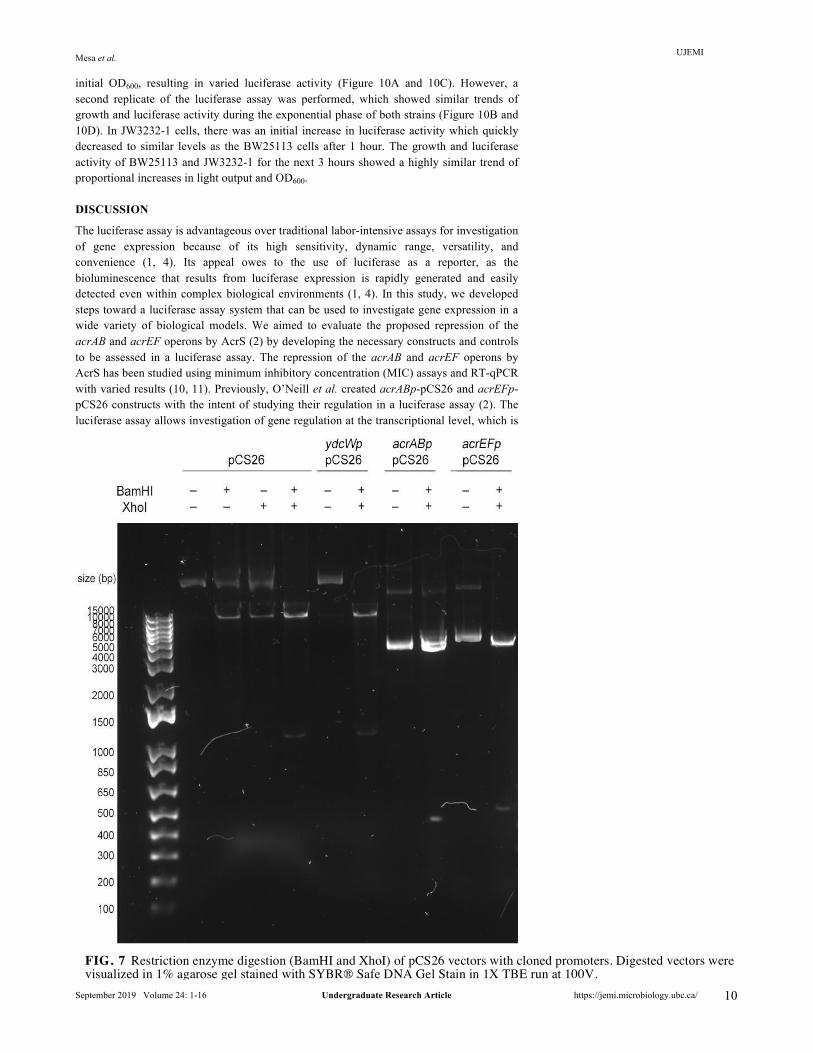
Partial degradation of pCS26 vector unlinked to restriction enzyme digestion was observed in Gibson cloning constructs. Gibson assembly protocol adapted from O’Neill et al. (2) was used to clone the promoters of the acrAB and acrEF operons upstream of the luxCDABE operon in pCS26. The cloned constructs were transformed into chemically competent E. coli DH5⍺ and allowed to grow overnight on LB plates supplemented with kanamycin for propagation and screening. No colonies were observed after transformation of the positive control supplied in the New England Biolabs Gibson Assembly Cloning Kit, suggesting cloning of the positive control was not successful. Subsequently, colonies carrying the acrAB and acrEF constructs were analyzed by restriction enzyme digestion (BamHI and XhoI). Successfully cloned constructs are expected to be digested into two fragments: about 500 bp acrAB/acrEF promoter and 9366 bp pCS26 vector. As shown in Figure 7, both acrABp-pCS26 and acrEFp-pCS26 digested by BamHI and XhoI separate into two fragments with approximate sizes of 4000 bp and 500 bp. The 500 bp fragment matches the expected size of the acrAB and acrEF operon promoters.
Transformation of ydcWp-pCS26 into E. coli BW25113 and JW3232-1 was also performed. ydcWp-pCS26 isolated from E. coli DH5⍺ was transformed into BW25113 and JW3232-1 to examine the validity of ydcWp-pCS26 as a positive control in the subsequent luciferase assay. After overnight growth, the transformed cells were visualized under a gel imager in the Chemiluminescence mode as an initial screen. All colonies appeared to emit light, suggesting that the transformation with ydcWp-pCS26 was successful (Figure 5). The darker spots in both plates in Figure 5 are due to confluent growth of cells in those areas.
FIG. 5 Chemiluminescence image of E. coli BW25113 and JW3232-1 cells transformed with ydcW -pCS26 indicates light production in both strains. Cells were imaged with the Bio Rad ChemiDoc™ MP Imaging System under the Chemiluminescence blot setting to measure luminescence.

UJEMI Mesa et al.
September 2019 Volume 24: 1-16 Undergraduate Research Article https://jemi.microbiology.ubc.ca/ 9
Inverse PCR failed to construct a negative control for the luciferase assay. The sequencing results for pCS26 indicate that removal of the S. enterica insert is required to produce a negative control for the luciferase assay. Attempts to construct the negative control with restriction enzyme digestion by XhoI and BamHI failed due to the difficulty of purifying sufficient amounts of the digested vector backbone for downstream ligation because of the large insert size of ~1500 bp. Hence, inverse PCR was attempted to amplify the entire pCS26 vector with primers designed to insert a short, random non-promoter fragment in the promoter insertion site and a unique EcoRI restriction site at the 5’ ends of amplicon to allow circularization. A gradient reverse touchdown PCR was performed to assess the optimal annealing temperatures for the amplification of the vector backbone. Despite the lack of primer dimers and the successful amplification of the positive control, gel electrophoresis of the PCR products revealed that the none of the optimization conditions successfully yielded a product (Figure 8).
Growth curve analysis of E. coli BW25113 and JW3232-1 after transformation with ydcWp-pCS26. Growth curve analysis was performed on E. coli BW25113 and JW3232-1 prior to the luciferase assay (Figure 9). Both strains were expected to share similar growth phenotypes in growth yield and rate, ensuring that differences in luciferase activity are physiological instead of due to growth and metabolic state. The growth curve suggests that the strains have a similar growth rate, indicated by the slopes of the curves during the exponential phase of growth. However, BW25113 appears to have a slightly higher growth yield than JW3232-1, as BW25113 and JW3232-1 in stationary phase have OD600 values of 1.14 and 1.06 respectively (Figure 9). Since the overall growth phenotype is comparable, the luciferase assay can still be performed to compare light output between the two strains. To account for the discrepancy in growth yield, optimization of the luciferase assay protocol was conducted to compare luciferase activity during the exponential phase of growth (about 0 to 4 hours), where both strains have similar growth rate and yield.
Luciferase assay to assess the suitability of ydcWp-pCS26 as a positive control. ydcWp-pCS26 was transformed into the E. coli wild-type BW25113 and ΔacrS JW3232-1 cells. Luciferase assay analysis was performed to compare the luciferase activity between the two strains in the exponential phase of growth. To optimize the assay, a 16-hour measurement of luminescence (cps) and OD600 was conducted to determine the exponential phase and optimal concentration of the cell cultures. Data for the first four hours of growth of the 1/10 diluted cells were used to compare luciferase activity between the strains. We see in Figure 10 that luciferase activity of ydcWp-pCS26 was similar in BW25113 and ΔacrS JW3232-1 during the exponential phase of growth. In the first assay replicate, BW25113 had a higher
FIG. 6 Characterization of a S. enterica insert within the promoter insertion site of pCS26. 1.5% agarose gel electrophoresis of the colony PCR product resulting from amplification of the promoter insertion region between the BamHI and XhoI sites in pCS26. Sequencing results identified the presence of the promoter of a S. enterica gene.
UJEMI Mesa et al.
September 2019 Volume 24: 1-16 Undergraduate Research Article https://jemi.microbiology.ubc.ca/ 10
initial OD600, resulting in varied luciferase activity (Figure 10A and 10C). However, a second replicate of the luciferase assay was performed, which showed similar trends of growth and luciferase activity during the exponential phase of both strains (Figure 10B and 10D). In JW3232-1 cells, there was an initial increase in luciferase activity which quickly decreased to similar levels as the BW25113 cells after 1 hour. The growth and luciferase activity of BW25113 and JW3232-1 for the next 3 hours showed a highly similar trend of proportional increases in light output and OD600.
DISCUSSION
The luciferase assay is advantageous over traditional labor-intensive assays for investigation of gene expression because of its high sensitivity, dynamic range, versatility, and convenience (1, 4). Its appeal owes to the use of luciferase as a reporter, as the bioluminescence that results from luciferase expression is rapidly generated and easily detected even within complex biological environments (1, 4). In this study, we developed steps toward a luciferase assay system that can be used to investigate gene expression in a wide variety of biological models. We aimed to evaluate the proposed repression of the acrAB and acrEF operons by AcrS (2) by developing the necessary constructs and controls to be assessed in a luciferase assay. The repression of the acrAB and acrEF operons by AcrS has been studied using minimum inhibitory concentration (MIC) assays and RT-qPCR with varied results (10, 11). Previously, O’Neill et al. created acrABp-pCS26 and acrEFp-pCS26 constructs with the intent of studying their regulation in a luciferase assay (2). The luciferase assay allows investigation of gene regulation at the transcriptional level, which is
FIG. 7 Restriction enzyme digestion (BamHI and XhoI) of pCS26 vectors with cloned promoters. Digested vectors were visualized in 1% agarose gel stained with SYBR® Safe DNA Gel Stain in 1X TBE run at 100V.

UJEMI Mesa et al.
September 2019 Volume 24: 1-16 Undergraduate Research Article https://jemi.microbiology.ubc.ca/ 11
more representative than analysis at the protein level such as in MIC assays, as post-translational effects can interfere with measurements. The hypothesized repression can be investigated using E. coli wild-type BW25113 and ΔacrS JW3232-1 strains from the Keio collection. The Keio collection is comprised of single-gene deletion mutants from the parent E. coli K-12 strain BW25113 (5). We hypothesized that ydcW-pCS26 and promoterless pCS26 are universal as positive and negative controls respectively and thus can be used to study gene expression within complex biological contexts such as using Keio strains in future luciferase assay experiments.
Our attempts at creating the pCS26 constructs with acrAB and acrEF promoters required to evaluate the biological question of AcrS repression failed. We see in Figure 7 that both digested acrABp-pCS26 and acrEFp-pCS26 have a 4000 bp fragment which is smaller than the expected 9366 bp pCS26 vector, suggesting it is partially degraded. This was not observed in digestion of pCS26 containing the S. enterica promoter or ydcWp-pCS26, which both produce an approximately 10 kb pCS26 fragment after digestion by BamHI and XhoI (Figure 7). Hence, the observed partial degradation of the pCS26 vector is not a result of restriction enzyme digestion. Since O’Neill et al. (2) previously used the same Gibson cloning protocol without observation of unexpected cleavage of the vector, it is possible that failure occurred due to expiration of the cloning kit. This may have interfered with the interactions between the three enzymes involved in Gibson assembly cloning, causing a larger than expected portion of pCS26 to be cleaved (Figure 7). This is supported by the failed cloning of the positive control supplied by the manufacturers. While plasmid secondary structures may cause the appearance of different sizes in gel electrophoresis, the approximately 10kb fragment expected was observed in digested ydcWp-pCS26 and pCS26 carrying the S. enterica insert. Thus, we suspect that partial degradation of pCS26 occurred during Gibson cloning. Additional analysis of the primers used during Gibson cloning revealed that the primers were designed to insert the promoter of acrAB into pCS26 in an incorrect orientation. Hence, the primers need to be redesigned in the correct direction in future studies.
Since sequencing revealed a S. enterica promoter in pCS26, a promoterless pCS26 vector must be constructed to act as a negative control for the luciferase assay. Inverse PCR was attempted using primers designed to amplify the entire pCS26 vector and introduce both a non-promoter insert and EcoRI restriction sites for subsequent circularization of the plasmid. As seen in Figure 8, PCR amplification failed. Gel electrophoresis of the PCR products revealed that none of the optimization conditions allowed yield of amplified pCS26 containing a non-promoter insert. No primer dimers were observed, and the positive control was successfully amplified. Therefore, the inverse PCR
FIG. 8 Inverse PCR attempt to create pCS26 with non-promoter insert as a negative control for the luciferase assay. PCR products were visualized in 0.8% agarose gel stained with SYBR® Safe DNA Gel Stain in 1X TBE run at 90V. Lane 1 to Lane 5 correspond to annealing temperature at a gradient of 60°C to 75°C.

UJEMI Mesa et al.
September 2019 Volume 24: 1-16 Undergraduate Research Article https://jemi.microbiology.ubc.ca/ 12
protocol was not the cause of amplification failure. Instead, low processivity of DNA polymerase may have caused failure, as pCS26 is a large, low copy-number plasmid, which can cause difficulties during cloning. Its large size of approximately 10kb results in an increased chance of unexpected reactions such as non-specific interactions and interference of DNA polymerase by secondary structures. In addition, pCS26 is difficult to propagate due to its low yield, as seen in the failed gel extraction of the digested vector. Other methods of creating a negative control may be possible, such as repressing ydcW. However, this may have significant consequences on cell growth, as ydcW allows optimal growth (6). Despite failures in Gibson cloning, this method should be reattempted to construct acrABp-pCS26, acrEFp-pCS26, and promoterless pCS26 as a negative control, as it was successfully performed by O’Neill et al. (2) and the size and yield of pCS26 can cause difficulties in other methods of cloning.
To determine whether AcrS repression can be assessed using E. coli BW25113 and JW3232-1, we performed growth curve analysis to evaluate whether they are comparable in growth rate and yield. We determined that growth rate and yield of the two strains following exponential phase are not similar enough for accurate comparison. After 4 hours of growth, the growth rate of E. coli BW25113 is greater than that of JW3232-1 (Figure 9). Both strains enter stationary phase at approximately 15 hours of growth, but JW3232-1 has a lower growth yield compared to BW25113. These growth yields are compatible with past studies (5). Large discrepancies in growth phenotype could cause differences in light output that may be confused for effects of gene regulation and promoter activity. To mitigate this, the luciferase assay protocol was optimized to perform during the exponential phase (first four hours) in which negligible discrepancies in growth phenotype between BW25113 and JW3232-1 are observed. Optimization of cell concentration and normalization of light output to OD600 further minimized the effect of extraneous factors and differences in cell growth. Overall, BW25113 and JW3232-1 can be compared in a luciferase assay to investigate AcrS repression.
The luciferase assay was performed to determine whether ydcWp-pCS26 is an appropriate positive control for investigating AcrS repression of the acrAB and acrEF
FIG. 9 Growth curve of undiluted E. coli BW25113 and JW3232-1 transformed with ydcWp-pCS26. OD600 of the cells was normalized to that of empty wells and plotted on a logarithmic scale to visualize the growth of the cells over 22 hours. Growth rate and yield are represented by the slope and the maximum y-value of the growth curve respectively.

UJEMI Mesa et al.
September 2019 Volume 24: 1-16 Undergraduate Research Article https://jemi.microbiology.ubc.ca/ 13
operons. The assay was repeated twice with measurements taken every 10 minutes to ensure reproducibility of the data. We see in Figure 10 that light output increases proportionally as cells grow, which supports that the luciferase reaction generates sustained light (1). Both replicates generally suggested that luciferase activity generated by ydcWp-pCS26 in E. coli BW25113 and JW3232-1 are comparable in the exponential phase of growth. Luciferase activity in JW3232-1 appears low from 1 to 3 hours of growth during the first assay, but we suspect that this is caused by the lower relative growth of the strain (Figure 10A and 10C). However, luciferase activity in JW3232-1 quickly increases during the fourth hour of growth, suggesting that ydcW may be repressed by AcrS in this time period. The lower growth rate and yield of JW3232-1 in the exponential phase of growth compared to BW25113 was not previously observed in the growth curve analysis (Figure 9), potentially affecting measurements of luciferase activity. Thus, a second replicate was performed to investigate the reproducibility of the trend. During the second replicate, an early peak in
FIG. 10 Normalized luciferase activity and optical density of 1/10 diluted E. coli BW25113 and JW3232-1 cells carrying ydcWp-pCS26 during exponential phase of growth. Luminescence of samples was normalized to the luminescence of empty wells and subsequently to OD600 at each time point. The OD600 readings of the two strains were normalized to empty wells and plotted on a logarithmic scale. (A) Luciferase activity of the first replicate. (B) Luciferase activity of the second replicate. (C) OD600 of the first replicate. (D) OD600 of the second replicate.

UJEMI Mesa et al.
September 2019 Volume 24: 1-16 Undergraduate Research Article https://jemi.microbiology.ubc.ca/ 14
luciferase activity was observed in the JW3232-1 strain which is much greater than that of the BW25113 strain (Figure 10B). After the first hour, luminescence returned to similar levels as BW25113. We hypothesize that this is a result of placing JW3232-1 cells on ice prior to the luciferase assay. The cold temperatures induced a stress response in cells, which in turn overactivated the ydcW promoter and thus luciferase expression. It is also possible that luciferase activity is activated by cold temperatures, but this is not supported by literature (12). ydcW is an aminobutyraldehyde dehydrogenase which catalyzes the oxidation of 1-pyrroline as part of putrescine degradation (7). Putrescine degradation contributes to proline accumulation (13), which is thought to be protective in stressful environmental conditions such as cold temperatures (13, 14). We suspect that cold temperatures induced ydcW expression for proline accumulation, leading to increased promoter activity and thus luciferase expression in the form of light. As cells were returned to optimal temperatures (37°C) during the luciferase assay, ydcW expression normalized and luciferase activity decreased to similar levels with the BW25113 cells that were not placed on ice. Since the luciferase activities in the wild-type and ΔacrS strains are highly similar during the rest of the exponential phase of growth, we suggest that ydcW is not repressed by the presence of AcrS. Thus, ydcWp-pCS26 is a suitable positive control for the luciferase assay in investigating acrAB and acrEF regulation.
Since ydcW is involved in metabolism, it has the potential to be a universal positive control that can be used in luciferase assay experiments in the context of a wide variety of biological systems. Similar to constitutively expressed housekeeping genes which are commonly used as positive controls, ydcW is critical for optimal cell growth; ydcW deletion mutants survive with slowed growth (6, 13, 14). Thus, promoter activity varies with growth state, in that it is enhanced in the exponential phase and reduced during stationary phase as reflected by the luciferase assays performed (5). Use of ydcWp-pCS26 as a positive control may be limited to investigations of genes expressed during the exponential phase of growth or strains that have highly similar growth phenotypes. Future experiments should first confirm the suitability of ydcWp-pCS26 in a luciferase assay as performed in this study prior to use as a positive control. Overall, with construction of pCS26 containing a non-promoter insert, these positive and negative controls form a luciferase assay system which can be applied to future investigations of regulation of acrAB and acrEF or a variety of other biological models.
Conclusions In this study, we developed steps toward a luciferase assay system that can be used to investigate gene regulation in a variety of biological models, such as the repression of the acrAB and acrEF operons by AcrS. We characterized a S. enterica promoter insert in pCS26 vector which drives luciferase expression. We failed to create the acrABp-pCS26 and acrEFp-pCS26 constructs required to evaluate the biological question. However, the Gibson cloning technique we used should be optimized in future experiments, as it is suited to cloning of complex vectors such as pCS26. This technique can also be applied to construct a negative control for the luciferase assay consisting of pCS26 with a non-promoter insert. We confirmed the comparability of growth phenotype in E. coli wild-type BW25113 and ΔacrS JW3232-1, determining that these strains can be effectively compared in a luciferase assay. We concluded that ydcWp-pCS26 is a suitable positive control for investigating AcrS repression in a luciferase assay. Furthermore, we proposed that ydcWp-pCS26 is a universal positive control that can be used to investigate gene expression for other biological models. Future experiments can use ydcWp-pCS26 and an appropriate negative control in an adaptable luciferase assay system. Future Directions Using AcrS repression of the acrAB and acrEF operons as a biological model, we have attempted to develop a luciferase assay system that can be used to investigate gene expression. We concluded that the growth phenotypes of the E. coli wild-type BW25113 and ΔacrS JW3232-1 strains are comparable, suggesting future studies can use these strains to evaluate the repression of gene expression by AcrS such as acrAB and acrEF operons. We determined that ydcWp-pCS26 is a suitable positive control for investigating AcrS repression using a luciferase assay, as it produces consistent levels of light in the presence and absence of AcrS. We propose that it can be used universally for
UJEMI Mesa et al.
September 2019 Volume 24: 1-16 Undergraduate Research Article https://jemi.microbiology.ubc.ca/ 15
assays studying other biological systems during the exponential phase. Future studies can similarly assess its suitability in the context of the biological model of interest as a potential positive control for the luciferase assay. We were not successful in constructing pCS26 with a promoterless luxCDABE operon. Future studies that plan to use a luciferase assay should first construct and characterize this negative control. This construct should not produce light in any condition in the luciferase assay. Once constructed, the promoterless pCS26 can also serve as a backbone for subsequent cloning, mitigating potential interference due to the large S. enterica promoter insert. The positive and negative controls described in this study effectively create a luciferase assay system that can be used by future studies to investigate gene expression in a wide variety of biological models. To complete the assessment of AcrS repression of the acrAB and acrEF operons, future studies should clone the acrAB and acrEF promoters into pCS26, transform these constructs into the E. coli ∆acrS JW3232-1 and wild-type BW25113 strains, and perform the luciferase assay with the positive and negative controls as outlined in this study. If AcrS represses the acrAB and acrEF operons, the light output measured in the luciferase assay will be reduced in the BW25113 strain compared to the JW3232-1 strain, since promoter activity is suppressed. The luciferase assay can be repeated to ensure reliability and reproducibility of the results. Studies that desire to use the assay to investigate gene expression in other biological systems can follow the same general methods, where the promoter of interest is cloned into pCS26, constructs are transformed into the appropriate strains, and the luciferase assay is performed as described or with a commercial kit. Light output can then be correlated to promoter activity.
To construct the negative control, acrABp-pCS26, and acrEFp-pCS26, future studies should use the Gibson cloning method mentioned in this study. While we failed to produce constructs using this protocol due to time constraints, it should be attempted with a non-expired commercial Gibson assembly cloning kit to ensure the functioning of the enzymes involved. Additionally, primers targeting the acrAB should be designed to insert the promoter in the correct orientation, ensuring that it is able to drive expression of the luxCDABE operon. pCS26 is a large plasmid with low yield, creating challenges during cloning. Unlike other cloning methods such as inverse PCR, Gibson assembly cloning is feasible as it is more specific and suitable for working with large plasmids. Although further characterization of the vector backbone may allow successful cloning using other cloning methods, our results suggested that Gibson cloning inserted the promoters of the acrAB and acrEF operons into pCS26. Successful construction of the negative control, acrABp-pCS26, and acrEFp-pCS26 can be achieved by optimizing the protocol with new reagents. To create the negative control, primers similar to PacrA and PacrE (Table 2) can be designed, where the region binding the BW25113 acrAB or acrEF promoters is changed to a random non-promoter sequence which maintains the XhoI and BamHI restriction sites (for convenient subsequent cloning) and allows formation of primer dimers. The product can then be ligated into the digested pCS26 vector using Gibson cloning. This should generate pCS26 carrying a non-promoter insert that does not drive expression of the luxCDABE operon and can be used for future cloning and as a negative control in the luciferase assay. ACKNOWLEDGEMENTS
We would like to acknowledge our instructor Dr. David Oliver and teaching assistants Aaron Liu and Jessica Tuengel for their support in the planning and troubleshooting of this experiment. We would like to thank MICB421 students for sharing lab resources and advice throughout the term. We would also like to acknowledge Ryan Nah from the O’Neill et al. MICB421/447 group for his help during the planning of the project. Finally, we would like to thank Shiela and the rest of the Wesbrook Building media room staff for preparing and supplying equipment for us. CONTRIBUTIONS
The execution of the experiments and writing of assignments throughout the term was a collective effort of all authors. All authors helped in the attempt to construct acrABp-pCS26 and acrEFp-pCS26. Generally, negative control construction was planned and attempted by Annie Yip and Ada Zhang. Assessment of the positive control and growth phenotypes of the Keio strains was primarily conducted by Ru Lan Xu and Adam Mesa. All authors contributed to editing of this manuscript.

UJEMI Mesa et al.
September 2019 Volume 24: 1-16 Undergraduate Research Article https://jemi.microbiology.ubc.ca/ 16
REFERENCES
1. Allard, STM, Kopish, K. 2008. Luciferase reporter assays: powerful, adaptive tools for cell biology research. Promega Corporation.
2. O’Neill, Z, Bulka, O, Marziali, M, Nah, R. 2018. Construction of luxCDABE reporter plasmids to investigate regulation of the acrAB and acrEF operons by the AcrS repressor in Escherichia coli BW25113. JEMI 4:1-9.
3. Craney, A, Hohenauer, T, Xu, Y, Navani, NK, Li, Y, Nodwell, J. 2007. A synthetic luxCDABE gene cluster optimized for expression in high-GC bacteria. Nucleic Acids Res 35:e46.
4. Ávila-Flores, A, Arranz-Nicolás, J, Mérida, I. 2019. Transcriptional activity of FOXO transcription factors measured by luciferase assays. Methods Mol Biol 1890:91–102.
5. Baba, T, Ara, T, Hasegawa, M, Takai, Y, Okumura, Y, Baba, M, Datsenko, KA, Tomita, M, Wanner, BL, Mori, H. 2006. Construction of Escherichia coli K-12 in-frame, single-gene knockout mutants: the Keio collection. Mol Syst Biol 2:2006.0008.
6. Prieto, MI, Martin, J, Balaña-Fouce, R, Garrido-Pertierra, A. 1987. Properties of γ-aminobutyraldehyde dehydrogenase from Escherichia coli. Biochimie 69:1161-1168.
7. Samsonova, NN, Smirnov, SV, Novikova, AE, Ptitsyn, LR. 2005. Identification of Escherichia coli K12 YdcW protein as a gamma-aminobutyraldehyde dehydrogenase. FEBS Lett 579:4107–4112.
8. Beeston, AL, Surette, MG. 2002. pfs-dependent regulation of autoinducer 2 production in Salmonella enterica serovar Typhimurium. J Bacteriol 184:3450–3456.
9. Yim, G, de la Cruz, F, Spiegelman, GB, Davies J. 2006. Transcription modulation of Salmonella enterica serovar Typhimurium promoters by sub-MIC levels of rifampin. J Bacteriol 188:7988–7991.
10. Hay, M, Li, MY, Ma, Y. 2017. Deletion of AcrS Results in increased expression of acrE and confers an increase in kanamycin resistance in Escherichia coli BW25113. JEMI 3:63-69.
11. Hirakawa, H, Takumi-Kobayashi, A, Theisen, U, Hirata, T, Nishino, K, Yamaguchi, A. 2008. AcrS/EnvR represses expression of the acrAB multidrug efflux genes in Escherichia coli. J Bacteriol 190:6276–6279.
12. Dorn, JG, Frye, RJ, Maier, RM. 2003. Effect of temperature, pH, and initial cell number on luxCDABE and nah gene expression during naphthalene and salicylate catabolism in the bioreporter organism Pseudomonas putida RB1353. Appl Environ Microbiol 69:2209–2216.
13. Su, GX, Bai, X. 2008. Contribution of putrescine degradation to proline accumulation in soybean leaves under salinity. Biol Plantarum 52:796.
14. Singh, S, Brocker, C, Koppaka, V, Chen, Y, Jackson, BC, Matsumoto, A, Thompson, DC, Vasiliou, V. 2013. Aldehyde dehydrogenases in cellular responses to oxidative/electrophilic stress. Free Radic Biol Med 56:89-101.
15. Pos, KM. 2009. Trinity revealed: Stoichiometric complex assembly of a bacterial multidrug efflux pump. Proc Natl Acad Sci U S A 106:6893–6894.
16. Shong, J, Huang, Y-M, Bystroff, C, Collins, CH. 2013. Directed evolution of the quorum-sensing regulator EsaR for increased signal sensitivity. ACS Chem Biol 8:789-795.