Embed Size (px)
Citation preview
Case Report
Stiff Heart Syndrome
Satya S. Bhupathi, MD, MPH; Sreelatha Chalasani, MD, MPH; and Roxann Rokey, MD, FACC, FASE
Corresponding Author Received: November 10, 2010 Dr. Sreelatha Chalasani Revised: June 25, 2010 Department of Internal Medicine Accepted: August 4, 2010 Marshfield Clinic 1000 North Oak Avenue doi:10.3121/cmr.2010.899 Marshfield, WI 54449 Tel.: 715-387-5537 Fax: 715-389-5757 E-mail: [email protected]
. Published online ahead of print September 17, 2010 as doi:10.3121/cmr.2010.899Rapid ReleaseCM&R
Copyright 2010 by Marshfield Clinic.
Bhupathi et al. doi:10.3121/cmr.2010.899
Abstract
Isolated cardiac amyloidosis, or “Stiff Heart Syndrome”, is a rare manifestation of amyloidosis.
Some degree of cardiac amyloid deposition is common in elderly patients, as reported in prior
post-mortem studies; however, isolated cardiac involvement with predominantly cardiac
symptoms and no evidence of systemic disease is a rare presentation. Establishing the correct
diagnosis, even with the use of extensive testing including amyloid typing, understanding the
clinical significance, and management can be challenging in such cases.
Keywords: Cardiac amyloidosis, Stiff heart syndrome, Diagnosis, Management
Stiff heart syndrome Page 2
Bhupathi et al.
eart failure is a common medical problem that is increasing both in incidence and
prevalence. Ischemic cardiomyopathy resulting from coronary artery disease
(CAD) is the most common cause of heart failure in the United States1. Non-
ischemic cardiomyopathy is characterized by the absence of CAD and includes dilated,
hypertrophic, and restrictive causes of cardiomyopathy. Out of these three types, restrictive
cardiomyopathy (RCM) is rare in the United States and most other industrial nations. Restrictive
cardiomyopathy is characterized by stiffening of the ventricular walls and loss of myocardial
flexibility due to infiltration by abnormal tissue, resulting in inadequate ventricular filling with
blood and eventually the loss in its ability to pump properly.
doi:10.3121/cmr.2010.899
Stiff heart syndrome Page 3
H
Restrictive cardiomyopathy is involved in approximately 5% of all primary myocardial diseases
and can be either due to idiopathic or secondary causes. Amyloidosis, hemochromatosis, and
sarcoidosis are among the most frequently encountered causes of secondary RCM. Apart from
these, secondary RCM is also caused by primary systemic sclerosis, carcinoid heart disease,
glycogen storage disease of the heart, radiation-induced heart disease, metastatic malignancy,
anthracycline toxicity, endomyocardial fibrosis, and Loeffler eosinophilic endomyocardial
disease. Restrictive cardiomyopathy shares similarities in clinical and hemodynamic profile with
constrictive pericarditis. Due to the difference in management, accurate diagnosis and
differentiation of these two conditions is necessary. We present two cases of isolated cardiac
amyloidosis.
Bhupathi et al. doi:10.3121/cmr.2010.899
Case Presentations
Case 1
A 74-year-old male was referred to the cardiology clinic following a witnessed episode of
syncope. The patient described his syncopal episodes, which have been occurring over several
years, as sudden onset with occasional symptoms of light-headedness. He denied symptoms of
chest pain and/or discomfort, palpitations, orthopnea, paroxysmal nocturnal dyspnea, or recent
change in exercise tolerance. Significant past medical history includes only hyperlipidemia. He
does not smoke, uses alcohol rarely, and denies significant exposure to chemicals. Physical
examination was normal, except for the cardiac examination which showed cardiomegaly.
Laboratory evaluations were within normal limits. Transthorasic echocardiogram revealed mild
to moderate concentric left ventricular (LV) hypertrophy with no pericardial effusion,
intracardiac masses, shunts, clots or vegetation. Dimensions of the cardiac chambers showed
normal left atrium with enlarged right atrium. The inter-ventricular septum was slightly
thickened, while the left ventricular end diastolic dimension and right ventricular end systolic
dimensions were normal. A tilt-table test was performed, including infusion of isoproterenol, and
was non-diagnostic of orthostatic hypotension.
As the patient continued to experience light-headedness, a work-up for restrictive
cardiomyopathy was initiated. Serum protein immuno-electrophoresis and serum free light chain
analysis were normal. Fat-pad biopsy showed no histopathologic abnormality and the Congo red
stain for amyloid was negative. Subsequently, the patient underwent a coronary angiogram, left
ventriculography, and right ventricle biopsy along with right and left heart catheterization. Apart
from 30% stenotic lesions in the proximal right coronary, proximal and mid left anterior
Stiff heart syndrome Page 4
Bhupathi et al. doi:10.3121/cmr.2010.899
descending artery, the remaining coronary arteries were disease free. Biplane left
ventriculography showed mild global LV hypokinesia. The endomyocardial biopsy showed
diffusely infiltrated myocardium with waxy, pale, eosinophilic material showing green
birefringence under standard polarized light and red under fluorescent light with a Texas red
filter, characteristic of amyloid (figure 1). Positive Congo red and sulfated Alcian blue stains
confirmed the presence of amyloid deposition. Immunohistochemical studies were performed on
paraffin sections using antibodies directed against serum amyloid P component (SAP),
transthyretin, kappa and lambda immunoglobulin free light chains, and serum amyloid A (SAA).
The amyloid deposits showed strong staining for transthyretin, with negative staining for SAP,
kappa and lambda light chains and for SAA. These results were consistent with transthyretin-
type amyloid deposition, which could represent either senile or familial amyloidosis. Bone
marrow aspiration, performed to rule out plasma-cell dyscrasias, showed normocellular marrow
with no evidence of amyloid deposition.
The patient was diagnosed with “Isolated Cardiac Amyloidosis” or “Stiff Heart Syndrome”, with
senile amyloid deposition. He was told there was no evidence of light chain disease, and that this
amyloidosis does not respond to chemotherapeutic interventions, but does have a relatively
favorable prognosis compared to amyloid light-chain (AL) cardiac amyloid. Median survival in
this group of patients without any chemotherapy is about seven years. Due to the relatively
favorable prognosis in this patient, no chemotherapy was initiated, and he continues follow-up
care in the cardiology clinic.
Stiff heart syndrome Page 5
Bhupathi et al. doi:10.3121/cmr.2010.899
Case 2
A 74-year-old male was referred to the cardiology clinic for a newly diagnosed congestive heart
failure (CHF). The patient presented with a three month history of progressively worsening
exertional dyspnea with marked physical activity limitation and bilateral symmetric lower
extremity edema. The patient denied symptoms of chest pain, palpitations, orthopnea,
paroxysmal nocturnal dyspnea, presyncopal symptoms or syncope. He had no history of
significant occupational chemical exposure and did not smoke or use alcohol. He had no history
of hypertension, diabetes mellitus, hyperlipidemia, or thyroid dysfunction.
Pulmonary examination revealed decreased breath sounds at the bilateral bases, with the left
more significant than the right. Cardiac examination showed a non-displaced point of maximal
impact. In the sitting position on forced expiration, a mild aortic insufficiency murmur was
auscultated. The remainder of his system review was normal.
An echocardiography (2D-Echo) showed depressed LV systolic function with regional wall
motion abnormalities involving the septal and inferior walls. Chest radiograph showed bilateral
pleural effusions with non-acute pulmonary parenchymal disease. He was started on an
angiotensin converting enzyme (ACE) inhibitor (lisinopril) and diuretic (torsemide) prior to this
referral.
The patient underwent elective coronary angiography and left ventriculography with right and
left heart catheterization. Biplane left ventriculography showed diffuse LV hypokinesia. Overall,
the findings were non-diagnostic for either pure restrictive or constrictive physiology, although
Stiff heart syndrome Page 6
Bhupathi et al. doi:10.3121/cmr.2010.899
mild restrictive physiology could not be completely ruled out. The patient was diagnosed with
idiopathic cardiomyopathy and started on a beta blocker (metoprolol succinate) and digoxin
along with his prior medications. A 2-month follow-up echocardiogram showed normal LV
function, normal regional wall motion, normal LV ejection fraction (LVEF), and moderate LV
hypertrophy. He returned to his primary care physician for heart failure management.
Three years after being diagnosed with idiopathic cardiomyopathy, the patient was re-referred to
cardiology with a 2-month history of acutely decompensated heart failure symptoms and
epigastric discomfort. Resting EKG showed atrial fibrillation (AF) with ventricular rate of 93
bpm and no associated sign of ischemia, injury, or infraction. Repeat laboratory evaluations were
within normal limits. 2D-echocardiography was repeated, showing mean right heart pressures,
left heart pressures, cardiac output, and cardiac index were comparably similar to earlier
findings. These findings suggested that the patient had a repeat myopathic process of unclear
etiology, now in the presence of AF.
After one month of therapeutic anticoagulation with warfarin, the patient underwent trans-
esophageal echocardiography (TEE). The TEE revealed LVEF of 35% and no thrombus. The
patient’s rhythm was converted to normal sinus rhythm by electric-cardio-version using a single
shock of 300-joules of synchronized direct current. Also, an abdominal fat pad biopsy was
performed, which showed normal adipose tissue and negative Congo red staining with no
histological evidence for other infiltrative diseases.
Stiff heart syndrome Page 7
Bhupathi et al. doi:10.3121/cmr.2010.899
This patient continued to have decompensated CHF with NYHA Class III-IV symptoms. Repeat
right heart catheterization with endomyocardial biopsy was performed. The histopathological
findings of the biopsy were highly consistent for amyloidosis (figure 2) with a positive Congo
red staining and apple green birefringence (figure 3). The patient was diagnosed with “Isolated
Cardiac Amyloidosis” or “Stiff Heart Syndrome”.
Considering the patient’s age and co-morbidities, he was not found to be an appropriate
candidate for heart-lung transplant. He underwent chemotherapy with melphalan and
dexamathasone (MDex) therapy. Chemotherapy with MDex is standard therapy for primary AL
amyloidosis patients who have not undergone or are not appropriate candidates for stem cell
transplant.2 Unfortunately, after the first cycle of chemotherapy, he developed melena requiring
blood transfusion. After considering risk versus benefit with poor tolerance to chemotherapy, it
was agreed to discontinue further chemotherapy and continue medical monitoring.
Five years after the initial onset of heart failure and twelve months after the biopsy-proven
diagnosis of isolated cardiac amyloidosis, or stiff heart syndrome complicated by AF, he
continues to follow-up in the cardiology clinic. His heart failure is compensated with NYHA
Heart Failure Class II, currently on furosemide, lisinopril and metolazone. For AF, rate control is
achieved with carvedilol along with continuation of anticoagulation.
Discussion
Amyloidosis is a relatively uncommon, mysterious, and under-diagnosed disease associated with
extracellular deposition of amorphous, homogenous, insoluble, fibrillar proteinaceous material in
Stiff heart syndrome Page 8
Bhupathi et al. doi:10.3121/cmr.2010.899
various organs of the body. Several types of proteins including light chain immunoglobulins,
transthyretin, acute-phase reactants, and apoprotein A have been reported to cause amyloidosis.3
All amyloid deposits share a characteristic microscopic beta pleated structure with positive
Congo red staining and apple green bifringence under polarizing light.3 Liver, spleen, and kidney
are the most commonly associated organs, however amyloid depositions have been identified in
virtually every organ of the body. Symptoms and clinical presentation are characterized by the
type of deposition and organ of significant involvement.
Cardiac amyloidosis or “stiff heart syndrome” is characterized by extracellular amyloid
infiltration of the myocardial tissue. Though deposition of amyloid in the hearts of elderly
asymptomatic populations is not uncommon, symptomatic isolated cardiac amyloidosis is
infrequent.4 Frequently, cardiac amyloidosis coexists with evident dysfunction of other major
organs and is identified with multi-organ involvement. Symptoms associated with cardiac
amyloidosis simulate cardiomyopathy, coronary heart disease, valvular heart disease, coronary
artery disease, or arrhythmia.5-7 It is estimated that 10% of cases of non-ischemic
cardiomyopathy are secondary to cardiac amyloidosis.8 Frequently reported symptoms of cardiac
amyloidosis are predominantly associated with right heart failure, including atypical chest pain,
dyspnea on exertion, fatigue, peripheral edema, and palpitations.9 Purpura is another frequently
reported symptom of cardiac amyloidosis, but typically only in primary AL amyloidosis
patients.10 By the time a patient becomes symptomatic, it is estimated that 25% of the myocardial
mass is infiltrated with amyloid deposition.11 However, due to the high prevalence of coronary
heart disease, it is imperative to rule this out prior to initiating workup for cardiac amyloidosis.
Stiff heart syndrome Page 9
Bhupathi et al. doi:10.3121/cmr.2010.899
Isolated cardiac amyloidosis, without clear cut evidence of other organ involvement, is possible.9
Iqbal et al12 reported a unique case with amyloid deposition on the aortic valve, with no evidence
of systemic disease. In an autopsy study of 100 patients, isolated atrial amyloid deposition was
identified in more than 75% of patients over age 50.13 These depositions were of a higher grade
in females when compared to males and associated with a risk of chronic AF.14 Chronic AF is
noted in 10% to 20% of cases with cardiac amyloidosis.14
Over the years, several types of protein depositions have been identified and linked to subtypes
of amyloidosis.7 Almost all forms of amyloidosis, including primary AL, hereditary, and “senile”
systemic can cause cardiomyopathy and/or heart failure. The most common type associated with
cardiac involvement (up to 85% ) is primary amyloidosis, associated with the deposition of AL
amyloid protein. This protein is made of either lambda-(Igλ) or kappa-(Igκ) light chain
immunoglobulins.14 Primary amyloidosis is the most common form of amyloidosis, involves
more organ disease, progresses quickly, and is commonly seen in patients with multiple
myeloma.15 Familial amyloidosis, such as ApoA1 type, and “senile” systemic amyloidosis, such
as TTR type, are the second most frequent types and are caused by the deposition of transthyretin
or ATTR amyloid protein, and are associated with 10% of cardiac amyloidosis cases.16,17 Cardiac
amyloidosis secondary to chronic inflammation due to diseases like rheumatoid arthritis,
tuberculosis, and Crohn’s disease is rare. This form of amyloidosis is associated with the
deposition of an acute phase reactant protein called serum amyloid A (SAA) protein.7,18
Secondary amyloidosis almost never affects the heart in a clinically significant manner.7,18,19
Isolated atrial amyloidosis (IAA) results in amyloid depositions which are immuno-reactive for
atrial natriuretic peptide (ANP). In one study, out of 245 atrial appendages isolated from patients
Stiff heart syndrome Page 10
Bhupathi et al. doi:10.3121/cmr.2010.899
undergoing open heart surgery, Rocken et al20 reports 16.3% (40 of 245 cases) of cases as having
amyloid deposition. Of these 245 patients, 38 (15.5%) had AF with a statistically significant
correlation independent of age and gender for the association of amyloid deposition and atrial
fibrillation.20 Cardiac involvement in the other subtypes of amyloidosis, including dialysis-
associated amyloidosis, familial Mediterranean fever, and familial corneal amyloidosis, is rare
and generally a late development in the disease process. 21-24
Increased risk of AF has been reported with AL amyloidosis, senile cardiovascular amyloidosis
and IAA. Interstitial, perivascular, or endocardial deposits of amyloid proteins causes atrial
fibrosis, which is known to increase the risk of AF. Atrial natriuretic peptide deposited in IAA
has been shown to have arrythmogenic properties increasing risk of atrial fibrillation, especially
as it leads to the prolongation of the P-wave duration.25
Symptoms and signs of systemic, as well as cardiac, amyloidosis are mostly non-specific. This is
further confounded by the high prevalence of other cardiovascular diseases such as coronary
heart disease, arrhythmias, and valvular heart disease. High clinical suspicion for restrictive
etiology of unexplained cardiomyopathy is essential and requires a multidisciplinary approach
for accurate diagnosis and treatment. A thorough clinical history, physical examination,
biochemical analysis, imaging studies, genetic analysis, and histopathological evaluation are
essential. Due to the similarity in clinical presentation to constrictive pericarditis and the
differences in management, accurate time-sensitive diagnosis is important. Physical findings like
distant heart sounds, elevated jugular pressures, blunted y-descent, pulses paradoxus, and other
right heart failure signs must be evaluated with high clinical suspicion. Electrocardiogram with
Stiff heart syndrome Page 11
Bhupathi et al. doi:10.3121/cmr.2010.899
low voltage in limb leads, Q waves, arrhythmias, and signs of increased left ventricular mass
have moderate sensitivity of 63% to 80%.26 Echocardiographic evidence of dilated atria,
interatrial septum >7mm, thickened interventricular septum, sparkled appearance of
myocardium, and presence of pericardial effusion have varying sensitivity for the diagnosis of
cardiac amyloidosis.18,27 Pulse and color-coded tissue Doppler imaging has been used to
measure myocardial function and myocardial velocity profile. Tissue Doppler imaging can also
calculate the myocardial velocity gradient across the interventricular septum and posterior left
ventricular walls. Characteristic serrated appearance in the M-mode has been shown to have high
diagnostic value in patients with cardiac amyloidosis.28
Coronary angiography is primarily intended to rule out coronary artery disease, but should not be
used if the patient is experiencing cardiorenal problems.29 Cardiac catheterization with full
hemodynamic evaluation is essential, but is generally done at the time of coronary artery
evaluation or endomyocardial biopsy. Impaired left ventricular relaxation with elevated left
ventricular end-diastolic pressure is a common association.30 Hemodynamic tracings help in
making the distinction from constrictive pericarditis, which is often associated with equalization
of the right and left ventricular pressures.
Basic blood counts, inflammatory markers like erythrocyte sedimentation rate, serum cardiac
biomarkers (BNP and troponin), chest radiography, urine and serum protein electrophoresis,
urine and serum immunofixation electrophoresis, thyroid function testing, SAA protein,
transthyretin level, and serum free light chain levels all have diagnostic value.7,9,31-33
Endomyocardial biopsy has 75% sensitivity in accurately diagnosing cardiac amyloidosis;
Stiff heart syndrome Page 12
Bhupathi et al. doi:10.3121/cmr.2010.899
however, this can be avoided in the presence of other diagnostic evidence, except in the case of
isolated cardiac amyloidosis.17,34 According to the American College of Cardiology (ACC),
American Heart Association (AHA), and the European Society of Cardiology (ESC) 2007 heart
failure guidelines, endomyocardial biopsy is a class IIa recommendation for assessing heart
failure patients.29 Endomyocardial biopsy should not be performed in the routine evaluation of
patients with heart failure, but can be useful in patients presenting with heart failure when a
specific diagnosis, such as heart failure associated with unexplained restrictive cardiomyopathy,
is suspected that would influence therapy.29,35
Fat pad aspiration, gingival, rectal, bone marrow, sural nerve, skeletal muscle, and
gastrointestinal biopsies can be used for the diagnosis of systemic amyloidosis.36-40 Fat pad
biopsy is a useful and important screening procedure for systemic amyloidosis. In a retrospective
analysis published by Dhingra et al,41 abdominal fat pad fine needle aspiration cytology showed
a sensitivity of 78%, specificity of 93%, positive predictive value of 84%, and negative
predictive value of 90%. Sensitivity is further increased by evaluating multiple smears. Gameren
et al42 reported that routine assessment of single fat pad smear resulted in sensitivity of 80%.
However, by using an approach to thoroughly examine three smears by two observers, the
sensitivity increased to 93%.
For typing amyloidosis, immunofixation (IF) studies have better diagnostic value in
differentiating from plasma cell dyscrias than immunohistochemistry.43 Further improvement in
sensitivity is achieved by testing with the quantitative serum free light chains (FLC) assay which
has a higher sensitivity compared to immunofixation.44 The kappa/lambda free light chain (LC)
Stiff heart syndrome Page 13
Bhupathi et al. doi:10.3121/cmr.2010.899
ratio obtained in this test helps in establishing the diagnosis, especially in cases with co-existing
renal impairment. Since both IF and the FLC assay detect a monoclonal protein, but cannot
prove that it is responsible for the formation of the amyloid deposits, they should both be used to
maximize diagnostic sensitivity.15 The availability of genetic testing has also improved the
ability to type amyloidosis.
A highly specific and sensitive test for typing amyloidosis was recently developed for clinical
biopsy specimens, combining laser microdissection sampling with tandem mass spectroscopy-
based proteomics analysis.45 These newer techniques enhance the ability to accurately type
amyloidosis, which is required in the determination of the most effective treatment regimens.
Morphologically, amyloid hearts are heavier, enlarged, waxy, firm, and rubbery with thickened
walls. Light microscopy shows amorphorous deposits by hematoxylin and eosin staining. The
two characteristic features of amyloid deposition are orange- or red-colored tinge with congo red
and apple green bifringence with polarized light. To type the amyloid deposits after positive
congo red staining, special immunohistochemical staining studies are recommended.
Commercially available anti-amyloid fibril protein antibodies against κ light chains, λ light
chains, AA amyloid, β2 macroglobulin, ANP, transthyretin are used.46,47
It is postulated that deposition of human amyloidogenic light chain in the myocytes leads to
increased oxidative stress, resulting in reduced contractility, reduced relaxation, and alteration in
intracellular calcium handling.48 Liao et al49 isolated light chains from patients with primary
amyloidosis (AL), with and without cardiac involvement, to study their effect on isolated mouse
Stiff heart syndrome Page 14
Bhupathi et al. doi:10.3121/cmr.2010.899
hearts. The study results showed that infusion of LC from patients without cardiac involvement
resulted in the development of diastolic dysfunction similar to patients with cardiac involved AL
amyloidosis. These results suggest that amyloid LC proteins deposition can cause rapid
progression of cardiomyopathy, independent of extracellular amyloid fibrillar deposition. Even
in primary systemic amyloidosis, CHF remains the primary cause of death.50
Once the diagnosis of cardiac amyloidosis is established, management of these patients involves
symptom control and treatment of underlying disease process. Congestive heart failure
symptoms are treated with diuretics. Calcium channel blockers are contraindicated as they are
associated with increased toxicity and significant ionotropic effect.51 Angiotensin-converting
enzyme inhibitors and angiotensin receptors blockers (ARB) have been associated with increased
risk of hypotension and should be used with extreme caution. Βeta blockers have not been shown
to improve survival and have side effects similar to ACE inhibitors and ARB. Midodrine has
been used successfully to treat orthostatic hypotension in patients with cardiac amyloidosis.52 In
cases with concomitant AF, digoxin can be used with caution.53 However, the propensity of
digoxin to bind with amyloid protein fibrils increases its availability and causes digoxin toxicity,
even at therapeutic digoxin levels.54 Pacemakers have been used in patients with symptomatic
bradycardia. The atrial and ventricular pacing thresholds have been shown to be higher in these
patients. In patients with AF, aspirin and anticoagulation are strongly recommended due to the
increased propensity to form atrial thrombi even after converting to sinus rhythm. The poor atrial
function due to infiltration with amyloid material and small mitral A wave noted on TEE (≤
20cm/sec) are strong predictors for this recommendation.55,56 The safety and efficacy of
Stiff heart syndrome Page 15
Bhupathi et al. doi:10.3121/cmr.2010.899
antiarrythemic agents and implantable defibrillators in preventing malignant arrhythmias has not
been studied in cardiac amyloidosis patients.
Chemotherapy regimens to treat amyloidosis have been evolving over the last few decades,
however, it has better efficacy in treating AL type amyloidosis with evidence of an underlying
amyloid-forming plasma cell clone.43 In the period known as the “colchicine era” (1980-1987),
the mainstay of treatment was monotherapy with colchicine.57 Subsequently, the MPC trial
results led to the use of melphalan, prednisone and colchicine as a combined regimen. In recent
years, melphalan along with stem cell transplant has become the mainstay of treatment in
patients who can tolerate this treatment.58 The same regimens have been used in cardiac
amyloidosis. Patients with severe symptoms with no improvement with the above therapy have
received palliative heart transplants or cardiac allograft followed by stem cell transplant.2 This is
more common in cardiac amyloidosis with primary AL amylodosis and less so with secondary or
familial subtypes of the disease.59 Although, in the US and Europe, patients with hereditary
amyloidosis caused by variant transthyretin who are young and have minimal symptoms, are
usually considered candidates for organ transplant.2 Ongoing studies are evaluating
immunotherapeutic agents, such as monoclonal antibodies, to treat this disease. It is postulated
that an amyloid-reactive monoclonal antibody should stimulate fibril disassembly.60 Etanercerpt
has been shown to provide limited benefits in cardiac amyloidosis.60 Use of similar agents with
mixed results has been reported, and these agents have limited efficacy in cases with cardiac
amyloidosis. Other new therapies, especially for secondary amyloidosis, are currently being
evaluated in clinical trials.
Stiff heart syndrome Page 16
Bhupathi et al. doi:10.3121/cmr.2010.899
Prognosis in patients with cardiac amyloidosis depends on the subtype. Amyloid light-chain
amyloidosis has a poor prognosis with median survival of 13 months without treatment. After the
onset of CHF in AL amyloidosis, median survival is only six months.61 Poor prognostic factors
include increased left ventricular wall thickness, syncope, and elevated troponin I and T. In
patients with cardiac amyloidosis, median survival improves to 18 months with treatment. Wide
variations of average survival after heart transplantation have been reported, ranging from 32 to
118 months in various case reports and case series.62 Prognosis in secondary amyloidosis with
cardiac involvement is dependent on the treatment of the primary disease and has an average five
year survival of 31%.63 Prognosis with isolated atrial amyloidosis is not documented.
Conclusion
Amyloidosis is a mysterious and complex disease that requires a high clinical suspicion for
diagnosis. Earlier diagnosis would mitigate end organ dysfunction and improve prognosis.
Cardiac amyloidosis should be considered in the differential diagnosis of all patients with
characteristic echocardiographic findings and correlating symptoms of right heart, especially in
the absence of coronary artery disease. In our two cases reports, abdominal fat pad biopsy was
negative for systemic amyloidosis. However, endomyocardial biopsy was pursued due to high
clinical suspicion for infiltrative cardiomyopathy, resulting in the final diagnosis of isolated
cardiac amyloidosis. Endomyocardial biopsy with appropriate immunohistochemical staining is a
useful tool for diagnosis of this condition. Recent advances in chemotherapy, stem cell
transplantation, and immunotherapy are not curative but help improve survival.
Stiff heart syndrome Page 17
Bhupathi et al. doi:10.3121/cmr.2010.899
Stiff heart syndrome Page 18
Acknowledgments
The authors thank Dr. Gene R. Shaw of the Marshfield Clinic Pathology Department for
supplying the pathology photos and the Marshfield Clinic Research Foundation’s Office of
Scientific Writing and Publication for writing and editorial assistance in the preparation of this
article.
Bhupathi et al. doi:10.3121/cmr.2010.899
References: 1. Massie B, Shah N. Evolving trends in the epidemiologic factors of heart failure: rationale for
preventive strategies and comprehensive disease management. Am Heart J
1997;133:703-12.
2. Comenzo RL. Managing systemic light-chain amyloidosis. J Natl Compr Canc Netw
2007;5:179-187.
3. Merlini G, Westermark P. The systemic amyloidoses: clearer understanding of the molecular
mechanisms offers hope for more effective therapies. J Intern Med 2004;255:159-78.
4. Sugiura M. Characteristic features of the heart disease in the elderly. [Article in Japanese]
Nippon Ronen Igakkai Zasshi 1994;31:182-6.
5. Kholova I, Niessen HW. Amyloid in the cardiovascular system: a review. J Clin Pathol
2005;58:125-33.
6. Kyle RA, Bayrd ED. Amyloidosis: a review of 236 cases. Medicine (Baltimore)
1975;54:271-99.
7. Falk RH, Comenzo RL, Skinner M. The systemic amyloidosis. N Engl J Med
1997;337:898-909.
8. Kingman A, Pereira NL. Cardiac amyloidosis. J S C Med Assoc 2001;97: 201–206.
9. Lindholm PF, Wick MR Isolated cardiac amyloidosis associated with sudden death. Arch
Pathol Lab Med 1986;110:243-245.
10. Kyle RA, Bayrd ED. Amyloidosis: review of 236 cases. Medicine (Baltimore)
1975;54:271-299.
11. Roberts WC, Waller BF. Cardiac amyloidosis causing cardiac dysfunction: analysis of 54
necropsy patients. Am J Cardiol 1983;52:137–146.
Stiff heart syndrome Page 19
Bhupathi et al. doi:10.3121/cmr.2010.899
12. Iqbal S, Reehana S, Lawrence D. Unique type of isolated cardiac valvular amyloidosis. J
Cardiothorac Surg 2006;1:38.
13. Steiner I, Hajkova P. Patterns of isolated atrial amyloid: a study of 100 hearts on autopsy.
Cardiovasc Pathol 2006;15:287-290.
14. Dubrey SW, Cha K, Anderson J, Chamarthi B, Reisinger J, Skinner, Falk RH. The clinical
features of immunoglobulin light-chain (AL) amyloidosis with heart involvement. QJM
1998;91:141–157.
15. Comenzo RL. How I treat amyloidosis. Blood 2009;114:3147-3157.
16. Hattori T, Takei Y, Koyama J, Nakazato M, Ikeda S. Clinical and pathological studies of
cardiac amyloidosis in transthyretin type familial amyloid polyneuropathy. Amyloid
2003;10:229–239.
17. Kothari SS, Ramakrishnan S, Bahl VK. Cardiac amyloidosis--an update. Indian Heart J
2004;56:197-203.
18. Dubrey SW, Cha K, Simms RW, Skinner M, Falk RH. Electrocardiography and Doppler
echocardiography in secondary (AA) amyloidosis. Am J Cardiol 1996;77:313–315.
19. Falk R. Diagnosis and management of the cardiac amyloidoses. Circulation
2005;112:2047-2060.
20. Rocken C, Peters B, Juenemann G, Saeger W, Klein HU, Huth C, Roessner A, Goette A.
Atrial amyloidosis: an arrhythmogenic substrate for persistent atrial fibrillation.
Circulation. 2002;106:2091-2097.
21. Takayama F, Miyazaki S, Morita T, Hirasawa Y, Niwa T. Dialysis-related amyloidosis of the
heart in long-term hemodialysis patients. Kidney Int Suppl 2001;78:S172–S176.
Stiff heart syndrome Page 20
Bhupathi et al. doi:10.3121/cmr.2010.899
22. Varga J, Wohlgethan JR. The clinical and biochemical spectrum of hereditary amyloidosis.
Semin Arthritis Rheum 1988;18:14-28.
23. Sohar E, Pras M, Heller J, Heller H. Genetics of familial Mediterranean fever. Arch Intern
Med 1961;107:529-538.
24. Gorevic PD, Rodrigues MM. Ocular amyloidosis. Am J Opthhalmol 1994;117:529-532.
25. Röcken C, Peters B, Juenemann G, Saeger W, Klein HU, Huth C, Roessner A, Goette A.
Atrial amyloidosis: an arrhythmogenic substrate for persistent atrial fibrillation.
Circulation. 2002;106:2091-2097.
26. Rahman JE, Helou EF, Gelzer-Bell R, Thompson RE, Kuo C, Rodriguez ER, Hare JM,
Baughman KL, Kasper EK. Noninvasive diagnosis of biopsy-proven cardiac
amyloidosis. J Am Coll Cardiol 2004;43:410–415.
27. Cueto-Garcia L, Reeder GS, Kyle RA, Wood DL, Seward JB, Naessens J, Offord KP, Greipp
PR, Edwards WD, Tajik AJ. Echocardiographic findings in systemic amyloidosis:
spectrum of cardiac involvement and relation to survival. J Am Coll Cardiol
1985;6:737-743.
28. Oki T, Tanaka H, Yamada H, Tabata T, Oishi Y, Ishimoto T, Nagase N, Shinohara H,
Sakabe K, Fukuda N. Diagnosis of cardiac amyloidosis based on the myocardial velocity
profile in the hypertrophied left ventricular wall. Am J Cardiol 2004;93:864-869.
29. Hunt SA, Abraham WT, Chin MH, Feldman AM, Francis GS, Ganiats TG, Jessup M,
Konstam MA, Mancini DM, Michl K, Oates JA, Rahko PS, Silver MA, Stevenson LW,
Yancy CW, Antman EM, Smith SC Jr, Adams CD, Anderson JL, Faxon DP, Fuster V,
Halperin JL, Hiratzka LF, Jacobs AK, Nishimura R, Ornato JP, Page RL, Riegel B;
American College of Cardiology; American Heart Association Task Force on Practice
Stiff heart syndrome Page 21
Bhupathi et al. doi:10.3121/cmr.2010.899
Guidelines; American College of Chest Physicians; International Society for Heart and
Lung Transplantation; Heart Rhythm Society. ACC/AHA 2005 guideline update for the
diagnosis and management of chronic heart failure in the adult: a report of the American
College of Cardiology/American Heart Association Task Force on Practice Guidelines
(Writing Committee to Update the 2001 Guidelines for the Evaluation and Management
of Heart Failure). J Am Coll Cardiol 2005; 46(6): 1116-1143.
30. Swanton RH, Brooksby IA, Davies MJ, Coltart DJ, Jenkins BS, Webb-Peploe MM. Systolic
and diastolic ventricular function in cardiac amyloidosis. Studies in six cases diagnosed
with endomyocardial biopsy. Am J Cardiol 1977;39:658–664.
31. Pascali E. Diagnosis and treatment of Primary Amyloidosis. Crit Rev Oncol Hematol
1995;19:149-181.
32. Dubrey SW, Cha K, Andersen J, Chamarthi B, Reisinger J, Skinner M, Falk RH. The clinical
features of immunoglobulin light-chain (AL) amyloidosis with heart involvement. QJM
1998;91:141-157.
33. O’Hara CJ, Falk RH. The diagnosis and typing of cardiac amyloidosis. Amyloid
2003;10:120-129.
34. Ardehali H, Qasim A, Cappola T, Howard D, Hruban R, Hare JM, Baughman KL, Kasper
EK. Endomyocardial biopsy plays a role in diagnosing patients with unexplained
cardiomyopathy. Am Heart J 2004;147:919-923.
35. Hunt SA, Abraham WT, Chin MH, Feldman AM, Francis GS, Ganiats TG, Jessup M,
Konstam MA, Mancini DM, Michl K, Oates JA, Rahko PS, Silver MA, Stevenson LW,
Yancy CW, Antman EM, Smith SC Jr, Adams CD, Anderson JL, Faxon DP, Fuster V,
Halperin JL, Hiratzka LF, Jacobs AK, Nishimura R, Ornato JP, Page RL, Riegel B;
Stiff heart syndrome Page 22
Bhupathi et al. doi:10.3121/cmr.2010.899
American College of Cardiology; American Heart Association Task Force on Practice
Guidelines; American College of Chest Physicians; International Society for Heart and
Lung Transplantation; Heart Rhythm Society. ACC/AHA 2005 guideline update for the
diagnosis and management of chronic heart failure in the adult: a report of the American
College of Cardiology/American Heart Association Task Force on Practice Guidelines
(Writing Committee to Update the 2001 Guidelines for the Evaluation and Management
of Heart Failure). J Am Coll Cardiol 2005; 46(6): 1116-1143.
36. Hunt SA, Hutter Jr AM. Heart failure: Best inotrope, best vasodilator, indications for biopsy.
Am Coll Cardiol Cardiosource 2009; 01/27/2009. Available at: www.medscape.com/
viewarticle/586488. Accessed October 20, 2009.
37. Gertz MA. Diagnosing primary amyloidosis. Mayo Clin Proc 2002;77:1278-1279.
38. Kuroda T, Tanabe N, Sakatsume M, Nozawa S, Mitsuka T, Ishikawa H, Tohyama CT,
Nakazono K, Murasawa A, Nakano M, Gejyo F. Comparison of gastroduodenal, renal
and abdominal fat biopsies for diagnosing amyloidosis in rheumatoid arthritis. Clin
Rheumatol 2002;21:123-128.
39. Gafni J, Sohar E. Rectal biopsy for the diagnosis of amyloidosis.Am J Med Sci
1960;240:332-336.
40. Rajani B, Rajani V, Prayson RA. Peripheral nerve amyloidosis in sural nerve biopsies: a
clinicopathologic analysis of 13 cases. Arch Pathol Lab Med 2000;124:114-118.
41. Dhingra S, Krishnani N, Kumari N, Pandey R . Evaluation of abdominal fat pad aspiration
cytology and grading for detection in systemic amyloidosis. Acta Cytol 2007;51:860-864.
Stiff heart syndrome Page 23
Bhupathi et al. doi:10.3121/cmr.2010.899
42. van Gameren II, Hazenberg BP, Bijzet J, van Rijswijk MH. Diagnostic accuracy of
subcutaneous abdominal fat tissue aspiration for detecting systemic amyloidosis and its
utility in clinical practice. Arthritis Rheum 2006;54:2015-2021.
43. Sedaghat D, Zakir RM, Choe J, Klapholz M, Saric M. Cardiac amyloidosis in a patient with
multiple myeloma: a case report and review of literature. J Clin Ultrasound
2009;37:179-184.
44. Abraham RS, Katzmann JA, Clark RJ, Bradwell AR, Kyle RA, Gertz MA. Quantitative
analysis of serum free light chains. A new marker for the diagnostic evaluation of
primary systemic amyloidosis. Am J Clin Pathol 2003;119:274–278.
45. Vrana JA, Gamez JD, Madden BJ, Theis JD, Bergen III R, Dogan A. Classification of
amyloidosis by laser dissection and mass spectroscopy-based proteomic analysis in
clinical biopsy specimens. Blood 2009; 114(24): 4957-4959.
46. Arbustini E, Merlini G, Gavazzi A, Grasso M, Diegoli M, Fasani R, Bellotti V, Marinone G,
Morbini P, Dal Bello B, et al. Cardiac immunocyte-derived (AL) amyloidosis: an
endomyocardial biopsy study in 11 patients. Am Heart J 1995 Sep;130:528-536.
47. Strege RJ, Saeger W, Linke RP. Diagnosis and immunohistochemical classification of
systemic amyloidoses. Report of 43 cases in an unselected autopsy series. Virchows Arch
1998;433:19-27.
48. Brenner DA, Jain M, Pimentel DR, Wang B, Connors LH, Skinner M, Apstein CS, Liao R.
Human amyloidogenic light chains directly impair cardiomyocyte function through an
increase in cellular oxidant stress. Circ Res 2004;94:1008-1010.
Stiff heart syndrome Page 24
Bhupathi et al. doi:10.3121/cmr.2010.899
49. Liao R, Jain M, Teller P, Connors LH, Ngoy S, Skinner M, Falk RH, Apstein CS. Infusion of
light chains from patients with cardiac amyloidosis causes diastolic dysfunction in
isolated mouse hearts. Circulation 2001;104:1594-1597.
50. Dispenzieri A, Kyle RA, Gertz MA, Therneau TM, Miller WL, Chandrasekaran K,
McConnell JP, Burritt MF, Jaffe AS. Survival in patients with primary systemic
amyloidosis and raised serum cardiac troponins. Lancet 2003;361:1787-1789.
51. Gertz MA, Falk RH, Skinner M, Cohen AS, Kyle RA. Worsening of congestive heart failure
in amyloid heart disease treated by calcium channel–blocking agents. Am J Cardiol
1985;55:1645.
52. Khan MF, Falk RH. Amyloidosis. Postgrad Med J 2001;77:686-693.
53. Falk RH, Comenzo RL, Skinner M The systemic amyloidosis. N Engl J Med
1997;337:898-909.
54. Rubinow A, Skinner M, Cohen AS. Digoxin sensitivity in amyloid cardiomyopathy.
Circulation. 1981;63:1285–1288.
55. Dubrey S, Pollak A, Skinner M, Falk RH. Atrial thrombi occurring during sinus rhythm in
cardiac amyloidosis: evidence for atrial electromechanical dissociation. Br Heart J
1995;74:541–544.
56. Santarone M, Corrado G, Tagliagambe LM, Manzillo GF, Tadeo G, Spata M, Longhi M.
Atrial thrombosis in cardiac amyloidosis: diagnostic contribution of transesophageal
echocardiography. J Am Soc Echocardiogr 1999;12:533–536.
57. Cohen AS, Rubinow A, Anderson JJ, Skinner M, Mason JH, Libbey C, Kayne H. Survival of
patients with primary (AL) amyloidosis. Colchicine-treated cases from 1976 to 1983
Stiff heart syndrome Page 25
Bhupathi et al. doi:10.3121/cmr.2010.899
compared with cases seen in previous years (1961 to 1973). Am J Med
1987;82:1182-1190.
58. Comenzo RL, Vosburgh E, Falk RH, Sanchorawala V, Reisinger J, Dubrey S, Dember LM,
Berk JL, Akpek G, LaValley M, O'hara C, Arkin CF, Wright DG, Skinner M. Dose-
intensive melphalan with blood stem-cell support for the treatment of AL (amyloid light-
chain) amyloidosis: survival and responses in 25 patients. Blood 1998;91:3662-3670.
59. Kholova I, Kautzner J. Current treatment in cardiac amyloidosis. Curr Treat Options
Cardiovasc Med 2006;8:468-473.
60. Hrncic R, Wall J, Wolfenbarger DA, Murphy CL, Schell M, Weiss DT, Solomon A.
Antibody-mediated resolution of light chain-associated amyloid deposits. Am J Pathol
2000;157:1239-1246.
61. Grogan M, Gertz MA, Kyle RA, Tajik AJ. Five or more years of survival in patients with
primary systemic amyloidosis and biopsy-proven cardiac involvement. Am J Cardiol
2000;85:664-665.
62. Hassan W, Al-Sergani H, Mourad W, Tabbaa R. Amyloid heart disease. New frontiers and
insights in pathophysiology, diagnosis, and management. Tex Heart Inst J
2005;32:178-184.
63. Tanaka F, Migita K, Honda S, Fukuda T, Mine M, Nakamura T, Yamasaki S, Ida H,
Kawakami A, Origuchi T, Eguchi K. Clinical outcome and survival of secondary (AA)
amyloidosis. Clin Exp Rheumatol 2003;21:343-346.
Stiff heart syndrome Page 26
Bhupathi et al. doi:10.3121/cmr.2010.899
Author Affiliations Satya S. Bhupathi, MD, MPH*; Sreelatha Chalasani, MD, MPH*; Roxann Rokey, MD, FACC, FASE† Departments of General Internal Medicine* and Cardiology†, Marshfield Clinic, Marshfield, Wisconsin USA
Stiff heart syndrome Page 27

Bhupathi et al. doi:10.3121/cmr.2010.899
Figure legends
Figure 1. Endomyocardial biopsy showing diffusely infiltrated myocardium with waxy, pale,
eosinophilic material indicated by the green birefringence under standard polarized light and red
under fluorescent light with a Texas red filter, characteristic of amyloid.
Stiff heart syndrome Page 28
Bhupathi et al. doi:10.3121/cmr.2010.899
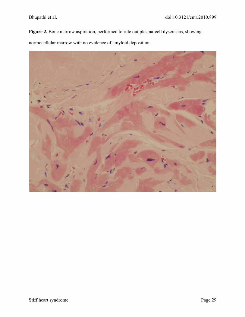
Figure 2. Bone marrow aspiration, performed to rule out plasma-cell dyscrasias, showing
normocellular marrow with no evidence of amyloid deposition.
Stiff heart syndrome Page 29
Bhupathi et al. doi:10.3121/cmr.2010.899
Stiff heart syndrome Page 30
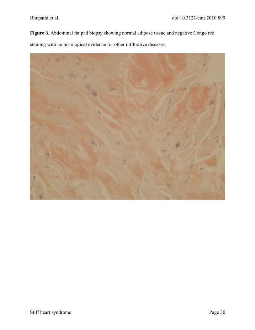
Figure 3. Abdominal fat pad biopsy showing normal adipose tissue and negative Congo red
staining with no histological evidence for other infiltrative diseases.