Embed Size (px)
Citation preview
Have you seen?
Stop competing, start talking!Luca L Fava & Andreas Villunger
According to current belief, the molecularnetworks orchestrating cell death or exitfrom mitosis upon extended mitoticarrest do not interact, stubbornly execut-ing two parallel biological programs andcompeting to define a stochastic decisionbetween death and a chance for survivalwith uncertain destiny. However, recentfindings by Diaz-Martinez et al (2014) inthis issue of The EMBO Journal now callfor a reassessment of the “competingnetwork” hypothesis.
See also: LA Diaz-Martinez et al(September 2014)
A nti-mitotic drugs are essential ingre-
dients of current anti-cancer therapy.
While the molecular basis of the
clinical benefit elicited by these drugs is still
debated (Mitchison, 2012), cells exposed to
taxanes or vinca-alkaloids in experimental
settings usually undergo one of two fates after
prolonged mitotic arrest: cell death, usually
by apoptosis, or adaptation, that is exit from
mitosis without cellular division, a process
also known as mitotic slippage and a possible
cause for long-term treatment failure.
The “competing network” hypothesis
developed by Gascoigne and Taylor suggests
that two independent molecular circuits
control cell death or slippage upon extended
mitotic arrest (Gascoigne & Taylor, 2008). In
this model, cell fate solely depends on the
time needed by either program to reach a
critical threshold. Gradual decline in cyclin
B1 levels defines the time period to mitotic
exit, since even in arrested cells, the spindle
assembly checkpoint (SAC) is unable to
fully restrain the activity of the APC/CCdc20
ubiquitin ligase toward cyclin B1 (Brito &
Rieder, 2006). In parallel, apoptotic cell
death is initiated through the integration of
largely undefined signals leading to activa-
tion of the two key pro-apoptotic effectors
within the Bcl-2 family, Bax and/or Bak,
required for mitochondrial outer membrane
permeabilization (MOMP) and subsequent
caspase activation (reviewed in Czabotar
et al, 2013). Different cancer cells and cell
lines vary considerably in their responsive-
ness to anti-mitotic drug treatment, with, for
example, HeLa and RKO cells being highly
apoptosis-prone, while U2OS or DLD1 cells
tend to adapt. This has been in part
explained by differences in individual apop-
tosis and adaptation thresholds, which may
in turn be based on different expression
levels and activities of key components of
the respective pathways (Gascoigne &
Taylor, 2008).
Several studies have highlighted possible
molecular crosstalk between the cell death
and cell cycle machineries: on one hand,
pro-survival Bcl-2 family members and
initiating cell death caspases are targets of
Cdk1-dependent phosphorylation (reviewed
in Topham & Taylor, 2013); on the other
hand, the SAC protein BubR1 is cleaved by
caspases during apoptosis (Kim et al,
2005). Despite such “opportunities to
communicate”, negatively interfering with
adaptation (e.g. by overexpressing non-
degradable cyclin B1 or by depleting the
APC/C activator Cdc20) leaves mitotic cell
death unaffected, and similarly, inhibiting
caspases does not affect the kinetics of
checkpoint adaptation (Huang et al, 2010).
The further fate of such post-slippage cells
can be highly variable, but this will not be
discussed here in more detail (for review,
please refer to Vitale et al, 2011). Although
well-supported by the above observations,
the “competing network” model becomes
less coherent upon interference with cell
death upstream of mitochondria, as Diaz-
Martinez et al now clearly show that inhibi-
tion of MOMP impacts on the timing by
which cells adapt, in direct contradiction to
the model.
Performing a genome-wide RNAi screen
and monitoring survival of Taxol-treated
HeLa cells, Hongtao Yu’s laboratory identi-
fied a number of known and novel candidate
genes involved in mitotic cell death and
adaptation (Diaz-Martinez et al, 2014).
Knockdown of regulators of mitochondrial
apoptosis (Bad, Noxa, and Bax) and SAC
fidelity (Mps1, Mad2, and BubR1) increased
HeLa cell viability, as did depletion of
factors that delayed mitotic entry. In
contrast, knockdown of APC/C components
(ANAPC1/5/13, CDC23, and CDC26), which
participate in the adaptation network,
reduced survival, as did knockdown of the
mitotic regulator Plk1 or the MAD2-inhibitor
p31comet (Diaz-Martinez et al, 2014).
p31comet prevents conformational activa-
tion of MAD2, a key SAC protein and
component of the mitotic checkpoint
complex (MCC), and by promoting MCC
disassembly both during and after check-
point arrest controls the amount of assem-
bled MCC, favouring APC/CCdc20 activation
and mitotic exit (Varetti et al, 2011). Consis-
tently, p31comet silencing was reported to
sensitize cancer cells to anti-mitotic drugs,
suggesting critical roles in adaptation. In
slippage-prone U2OS cells, p31comet knock-
down readily reduced adaptation and
increased rates of mitotic cell death. As
predicted, enforced arrest upon p31comet
knockdown was associated with prolonged
APC/CCdc20 inhibition reflected by reduced
cyclin B1 degradation. Surprisingly however,
p31comet knockdown also significantly short-
ened the time to cell death, and in cell
death-prone HeLa cells accelerated caspase
activation, with neither effect being pheno-
copied when APC/C activation was
prevented by Cdc20 knockdown (Diaz-
Martinez et al, 2014). Taken together, these
findings thus demonstrate that p31comet
exerts a previously unappreciated anti-
apoptotic function in mitotically arrested
Division of Developmental Immunology, Biocenter, Innsbruck Medical University, Innsbruck, Austria. E-mail: [email protected] 10.15252/embj.201489466 | Published online 25 July 2014
ª 2014 The Authors The EMBO Journal Vol 33 | No 17 | 2014 1849
cells, independent of its role in promoting
mitotic slippage by SAC inhibition and
APC/C activation.
The comparable death-prone phenotype
seen when depleting only p31comet or co-
depleting p31comet and CDC20 may suggest
that p31comet affects apoptosis independently
of APC/CCdc20; however, since Bcl-2 family
members such as the pro-survival molecule
Mcl-1 (Harley et al, 2010) and the pro-
apoptotic BH3-only protein Bim (Wan et al,
2014) have also been reported to be APC/C
degradation targets, it can at this point not
yet be excluded that p31comet acts by indi-
rectly controlling the protein abundance of
cell death regulators. As such, it would have
been interesting to see whether depletion of
p31comet, Cdc20, or both, in Taxol-arrested
cells would differentially affect Bim and
Mcl-1 levels, something that was, however,
not pursued in more detail. In the case of
Bim, the authors observed insignificant cell
death protection upon knockdown to begin
with, and they also failed to reproduce Mcl-1
stabilization upon Cdc20 depletion in their
experimental conditions, consistent with the
existence of multiple redundant mechanisms
acting to degrade pro-survival Mcl-1 during
mitotic arrest (Topham & Taylor, 2013).
Hence, it remains possible that p31comet
controls additional pro-apoptotic targets that
act in concert with Bim to kill mitotically
arrested cells, given that even the most
potent BH3-only proteins usually display
significant redundancy with other members
of this group.
Amongst all pro-survival Bcl-2 family
members, Mcl-1 is the one that displays the
highest affinity for the BH3-only protein
Noxa, while it does not interact with Bad,
another pro-apoptotic homolog picked up by
the authors’ initial screen. Although
reported to be a critical effector of p53-medi-
ated cell death upon DNA damage, recent
studies suggest Noxa roles also in cell death
upon glucose-deprivation or proteasome
inhibition, where it neutralizes Mcl-1
(reviewed in Ploner et al, 2008). Strikingly,
Noxa knockdown in HeLa cells proved as
efficient in preventing Taxol-mediated apop-
tosis and increasing adaptation rate as did
combined Bax/Bak knockdown or Mcl-1
overexpression. Moreover, inhibition of the
Noxa/Mcl-1/Bax/Bak axis in slippage-prone
U2OS cells resulted not only in suppression
of apoptosis but also in a reduction of
mitotic adaptation, demonstrating once
more how key players of one pathway
impact on the other (Diaz-Martinez et al,
2014). Although the lack of suitable reagents
(in particular anti-Noxa antibodies of suffi-
cient quality) prevented the authors from
showing Noxa accumulation or a direct
interaction between p31comet and Noxa in
mitotically arrested cells, it remains of inter-
est to test whether and how components of
the p31comet-targeted MCC or mitotic kinases
might control Noxa abundance and thereby
regulate Mcl-1 levels upon enforced mitotic
arrest.
Of note, Mcl-1 inhibition by Noxa may
not suffice to prime all cell types stalled in
mitosis to apoptosis nor may Bim accumula-
tion be rate-limiting in all settings. Accordingly,
RKO cell sensitisation to Taxol-induced
death was recently reported to involve a
Cdk1-dependent mitotic priming phosphory-
lation on Bid (Wang et al, 2014). Similarly,
direct inhibitory phosphorylation of Bcl-2
and Bcl-x by Cdk1 (Topham & Taylor, 2013)
may be more or less critical dependent on a
given cell type or stimulus. Together, this
suggests that cells during mitosis become
highly primed to apoptosis and ready to self-
destruct any time in the face of trouble, with
timely exit from mitosis seemingly the only
way to survive.
While these findings together with the
new results presented here provide a plausi-
ble model for mitotic cell death (Fig 1), it
remains enigmatic how upstream elements
of the apoptotic machinery may directly
interfere with mitotic adaptation. Having
excluded several proteins acting down-
stream of MOMP, Diaz-Martinez and collea-
gues propose non-apoptotic Bax/Bak
functions in mitochondrial dynamics as a
possible cause for this phenomenon. Bax/
Bak impinge on mitochondrial fission by
facilitating activation of dynamin-related
protein 1 (Drp1), and concerted fission is a
prerequisite for the successful completion of
mitosis (Kashatus et al, 2011; and references
therein). Consistent with this hypothesis,
Drp1 knockdown caused mitotic phenotypes
comparable to those caused by Bax/Bak
depletion but did not affect cell death (Diaz-
Martinez et al, 2014). In the absence of
Noxa, increased availability of Mcl-1 may
keep Bax/Bak from interacting with the
fission machinery and hence phenocopy the
effect of Bax/Bak depletion. Whether mito-
chondrial fission, as speculated by the
authors, weakens the spindle assembly
checkpoint through a drop in ATP levels and
subsequent global reduction of protein
synthesis affecting cyclin B1 levels remains
to be experimentally established.
Collectively, these observations argue
against a strict separation of two competing
molecular networks that would act indepen-
dently of each other to define the fate of
mitotically arrested cells. Instead, Diaz-
Martinez et al demonstrate how individual
proteins thought to belong to one given
network actually act in both, and ultimately
that components of both circuitries are hard-
wired together, even if they may not exclusively
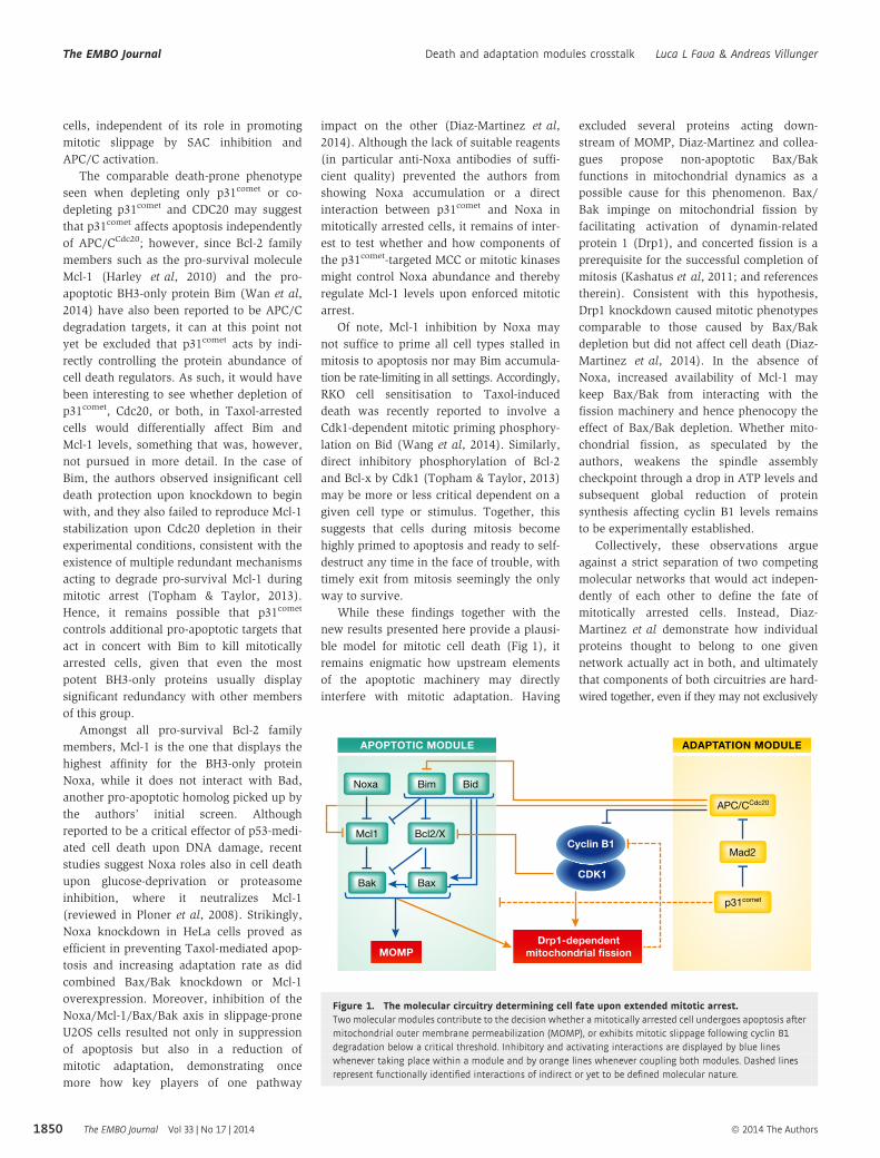
ADAPTATION MODULE
MOMPDrp1-dependent
mitochondrial fission
APOPTOTIC MODULE
APC/CCdc20
p31comet
Mad2Cyclin B1
CDK1
Noxa Bim Bid
Mcl1 Bcl2/X
Bak Bax
Figure 1. The molecular circuitry determining cell fate upon extended mitotic arrest.Two molecular modules contribute to the decision whether a mitotically arrested cell undergoes apoptosis aftermitochondrial outer membrane permeabilization (MOMP), or exhibits mitotic slippage following cyclin B1degradation below a critical threshold. Inhibitory and activating interactions are displayed by blue lineswhenever taking place within a module and by orange lines whenever coupling both modules. Dashed linesrepresent functionally identified interactions of indirect or yet to be defined molecular nature.
The EMBO Journal Vol 33 | No 17 | 2014 ª 2014 The Authors
The EMBO Journal Death and adaptation modules crosstalk Luca L Fava & Andreas Villunger
1850
execute their presumed bona fide biological
functions.
AcknowledgementsThe work in our laboratory is supported by
grants from the Austrian Science Fund (FWF),
Tiroler Wissenschaftsfond (TWF), and Krebshilfe
Tirol. LLF was supported by the EMBO-LTF
program. We apologize to all scientists in the
field whose work could not be cited due to space
constraints.
ReferencesBrito DA, Rieder CL (2006) Mitotic checkpoint
slippage in humans occurs via cyclin B
destruction in the presence of an active
checkpoint. Curr Biol 16: 1194 – 1200
Czabotar PE, Lessene G, Strasser A, Adams JM
(2013) Control of apoptosis by the BCL-2
protein family: implications for physiology and
therapy. Nat Rev Mol Cell Biol 15: 49 – 63
Diaz-Martinez LA, Karamysheva Z, Warrington R,
Li B, Wei S, Xie XJ, Roth MG, Yu H (2014)
Genome-wide siRNA screen reveals coupling
between mitotic apoptosis and adaptation.
EMBO J 33: 1960 – 1976
Gascoigne KE, Taylor SS (2008) Cancer cells display
profound intra- and interline variation
following prolonged exposure to antimitotic
drugs. Cancer Cell 14: 111 – 122
Harley ME, Allan LA, Sanderson HS, Clarke PR
(2010) Phosphorylation of Mcl-1 by
CDK1-cyclin B1 initiates its Cdc20-dependent
destruction during mitotic arrest. EMBO J 29:
2407 – 2420
Huang HC, Mitchison TJ, Shi J (2010) Stochastic
competition between mechanistically
independent slippage and death pathways
determines cell fate during mitotic arrest. PLoS
ONE 5: e15724
Kashatus DF, Lim KH, Brady DC, Pershing NL, Cox
AD, Counter CM (2011) RALA and RALBP1
regulate mitochondrial fission at mitosis. Nat
Cell Biol 13: 1108 – 1115
Kim M, Murphy K, Liu F, Parker SE, Dowling ML,
Baff W, Kao GD (2005) Caspase-mediated
specific cleavage of BubR1 is a determinant of
mitotic progression. Mol Cell Biol 25:
9232 – 9248
Mitchison TJ (2012) The proliferation rate paradox
in antimitotic chemotherapy. Mol Biol Cell 23:
1 – 6
Ploner C, Kofler R, Villunger A (2008) Noxa: at the
tip of the balance between life and death.
Oncogene 27(Suppl. 1): S84 – S92
Topham CH, Taylor SS (2013) Mitosis and
apoptosis: how is the balance set? Curr Opin
Cell Biol 25: 780 – 785
Varetti G, Guida C, Santaguida S, Chiroli E,
Musacchio A (2011) Homeostatic control of
mitotic arrest. Mol Cell 44: 710 – 720
Vitale I, Galluzzi L, Castedo M, Kroemer G (2011)
Mitotic catastrophe: a mechanism for avoiding
genomic instability. Nat Rev Mol Cell Biol 12:
385 – 392
Wan L, Tan M, Yang J, Inuzuka H, Dai X, Wu T, Liu
J, Shaik S, Chen G, Deng J, Malumbres M, Letai
A, Kirschner MW, Sun Y, Wei W (2014) APC
(Cdc20) suppresses apoptosis THROUGH
targeting bim for ubiquitination and
destruction. Dev Cell 29: 377 – 391
Wang P, Lindsay J, Owens TW, Mularczyk EJ,
Warwood S, Foster F, Streuli CH,
Brennan K, Gilmore AP (2014) Phosphorylation
of the proapoptotic BH3-only protein
bid primes mitochondria for apoptosis
during mitotic arrest. Cell Rep 7:
661 – 671
ª 2014 The Authors The EMBO Journal Vol 33 | No 17 | 2014
Luca L Fava & Andreas Villunger Death and adaptation modules crosstalk The EMBO Journal
1851