Embed Size (px)
Citation preview
Strain analysis of protein structures and lowdimensionality of mechanical allosteric couplingsMichael R. Mitchella,b,1, Tsvi Tlustyc,d,e,1, and Stanislas Leiblera,b,c,1
aLaboratory of Living Matter, The Rockefeller University, New York, NY 10065; bCenter for Studies in Physics and Biology, The Rockefeller University, New York,NY 10065; cThe Simons Center for Systems Biology, School of Natural Sciences, Institute for Advanced Study, Princeton, NJ 08540; dCenter for Soft andLiving Matter, Institute for Basic Science, Ulsan 689-798, South Korea; and eDepartment of Physics, Ulsan National Institute of Science and Technology, Ulsan689-798, South Korea
Contributed by Stanislas Leibler, August 2, 2016 (sent for review June 13, 2016; reviewed by Alexander Y. Grosberg and Henri Orland)
In many proteins, especially allosteric proteins that communicateregulatory states from allosteric to active sites, structural defor-mations are functionally important. To understand these defor-mations, dynamical experiments are ideal but challenging. Usingstatic structural information, although more limited than dynam-ical analysis, is much more accessible. Underused for proteinanalysis, strain is the natural quantity for studying local deforma-tions. We calculate strain tensor fields for proteins deformed byligands or thermal fluctuations using crystal and NMR structureensembles. Strains—primarily shears—show deformations aroundbinding sites. These deformations can be induced solely by ligandbinding at distant allosteric sites. Shears reveal quasi-2D paths ofmechanical coupling between allosteric and active sites that mayconstitute a widespread mechanism of allostery. We argue thatstrain—particularly shear—is the most appropriate quantity foranalysis of local protein deformations. This analysis can revealmechanical and biological properties of many proteins.
strain | protein mechanics | protein allostery | elasticity
Although many proteins fold into well-defined, stable struc-tures, their internal deformations around such average
structures often play a vital role in their functions. In allostericprotein regulation, a protein’s ability to catalyze reactions or toassociate with binding partners is influenced by binding of anallosteric regulator to a spatially distinct site. For example,subunits of the tetrameric protein hemoglobin, which transportsoxygen in the bloodstream, undergo allosteric structural shifts asthey bind O2 (1). These allosteric shifts alter the protein’s O2binding affinity (2), enabling hemoglobin to deliver nearly twiceas much oxygen to tissues as it could were it not allosteric (3).Elsewhere, allostery enables cells to modulate the activity of pro-teins more quickly than other regulatory mechanisms like control ofprotein synthesis and degradation would permit (e.g., ref. 4). Thisregulation enables cells to respond rapidly to changing conditions.Given the significant role that structural shifts of proteins play
in their function, there has been substantial effort toward betterunderstanding them. Several models have attempted to explainthe mechanism of allostery. The most prominent have been thewell-known concerted model of Monod, Wyman, and Changeux(5) and the sequential model of Koshland, Nemethy, and Filmer(6). Recently, there has been increasing consideration of thethermodynamic nature of allostery; allosteric proteins’ transi-tions between different functional states can correspond not todiscrete switching between states, but rather to a statistical shiftin a population distribution of structural states (reviewed in, e.g.,refs. 7–11). In the present work, we remain largely agnostic amongthese models and simply attempt to exploit experimental data tounderstand the mechanical properties of allosteric proteins.Dynamical experiments can provide the most direct informa-
tion about the conduction of allosteric signals and about proteinstructural dynamics more generally. Studies using methods in-cluding room-temperature crystallography (e.g., ref. 12), time-resolved crystallography (e.g., ref. 13), FRET (e.g., ref. 14), anddirect pulling measurements (15, 16) have been published. However,
these techniques are often labor-intensive and technically challenging.Some are applicable only to certain experimentally amenable pro-teins while some provide valuable but incomplete information. Thus,although such methods provide highly valuable information, manyproteins are resistant to study using these methods.In contrast, thousands of crystallography and NMR structures
are publicly available for many proteins in multiple ligand-binding states. Furthermore, standard crystallography and NMRtechniques are generally more accessible for the study of newproteins than are methods for direct dynamical measurements.Although these data, being static, cannot provide the richness offull dynamical experiments, they nevertheless contain valuableinformation concerning the net deformations that take placewithin proteins. It would therefore be highly desirable to extractuseful insights regarding allosteric and other structural proper-ties of proteins solely from datasets comprising several staticcrystal or NMR structures.
Comparison of Protein Structural StatesThe simplest and most widespread method used in detailedanalysis of related crystal structures is direct, manual inspectionof positions, distances, and angles of specific atoms, residues,and bonds in protein structures using molecular viewer programssuch as PyMOL, VMD, or Chimera. Such inspection can providedetailed information about differences between a few relatedstructures and may be appropriate for analysis of a small regionwithin a protein such as an enzyme’s active site. In such a situ-ation, analysis is confined to a small region of the protein known
Significance
Regulation of biochemical activity is essential for proper cellgrowth and metabolism. Many proteins’ activities are regu-lated by interactions with other molecules binding some dis-tance away from the proteins’ active sites. In such allostericproteins, active sites should thus be mechanically coupled tospatially removed regulatory regions. We studied crystal andNMR structures of proteins in various regulatory and ligand-binding states. We calculated and analyzed distributions ofstrains throughout several proteins. Strains reveal allosteric andactive sites and suggest that quasi-two-dimensional strainedsurfaces mediate mechanical couplings between them. Strainanalysis of widely available structural data can illuminate pro-tein function and guide future experimental investigation.
Author contributions: M.R.M., T.T., and S.L. designed research; M.R.M. performed re-search; M.R.M. contributed new reagents/analytic tools; M.R.M., T.T., and S.L. analyzeddata; and M.R.M., T.T., and S.L. wrote the paper.
Reviewers: A.Y.G., New York University; and H.O., Alternative Energies and AtomicEnergy Commission.
The authors declare no conflict of interest.1To whom correspondence may be addressed. Email: [email protected], [email protected], or [email protected].
This article contains supporting information online at www.pnas.org/lookup/suppl/doi:10.1073/pnas.1609462113/-/DCSupplemental.
www.pnas.org/cgi/doi/10.1073/pnas.1609462113 PNAS | Published online September 21, 2016 | E5847–E5855
BIOPH
YSICSAND
COMPU
TATIONALBIOLO
GY
PNASPL
US
Dow
nloa
ded
by g
uest
on
Feb
ruar
y 16
, 202
2
to be functionally important and measurements can be informedby knowledge of the relevant reaction chemistry. However, thisapproach is labor-intensive, particularly for analysis of many or largestructures. In addition, it does not provide an unbiased frameworkfor analyzing large-scale structural changes or distinguishing isolatedfluctuations from biologically important deformations.Other methods have been applied to analyze large-scale protein
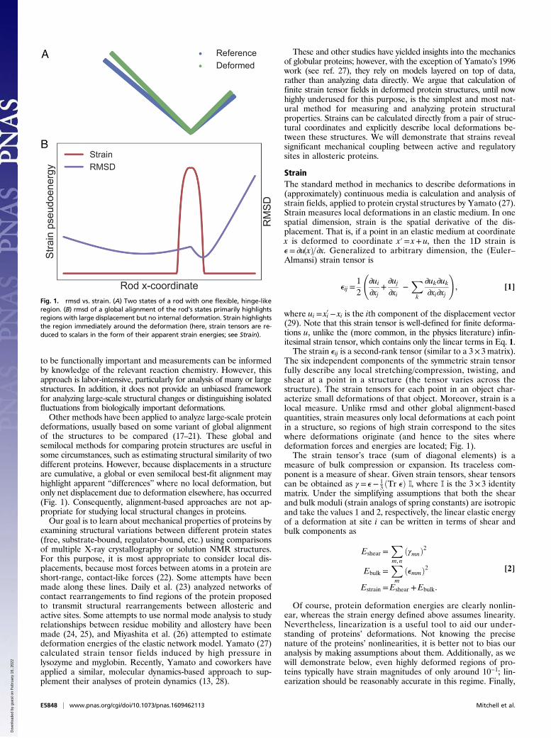
deformations, usually based on some variant of global alignmentof the structures to be compared (17–21). These global andsemilocal methods for comparing protein structures are useful insome circumstances, such as estimating structural similarity of twodifferent proteins. However, because displacements in a structureare cumulative, a global or even semilocal best-fit alignment mayhighlight apparent “differences” where no local deformation, butonly net displacement due to deformation elsewhere, has occurred(Fig. 1). Consequently, alignment-based approaches are not ap-propriate for studying local structural changes in proteins.Our goal is to learn about mechanical properties of proteins by
examining structural variations between different protein states(free, substrate-bound, regulator-bound, etc.) using comparisonsof multiple X-ray crystallography or solution NMR structures.For this purpose, it is most appropriate to consider local dis-placements, because most forces between atoms in a protein areshort-range, contact-like forces (22). Some attempts have beenmade along these lines. Daily et al. (23) analyzed networks ofcontact rearrangements to find regions of the protein proposedto transmit structural rearrangements between allosteric andactive sites. Some attempts to use normal mode analysis to studyrelationships between residue mobility and allostery have beenmade (24, 25), and Miyashita et al. (26) attempted to estimatedeformation energies of the elastic network model. Yamato (27)calculated strain tensor fields induced by high pressure inlysozyme and myglobin. Recently, Yamato and coworkers haveapplied a similar, molecular dynamics-based approach to sup-plement their analyses of protein dynamics (13, 28).
These and other studies have yielded insights into the mechanicsof globular proteins; however, with the exception of Yamato’s 1996work (see ref. 27), they rely on models layered on top of data,rather than analyzing data directly. We argue that calculation offinite strain tensor fields in deformed protein structures, until nowhighly underused for this purpose, is the simplest and most nat-ural method for measuring and analyzing protein structuralproperties. Strains can be calculated directly from a pair of struc-tural coordinates and explicitly describe local deformations be-tween these structures. We will demonstrate that strains revealsignificant mechanical coupling between active and regulatorysites in allosteric proteins.
StrainThe standard method in mechanics to describe deformations in(approximately) continuous media is calculation and analysis ofstrain fields, applied to protein crystal structures by Yamato (27).Strain measures local deformations in an elastic medium. In onespatial dimension, strain is the spatial derivative of the dis-placement. That is, if a point in an elastic medium at coordinatex is deformed to coordinate x′= x+ u, then the 1D strain ise= ∂uðxÞ=∂x. Generalized to arbitrary dimension, the (Euler–Almansi) strain tensor is
eij =12
∂ui∂xj
+∂uj∂xi
−Xk
∂uk∂uk∂xi∂xj
!, [1]
where ui = xi′− xi is the ith component of the displacement vector(29). Note that this strain tensor is well-defined for finite deforma-tions u, unlike the (more common, in the physics literature) infin-itesimal strain tensor, which contains only the linear terms in Eq. 1.The strain eij is a second-rank tensor (similar to a 3× 3matrix).
The six independent components of the symmetric strain tensorfully describe any local stretching/compression, twisting, andshear at a point in a structure (the tensor varies across thestructure). The strain tensors for each point in an object char-acterize small deformations of that object. Moreover, strain is alocal measure. Unlike rmsd and other global alignment-basedquantities, strain measures only local deformations at each pointin a structure, so regions of high strain correspond to the siteswhere deformations originate (and hence to the sites wheredeformation forces and energies are located; Fig. 1).The strain tensor’s trace (sum of diagonal elements) is a
measure of bulk compression or expansion. Its traceless com-ponent is a measure of shear. Given strain tensors, shear tensorscan be obtained as γ = e− 1
3 ðTr eÞ I, where I is the 3× 3 identitymatrix. Under the simplifying assumptions that both the shearand bulk moduli (strain analogs of spring constants) are isotropicand take the values 1 and 2, respectively, the linear elastic energyof a deformation at site i can be written in terms of shear andbulk components as
Eshear =Xm, n
ðγmnÞ2
Ebulk =Xm
ðemmÞ2
Estrain =Eshear +Ebulk.
[2]
Of course, protein deformation energies are clearly nonlin-ear, whereas the strain energy defined above assumes linearity.Nevertheless, linearization is a useful tool to aid our under-standing of proteins’ deformations. Not knowing the precisenature of the proteins’ nonlinearities, it is better not to bias ouranalysis by making assumptions about them. Additionally, as wewill demonstrate below, even highly deformed regions of pro-teins typically have strain magnitudes of only around 10−1; lin-earization should be reasonably accurate in this regime. Finally,
Fig. 1. rmsd vs. strain. (A) Two states of a rod with one flexible, hinge-likeregion. (B) rmsd of a global alignment of the rod’s states primarily highlightsregions with large displacement but no internal deformation. Strain highlightsthe region immediately around the deformation (here, strain tensors are re-duced to scalars in the form of their apparent strain energies; see Strain).
E5848 | www.pnas.org/cgi/doi/10.1073/pnas.1609462113 Mitchell et al.
Dow
nloa
ded
by g
uest
on
Feb
ruar
y 16
, 202
2
other linear methods, such as those of refs. 24 and 25, have beenable to generate insights despite their use of this approximation.In addition, calculation of energies requires knowledge of
strain moduli that are not known for proteins and surely varythroughout each protein. An important source of anisotropy inprotein strain moduli are forces produced by covalent bondsbetween backbone atoms, which are much stronger than non-covalent interactions between neighboring atoms and residues.Our approach is to neglect both the nonlinearities and the an-isotropies present in the proteins. In this way, we can avoid bi-asing our analysis with (necessarily incorrect) assumptionsregarding the actual distribution of strain moduli in the proteinswe study. Instead, we interpret the strain tensors we estimate andthe corresponding “pseudoenergies” as aggregate indications ofthe type and magnitude of local deformations occurring in theproteins under study, and of the local amino acid interactionspresent throughout those proteins. In doing so, we can use strainsto analyze proteins’ deformations and identify residues essentialfor allostery and other protein functions.Finally, the concept of strain is typically applied in the context
of continuous media. Proteins are clearly not continuous, beingcomposed of residues themselves made up of atoms. However,the theory of elasticity has been remarkably successful in de-scribing the mechanical properties of real materials, all discretelycomposed of atoms. Indeed, much of the early development ofthe theory of elasticity—including the models of Fresnel, Navier,Cauchy, Poisson, Voigt, Born, and von Karman—occurred in thecontext of discrete, atomic media (30, 31), with continuum the-ories arising as a limit of molecular models. Moreover, the cal-culation of strain tensors can readily be extended to granularmedia (32), and strains remain highly useful in describing gran-ular materials’ deformations. Ultimately, our calculation of straintensors in protein structures is equivalent to calculation of thespatial derivatives of the displacement fields between pairs ofstructures; these spatial derivatives (i.e., strains) indicate thestructure of local deformations independently of any interpre-tation under the theory of continuum elasticity.Taken together, the simplifying assumptions of linearity and
homogeneous strain moduli mean that, while the calculatedpseudoenergy values have the mathematical form of an energy,they can alternatively be interpreted as merely the magnitude oflocal deformation at each site in the protein. This interpretationassumes neither linearity of strain energies nor homogeneity ofstrain moduli. In this sense, our analysis is closely analogous tothe use of rmsd and related measures for comparing two or morestructures, except that where rmsd identifies large global dis-placements but not their structural origin, our approach identifieslocal deformations that underlie proteins’ structural variations(Fig. 1).
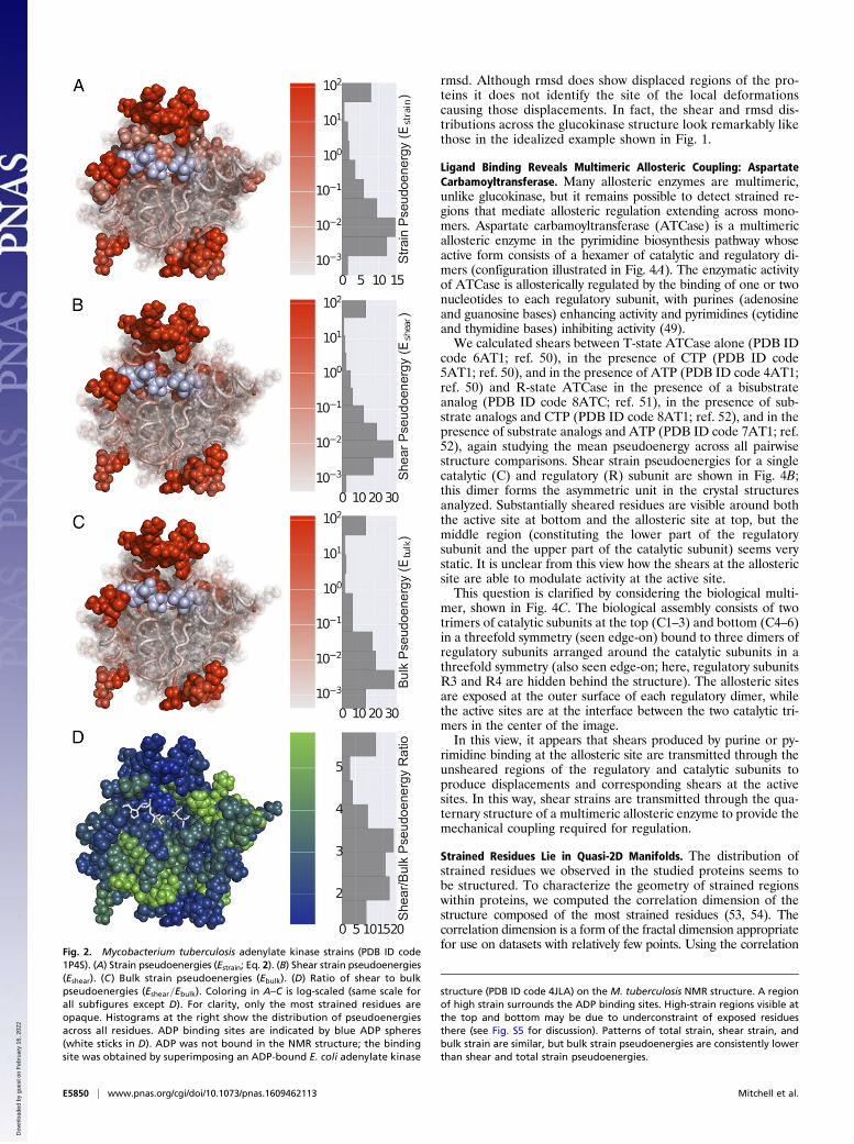
ResultsProtein Strains Are Primarily Shears and Reveal an Enzyme’s ActiveSite: Adenylate Kinase. We first illustrate the use of strain calcu-lations to describe the equilibrium structure of adenylate kinasemeasured by solution NMR. We computed deformations be-tween an ensemble of 20 solution NMR structures [Protein DataBank (PDB) ID code 1P4S; ref. 33]. The NMR structures wereobtained using free adenylate kinase, with no ligands present.Because there is no natural reference structure, we calculatedthe mean strain pseudoenergies across strains for all pairwisestructure comparisons. Although the NMR structure ensemblecorresponds to a collection of structures consistent with mea-sured NOE constraints rather than a directly measured set ofconformations, strains between the calculated structures shouldprovide information about local deformations within the protein.Strain, shear strain, and bulk strain pseudoenergies for eachresidue in the protein are shown in Fig. 2 A–C. The strain dis-tribution shows that relatively unconstrained loops at the top and
bottom undergo very high shears; in addition, a small pocketbetween the two loops undergoes relatively high shear defor-mations. Comparison with a crystal structure of the Escherichiacoli adenylate kinase (PDB ID code 4JLA; ref. 34) shows thatthis sheared pocket corresponds to the ADP binding sites (ADPshown in light blue). Here we observe that strains correspondingto structural fluctuations captured by the NMR structure en-semble reveal functionally significant features in the protein,even without perturbation of those sites by ligand binding.As seen in Fig. 2, distributions of total strain, shear strain, and
bulk strain throughout adenylate kinase are quite similar; thisobservation holds true also for other proteins we studied. Be-cause of this similarity and because globular proteins are gen-erally not very compressible (i.e., bulk strain is a relatively smallfraction of total strain), we will show only shear strains for sub-sequent proteins. Shear pseudoenergies were generally higherthan bulk pseudoenergies, but they were typically within an orderof magnitude of each other. The ratios of shear pseudoenergy tobulk pseudoenergy for adenylate kinase are shown in Fig. 2D; for allresidues, shear pseudoenergy was larger than bulk pseudoenergy,but underconstrained surface sites tended to have lower shear/bulk ratios.We also calculated strains in a closely related protein, gua-
nylate kinase, based on X-ray structures (PDB ID codes 1ZNW,1ZNX, and 1ZNY; ref. 35), with similar results (Fig. S1).
Strain Analysis of an Allosteric Protein Reveals Mechanical CouplingBetween Allosteric and Active Sites Not Demonstrated by rmsd:Glucokinase. The previous example has shown that analyzingstrain distributions in proteins can highlight enzymes’ active sites,but adenylate kinase is not known to be allosteric. As a result thestrained region in that protein seems to be confined to the activesite. In the case of allosteric proteins, it can be possible to ob-serve strained regions connecting active and allosteric sites,demonstrating a direct mechanical coupling between these sites.We now consider glucokinase, a monomeric allosteric enzyme
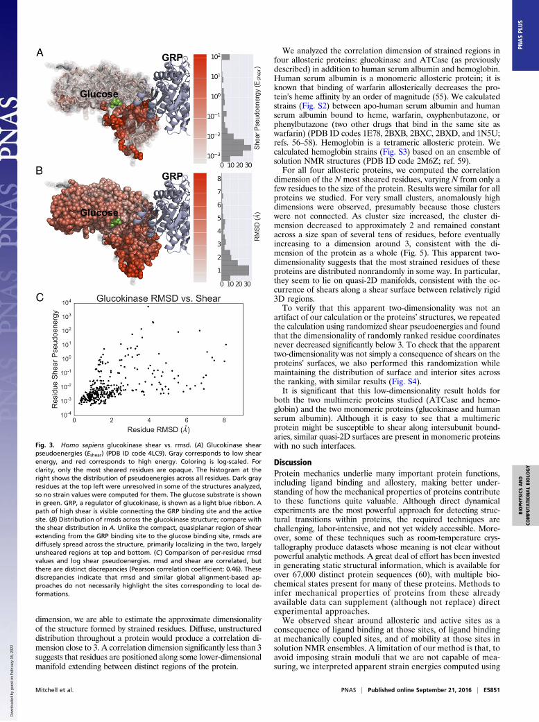
that phosphorylates glucose to glucose-6-phosphate. Among itsregulators is glucokinase regulatory protein (GRP), which acts asan inhibitor. There are also numerous synthetic activators andinhibitors. We computed shears across 26 different structuresof glucokinase including free enzyme, glucose-bound enzyme,GRP-bound enzyme, and multiple synthetic inhibitor- and acti-vator-bound states (PDB ID codes 1V4S, 1V4T, 3A0I, 3F9M,3FGU, 3FR0, 3GOI, 3H1V, 3ID8, 3IDH, 3IMX, 3S41, 3VEV,3VEY, 3VF6, 4DCH, 4DHY, 4ISE, 4ISF, 4ISG, 4L3Q, 4LC9,4MLE, 4MLH, 4NO7, and 4RCH; refs. 36–48), again studying themean pseudoenergy across all pairwise structure comparisons.Shear pseudoenergies are shown in Fig. 3A. There is a qua-
siplanar region of high shear connecting the GRP binding siteand the glucose binding site. This finding suggests that in glu-cokinase information about the GRP binding state is transmittedalong the observed manifold of shear strain. This observation isconsistent with previous reports that the two domains of gluco-kinase undergo relative displacement during regulation (43), butby examining strains we can see that this displacement corre-sponds to a deformation propagated across the protein to theactive site by the strained residues shown in Fig. 3. Thus, for thisallosteric protein, strain-based analysis reveals not only bindingsites (which, in this case, were apparent from the structures) butalso how functionally coupled sites are mechanically connected.We compared the distribution of shears across the glucoki-
nase structure to the distribution of rmsds across the structure.The rmsd distribution is shown in Fig. 3B; it is distinctly dif-ferent from the distribution of shears. It is diffusely spreadacross the structure, and the regions of largest rmsd are the topand bottom lobes of the protein, which are largely free of shear.The sheared region extending between the GRP binding siteand the glucose binding site, in fact, is near the region of lowest
Mitchell et al. PNAS | Published online September 21, 2016 | E5849
BIOPH
YSICSAND
COMPU
TATIONALBIOLO
GY
PNASPL
US
Dow
nloa
ded
by g
uest
on
Feb
ruar
y 16
, 202
2
rmsd. Although rmsd does show displaced regions of the pro-teins it does not identify the site of the local deformationscausing those displacements. In fact, the shear and rmsd dis-tributions across the glucokinase structure look remarkably likethose in the idealized example shown in Fig. 1.
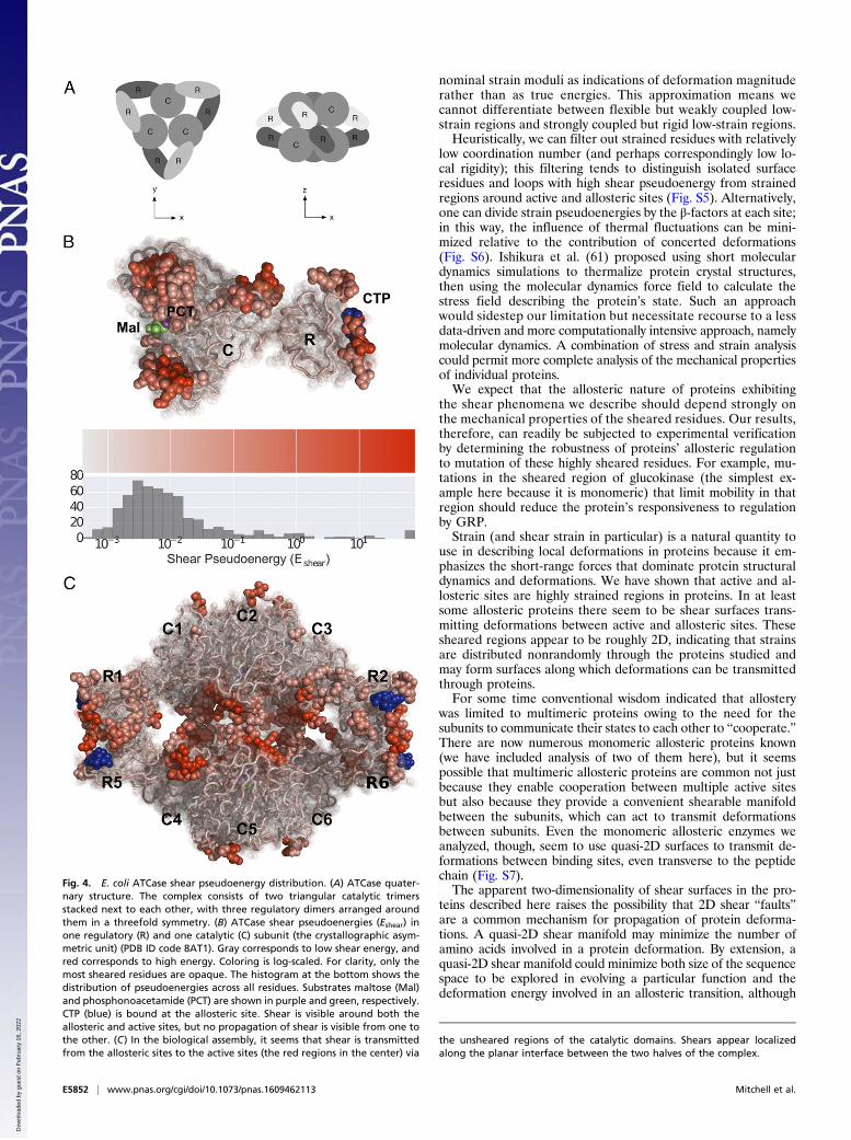
Ligand Binding Reveals Multimeric Allosteric Coupling: AspartateCarbamoyltransferase. Many allosteric enzymes are multimeric,unlike glucokinase, but it remains possible to detect strained re-gions that mediate allosteric regulation extending across mono-mers. Aspartate carbamoyltransferase (ATCase) is a multimericallosteric enzyme in the pyrimidine biosynthesis pathway whoseactive form consists of a hexamer of catalytic and regulatory di-mers (configuration illustrated in Fig. 4A). The enzymatic activityof ATCase is allosterically regulated by the binding of one or twonucleotides to each regulatory subunit, with purines (adenosineand guanosine bases) enhancing activity and pyrimidines (cytidineand thymidine bases) inhibiting activity (49).We calculated shears between T-state ATCase alone (PDB ID
code 6AT1; ref. 50), in the presence of CTP (PDB ID code5AT1; ref. 50), and in the presence of ATP (PDB ID code 4AT1;ref. 50) and R-state ATCase in the presence of a bisubstrateanalog (PDB ID code 8ATC; ref. 51), in the presence of sub-strate analogs and CTP (PDB ID code 8AT1; ref. 52), and in thepresence of substrate analogs and ATP (PDB ID code 7AT1; ref.52), again studying the mean pseudoenergy across all pairwisestructure comparisons. Shear strain pseudoenergies for a singlecatalytic (C) and regulatory (R) subunit are shown in Fig. 4B;this dimer forms the asymmetric unit in the crystal structuresanalyzed. Substantially sheared residues are visible around boththe active site at bottom and the allosteric site at top, but themiddle region (constituting the lower part of the regulatorysubunit and the upper part of the catalytic subunit) seems verystatic. It is unclear from this view how the shears at the allostericsite are able to modulate activity at the active site.This question is clarified by considering the biological multi-
mer, shown in Fig. 4C. The biological assembly consists of twotrimers of catalytic subunits at the top (C1–3) and bottom (C4–6)in a threefold symmetry (seen edge-on) bound to three dimers ofregulatory subunits arranged around the catalytic subunits in athreefold symmetry (also seen edge-on; here, regulatory subunitsR3 and R4 are hidden behind the structure). The allosteric sitesare exposed at the outer surface of each regulatory dimer, whilethe active sites are at the interface between the two catalytic tri-mers in the center of the image.In this view, it appears that shears produced by purine or py-
rimidine binding at the allosteric site are transmitted through theunsheared regions of the regulatory and catalytic subunits toproduce displacements and corresponding shears at the activesites. In this way, shear strains are transmitted through the qua-ternary structure of a multimeric allosteric enzyme to provide themechanical coupling required for regulation.
Strained Residues Lie in Quasi-2D Manifolds. The distribution ofstrained residues we observed in the studied proteins seems tobe structured. To characterize the geometry of strained regionswithin proteins, we computed the correlation dimension of thestructure composed of the most strained residues (53, 54). Thecorrelation dimension is a form of the fractal dimension appropriatefor use on datasets with relatively few points. Using the correlation
Fig. 2. Mycobacterium tuberculosis adenylate kinase strains (PDB ID code1P4S). (A) Strain pseudoenergies (Estrain; Eq. 2). (B) Shear strain pseudoenergies(Eshear). (C) Bulk strain pseudoenergies (Ebulk). (D) Ratio of shear to bulkpseudoenergies (Eshear=Ebulk). Coloring in A–C is log-scaled (same scale forall subfigures except D). For clarity, only the most strained residues areopaque. Histograms at the right show the distribution of pseudoenergiesacross all residues. ADP binding sites are indicated by blue ADP spheres(white sticks in D). ADP was not bound in the NMR structure; the bindingsite was obtained by superimposing an ADP-bound E. coli adenylate kinase
structure (PDB ID code 4JLA) on the M. tuberculosis NMR structure. A regionof high strain surrounds the ADP binding sites. High-strain regions visible atthe top and bottom may be due to underconstraint of exposed residuesthere (see Fig. S5 for discussion). Patterns of total strain, shear strain, andbulk strain are similar, but bulk strain pseudoenergies are consistently lowerthan shear and total strain pseudoenergies.
E5850 | www.pnas.org/cgi/doi/10.1073/pnas.1609462113 Mitchell et al.
Dow
nloa
ded
by g
uest
on
Feb
ruar
y 16
, 202
2
dimension, we are able to estimate the approximate dimensionalityof the structure formed by strained residues. Diffuse, unstructureddistribution throughout a protein would produce a correlation di-mension close to 3. A correlation dimension significantly less than 3suggests that residues are positioned along some lower-dimensionalmanifold extending between distinct regions of the protein.
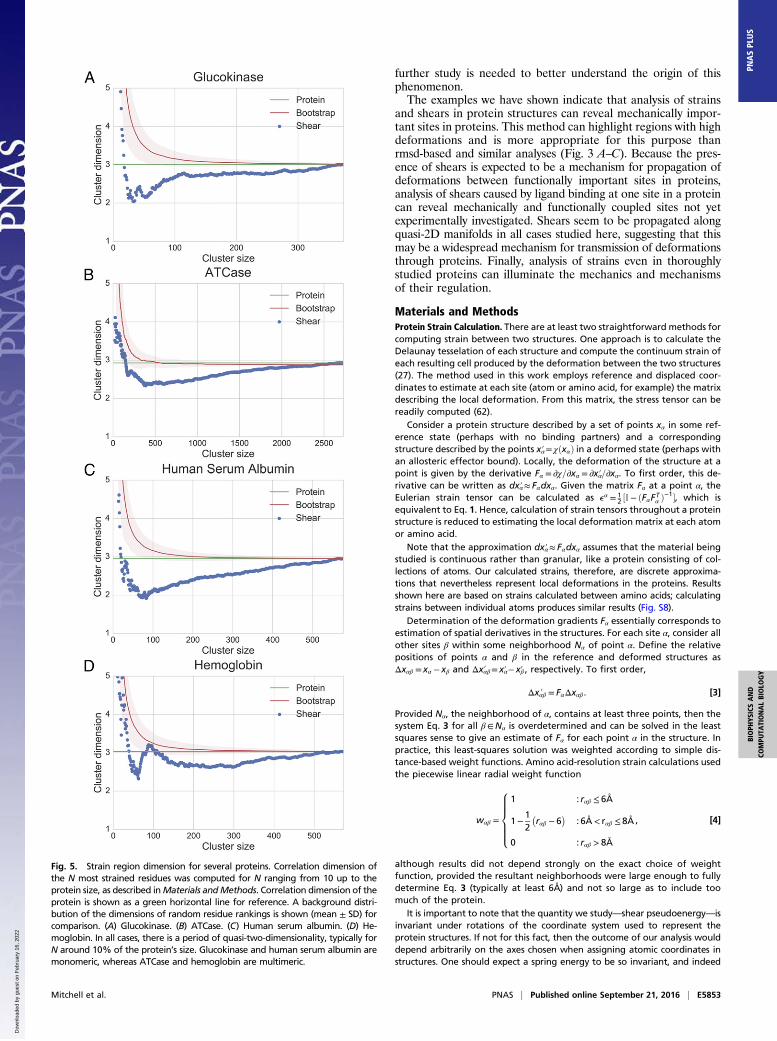
We analyzed the correlation dimension of strained regions infour allosteric proteins: glucokinase and ATCase (as previouslydescribed) in addition to human serum albumin and hemoglobin.Human serum albumin is a monomeric allosteric protein; it isknown that binding of warfarin allosterically decreases the pro-tein’s heme affinity by an order of magnitude (55). We calculatedstrains (Fig. S2) between apo-human serum albumin and humanserum albumin bound to heme, warfarin, oxyphenbutazone, orphenylbutazone (two other drugs that bind in the same site aswarfarin) (PDB ID codes 1E78, 2BXB, 2BXC, 2BXD, and 1N5U;refs. 56–58). Hemoglobin is a tetrameric allosteric protein. Wecalculated hemoglobin strains (Fig. S3) based on an ensemble ofsolution NMR structures (PDB ID code 2M6Z; ref. 59).For all four allosteric proteins, we computed the correlation
dimension of the N most sheared residues, varying N from only afew residues to the size of the protein. Results were similar for allproteins we studied. For very small clusters, anomalously highdimensions were observed, presumably because those clusterswere not connected. As cluster size increased, the cluster di-mension decreased to approximately 2 and remained constantacross a size span of several tens of residues, before eventuallyincreasing to a dimension around 3, consistent with the di-mension of the protein as a whole (Fig. 5). This apparent two-dimensionality suggests that the most strained residues of theseproteins are distributed nonrandomly in some way. In particular,they seem to lie on quasi-2D manifolds, consistent with the oc-currence of shears along a shear surface between relatively rigid3D regions.To verify that this apparent two-dimensionality was not an
artifact of our calculation or the proteins’ structures, we repeatedthe calculation using randomized shear pseudoenergies and foundthat the dimensionality of randomly ranked residue coordinatesnever decreased significantly below 3. To check that the apparenttwo-dimensionality was not simply a consequence of shears on theproteins’ surfaces, we also performed this randomization whilemaintaining the distribution of surface and interior sites acrossthe ranking, with similar results (Fig. S4).It is significant that this low-dimensionality result holds for
both the two multimeric proteins studied (ATCase and hemo-globin) and the two monomeric proteins (glucokinase and humanserum albumin). Although it is easy to see that a multimericprotein might be susceptible to shear along intersubunit bound-aries, similar quasi-2D surfaces are present in monomeric proteinswith no such interfaces.
DiscussionProtein mechanics underlie many important protein functions,including ligand binding and allostery, making better under-standing of how the mechanical properties of proteins contributeto these functions quite valuable. Although direct dynamicalexperiments are the most powerful approach for detecting struc-tural transitions within proteins, the required techniques arechallenging, labor-intensive, and not yet widely accessible. More-over, some of these techniques such as room-temperature crys-tallography produce datasets whose meaning is not clear withoutpowerful analytic methods. A great deal of effort has been investedin generating static structural information, which is available forover 67,000 distinct protein sequences (60), with multiple bio-chemical states present for many of these proteins. Methods toinfer mechanical properties of proteins from these alreadyavailable data can supplement (although not replace) directexperimental approaches.We observed shear around allosteric and active sites as a
consequence of ligand binding at those sites, of ligand bindingat mechanically coupled sites, and of mobility at those sites insolution NMR ensembles. A limitation of our method is that, toavoid imposing strain moduli that we are not capable of mea-suring, we interpreted apparent strain energies computed using
Fig. 3. Homo sapiens glucokinase shear vs. rmsd. (A) Glucokinase shearpseudoenergies (Eshear) (PDB ID code 4LC9). Gray corresponds to low shearenergy, and red corresponds to high energy. Coloring is log-scaled. Forclarity, only the most sheared residues are opaque. The histogram at theright shows the distribution of pseudoenergies across all residues. Dark grayresidues at the top left were unresolved in some of the structures analyzed,so no strain values were computed for them. The glucose substrate is shownin green. GRP, a regulator of glucokinase, is shown as a light blue ribbon. Apath of high shear is visible connecting the GRP binding site and the activesite. (B) Distribution of rmsds across the glucokinase structure; compare withthe shear distribution in A. Unlike the compact, quasiplanar region of shearextending from the GRP binding site to the glucose binding site, rmsds arediffusely spread across the structure, primarily localizing in the two, largelyunsheared regions at top and bottom. (C) Comparison of per-residue rmsdvalues and log shear pseudoenergies. rmsd and shear are correlated, butthere are distinct discrepancies (Pearson correlation coefficient: 0.46). Thesediscrepancies indicate that rmsd and similar global alignment-based ap-proaches do not necessarily highlight the sites corresponding to local de-formations.
Mitchell et al. PNAS | Published online September 21, 2016 | E5851
BIOPH
YSICSAND
COMPU
TATIONALBIOLO
GY
PNASPL
US
Dow
nloa
ded
by g
uest
on
Feb
ruar
y 16
, 202
2
nominal strain moduli as indications of deformation magnituderather than as true energies. This approximation means wecannot differentiate between flexible but weakly coupled low-strain regions and strongly coupled but rigid low-strain regions.Heuristically, we can filter out strained residues with relatively
low coordination number (and perhaps correspondingly low lo-cal rigidity); this filtering tends to distinguish isolated surfaceresidues and loops with high shear pseudoenergy from strainedregions around active and allosteric sites (Fig. S5). Alternatively,one can divide strain pseudoenergies by the β-factors at each site;in this way, the influence of thermal fluctuations can be mini-mized relative to the contribution of concerted deformations(Fig. S6). Ishikura et al. (61) proposed using short moleculardynamics simulations to thermalize protein crystal structures,then using the molecular dynamics force field to calculate thestress field describing the protein’s state. Such an approachwould sidestep our limitation but necessitate recourse to a lessdata-driven and more computationally intensive approach, namelymolecular dynamics. A combination of stress and strain analysiscould permit more complete analysis of the mechanical propertiesof individual proteins.We expect that the allosteric nature of proteins exhibiting
the shear phenomena we describe should depend strongly onthe mechanical properties of the sheared residues. Our results,therefore, can readily be subjected to experimental verificationby determining the robustness of proteins’ allosteric regulationto mutation of these highly sheared residues. For example, mu-tations in the sheared region of glucokinase (the simplest ex-ample here because it is monomeric) that limit mobility in thatregion should reduce the protein’s responsiveness to regulationby GRP.Strain (and shear strain in particular) is a natural quantity to
use in describing local deformations in proteins because it em-phasizes the short-range forces that dominate protein structuraldynamics and deformations. We have shown that active and al-losteric sites are highly strained regions in proteins. In at leastsome allosteric proteins there seem to be shear surfaces trans-mitting deformations between active and allosteric sites. Thesesheared regions appear to be roughly 2D, indicating that strainsare distributed nonrandomly through the proteins studied andmay form surfaces along which deformations can be transmittedthrough proteins.For some time conventional wisdom indicated that allostery
was limited to multimeric proteins owing to the need for thesubunits to communicate their states to each other to “cooperate.”There are now numerous monomeric allosteric proteins known(we have included analysis of two of them here), but it seemspossible that multimeric allosteric proteins are common not justbecause they enable cooperation between multiple active sitesbut also because they provide a convenient shearable manifoldbetween the subunits, which can act to transmit deformationsbetween subunits. Even the monomeric allosteric enzymes weanalyzed, though, seem to use quasi-2D surfaces to transmit de-formations between binding sites, even transverse to the peptidechain (Fig. S7).The apparent two-dimensionality of shear surfaces in the pro-
teins described here raises the possibility that 2D shear “faults”are a common mechanism for propagation of protein deforma-tions. A quasi-2D shear manifold may minimize the number ofamino acids involved in a protein deformation. By extension, aquasi-2D shear manifold could minimize both size of the sequencespace to be explored in evolving a particular function and thedeformation energy involved in an allosteric transition, although
Fig. 4. E. coli ATCase shear pseudoenergy distribution. (A) ATCase quater-nary structure. The complex consists of two triangular catalytic trimersstacked next to each other, with three regulatory dimers arranged aroundthem in a threefold symmetry. (B) ATCase shear pseudoenergies (Eshear) inone regulatory (R) and one catalytic (C) subunit (the crystallographic asym-metric unit) (PDB ID code 8AT1). Gray corresponds to low shear energy, andred corresponds to high energy. Coloring is log-scaled. For clarity, only themost sheared residues are opaque. The histogram at the bottom shows thedistribution of pseudoenergies across all residues. Substrates maltose (Mal)and phosphonoacetamide (PCT) are shown in purple and green, respectively.CTP (blue) is bound at the allosteric site. Shear is visible around both theallosteric and active sites, but no propagation of shear is visible from one tothe other. (C) In the biological assembly, it seems that shear is transmittedfrom the allosteric sites to the active sites (the red regions in the center) via
the unsheared regions of the catalytic domains. Shears appear localizedalong the planar interface between the two halves of the complex.
E5852 | www.pnas.org/cgi/doi/10.1073/pnas.1609462113 Mitchell et al.
Dow
nloa
ded
by g
uest
on
Feb
ruar
y 16
, 202
2
further study is needed to better understand the origin of thisphenomenon.The examples we have shown indicate that analysis of strains
and shears in protein structures can reveal mechanically impor-tant sites in proteins. This method can highlight regions with highdeformations and is more appropriate for this purpose thanrmsd-based and similar analyses (Fig. 3 A–C). Because the pres-ence of shears is expected to be a mechanism for propagation ofdeformations between functionally important sites in proteins,analysis of shears caused by ligand binding at one site in a proteincan reveal mechanically and functionally coupled sites not yetexperimentally investigated. Shears seem to be propagated alongquasi-2D manifolds in all cases studied here, suggesting that thismay be a widespread mechanism for transmission of deformationsthrough proteins. Finally, analysis of strains even in thoroughlystudied proteins can illuminate the mechanics and mechanismsof their regulation.
Materials and MethodsProtein Strain Calculation. There are at least two straightforward methods forcomputing strain between two structures. One approach is to calculate theDelaunay tesselation of each structure and compute the continuum strain ofeach resulting cell produced by the deformation between the two structures(27). The method used in this work employs reference and displaced coor-dinates to estimate at each site (atom or amino acid, for example) the matrixdescribing the local deformation. From this matrix, the stress tensor can bereadily computed (62).
Consider a protein structure described by a set of points xα in some ref-erence state (perhaps with no binding partners) and a correspondingstructure described by the points xα′= χðxαÞ in a deformed state (perhaps withan allosteric effector bound). Locally, the deformation of the structure at apoint is given by the derivative Fα = ∂χ=∂xα = ∂xα′=∂xα. To first order, this de-rivative can be written as dxα′≈ Fαdxα. Given the matrix Fα at a point α, theEulerian strain tensor can be calculated as eα = 1
2 ½I− ðFαFTα Þ−1�, which is
equivalent to Eq. 1. Hence, calculation of strain tensors throughout a proteinstructure is reduced to estimating the local deformation matrix at each atomor amino acid.
Note that the approximation dxα′≈ Fαdxα assumes that the material beingstudied is continuous rather than granular, like a protein consisting of col-lections of atoms. Our calculated strains, therefore, are discrete approxima-tions that nevertheless represent local deformations in the proteins. Resultsshown here are based on strains calculated between amino acids; calculatingstrains between individual atoms produces similar results (Fig. S8).
Determination of the deformation gradients Fα essentially corresponds toestimation of spatial derivatives in the structures. For each site α, consider allother sites β within some neighborhood Nα of point α. Define the relativepositions of points α and β in the reference and deformed structures asΔxαβ = xα − xβ and Δxαβ′ = xα′− xβ′ , respectively. To first order,
Δxαβ′ = FαΔxαβ . [3]
Provided Nα, the neighborhood of α, contains at least three points, then thesystem Eq. 3 for all β∈Nα is overdetermined and can be solved in the leastsquares sense to give an estimate of Fα for each point α in the structure. Inpractice, this least-squares solution was weighted according to simple dis-tance-based weight functions. Amino acid-resolution strain calculations usedthe piecewise linear radial weight function
wαβ =
8>>>><>>>>:
1 : rαβ ≤ 6Å
1−12
�rαβ − 6
�: 6Å< rαβ ≤ 8Å
0 : rαβ > 8Å
, [4]
although results did not depend strongly on the exact choice of weightfunction, provided the resultant neighborhoods were large enough to fullydetermine Eq. 3 (typically at least 6Å) and not so large as to include toomuch of the protein.
It is important to note that the quantity we study—shear pseudoenergy—isinvariant under rotations of the coordinate system used to represent theprotein structures. If not for this fact, then the outcome of our analysis woulddepend arbitrarily on the axes chosen when assigning atomic coordinates instructures. One should expect a spring energy to be so invariant, and indeed
Fig. 5. Strain region dimension for several proteins. Correlation dimension ofthe N most strained residues was computed for N ranging from 10 up to theprotein size, as described inMaterials andMethods. Correlation dimension of theprotein is shown as a green horizontal line for reference. A background distri-bution of the dimensions of random residue rankings is shown (mean ± SD) forcomparison. (A) Glucokinase. (B) ATCase. (C) Human serum albumin. (D) He-moglobin. In all cases, there is a period of quasi-two-dimensionality, typically forN around 10% of the protein’s size. Glucokinase and human serum albumin aremonomeric, whereas ATCase and hemoglobin are multimeric.
Mitchell et al. PNAS | Published online September 21, 2016 | E5853
BIOPH
YSICSAND
COMPU
TATIONALBIOLO
GY
PNASPL
US
Dow
nloa
ded
by g
uest
on
Feb
ruar
y 16
, 202
2
the shear strain energy is rotationally invariant. The three invariants of anysymmetric second-order tensor A are
IA = TrðAÞ
IIA =12 �TrðAÞ2 − Tr
�A2��
IIIA =detðAÞ.
It should therefore be possible to write the shear strain energy as a functionof these invariants. Shear strain energy can be written as Trðe2Þ− 1
3 TrðeÞ2,which is a function of IA and IIA, by observing that the energy is the squaredFrobenius norm of the shear tensor, which can be written Trðγ2Þ. Substitutingthe definition of the shear tensor γ yields the previous expression in terms ofthe first two symmetric tensor invariants. Although we could analyze otherinvariants of the shear tensor such as the trace or determinant, we are in-terested in a quadratic spring energy and so consider a function involvingthe second tensor invariant.
In analysis of experimental data, the lower limit for meaningful mea-surement of strain pseudoenergies is an important consideration. That is, verysmall apparent strain pseudoenergies might derive from the resolution limitof experimental data. We calculated shear pseudoenergies across an en-semble of five crystal structures of similarly prepared Bos taurus trypsin,which had been produced for the purpose of analyzing reproducibility ofcrystallography data (PDB ID codes 4I8G, 4I8H, 4I8J, 4I8K, and 4I8L; ref. 63).Typical shear pseudoenergies in that dataset were 10−5 ∼ 10−4, suggestingthat shear pseudoenergies larger than this are most likely due to meaningfuldifferences between structures (Fig. S9).
Protein structure files were parsed using the Biopython PDBmodule (64, 65).Where PDB residue numbering differed between structure files for the sameprotein, residues were aligned using the EMBOSS water Smith–Watermanalignment program (66).
Tables of raw strain, shear strain, and bulk strain pseudoenergies in theproteins described here will be provided on request.
Protein rmsd Calculation. To compare strain calculations to rmsd-basedstructural comparison, we calculated per-residue rmsds for some proteins.Wealigned ensembles of structures to globally minimize total rmsd using theiterative method of Wang and Snoeyink (67). We then computed the rmsdfor each residue relative to the average structure.
Protein Structure Selection. Because our analysis made use of preexistingstructural data, comparability of the structures involved is an importantconsideration; experimental conditions such as pH, salt content, and crystalsymmetry could affect structures and produce spurious results. For ouranalysis of adenylate kinase, we made use of a solution NMR structure en-semble; experimental variability of this type is not a concern for NMR data,because all structures were obtained in the same solution. For our analysis ofguanylate kinase, we used three structures crystallized by the same group forthe same publication; all structures had the same symmetry group and verysimilar unit cell parameters, and chemical conditions were quite similar for allthree. In the case of glucokinase, more variability between crystal propertieswas unavoidable, because some structures were of free glucokinase whereasothers were of glucokinase complexed with other proteins. However, in thiscase, we were able to average over 325 pairwise comparisons between 26distinct structures; any idiosyncratic variations due to experimental condi-tions should contribute negligibly to the final result in this case. Our analysisof ATCase made use of six crystal structures, all obtained under as similar
chemical conditions as could be expected, and all exhibiting the same sym-metry group and very similar unit cell parameters. Our analysis of hemoglobinwas based on analysis of solution NMR structures, for which these concernsare not relevant. The human serum albumin structures were based on crystalsproduced under similar chemical conditions, and four of the five structureswe analyzed were based on crystals with the same space group. Finally, theconsistency of our results across all of the proteins studied suggests thatexperimental variations contributed negligibly to the phenomena we de-scribe. The care that must be exercised in this matter does emphasize theimportance of additional, carefully consistent experimentation.
Strain Region Dimension. The fractal dimensions of point clouds corre-sponding to the most strained protein residues were estimated using thecorrelation dimension (53, 54). For a set of points f~Xig, the correlation di-mension is determined from the correlation integral
CðrÞ= limM→∞
1M2
XMi, j=1
θ�r −���Xi!
−Xj!����
=Z r
0ddr′c
�r′!�,
[5]
which can be approximated for a finite set of size N (68) by
CðN, rÞ≊ 1NðN− 1Þ
Xi≠j
θ �r −���Xi!
−Xj!����. [6]
The correlation dimension ν, then, is
ν= limr→0
logCðN, rÞlog r
. [7]
The correlation integral was computed across a range of radii spanning thesmallest and largest pairwise distances in the protein structure. The gradientd logCðN, rÞ=d log r was estimated using the first-order central difference.The dimension was estimated as the maximum of the smoothed gradient forr > 8 Å.
To explore the relationship between strain cluster size and dimension,correlation dimension of the Nmost strained residues (by strain, shear strain,or bulk strain) was estimated for N from 10 residues up to the protein size.
To help interpret the results, the correlation dimension of the entire proteinwas also computed. Additionally, a background dimension distribution wasbootstrapped by repeatedly computing dimension as a function of cluster sizeusing random residue rankings rather than strain pseudoenergies. To ensurethe apparent dimensionality of strain clusters was not a surface artifact, thisbootstrappingwas also performedby randomly permuting strain labels amongsurface and interior residues separately; results were very similar (Fig. S4).Surface residues were defined using the Biopython PDB module in conjunc-tion with the MSMS tool (64, 65, 69).
ACKNOWLEDGMENTS. We thank Jean-Pierre Eckmann, Doeke Hekstra,David Huse, and Olivier Rivoire for discussions, comments, and suggestions.M.R.M. thanks the Institute for Advanced Study for its hospitality. This re-search has been partly supported by grants from the Simons Foundation toS.L. through The Rockefeller University (Grant 345430) and the Institute forAdvanced Study (Grant 345801). This material is based upon work supportedby National Science Foundation Graduate Research Fellowship Grant DGE-1325261. T.T. has been partly supported by the Institute for Basic ScienceGrant IBS-R020-D1 and The Simons Center for Systems Biology at the Institutefor Advanced Study.
1. Perutz MF (1970) Stereochemistry of cooperative effects in haemoglobin. Nature
228(5273):726–739.2. Hill AV (1910) The possible effects of the aggregation of the molecules of haemo-
globin on its dissociation curves. J Physiol 40(suppl):iv–vii.3. Stryer L (1995) Biochemistry (Freeman, New York), 4th Ed.4. Van Schaftingen E (1989) A protein from rat liver confers to glucokinase the property
of being antagonistically regulated by fructose 6-phosphate and fructose 1-phos-
phate. Eur J Biochem 179(1):179–184.5. Monod J, Wyman J, Changeux JP (1965) On the nature of allosteric transitions: A
plausible model. J Mol Biol 12:88–118.6. Koshland DE, Jr, Némethy G, Filmer D (1966) Comparison of experimental binding data
and theoretical models in proteins containing subunits. Biochemistry 5(1):365–385.7. Kern D, Zuiderweg ER (2003) The role of dynamics in allosteric regulation. Curr Opin
Struct Biol 13(6):748–757.8. Cui Q, Karplus M (2008) Allostery and cooperativity revisited. Protein Sci 17(8):
1295–1307.
9. Tsai CJ, Del Sol A, Nussinov R (2009) Protein allostery, signal transmission and dy-
namics: A classification scheme of allosteric mechanisms. Mol Biosyst 5(3):207–216.10. Changeux JP (2012) Allostery and the Monod-Wyman-Changeux model after 50 years.
Annu Rev Biophys 41:103–133.11. Motlagh HN, Wrabl JO, Li J, Hilser VJ (2014) The ensemble nature of allostery. Nature
508(7496):331–339.12. Fraser JS, et al. (2009) Hidden alternative structures of proline isomerase essential for
catalysis. Nature 462(7273):669–673.13. Tomita A, et al. (2009) Visualizing breathing motion of internal cavities in concert
with ligand migration in myoglobin. Proc Natl Acad Sci USA 106(8):2612–2616.14. Rice S, et al. (1999) A structural change in the kinesin motor protein that drives
motility. Nature 402(6763):778–784.15. Choi B, et al. (2005) Artificial allosteric control of maltose binding protein. Phys Rev
Lett 94(3):038103.16. Wang Y, Zocchi G (2010) Elasticity of globular proteins measured from the ac sus-
ceptibility. Phys Rev Lett 105(23):238104.
E5854 | www.pnas.org/cgi/doi/10.1073/pnas.1609462113 Mitchell et al.
Dow
nloa
ded
by g
uest
on
Feb
ruar
y 16
, 202
2
17. Nichols WL, Rose GD, Ten Eyck LF, Zimm BH (1995) Rigid domains in proteins: an al-gorithmic approach to their identification. Proteins 23(1):38–48.
18. Kelley LA, Gardner SP, Sutcliffe MJ (1997) An automated approach for defining coreatoms and domains in an ensemble of NMR-derived protein structures. Protein Eng10(6):737–741.
19. Wriggers W, Schulten K (1997) Protein domain movements: Detection of rigid do-mains and visualization of hinges in comparisons of atomic coordinates. Proteins29(1):1–14.
20. Schneider TR (2002) A genetic algorithm for the identification of conformationallyinvariant regions in protein molecules. Acta Crystallogr D Biol Crystallogr 58(Pt 2):195–208.
21. Damm KL, Carlson HA (2006) Gaussian-weighted RMSD superposition of proteins: Astructural comparison for flexible proteins and predicted protein structures. Biophys J90(12):4558–4573.
22. Finkelstein A, Ptitsyn O (2002) Protein Physics: A Course of Lectures, Soft CondensedMatter, Complex Fluids and Biomaterials (Elsevier, Amsterdam).
23. Daily MD, Upadhyaya TJ, Gray JJ (2008) Contact rearrangements form coupled net-works from local motions in allosteric proteins. Proteins 71(1):455–466.
24. Hinsen K, Thomas A, Field MJ (1999) Analysis of domain motions in large proteins.Proteins 34(3):369–382.
25. Panjkovich A, Daura X (2012) Exploiting protein flexibility to predict the location ofallosteric sites. BMC Bioinformatics 13(1):273.
26. Miyashita O, Onuchic JN, Wolynes PG (2003) Nonlinear elasticity, proteinquakes, andthe energy landscapes of functional transitions in proteins. Proc Natl Acad Sci USA100(22):12570–12575.
27. Yamato T (1996) Strain tensor field in proteins. J Mol Graph 14(2):105–107, 98–99.28. Koike K, Kawaguchi K, Yamato T (2008) Stress tensor analysis of the protein quake of
photoactive yellow protein. Phys Chem Chem Phys 10(10):1400–1405.29. Landau L, Lifshitz E, Kosevich A, Pitaevskiı̆ L (1986) Theory of Elasticity, Course of
Theoretical Physics (Butterworth-Heinemann, Amsterdam).30. Born M, Huang K (1954) Dynamical Theory of Crystal Lattices (Clarendon, London).31. Capecchi D, Ruta G, Trovalusci P (2010) From classical to Voigt’s molecular models in
elasticity. Arch Hist Exact Sci 64(5):525–559.32. Bagi K (1996) Stress and strain in granular assemblies. Mech Mater 22(3):165–177.33. Miron S, Munier-Lehmann H, Craescu CT (2004) Structural and dynamic studies on
ligand-free adenylate kinase from Mycobacterium tuberculosis revealed a closedconformation that can be related to the reduced catalytic activity. Biochemistry 43(1):67–77.
34. Kerns SJ, et al. (2015) The energy landscape of adenylate kinase during catalysis. NatStruct Mol Biol 22(2):124–131.
35. Hible G, et al. (2006) Unique GMP-binding site in Mycobacterium tuberculosis gua-nosine monophosphate kinase. Proteins 62(2):489–500.
36. Kamata K, Mitsuya M, Nishimura T, Eiki JI, Nagata Y (2004) Structural basis for allo-steric regulation of the monomeric allosteric enzyme human glucokinase. Structure12(3):429–438.
37. Mitsuya M, et al. (2009) Discovery of novel 3,6-disubstituted 2-pyridinecarboxamidederivatives as GK activators. Bioorg Med Chem Lett 19(10):2718–2721.
38. Nishimura T, et al. (2009) Identification of novel and potent 2-amino benzamidederivatives as allosteric glucokinase activators. Bioorg Med Chem Lett 19(5):1357–1360.
39. Petit P, et al. (2011) The active conformation of human glucokinase is not altered byallosteric activators. Acta Crystallogr D Biol Crystallogr 67(Pt 11):929–935.
40. Bebernitz GR, et al. (2009) Investigation of functionally liver selective glucokinaseactivators for the treatment of type 2 diabetes. J Med Chem 52(19):6142–6152.
41. Takahashi K, et al. (2009) The design and optimization of a series of 2-(pyridin-2-yl)-1H-benzimidazole compounds as allosteric glucokinase activators. Bioorg Med Chem17(19):7042–7051.
42. Pfefferkorn JA, et al. (2011) Designing glucokinase activators with reduced hypo-glycemia risk: Discovery of N,N-dimethyl-5-(2-methyl-6-((5-methylpyrazin-2-yl)-carba-moyl)benzofuran-4-yloxy)pyrimidine-2-carboxamide as a clinical candidate for thetreatment of type 2 diabetes mellitus. Med Chem Commun 2(9):828–839.
43. Liu S, et al. (2012) Insights into mechanism of glucokinase activation: Observation ofmultiple distinct protein conformations. J Biol Chem 287(17):13598–13610.
44. Cheruvallath ZS, et al. (2013) Design, synthesis and SAR of novel glucokinase activa-tors. Bioorg Med Chem Lett 23(7):2166–2171.
45. Filipski KJ, et al. (2013) Pyrimidone-based series of glucokinase activators with alter-native donor-acceptor motif. Bioorg Med Chem Lett 23(16):4571–4578.
46. Beck T, Miller BG (2013) Structural basis for regulation of human glucokinase byglucokinase regulatory protein. Biochemistry 52(36):6232–6239.
47. Hinklin RJ, et al. (2013) Identification of a new class of glucokinase activators throughstructure-based design. J Med Chem 56(19):7669–7678.
48. Hinklin RJ, et al. (2014) Discovery of 2-pyridylureas as glucokinase activators. J MedChem 57(19):8180–8186.
49. Cockrell GM, et al. (2013) New paradigm for allosteric regulation of Escherichia coliaspartate transcarbamoylase. Biochemistry 52(45):8036–8047.
50. Stevens RC, Gouaux JE, Lipscomb WN (1990) Structural consequences of effectorbinding to the T state of aspartate carbamoyltransferase: Crystal structures of theunligated and ATP- and CTP-complexed enzymes at 2.6-A resolution. Biochemistry29(33):7691–7701.
51. Ke HM, Lipscomb WN, Cho YJ, Honzatko RB (1988) Complex of N-phosphonacetyl-L-aspartate with aspartate carbamoyltransferase. X-ray refinement, analysis of con-formational changes and catalytic and allosteric mechanisms. J Mol Biol 204(3):725–747.
52. Gouaux JE, Stevens RC, Lipscomb WN (1990) Crystal structures of aspartate carba-moyltransferase ligated with phosphonoacetamide, malonate, and CTP or ATP at2.8-A resolution and neutral pH. Biochemistry 29(33):7702–7715.
53. Grassberger P, Procaccia I (1983) Characterization of strange attractors. Phys Rev Lett50(5):346–349.
54. Grassberger P, Procaccia I (1983) Measuring the strangeness of strange attractors.Physica D 9(1–2):189–208.
55. Baroni S, et al. (2001) Effect of ibuprofen and warfarin on the allosteric properties ofhaem-human serum albumin. A spectroscopic study. Eur J Biochem 268(23):6214–6220.
56. Bhattacharya AA, Curry S, Franks NP (2000) Binding of the general anesthetics pro-pofol and halothane to human serum albumin. High resolution crystal structures.J Biol Chem 275(49):38731–38738.
57. Ghuman J, et al. (2005) Structural basis of the drug-binding specificity of human se-rum albumin. J Mol Biol 353(1):38–52.
58. Wardell M, et al. (2002) The atomic structure of human methemalbumin at 1.9 A.Biochem Biophys Res Commun 291(4):813–819.
59. Fan JS, et al. (2013) Solution structure and dynamics of human hemoglobin in thecarbonmonoxy form. Biochemistry 52(34):5809–5820.
60. Berman HM, et al. (2000) The Protein Data Bank. Nucleic Acids Res 28(1):235–242.61. Ishikura T, Hatano T, Yamato T (2012) Atomic stress tensor analysis of proteins. Chem
Phys Lett 539–540:144–150.62. Gullett PM, Horstemeyer MF, Baskes MI, Fang H (2007) A deformation gradient tensor
and strain tensors for atomistic simulations. Model Simul Mater Sci Eng 16(1):015001.63. Liebschner D, Dauter M, Brzuszkiewicz A, Dauter Z (2013) On the reproducibility of
protein crystal structures: five atomic resolution structures of trypsin. Acta CrystallogrD Biol Crystallogr 69(Pt 8):1447–1462.
64. Cock PJA, et al. (2009) Biopython: Freely available Python tools for computationalmolecular biology and bioinformatics. Bioinformatics 25(11):1422–1423.
65. Hamelryck T, Manderick B (2003) PDB file parser and structure class implemented inPython. Bioinformatics 19(17):2308–2310.
66. Rice P, Longden I, Bleasby A (2000) EMBOSS: The European Molecular Biology OpenSoftware Suite. Trends Genet 16(6):276–277.
67. Wang X, Snoeyink J (2006) Multiple structure alignment by optimal RMSD impliesthat the average structure is a consensus. Comput Syst Bioinformatics Conf 2006:79–87.
68. Theiler J (1990) Estimating fractal dimension. J Opt Soc Am A Opt Image Sci Vis 7(6):1055–1073.
69. Sanner MF, Olson AJ, Spehner JC (1996) Reduced surface: An efficient way to computemolecular surfaces. Biopolymers 38(3):305–320.
Mitchell et al. PNAS | Published online September 21, 2016 | E5855
BIOPH
YSICSAND
COMPU
TATIONALBIOLO
GY
PNASPL
US
Dow
nloa
ded
by g
uest
on
Feb
ruar
y 16
, 202
2