Embed Size (px)
Citation preview
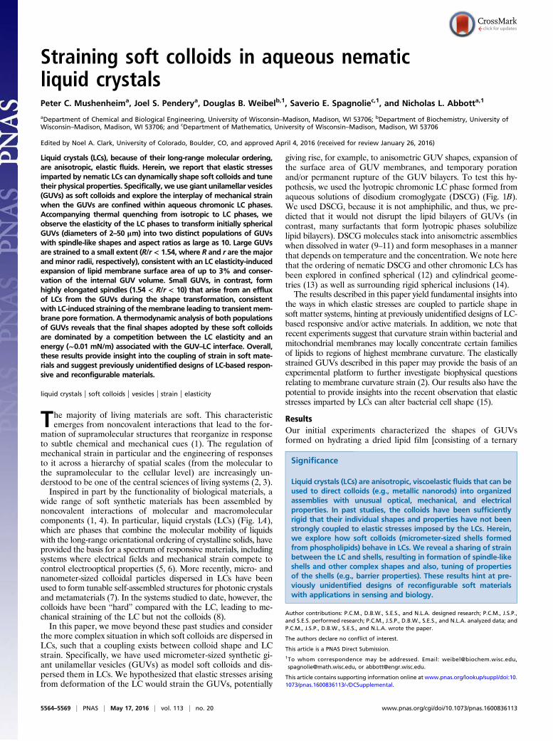
Straining soft colloids in aqueous nematicliquid crystalsPeter C. Mushenheima, Joel S. Penderya, Douglas B. Weibelb,1, Saverio E. Spagnoliec,1, and Nicholas L. Abbotta,1
aDepartment of Chemical and Biological Engineering, University of Wisconsin–Madison, Madison, WI 53706; bDepartment of Biochemistry, University ofWisconsin–Madison, Madison, WI 53706; and cDepartment of Mathematics, University of Wisconsin–Madison, Madison, WI 53706
Edited by Noel A. Clark, University of Colorado, Boulder, CO, and approved April 4, 2016 (received for review January 26, 2016)
Liquid crystals (LCs), because of their long-range molecular ordering,are anisotropic, elastic fluids. Herein, we report that elastic stressesimparted by nematic LCs can dynamically shape soft colloids and tunetheir physical properties. Specifically, we use giant unilamellar vesicles(GUVs) as soft colloids and explore the interplay of mechanical strainwhen the GUVs are confined within aqueous chromonic LC phases.Accompanying thermal quenching from isotropic to LC phases, weobserve the elasticity of the LC phases to transform initially sphericalGUVs (diameters of 2–50 μm) into two distinct populations of GUVswith spindle-like shapes and aspect ratios as large as 10. Large GUVsare strained to a small extent (R/r < 1.54, where R and r are the majorand minor radii, respectively), consistent with an LC elasticity-inducedexpansion of lipid membrane surface area of up to 3% and conser-vation of the internal GUV volume. Small GUVs, in contrast, formhighly elongated spindles (1.54 < R/r < 10) that arise from an effluxof LCs from the GUVs during the shape transformation, consistentwith LC-induced straining of themembrane leading to transient mem-brane pore formation. A thermodynamic analysis of both populationsof GUVs reveals that the final shapes adopted by these soft colloidsare dominated by a competition between the LC elasticity and anenergy (∼0.01 mN/m) associated with the GUV–LC interface. Overall,these results provide insight into the coupling of strain in soft mate-rials and suggest previously unidentified designs of LC-based respon-sive and reconfigurable materials.
liquid crystals | soft colloids | vesicles | strain | elasticity
The majority of living materials are soft. This characteristicemerges from noncovalent interactions that lead to the for-
mation of supramolecular structures that reorganize in responseto subtle chemical and mechanical cues (1). The regulation ofmechanical strain in particular and the engineering of responsesto it across a hierarchy of spatial scales (from the molecular tothe supramolecular to the cellular level) are increasingly un-derstood to be one of the central sciences of living systems (2, 3).Inspired in part by the functionality of biological materials, a
wide range of soft synthetic materials has been assembled bynoncovalent interactions of molecular and macromolecularcomponents (1, 4). In particular, liquid crystals (LCs) (Fig. 1A),which are phases that combine the molecular mobility of liquidswith the long-range orientational ordering of crystalline solids, haveprovided the basis for a spectrum of responsive materials, includingsystems where electrical fields and mechanical strain compete tocontrol electrooptical properties (5, 6). More recently, micro- andnanometer-sized colloidal particles dispersed in LCs have beenused to form tunable self-assembled structures for photonic crystalsand metamaterials (7). In the systems studied to date, however, thecolloids have been “hard” compared with the LC, leading to me-chanical straining of the LC but not the colloids (8).In this paper, we move beyond these past studies and consider
the more complex situation in which soft colloids are dispersed inLCs, such that a coupling exists between colloid shape and LCstrain. Specifically, we have used micrometer-sized synthetic gi-ant unilamellar vesicles (GUVs) as model soft colloids and dis-persed them in LCs. We hypothesized that elastic stresses arisingfrom deformation of the LC would strain the GUVs, potentially
giving rise, for example, to anisometric GUV shapes, expansion ofthe surface area of GUV membranes, and temporary porationand/or permanent rupture of the GUV bilayers. To test this hy-pothesis, we used the lyotropic chromonic LC phase formed fromaqueous solutions of disodium cromoglygate (DSCG) (Fig. 1B).We used DSCG, because it is not amphiphilic, and thus, we pre-dicted that it would not disrupt the lipid bilayers of GUVs (incontrast, many surfactants that form lyotropic phases solubilizelipid bilayers). DSCG molecules stack into anisometric assemblieswhen dissolved in water (9–11) and form mesophases in a mannerthat depends on temperature and the concentration. We note herethat the ordering of nematic DSCG and other chromonic LCs hasbeen explored in confined spherical (12) and cylindrical geome-tries (13) as well as surrounding rigid spherical inclusions (14).The results described in this paper yield fundamental insights into
the ways in which elastic stresses are coupled to particle shape insoft matter systems, hinting at previously unidentified designs of LC-based responsive and/or active materials. In addition, we note thatrecent experiments suggest that curvature strain within bacterial andmitochondrial membranes may locally concentrate certain familiesof lipids to regions of highest membrane curvature. The elasticallystrained GUVs described in this paper may provide the basis of anexperimental platform to further investigate biophysical questionsrelating to membrane curvature strain (2). Our results also have thepotential to provide insights into the recent observation that elasticstresses imparted by LCs can alter bacterial cell shape (15).
ResultsOur initial experiments characterized the shapes of GUVsformed on hydrating a dried lipid film [consisting of a ternary
Significance
Liquid crystals (LCs) are anisotropic, viscoelastic fluids that can beused to direct colloids (e.g., metallic nanorods) into organizedassemblies with unusual optical, mechanical, and electricalproperties. In past studies, the colloids have been sufficientlyrigid that their individual shapes and properties have not beenstrongly coupled to elastic stresses imposed by the LCs. Herein,we explore how soft colloids (micrometer-sized shells formedfrom phospholipids) behave in LCs. We reveal a sharing of strainbetween the LC and shells, resulting in formation of spindle-likeshells and other complex shapes and also, tuning of propertiesof the shells (e.g., barrier properties). These results hint at pre-viously unidentified designs of reconfigurable soft materialswith applications in sensing and biology.
Author contributions: P.C.M., D.B.W., S.E.S., and N.L.A. designed research; P.C.M., J.S.P.,and S.E.S. performed research; P.C.M., J.S.P., D.B.W., S.E.S., and N.L.A. analyzed data; andP.C.M., J.S.P., D.B.W., S.E.S., and N.L.A. wrote the paper.
The authors declare no conflict of interest.
This article is a PNAS Direct Submission.1To whom correspondence may be addressed. Email: [email protected],[email protected], or [email protected].
This article contains supporting information online at www.pnas.org/lookup/suppl/doi:10.1073/pnas.1600836113/-/DCSupplemental.
5564–5569 | PNAS | May 17, 2016 | vol. 113 | no. 20 www.pnas.org/cgi/doi/10.1073/pnas.1600836113
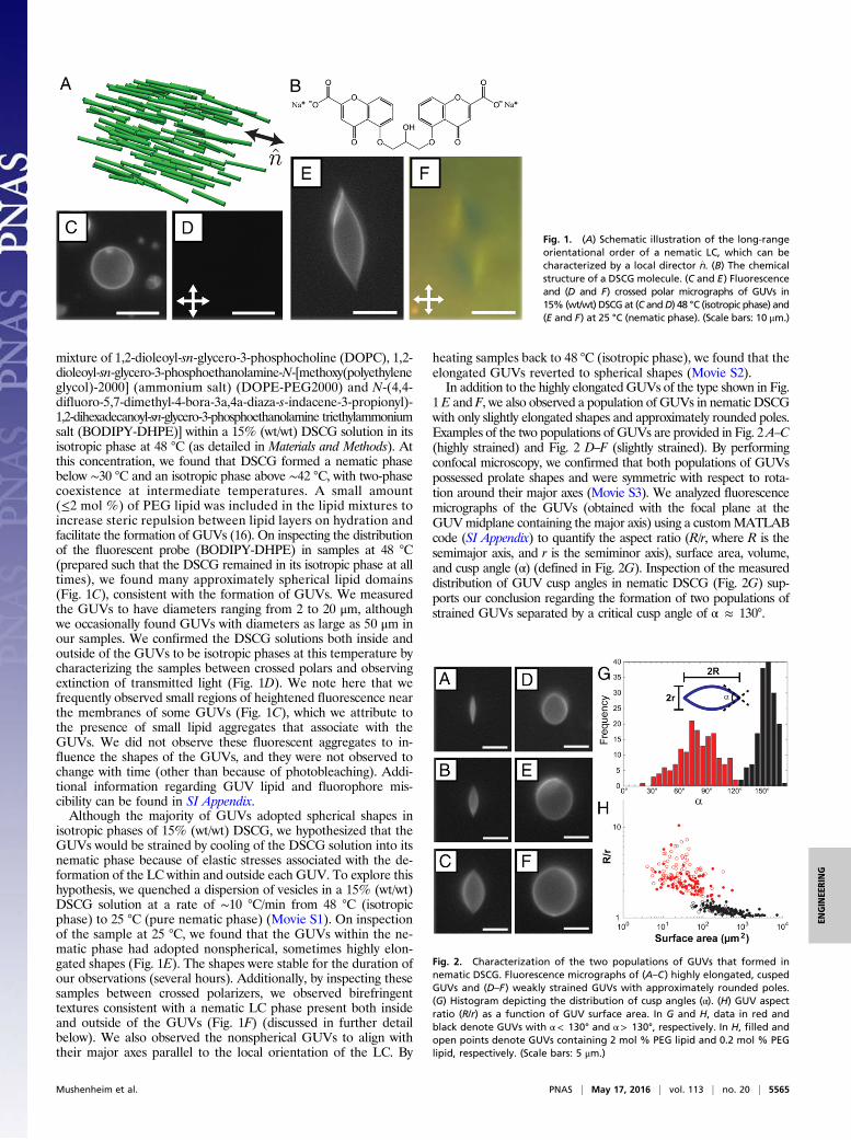
mixture of 1,2-dioleoyl-sn-glycero-3-phosphocholine (DOPC), 1,2-dioleoyl-sn-glycero-3-phosphoethanolamine-N-[methoxy(polyethyleneglycol)-2000] (ammonium salt) (DOPE-PEG2000) and N-(4,4-difluoro-5,7-dimethyl-4-bora-3a,4a-diaza-s-indacene-3-propionyl)-1,2-dihexadecanoyl-sn-glycero-3-phosphoethanolamine triethylammoniumsalt (BODIPY-DHPE)] within a 15% (wt/wt) DSCG solution in itsisotropic phase at 48 °C (as detailed in Materials and Methods). Atthis concentration, we found that DSCG formed a nematic phasebelow ∼30 °C and an isotropic phase above ∼42 °C, with two-phasecoexistence at intermediate temperatures. A small amount(≤2 mol %) of PEG lipid was included in the lipid mixtures toincrease steric repulsion between lipid layers on hydration andfacilitate the formation of GUVs (16). On inspecting the distributionof the fluorescent probe (BODIPY-DHPE) in samples at 48 °C(prepared such that the DSCG remained in its isotropic phase at alltimes), we found many approximately spherical lipid domains(Fig. 1C), consistent with the formation of GUVs. We measuredthe GUVs to have diameters ranging from 2 to 20 μm, althoughwe occasionally found GUVs with diameters as large as 50 μm inour samples. We confirmed the DSCG solutions both inside andoutside of the GUVs to be isotropic phases at this temperature bycharacterizing the samples between crossed polars and observingextinction of transmitted light (Fig. 1D). We note here that wefrequently observed small regions of heightened fluorescence nearthe membranes of some GUVs (Fig. 1C), which we attribute tothe presence of small lipid aggregates that associate with theGUVs. We did not observe these fluorescent aggregates to in-fluence the shapes of the GUVs, and they were not observed tochange with time (other than because of photobleaching). Addi-tional information regarding GUV lipid and fluorophore mis-cibility can be found in SI Appendix.Although the majority of GUVs adopted spherical shapes in
isotropic phases of 15% (wt/wt) DSCG, we hypothesized that theGUVs would be strained by cooling of the DSCG solution into itsnematic phase because of elastic stresses associated with the de-formation of the LC within and outside each GUV. To explore thishypothesis, we quenched a dispersion of vesicles in a 15% (wt/wt)DSCG solution at a rate of ∼10 °C/min from 48 °C (isotropicphase) to 25 °C (pure nematic phase) (Movie S1). On inspectionof the sample at 25 °C, we found that the GUVs within the ne-matic phase had adopted nonspherical, sometimes highly elon-gated shapes (Fig. 1E). The shapes were stable for the duration ofour observations (several hours). Additionally, by inspecting thesesamples between crossed polarizers, we observed birefringenttextures consistent with a nematic LC phase present both insideand outside of the GUVs (Fig. 1F) (discussed in further detailbelow). We also observed the nonspherical GUVs to align withtheir major axes parallel to the local orientation of the LC. By
heating samples back to 48 °C (isotropic phase), we found that theelongated GUVs reverted to spherical shapes (Movie S2).In addition to the highly elongated GUVs of the type shown in Fig.
1E and F, we also observed a population of GUVs in nematic DSCGwith only slightly elongated shapes and approximately rounded poles.Examples of the two populations of GUVs are provided in Fig. 2A–C(highly strained) and Fig. 2 D–F (slightly strained). By performingconfocal microscopy, we confirmed that both populations of GUVspossessed prolate shapes and were symmetric with respect to rota-tion around their major axes (Movie S3). We analyzed fluorescencemicrographs of the GUVs (obtained with the focal plane at theGUVmidplane containing the major axis) using a customMATLABcode (SI Appendix) to quantify the aspect ratio (R/r, where R is thesemimajor axis, and r is the semiminor axis), surface area, volume,and cusp angle (α) (defined in Fig. 2G). Inspection of the measureddistribution of GUV cusp angles in nematic DSCG (Fig. 2G) sup-ports our conclusion regarding the formation of two populations ofstrained GUVs separated by a critical cusp angle of α ≈ 130°.
Fig. 1. (A) Schematic illustration of the long-rangeorientational order of a nematic LC, which can becharacterized by a local director n̂. (B) The chemicalstructure of a DSCG molecule. (C and E) Fluorescenceand (D and F) crossed polar micrographs of GUVs in15% (wt/wt) DSCG at (C andD) 48 °C (isotropic phase) and(E and F) at 25 °C (nematic phase). (Scale bars: 10 μm.)
Fig. 2. Characterization of the two populations of GUVs that formed innematic DSCG. Fluorescence micrographs of (A–C) highly elongated, cuspedGUVs and (D–F) weakly strained GUVs with approximately rounded poles.(G) Histogram depicting the distribution of cusp angles (α). (H) GUV aspectratio (R/r) as a function of GUV surface area. In G and H, data in red andblack denote GUVs with α< 130° and α> 130°, respectively. In H, filled andopen points denote GUVs containing 2 mol % PEG lipid and 0.2 mol % PEGlipid, respectively. (Scale bars: 5 μm.)
Mushenheim et al. PNAS | May 17, 2016 | vol. 113 | no. 20 | 5565
ENGINEE
RING
We also quantified the change in aspect ratio of the vesicleswith size by plotting R/r as a function of GUV membrane surfacearea (SI Appendix, Eqs. S1 and S3) in nematic DSCG (Fig. 2H).In this plot, we distinguish between the population of GUVspossessing sharp cusps (α< 130°) and the population exhibitingapproximately rounded poles (α> 130°). Fig. 2H reveals thatsmall GUVs (surface area < 50 μm2) typically adopt highlyelongated shapes (R/r > 1.54) with sharp cusps in nematicDSCG, whereas larger GUVs (surface area > 500 μm2) adoptslightly elongated shapes (R/r < 1.54) with approximatelyrounded poles. We also note that there is far greater scatterwithin the data for GUVs with α< 130° and that GUVs withsurface areas between 50 and 500 μm2 exhibit either cusped orrounded poles. Fig. 2H includes data obtained from analysis ofGUVs containing 2 or 0.2 mol % PEG lipid plotted as filled andopen circles, respectively. Although the rigidity of lipid bilayersincreases with increasing PEG lipid content (17), we did notobserve the shapes of GUVs in nematic DSCG to depend onPEG lipid concentration. The trends discussed above in thecontext of Fig. 2H are also evident in a plot of cusp angle vs.GUV surface area (SI Appendix, Fig. S8).The above-described data provide evidence that GUVs be-
come strained in nematic DSCG, consistent with our initial hy-pothesis of the influence of LC elastic stresses on GUV shape.However, our observation of two distinct types of nonsphericalshapes was not anticipated. We have organized the remainder ofthis section into two parts, each focusing on one of the twopopulations of strained GUVs.
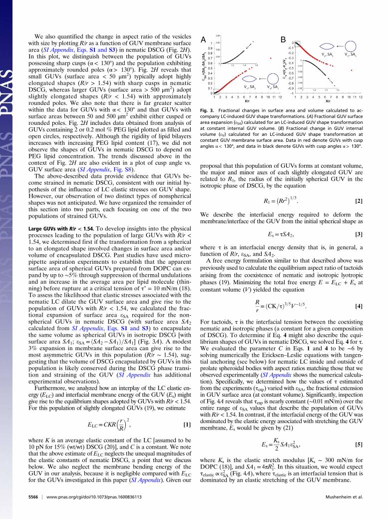
Large GUVs with R/r < 1.54. To develop insights into the physicalprocesses leading to the population of large GUVs with R/r <1.54, we determined first if the transformation from a sphericalto an elongated shape involved changes in surface area and/orvolume of encapsulated DSCG. Past studies have used micro-pipette aspiration experiments to establish that the apparentsurface area of spherical GUVs prepared from DOPC can ex-pand by up to ∼5% through suppression of thermal undulationsand an increase in the average area per lipid molecule (thin-ning) before rupture at a critical tension of τp = 10 mN/m (18).To assess the likelihood that elastic stresses associated with thenematic LC dilate the GUV surface area and give rise to thepopulation of GUVs with R/r < 1.54, we calculated the frac-tional expansion of surface area eSA required for the non-spherical GUVs in nematic DSCG (with surface area SA2calculated from SI Appendix, Eqs. S1 and S3) to encapsulatethe same volume as spherical GUVs in isotropic DSCG [withsurface area SA1; eSA = ðSA2 − SA1Þ=SA1] (Fig. 3A). A modest3% expansion in membrane surface area can give rise to themost asymmetric GUVs in this population (R/r ∼ 1.54), sug-gesting that the volume of DSCG encapsulated by GUVs in thispopulation is likely conserved during the DSCG phase transi-tion and straining of the GUV (SI Appendix has additionalexperimental observations).Furthermore, we analyzed how an interplay of the LC elastic en-
ergy (ELC) and interfacial membrane energy of the GUV (Es) mightgive rise to the equilibrium shapes adopted by GUVs with R/r < 1.54.For this population of slightly elongated GUVs (19), we estimate
ELC =CKR� rR
�2, [1]
where K is an average elastic constant of the LC [assumed to be10 pN for 15% (wt/wt) DSCG (20)], and C is a constant. We notethat the above estimate of ELC neglects the unequal magnitudes ofthe elastic constants of nematic DSCG, a point that we discussbelow. We also neglect the membrane bending energy of theGUV in our analysis, because it is negligible compared with ELCfor the GUVs investigated in this paper (SI Appendix). Given our
proposal that this population of GUVs forms at constant volume,the major and minor axes of each slightly elongated GUV arerelated to R1, the radius of the initially spherical GUV in theisotropic phase of DSCG, by the equation
R1 =�Rr2
�1=3. [2]
We describe the interfacial energy required to deform themembrane/interface of the GUV from the initial spherical shape as
Es = τSA2, [3]
where τ is an interfacial energy density that is, in general, afunction of R/r, eSA, and SA2.A free energy formulation similar to that described above was
previously used to calculate the equilibrium aspect ratio of tactoidsarising from the coexistence of nematic and isotropic lyotropicphases (19). Minimizing the total free energy E = ELC + Es atconstant volume (V) yielded the equation
Rr= ðCK=τÞ3=5V−1=5. [4]
For tactoids, τ is the interfacial tension between the coexistingnematic and isotropic phases (a constant for a given compositionof DSCG). To determine if Eq. 4 might also describe the equi-librium shapes of GUVs in nematic DSCG, we solved Eq. 4 for τ.We evaluated the parameter C in Eqs. 1 and 4 to be ∼6 bysolving numerically the Ericksen–Leslie equations with tangen-tial anchoring (see below) for nematic LC inside and outside ofprolate spheroidal bodies with aspect ratios matching those that weobserved experimentally (SI Appendix shows the numerical calcula-tion). Specifically, we determined how the values of τ estimatedfrom the experiments (τexp) varied with eSA, the fractional extensionin GUV surface area (at constant volume). Significantly, inspectionof Fig. 4A reveals that τexp is nearly constant (∼0.01 mN/m) over theentire range of eSA values that describe the population of GUVswith R/r < 1.54. In contrast, if the interfacial energy of the GUV wasdominated by the elastic energy associated with stretching the GUVmembrane, Es would be given by (21)
Es =Ks
2SA1e
2SA, [5]
where Ks is the elastic stretch modulus [Ks ∼ 300 mN/m forDOPC (18)], and SA1 = 4πR2
1. In this situation, we would expectτelastic ∝ e2SA (Fig. 4A), where τelastic is an interfacial tension that isdominated by an elastic stretching of the GUV membrane.
Fig. 3. Fractional changes in surface area and volume calculated to ac-company LC-induced GUV shape transformations. (A) Fractional GUV surfacearea expansion (eSA) calculated for an LC-induced GUV shape transformationat constant internal GUV volume. (B) Fractional change in GUV internalvolume (eV) calculated for an LC-induced GUV shape transformation atconstant GUV membrane surface area. Data in red denote GUVs with cuspangles α< 130°, and data in black denote GUVs with cusp angles α> 130°.
5566 | www.pnas.org/cgi/doi/10.1073/pnas.1600836113 Mushenheim et al.
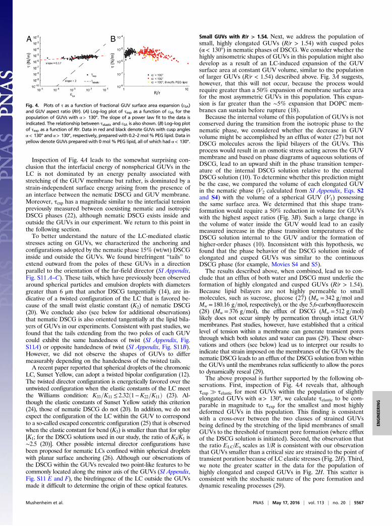
Inspection of Fig. 4A leads to the somewhat surprising con-clusion that the interfacial energy of nonspherical GUVs in theLC is not dominated by an energy penalty associated withstretching of the GUV membrane but rather, is dominated by astrain-independent surface energy arising from the presence ofan interface between the nematic DSCG and GUV membrane.Moreover, τexp has a magnitude similar to the interfacial tensionpreviously measured between coexisting nematic and isotropicDSCG phases (22), although nematic DSCG exists inside andoutside the GUVs in our experiment. We return to this point inthe following section.To better understand the nature of the LC-mediated elastic
stresses acting on GUVs, we characterized the anchoring andconfigurations adopted by the nematic phase 15% (wt/wt) DSCGinside and outside the GUVs. We found birefringent “tails” toextend outward from the poles of these GUVs in a directionparallel to the orientation of the far-field director (SI Appendix,Fig. S11 A–C). These tails, which have previously been observedaround spherical particles and emulsion droplets with diametersgreater than 6 μm that anchor DSCG tangentially (14), are in-dicative of a twisted configuration of the LC that is favored be-cause of the small twist elastic constant (K2) of nematic DSCG(20). We conclude also (see below for additional observations)that nematic DSCG is also oriented tangentially at the lipid bila-yers of GUVs in our experiments. Consistent with past studies, wefound that the tails extending from the two poles of each GUVcould exhibit the same handedness of twist (SI Appendix, Fig.S11A) or opposite handedness of twist (SI Appendix, Fig. S11B).However, we did not observe the shapes of GUVs to differmeasurably depending on the handedness of the twisted tails.A recent paper reported that spherical droplets of the chromonic
LC, Sunset Yellow, can adopt a twisted bipolar configuration (12).The twisted director configuration is energetically favored over theuntwisted configuration when the elastic constants of the LC meetthe Williams condition: K33=K11 ≤ 2.32ð1−K22=K11Þ (23). Al-though the elastic constants of Sunset Yellow satisfy this criterion(24), those of nematic DSCG do not (20). In addition, we do notexpect the configuration of the LC within the GUV to correspondto a so-called escaped concentric configuration (25) that is observedwhen the elastic constant for bend (K3) is smaller than that for splay[K1; for the DSCG solutions used in our study, the ratio of K3/K1 is∼2.5 (20)]. Other possible internal director configurations havebeen proposed for nematic LCs confined within spherical dropletswith planar surface anchoring (26). Although our observations ofthe DSCG within the GUVs revealed two point-like features to becommonly located along the minor axis of the GUVs (SI Appendix,Fig. S11 E and F), the birefringence of the LC outside the GUVsmade it difficult to determine the origin of these optical features.
Small GUVs with R/r > 1.54. Next, we address the population ofsmall, highly elongated GUVs (R/r > 1.54) with cusped poles(α< 130°) in nematic phases of DSCG. We consider whether thehighly anisometric shapes of GUVs in this population might alsodevelop as a result of an LC-induced expansion of the GUVsurface area at constant GUV volume, similar to the populationof larger GUVs (R/r < 1.54) described above. Fig. 3A suggests,however, that this will not occur, because the process wouldrequire greater than a 50% expansion of membrane surface areafor the most asymmetric GUVs in this population. This expan-sion is far greater than the ∼5% expansion that DOPC mem-branes can sustain before rupture (18).Because the internal volume of this population of GUVs is not
conserved during the transition from the isotropic phase to thenematic phase, we considered whether the decrease in GUVvolume might be accomplished by an efflux of water (27) but notDSCG molecules across the lipid bilayers of the GUVs. Thisprocess would result in an osmotic stress acting across the GUVmembrane and based on phase diagrams of aqueous solutions ofDSCG, lead to an upward shift in the phase transition temper-ature of the internal DSCG solution relative to the externalDSCG solution (10). To determine whether this prediction mightbe the case, we compared the volume of each elongated GUVin the nematic phase (V2 calculated from SI Appendix, Eqs. S2and S4) with the volume of a spherical GUV (V1) possessingthe same surface area. We determined that this shape trans-formation would require a 50% reduction in volume for GUVswith the highest aspect ratios (Fig. 3B). Such a large change inthe volume of water inside the GUV would lead to an easilymeasured increase in the phase transition temperatures of theDSCG solution internal to the GUV and/or the formation ofhigher-order phases (10). Inconsistent with this hypothesis, wefound that the phase behavior of the DSCG solution inside ofelongated and cusped GUVs was similar to the continuousDSCG phase (for example, Movies S4 and S5).The results described above, when combined, lead us to con-
clude that an efflux of both water and DSCG must underlie theformation of highly elongated and cusped GUVs (R/r > 1.54).Because lipid bilayers are not highly permeable to smallmolecules, such as sucrose, glucose (27) (Mw = 342 g=mol andMw = 180.16 g=mol, respectively), or the dye 5,6-carboxyfluorescein(28) (Mw = 376 g=mol), the efflux of DSCG (Mw = 512 g=mol)likely does not occur simply by permeation through intact GUVmembranes. Past studies, however, have established that a criticallevel of tension within a membrane can generate transient poresthrough which both solutes and water can pass (29). These obser-vations and others (see below) lead us to interpret our results toindicate that strain imposed on the membranes of the GUVs by thenematic DSCG leads to an efflux of the DSCG solution from withinthe GUVs until the membranes relax sufficiently to allow the poresto dynamically reseal (29).The above proposal is further supported by the following ob-
servations. First, inspection of Fig. 4A reveals that, althoughτexp � τelastic for most GUVs within the population of slightlyelongated GUVs with α> 130°, we calculate τelastic to be com-parable in magnitude to τexp for the smallest and most highlydeformed GUVs in this population. This finding is consistentwith a cross-over between the two classes of strained GUVsbeing defined by the stretching of the lipid membranes of smallGUVs to the threshold of transient pore formation (where effluxof the DSCG solution is initiated). Second, the observation thatthe ratio ELC/Es scales as 1/R is consistent with our observationthat GUVs smaller than a critical size are strained to the point oftransient poration because of LC elastic stresses (Fig. 2H). Third,we note the greater scatter in the data for the population ofhighly elongated and cusped GUVs in Fig. 2H. This scatter isconsistent with the stochastic nature of the pore formation anddynamic resealing processes (29).
Fig. 4. Plots of τ as a function of fractional GUV surface area expansion (eSA)and GUV aspect ratio (R/r). (A) Log–log plot of τexp as a function of eSA for thepopulation of GUVs with α> 130°. The slope of a power law fit to the data isindicated. The relationship between τelastic and eSA is also shown. (B) Log–log plotof τexp as a function of R/r. Data in red and black denote GUVs with cusp anglesα< 130° and α> 130°, respectively, prepared with 0.2–2mol% PEG lipid. Data inyellow denote GUVs prepared with 0 mol % PEG lipid, all of which had α< 130°.
Mushenheim et al. PNAS | May 17, 2016 | vol. 113 | no. 20 | 5567
ENGINEE
RING
Next, we determined if the equilibrium shapes adopted byGUVs with α< 130° are controlled by a similar interplay betweenthe LC elastic and interfacial energies, which was found for thepopulation of GUVs with α> 130°. By using experimental data inconjunction with Eq. 4 to generate a plot of τexp vs. R/r (Fig. 4B), wefound that τexp varied weakly as a function of R/r for the populationof GUVs with α< 130° (similar to the population of GUVs withα> 130°) with values that ranged between 0.01 and 0.001 mN/m.The data suggest that the same coupling between LC elastic energyand GUV–LC surface energy dictates the shapes adopted by all ofthe GUVs that we observe in nematic 15% (wt/wt) DSCG.A key finding of our experiments is that the shapes of the
GUVs are influenced by a shape-independent interfacial tension.The magnitude of the interfacial energy (∼0.01 mN/m) is similarto that which controls the shapes of tactoids of nematic DSCGdispersed in isotropic phases. We propose that the origin of theinterfacial energy associated with the GUVs dispersed in nematicDSCG may arise from depletion of the DSCG concentration nearthe surface of GUV membranes, effectively creating an “in-terface” between DSCG-rich and DSCG-poor solutions. Consis-tent with this interpretation, on heating GUV-containing samples,we observed isotropic domains to form first near the surfaces ofGUVs (SI Appendix, Fig. S6 C and D). We note that the PEGlipids are not responsible for this phenomenon, because similarresults were obtained in GUVs without PEG lipids (SI Appendix).We also performed detailed observations of the DSCG director
inside and outside of elongated GUVs as described in SI Appendix(a schematic illustration of the director configuration inside andaround an elongated GUV is found in SI Appendix, Fig. S12D).For reasons of brevity, we do not detail them here other than tonote that neither the nematic DSCG inside highly elongatedGUVs nor the nematic DSCG outside highly elongated GUVsshowed evidence of the presence of twist, consistent with pastobservations of nematic tactoids with high aspect ratios (9, 22, 30).
DiscussionThe results presented in this paper reveal that spherical GUVssuspended in isotropic 15% (wt/wt) DSCG deform into elon-gated, nonspherical shapes (spindles) on quenching of the solu-tion into a nematic LC phase. The observation that these shapetransformations accompany the DSCG phase transition suggeststhat elastic stresses imparted by the LC drive the GUV elonga-tion and that a key factor that controls the shapes of the GUVs isa strain-induced efflux of DSCG solution from the interior of thesmall GUVs. Here, we note that a prior study has documentedthat the addition of high concentrations [> 6% (wt/wt)] of sur-factants to isotropic phases of DSCG [5.5% (wt/wt) DSCG solutionsat room temperature] generated nematic domains (31). Additionalreports describe that similarly high concentrations of nonamphiphilicmolecules, such as poly(vinyl alcohol) (32), PEG (33), and spermine(33), condense DSCG solutions and lead to the formation of ne-matic and columnar phases. Condensation of DSCG into higher-order phases by concentrated surfactant and polymer solutions islikely the result of depletion effects, whereas electrostatic interac-tions also may contribute in the case of spermine (33). In contrast,our experiments use low lipid concentrations [∼0.04% (wt/wt)], andwe emphasize that the DSCG phase behavior is not substantiallyperturbed by the presence of the low concentrations. We also notethat we did not observe nematic LC phases to form inside or outsideGUVs prepared by gentle hydration of lipid films within aqueoussolutions of 5.5% (wt/wt) DSCG (SI Appendix, Fig. S1).The observations reported in our paper document the most
common behaviors of GUVs in nematic DSCG. We did, how-ever, observe several GUVs to exhibit behaviors that differedfrom those described above. First, we note that a small fraction(5–10%) of highly strained GUVs possessed optical featuresnear their poles that hinted at the possible presence of a subtlechange of membrane curvature (Fig. 1E). Additional studies are
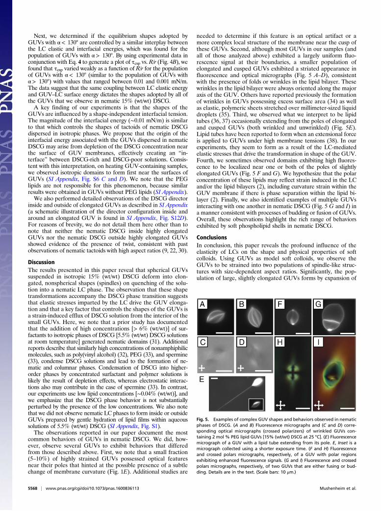
needed to determine if this feature is an optical artifact or amore complex local structure of the membrane near the cusp ofthese GUVs. Second, although most GUVs in our samples (andall of those analyzed above) exhibited a largely uniform fluo-rescence signal at their boundaries, a smaller population ofelongated and cusped GUVs exhibited a striated appearance influorescence and optical micrographs (Fig. 5 A–D), consistentwith the presence of folds or wrinkles in the lipid bilayer. Thesewrinkles in the lipid bilayer were always oriented along the majoraxis of the GUV. Others have reported previously the formationof wrinkles in GUVs possessing excess surface area (34) as wellas elastic, polymeric sheets stretched over millimeter-sized liquiddroplets (35). Third, we observed what we interpret to be lipidtubes (36, 37) occasionally extending from the poles of elongatedand cusped GUVs (both wrinkled and unwrinkled) (Fig. 5E).Lipid tubes have been reported to form when an extensional forceis applied to GUVs under high membrane tensions (38). In ourexperiments, they seem to form as a result of the LC-mediatedelastic stresses that drive the transformation in shape of the GUV.Fourth, we sometimes observed domains exhibiting high fluores-cence to be localized near one or both of the poles of slightlyelongated GUVs (Fig. 5 F and G). We hypothesize that the polarconcentration of these lipids may reflect strain induced in the LCand/or the lipid bilayers (2), including curvature strain within theGUV membrane if there is phase separation within the lipid bi-layer (2). Finally, we also identified examples of multiple GUVsinteracting with one another in nematic DSCG (Fig. 5 G and I) ina manner consistent with processes of budding or fusion of GUVs.Overall, these observations highlight the rich range of behaviorsexhibited by soft phospholipid shells in nematic DSCG.
ConclusionsIn conclusion, this paper reveals the profound influence of theelasticity of LCs on the shape and physical properties of softcolloids. Using GUVs as model soft colloids, we observe theGUVs to be strained into two populations of spindle-like struc-tures with size-dependent aspect ratios. Significantly, the pop-ulation of large, slightly elongated GUVs forms by expansion of
Fig. 5. Examples of complex GUV shapes and behaviors observed in nematicphases of DSCG. (A and B) Fluorescence micrographs and (C and D) corre-sponding optical micrographs (crossed polarizers) of wrinkled GUVs con-taining 2 mol % PEG lipid GUVs [15% (wt/wt) DSCG at 25 °C]. (E) Fluorescencemicrograph of a GUV with a lipid tube extending from its pole. E, Inset is amicrograph collected using a shorter exposure time. (F and H) Fluorescenceand crossed polars micrographs, respectively, of a GUV with polar regionsexhibiting enhanced fluorescence signals. (G and I) Fluorescence and crossedpolars micrographs, respectively, of two GUVs that are either fusing or bud-ding. Details are in the text. (Scale bars: 10 μm.)
5568 | www.pnas.org/cgi/doi/10.1073/pnas.1600836113 Mushenheim et al.

lipid membrane surface area at constant GUV volume, whereasthe smaller, highly elongated GUVs form through a mechanismthat involves a strain-induced transient pore formation in theGUV bilayer. A key conclusion is that the cross-over between thetwo populations is defined by the stretching of the lipid mem-branes of small GUVs to the threshold of transient pore for-mation (where efflux of the DSCG solution is initiated). Thisresult reveals that LC-induced straining of phospholipid shellsprovides a potentially useful strategy for controlling membraneproperties, including their transmembrane transport properties.Another key finding of our study is that the final shapes of bothpopulations of GUV are controlled by a strain-independentenergy associated with the area of contact between the GUV andthe nematic LC. The magnitude of the interfacial energy issimilar to the nematic–isotropic interface of DSCG solutions.Our observation that isotropic domains of DSCG nucleate nearthe surface of GUVs on heating hints that the concentration ofDSCG may be depleted near the GUV membrane surface. Wenote also that our study has focused on the influence of isotropicto nematic transitions on the GUVs and that additional studiesare required to fully elucidate processes that result in the ref-ormation of spherical GUVs accompanying the nematic to iso-tropic phase transition (as shown in Movie S2). More broadly,the coupling between LC elastic strain and strain within flexiblelipid membranes, as described in this study, may provide guid-ance to the design of reconfigurable soft materials with potentialapplication in sensing and biology. For example, we envision thatthese elastically strained GUVs might be attractive model cellmembranes and could serve as versatile synthetic platforms toinvestigate further, for example, how curvature strain influenceslocalization of lipids and proteins in biological membranes.
Materials and MethodsMaterials. A list of materials can be found in SI Appendix.
Lyotropic LC Preparation. Lyotropic LCs containing DSCG were prepared bymixing 15% (wt/wt) DSCG with 85% (wt/wt) water. At this concentration, theDSCG solution is nematic at room temperature and has a nematic–isotropiccoexistence temperature range between 29 °C and 35 °C (10). The mixturewas shaken for at least 12 h to ensure complete solubility and homogeneity.Before experimentation, the DSCG solution was heated at 65 °C for 10 minto erase any influence of the history of the sample on its properties.
GUV Preparation. To enable observations of the behaviors of isolated vesicles,dilute dispersions of GUVs were prepared through the gentle hydration of adried lipid films containing DOPC, 0.2–2 mol % DOPE-PEG 2000 [or 1,2-distearoyl-sn-glycero-3-phosphoethanolamine-N-[poly(ethylene glycol)2000-N′-carboxyfluorescein] (ammonium salt) (DSPE-PEG2000-CF)], and up to 0.5 mol %BODIPY-DHPE (1 mM total lipid concentration). We modified proceduresdetailed elsewhere (16) to permit hydration of the GUVs in isotropic phasesof DSCG (SI Appendix).
Preparation of Optical Cells. To investigate GUVs prepared in 15% (wt/wt)DSCG, we added a small volume (∼4.5 μL) of the GUV mixture to a cavitycreated using sheets of Mylar film (∼60 μm in thickness) placed between twoglass substrates. After assembly, the chamber was immediately sealed withepoxy glue to prevent water evaporation. Samples were prepared in theisotropic phase at 48 °C and quenched to the nematic phase at 25 °C toinvestigate the dynamical shape changes of GUVs that accompany this phasetransition. The glass surfaces of the substrates caused degenerate, planaranchoring of the nematic DSCG phase.
Microscopy. Information about the optical microscopes used in this study andour methods can be found in SI Appendix.
ACKNOWLEDGMENTS. Financial support fromWisconsin MRSEC Grant DMR-1121288 is acknowledged.
1. Aida T, Meijer EW, Stupp SI (2012) Functional supramolecular polymers. Science335(6070):813–817.
2. Renner LD, Weibel DB (2011) Cardiolipin microdomains localize to negatively curvedregions of Escherichia coli membranes. Proc Natl Acad Sci USA 108(15):6264–6269.
3. Wang N, Butler JP, Ingber DE (1993) Mechanotransduction across the cell surface andthrough the cytoskeleton. Science 260(5111):1124–1127.
4. Petka WA, Harden JL, McGrath KP, Wirtz D, Tirrell DA (1998) Reversible hydrogelsfrom self-assembling artificial proteins. Science 281(5375):389–392.
5. Ichimura K (2000) Photoalignment of liquid-crystal systems. Chem Rev 100(5):1847–1874.6. Kato T, Mizoshita N, Kishimoto K (2005) Functional liquid-crystalline assemblies: Self-
organized soft materials. Angew Chem Int Ed Engl 45(1):38–68.7. Musevic I, Skarabot M, Tkalec U, Ravnik M, Zumer S (2006) Two-dimensional nematic
colloidal crystals self-assembled by topological defects. Science 313(5789):954–958.8. Stark H (2001) Physics of colloidal dispersions in nematic liquid crystals. Phys Rep
351(6):387–474.9. Nastishin YA, et al. (2005) Optical characterization of the nematic lyotropic chromonic
liquid crystals: Light absorption, birefringence, and scalar order parameter. Phys Rev EStat Nonlin Soft Matter Phys 72(4 Pt 1):041711.
10. Lydon J (2010) Chromonic review. J Mater Chem 20(45):10071–10099.11. Collings PJ, Dickinson AJ, Smith EC (2010) Molecular aggregation and chromonic
liquid crystals. Liq Cryst 37(6-7):701–710.12. Jeong J, Davidson ZS, Collings PJ, Lubensky TC, Yodh AG (2014) Chiral symmetry
breaking and surface faceting in chromonic liquid crystal droplets with giant elasticanisotropy. Proc Natl Acad Sci USA 111(5):1742–1747.
13. Jeong J, et al. (2015) Chiral structures from achiral liquid crystals in cylindrical capil-laries. Proc Natl Acad Sci USA 112(15):E1837–E1844.
14. Nych A, et al. (2014) Chiral bipolar colloids from nonchiral chromonic liquid crystals.Phys Rev E Stat Nonlin Soft Matter Phys 89(6):062502.
15. Mushenheim PC, et al. (2015) Effects of confinement, surface-induced orientationsand strain on dynamical behaviors of bacteria in thin liquid crystalline films. SoftMatter 11(34):6821–6831.
16. Yamashita Y, Oka M, Tanaka T, Yamazaki M (2002) A new method for the prepa-ration of giant liposomes in high salt concentrations and growth of protein micro-crystals in them. Biochim Biophys Acta - Biomembr 1561(2):129–134.
17. Marsh D, Bartucci R, Sportelli L (2003) Lipid membranes with grafted polymers:Physicochemical aspects. Biochim Biophys Acta - Biomembr 1615(1-2):33–59.
18. Rawicz W, Olbrich KC, McIntosh T, Needham D, Evans E (2000) Effect of chain lengthand unsaturation on elasticity of lipid bilayers. Biophys J 79(1):328–339.
19. Prinsen P, van der Schoot P (2003) Shape and director-field transformation of tactoids.Phys Rev E Stat Nonlin Soft Matter Phys 68(2 Pt 1):021701.
20. Zhou S, et al. (2014) Elasticity, viscosity, and orientational fluctuations of a lyotropicchromonic nematic liquid crystal disodium cromoglycate. Soft Matter 10(34):6571–6581.
21. Idiart MA, Levin Y (2004) Rupture of a liposomal vesicle. Phys Rev E Stat Nonlin SoftMatter Phys 69(6 Pt 1):061922.
22. Kim Y-K, Shiyanovskii SV, Lavrentovich OD (2013) Morphogenesis of defects andtactoids during isotropic-nematic phase transition in self-assembled lyotropic chro-monic liquid crystals. J Phys Condens Matter 25(40):404202.
23. Williams RD (1986) Two transitions in tangentially anchored nematic droplets. J PhysA Math Gen 19(16):3211–3222.
24. Zhou S, et al. (2012) Elasticity of lyotropic chromonic liquid crystals probed by directorreorientation in a magnetic field. Phys Rev Lett 109(3):037801.
25. Drzaic PS (1995) Liquid Crystal Dispersions (World Scientific, Teaneck, NJ).26. Fernández-Nieves A, Link DR, Márquez M, Weitz DA (2007) Topological changes in
bipolar nematic droplets under flow. Phys Rev Lett 98(8):087801.27. Olbrich K, Rawicz W, Needham D, Evans E (2000) Water permeability and mechanical
strength of polyunsaturated lipid bilayers. Biophys J 79(1):321–327.28. Marmottant P, Biben T, Hilgenfeldt S (2008) Deformation and rupture of lipid vesicles
in the strong shear flow generated by ultrasound-driven microbubbles. Proc R SocLond A Math Phys Sci 464(2095):1781–1800.
29. Ohno M, Hamada T, Takiguchi K, Homma M (2009) Dynamic behavior of giant lipo-somes at desired osmotic pressures. Langmuir 25(19):11680–11685.
30. Kaznacheev AV, Bogdanov MM, Taraskin SA (2002) The nature of prolate shape oftactoids in lyotropic inorganic liquid crystals. J Exp Theor Phys 95(1):57–63.
31. Varghese N, et al. (2012) Emulsion of aqueous-based nonspherical droplets in aque-ous solutions by single-chain surfactants: Templated assembly by nonamphiphiliclyotropic liquid crystals in water. Langmuir 28(29):10797–10807.
32. Simon KA, Sejwal P, Gerecht RB, Luk Y-Y (2007) Water-in-water emulsions stabilizedby non-amphiphilic interactions: Polymer-dispersed lyotropic liquid crystals. Langmuir23(3):1453–1458.
33. Tortora L, et al. (2010) Self-assembly, condensation, and order in aqueous lyotropicchromonic liquid crystals crowded with additives. Soft Matter 6(17):4157–4167.
34. Knorr RL, Staykova M (2010) Wrinkling and electroporation of giant vesicles in the gelphase. Soft Matter 6(9):1990–1996.
35. Hohlfeld E, Davidovitch B (2015) Sheet on a deformable sphere: Wrinkle patternssuppress curvature-induced delamination. Phys Rev E Stat Nonlin Soft Matter Phys91(1):012407.
36. Mui BL-S, Döbereiner H-G, Madden TD, Cullis PR (1995) Influence of transbilayerarea asymmetry on the morphology of large unilamellar vesicles. Biophys J 69(3):930–941.
37. Bagatolli LA, Parasassi T, Gratton E (2000) Giant phospholipid vesicles: Comparisonamong the whole lipid sample characteristics using different preparation methods: Atwo photon fluorescence microscopy study. Chem Phys Lipids 105(2):135–147.
38. Sorre B, et al. (2012) Nature of curvature coupling of amphiphysin with membranesdepends on its bound density. Proc Natl Acad Sci USA 109(1):173–178.
Mushenheim et al. PNAS | May 17, 2016 | vol. 113 | no. 20 | 5569
ENGINEE
RING