Embed Size (px)
Citation preview
advance online publication nature clinical practice oncoloGY �
www.nature.com/clinicalpractice/onc
SuMMarY
Strategies for discovering novel cancer biomarkers through utilization of emerging technologiesVathany Kulasingam and Eleftherios P Diamandis*
Continuing Medical Education onlineMedscape, LLC is pleased to provide online continuing medical education (CME) for this journal article, allowing clinicians the opportunity to earn CME credit. Medscape, LLC is accredited by the Accreditation Council for Continuing Medical Education (ACCME) to provide CME for physicians. Medscape, LLC designates this educational activity for a maximum of 1.0 AMA PRA Category 1 CreditsTM. Physicians should only claim credit commensurate with the extent of their participation in the activity. All other clinicians completing this activity will be issued a certificate of participation. To receive credit, please go to http://www.medscape.com/cme/ncp and complete the post-test.
Learning objectivesUpon completion of this activity, participants should be able to: 1 Identify how cancer biomarkers are best applied
to clinical care.2 Describe the impact of biomarkers on specific
types of cancer.3 Describe the process and applicability of gene
expression profiling.4 List potential advantages of mass spectrometry-
based proteomic profiling.
Competing interestsThe authors and the Journal Editor L Hutchinson declared no competing interests. The CME questions author CP Vega declared that he has served as an advisor or consultant to Novartis, Inc.
INTRODUCTIONCancer continues to be a major cause of morbidity and mortality among men and women. In the US in 2006, over 1.4 million new cases of cancer were diagnosed and over half a million people died from this disease; the disease accounts for approxi-mately 25% of all deaths in the US each year.1 With increasing life expectancy, the prevalence of many cancers will probably increase. Early detection of various forms of cancer before they spread and become incurable is an important incentive for physicians and research scientists.2 One of the best ways to diagnose cancer early, aid in its prognosis, or predict therapeutic response, is to use serum or tissue biomarkers.
Cancer biomarkers can be DNA, mRNA, proteins, metabolites, or processes such as apop-tosis, angiogenesis or proliferation.3 The markers
The introduction of technologies such as mass spectrometry and protein and DNA arrays, combined with our understanding of the human genome, has enabled simultaneous examination of thousands of proteins and genes in single experiments, which has led to renewed interest in discovering novel biomarkers for cancer. The modern technologies are capable of performing parallel analyses as opposed to the serial analyses conducted with older methods, and they therefore provide opportunities to identify distinguishing patterns (signatures or portraits) for cancer diagnosis and classification as well as to predict response to therapies. Furthermore, these technologies provide the means by which new, single tumor markers could be discovered through use of reasonable hypotheses and novel analytical strategies. Despite the current optimism, a number of important limitations to the discovery of novel single tumor markers have been identified, including study design bias, and artefacts related to the collection and storage of samples. Despite the fact that new technologies and strategies often fail to identify well-established cancer biomarkers and show a bias toward the identification of high-abundance molecules, these technological advances have the capacity to revolutionize biomarker discovery. It is now necessary to focus on careful validation studies in order to identify the strategies and biomarkers that work and bring them to the clinic as early as possible.
keywoRds mass spectrometry, microarrays, multiparametric, proteomics, tumor markers
V Kulasingam is a postdoctoral trainee in clinical biochemistry, and EP Diamandis is Professor and Head of Clinical Biochemistry, Department of Laboratory Medicine and Pathobiology, University of Toronto, University Health Network and Toronto Medical Laboratories and Mount Sinai Hospital, Toronto, ON, Canada.
Correspondence*Department of Pathology and Laboratory Medicine, Mount Sinai Hospital, 600 University Avenue, Toronto, ON M5G 1X5, Canada [email protected]
Received 4 October 2007 Accepted 16 April 2008 Published online 12 August 2008
www.nature.com/clinicalpracticedoi:10.1038/ncponc1187
REvIEw CRITERIAThe information for this Review was compiled by searching the PubMed database for articles published up until 6 August 2007. Electronic early-release publications were included. Only articles published in English were considered. The search terms included “tumor markers” in association with the following search terms: “reviews”, “mass spectrometry”, “protein arrays”, “gene expression profiling”, “proteomics”, “molecular markers”, “cancer biomarker guidelines”, “peptidomics” and “microarrays”. When possible, primary sources have been quoted..
cMe
review
� nature clinical practice oncoloGY KulaSinGaM and diaMandiS advance online publication
www.nature.com/clinicalpractice/onc
are produced either by the tumor itself or by other tissues, in response to the presence of cancer or other associated conditions, such as inflamma-tion. Such biomarkers can be found in a variety of fluids, tissues and cell lines. Tumor markers can be used for screening the general population, for differential diagnosis in symptomatic patients, and for clinical staging of cancer. Additionally, these markers can be used to estimate tumor volume, to evaluate response to treatment, to assess disease recurrence through monitoring, or as prognostic indicators of disease progression (Box 1). Given
the low prevalence of cancer in any given popula-tion, no marker has yet been discovered that meets all of these criteria.
A number of different types and forms of tumor markers exist. These markers include hormones, as well as different functional subgroups of proteins such as enzymes, glycoproteins, oncofetal antigens and receptors. Furthermore, other changes in tumors, such as genetic mutations, amplifications or translocations, and changes in microarray-generated profiles (genetic signatures), are also forms of tumor markers. Regardless of the type of tumor marker or profile, the use of a tumor marker must be associated with proven improvements in patient outcomes, such as increased survival or enhanced quality of life, in order to be substanti-ated.3 An ideal tumor marker should be able to be measured easily, reliably and cost-effectively by use of an assay with high analytical sensitivity and specificity (Box 2). A caveat concerning currently used tumor markers is that, generally, they suffer from low diagnostic specificity and sensitivity (Table 1). Only a few markers have entered routine use, and then only for a limited number of cancer types and clinical settings. In the majority of cases, the current markers are used in conjunction with imaging, biopsy and associated clinicopathological information before a clinical decision is made.
The first cancer marker ever reported was the light chain of immunoglobulin in the urine, as identified in 75% of patients with myeloma in an 1848 study.4 The test for this marker is still employed by clinicians today, but with use of modern quantification techniques. From 1930 to 1960, scientists identified numerous hormones,
Box 1 Definitions and specifications of biomarkers.
diagnostic (screening) biomarker A marker that is used to detect and identify a given type of cancer in an individual. These markers are expected to have high specificity and sensitivity; for example, the presence of Bence–Jones protein in urine remains one of the strongest diagnostic indicators of multiple myeloma.
Prognostic biomarkerThis type of marker is used once the disease status has been established. These biomarkers are expected to predict the probable course of the disease including its recurrence, and they therefore have an important influence on the aggressiveness of therapy. For example, in testicular teratoma, human chorionic gonadotropin and alfa-fetoprotein levels can discriminate two groups with different survival rates.
stratification (predictive) biomarkerThis type of marker serves to predict the response to a drug before treatment is started. This marker classifies individuals as likely responders or nonresponders to a particular treatment. These biomarkers mainly arise from array-type experiments that make it possible to predict clinical outcome from the molecular characteristics of a patient’s tumor.
specificityThe proportion of control (normal) individuals who test negative for the biomarker.
sensitivityThe proportion of individuals with confirmed disease who test positive for the biomarker.
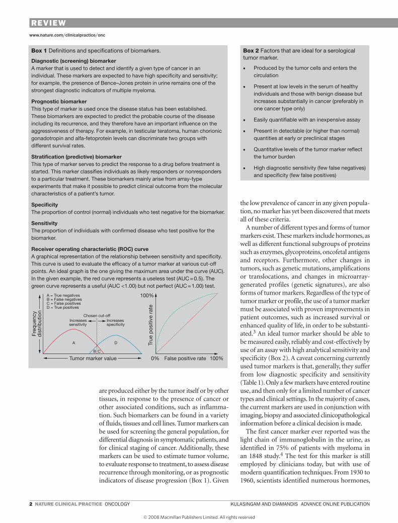
Receiver operating characteristic (RoC) curveA graphical representation of the relationship between sensitivity and specificity. This curve is used to evaluate the efficacy of a tumor marker at various cut-off points. An ideal graph is the one giving the maximum area under the curve (AUC). In the given example, the red curve represents a useless test (AUC = 0.5). The green curve represents a useful (AUC <1.00) but not perfect (AUC = 1.00) test.
ncponc_2007_214bx1.eps
Tumor marker value 0% 100%
100%
False positive rate
Chosen cut-off
A = True negativesB = False negativesC = False positivesD = True positives
A D
B C
Increasessensitivity
Increasesspecificity
Freq
uenc
yd
istr
ibut
ion
True
pos
itive
rat
e
Box 2 Factors that are ideal for a serological tumor marker.
■ Produced by the tumor cells and enters the circulation
■ Present at low levels in the serum of healthy individuals and those with benign disease but increases substantially in cancer (preferably in one cancer type only)
■ Easily quantifiable with an inexpensive assay
■ Present in detectable (or higher than normal) quantities at early or preclinical stages
■ Quantitative levels of the tumor marker reflect the tumor burden
■ High diagnostic sensitivity (few false negatives) and specificity (few false positives)
review review
advance online publication KulaSinGaM and diaMandiS nature clinical practice oncoloGY �
www.nature.com/clinicalpractice/onc
enzymes and other proteins, the concentration of which was altered in biological fluids from patients with cancer. The modern era of moni-toring malignant disease, however, began in the 1960s with the discovery of alfa-fetoprotein5 and carcinoembryonic antigen (CEA),6 which was facilitated by the introduction of immuno-logical techniques such as the radioimmunoassay. In the 1980s, the era of hybridoma technology enabled development of the ovarian epithelial cancer marker carbohydrate antigen (CA) 125.7 In 1980, prostate-specific antigen (PSA [KLK3]), considered one of the best cancer markers, was discovered.8 Table 2 summarizes some currently used markers and their clinical utility.
Every era of biomarker discovery seems to be associated closely with the emergence of a new and powerful analytical technology. The past decade has witnessed an impressive growth in the field of large-scale and high-throughput biology, which has contributed to an era of new techno-logy development. The completion of a number of genome-sequencing projects, the discovery of oncogenes and tumor-suppressor genes, and recent advances in genomic and proteomic techno-logies, together with powerful bioinformatics
tools, will have a direct and major impact on the way the search for cancer biomarkers is conducted. Early discoveries of cancer biomarkers were based mainly on empirical observations, such as the overexpression of CEA. The modern techno-logies are capable of performing parallel rather than serial analyses, and they can help to iden-tify distinguishing patterns and multiple markers rather than just a single marker; such strategies represent a central component and a paradigm shift in the search for novel biomarkers (Box 3).
These breakthroughs have paved the way for countless new avenues for biomarker identifica-tion. Very few serum tumor markers, however, have been introduced to the clinic over the past 15 years.9 In this Review, we highlight some of the mechanisms behind biomarker elevation in biological fluids, and outline strategies for novel marker identification. These strategies should facilitate the delivery of potential candidate mol-ecules for cancer diagnosis and prognosis and for prediction of therapy. These projected discoveries may be instrumental in substantially reducing the burden of cancer by providing prevention, indivi-dualized therapies, and improved monitoring following treatment.
Table 1 Current applications of tumor markers and their limitations.a
Application Current usefulness
Comments
Population screening
Limited A screening test should have very high sensitivity and exceptional specificity, to avoid too many false positives in populations with a low cancer prevalence. The test must demonstrate a benefit in terms of clinical outcome. Current biomarkers suffer from too low diagnostic sensitivity and specificity to serve as screening markers.Except for PSA, current tumor markers are more frequently elevated at late stages of disease.
Diagnosis Limited Current biomarkers suffer from too low diagnostic sensitivity and specificity to serve as diagnostic markers.
Prognosis Limited Most cancer markers have some prognostic value. Specific therapeutic interventions cannot be determined because the accuracy of prediction of current markers is rather poor.
Prediction of therapeutic response
High Very few markers have predictive power (exceptions include steroid hormone receptors and HER2 amplification for breast cancer), but the information they provide aids therapy selection.
Tumor staging Limited Besides AFP and HCG, the accuracy of the markers in determining tumor stage is poor.
Detecting early tumor recurrence
Controversial Lead time is short and does not considerably affect outcome. Clinical relapses could occur without biomarker elevation. Biomarker elevation can be nonspecific
Monitoring effectiveness of cancer therapy
High Current biomarkers provide information on therapeutic response (effective or noneffective) that is readily interpretable and more economical than imaging modalities.
aTable modified with permission from Diamandis EP et al. (2002) Tumor markers: past, present, and future. In: Diamandis EP et al., eds. Tumor Markers: Physiology, Pathobiology, Technology, and Clinical Applications. Washington DC: AACC Press.80 Abbreviations: AFP, alfa-fetoprotein; HCG, human chorionic gonadotropin; PSA, prostate-specific antigen.
review review
� nature clinical practice oncoloGY KulaSinGaM and diaMandiS advance online publication
www.nature.com/clinicalpractice/onc
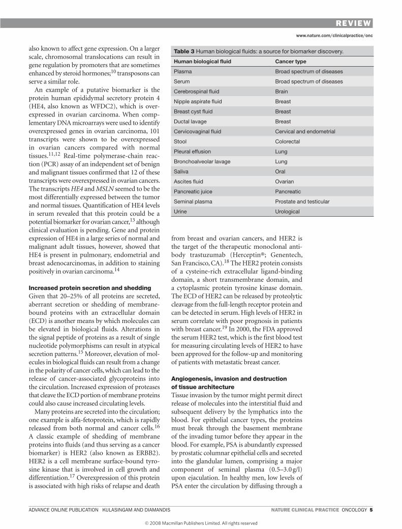
MECHANISMS OF BIOMARKER ELEvATION IN BIOLOGICAL FLUIDSFive major mechanisms exist by which molecules can be elevated in biological fluids during cancer initiation and progression. Such molecules could serve as effective cancer biomarkers. The mecha-nisms involved are outlined below; some of the different human body fluids that could be used as a source of biomarkers for specific types of cancers are shown in Table 3.
Gene overexpressionThe protein encoded by a gene can be expressed in increased quantities as a result of increases in gene or chromosome copy number (i.e. gene amplifica-tion) or through increased transcriptional activity. The latter process could be the result of imbal-ances between gene repressors and gene activators. Epigenetic changes, such as DNA methylation, are
Table 2 Cancer biomarkers that are currently in clinical use.
Tumor marker Cancer type year of discovery and reference
Application based on AsCo and/or NACB recommendations
Reference
Alfa-fetoprotein Germ-cellhepatoma
19635 DiagnosisDifferential diagnosis of NSGCTStagingDetecting recurrenceMonitoring therapy
80
Calcitonin Medullary thyroid carcinoma
1970s81 DiagnosisMonitoring therapy
82
CA125 Ovarian 19817 PrognosisDetecting recurrenceMonitoring therapy
80
CA 15-3 Breast 1984–583,84 Monitoring therapy 77
CA 19-9 Pancreatic 197985 Monitoring therapy 86
Carcinoembryonic antigen
Colon 196586 Monitoring therapyPrognosisDetecting recurrenceScreening for hepatic metastases
77,80
ER and PgR Breast 1970s87 Select patients for endocrine therapy 77
HER2 Breast 1985–688,89 Select patients for trastuzumab therapy 77
Human chorionic gonadotropin-β
Testicular 193890 DiagnosisStagingDetecting recurrenceMonitoring therapy
80
Lactate dehydrogenase
Germ cell 195491 DiagnosisPrognosisDetecting recurrenceMonitoring therapy
80
Prostate-specific antigen
Prostate 197992 Screening (with DRE)Diagnosis (with DRE)
80
Thyroglobulin Thyroid 195693 Monitoring 82
Abbreviations: DRE, digital rectal examination; ER, estrogen receptor; NACB, National Academy of Clinical Biochemistry; NSGCT, nonseminomatous germ cell tumor; PgR, progesterone receptor.
Box 3 Why the recent optimism for biomarker discovery?
The emergences of new technologies and new resources have created optimistic views that many more biomarkers will be discovered and validated. New technologies and resources include the following:
■ Completion of the Human Genome Project
■ Advanced bioinformatics
■ Array analysis (e.g. DNA, RNA, protein)
■ Mass-spectrometry-based profiling and identification
■ Laser-capture microdissection
■ Databases of single nucleotide polymorphisms
■ Comparative genomic hybridization
■ High-throughput sequencing
review review
advance online publication KulaSinGaM and diaMandiS nature clinical practice oncoloGY �
www.nature.com/clinicalpractice/onc
also known to affect gene expression. On a larger scale, chromosomal translocations can result in gene regulation by promoters that are sometimes enhanced by steroid hormones;10 transposons can serve a similar role.
An example of a putative biomarker is the protein human epididymal secretory protein 4 (HE4, also known as WFDC2), which is over-expressed in ovarian carcinoma. When comp-lementary DNA microarrays were used to identify overexpressed genes in ovarian carcinoma, 101 transcripts were shown to be overexpressed in ovarian cancers compared with normal tissues.11,12 Real-time polymerase-chain reac-tion (PCR) assay of an independent set of benign and malignant tissues confirmed that 12 of these transcripts were overexpressed in ovarian cancers. The transcripts HE4 and MSLN seemed to be the most differentially expressed between the tumor and normal tissues. Quantification of HE4 levels in serum revealed that this protein could be a potential biomarker for ovarian cancer,13 although clinical evaluation is pending. Gene and protein expression of HE4 in a large series of normal and malignant adult tissues, however, showed that HE4 is present in pulmonary, endometrial and breast adenocarcinomas, in addition to staining positively in ovarian carcinoma.14
Increased protein secretion and sheddingGiven that 20–25% of all proteins are secreted, aberrant secretion or shedding of membrane-bound proteins with an extracellular domain (ECD) is another means by which molecules can be elevated in biological fluids. Alterations in the signal peptide of proteins as a result of single nucleotide polymorphisms can result in atypical secretion patterns.15 Moreover, elevation of mol-ecules in biological fluids can result from a change in the polarity of cancer cells, which can lead to the release of cancer-associated glycoproteins into the circulation. Increased expression of proteases that cleave the ECD portion of membrane proteins could also cause increased circulating levels.
Many proteins are secreted into the circulation; one example is alfa-fetoprotein, which is rapidly released from both normal and cancer cells.16 A classic example of shedding of membrane proteins into fluids (and thus serving as a cancer biomarker) is HER2 (also known as ERBB2). HER2 is a cell membrane surface-bound tyro-sine kinase that is involved in cell growth and differentiation.17 Overexpression of this protein is associated with high risks of relapse and death
from breast and ovarian cancers, and HER2 is the target of the therapeutic monoclonal anti-body trastuzumab (Herceptin®; Genentech, San Francisco, CA).18 The HER2 protein consists of a cysteine-rich extracellular ligand-binding domain, a short transmembrane domain, and a cytoplasmic protein tyrosine kinase domain. The ECD of HER2 can be released by proteolytic cleavage from the full-length receptor protein and can be detected in serum. High levels of HER2 in serum correlate with poor prognosis in patients with breast cancer.19 In 2000, the FDA approved the serum HER2 test, which is the first blood test for measuring circulating levels of HER2 to have been approved for the follow-up and monitoring of patients with metastatic breast cancer.
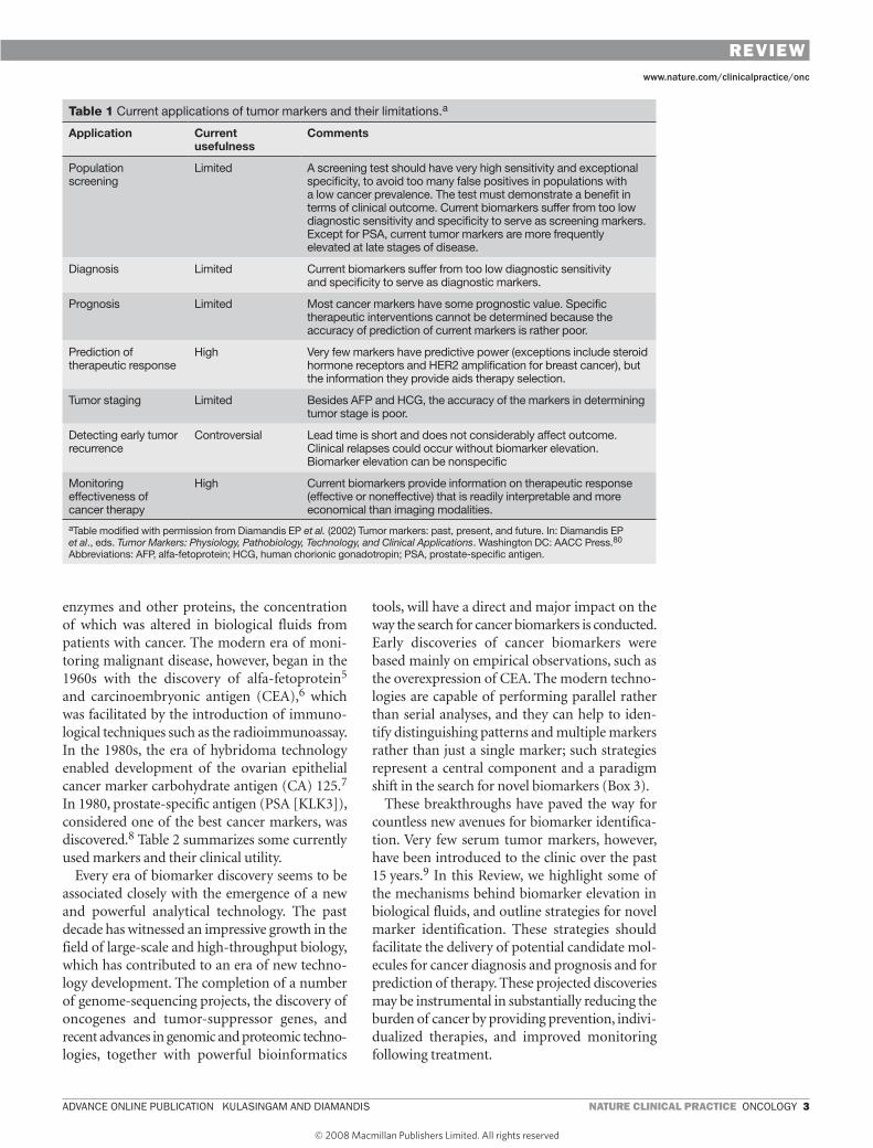
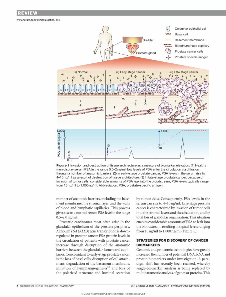
Angiogenesis, invasion and destruction of tissue architectureTissue invasion by the tumor might permit direct release of molecules into the interstitial fluid and subsequent delivery by the lymphatics into the blood. For epithelial cancer types, the proteins must break through the basement membrane of the invading tumor before they appear in the blood. For example, PSA is abundantly expressed by prostatic columnar epithelial cells and secreted into the glandular lumen, comprising a major component of seminal plasma (0.5–3.0 g/l) upon ejaculation. In healthy men, low levels of PSA enter the circulation by diffusing through a
Table 3 Human biological fluids: a source for biomarker discovery.
Human biological fluid Cancer type
Plasma Broad spectrum of diseases
Serum Broad spectrum of diseases
Cerebrospinal fluid Brain
Nipple aspirate fluid Breast
Breast cyst fluid Breast
Ductal lavage Breast
Cervicovaginal fluid Cervical and endometrial
Stool Colorectal
Pleural effusion Lung
Bronchoalveolar lavage Lung
Saliva Oral
Ascites fluid Ovarian
Pancreatic juice Pancreatic
Seminal plasma Prostate and testicular
Urine Urological
review review
� nature clinical practice oncoloGY KulaSinGaM and diaMandiS advance online publication
www.nature.com/clinicalpractice/onc
number of anatomic barriers, including the base-ment membrane, the stromal layer, and the walls of blood and lymphatic capillaries. This process gives rise to a normal serum PSA level in the range 0.5–2.0 ng/ml.
Prostatic carcinomas most often arise in the glandular epithelium of the prostate periphery. Although PSA (KLK3) gene transcription is down-regulated in prostate cancer, PSA protein levels in the circulation of patients with prostate cancer increase through disruption of the anatomic barriers between the glandular lumen and capil-laries. Concomitant to early-stage prostate cancer is the loss of basal cells, disruption of cell attach-ment, degradation of the basement membrane, initiation of lymphangiogenesis20 and loss of the polarized structure and luminal secretion
by tumor cells. Consequently, PSA levels in the serum can rise to 4–10 ng/ml. Late-stage prostate cancer is characterized by invasion of tumor cells into the stromal layers and the circulation, and by total loss of glandular organization. This situation enables considerable amounts of PSA to leak into the bloodstream, resulting in typical levels ranging from 10 ng/ml to 1,000 ng/ml (Figure 1).
STRATEGIES FOR DISCOvERY OF CANCER BIOMARKERSGenomic and proteomic technologies have greatly increased the number of potential DNA, RNA and protein biomarkers under investigation. A para-digm shift has recently been realized, whereby single-biomarker analysis is being replaced by multiparametric analysis of genes or proteins. This
10
1,000
10
42
0.50
1,000
PS
A L
evel
s (µ
g/L)
Columnar epithelial cell
Basal cell
Basement membrane
Blood/lymphatic capillary
Prostate cancer cells
Prostate specific antigen
ncponc_2007_214f1.eps
Bladder
Prostate gland
(iii) Late stage cancer(ii) Early stage cancer(i) Normal
Figure 1 Invasion and destruction of tissue architecture as a measure of biomarker elevation. (1) Healthy men display serum PSA in the range 0.5–2 ng/ml; low levels of PSA enter the circulation via diffusion through a number of anatomic barriers. (2) In early-stage prostate cancer, PSA levels in the serum rise to 4–10 ng/ml as a result of destruction of tissue architecture. (3) In late-stage prostate cancer, because of invasion of tumor cells, considerable amounts of PSA leak into the bloodstream; PSA levels typically range from 10 ng/ml to 1,000 ng/ml. Abbreviation: PSA, prostate-specific antigen.
review review
advance online publication KulaSinGaM and diaMandiS nature clinical practice oncoloGY �
www.nature.com/clinicalpractice/onc
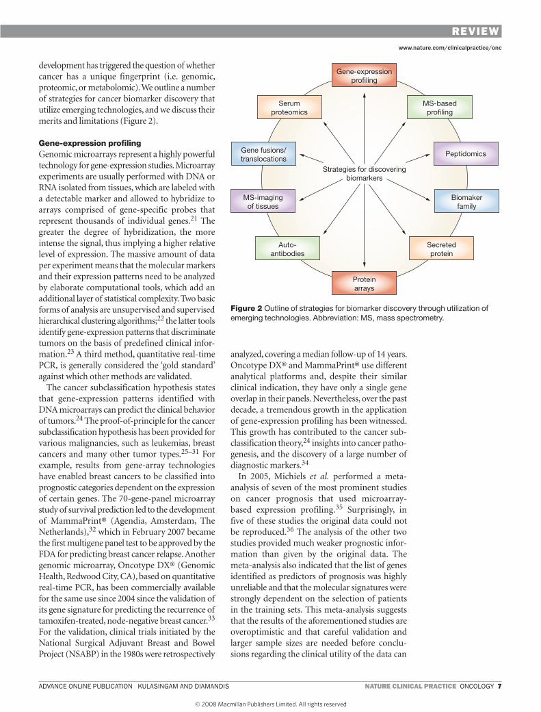
development has triggered the question of whether cancer has a unique fingerprint (i.e. genomic, proteomic, or metabolomic). We outline a number of strategies for cancer biomarker discovery that utilize emerging technologies, and we discuss their merits and limitations (Figure 2).
Gene-expression profilingGenomic microarrays represent a highly powerful technology for gene-expression studies. Microarray experiments are usually performed with DNA or RNA isolated from tissues, which are labeled with a detectable marker and allowed to hybridize to arrays comprised of gene-specific probes that represent thousands of individual genes.21 The greater the degree of hybridization, the more intense the signal, thus implying a higher relative level of expression. The massive amount of data per experiment means that the molecular markers and their expression patterns need to be analyzed by elaborate computational tools, which add an additional layer of statistical complexity. Two basic forms of analysis are unsupervised and supervised hierarchical clustering algorithms;22 the latter tools identify gene-expression patterns that discriminate tumors on the basis of predefined clinical infor-mation.23 A third method, quantitative real-time PCR, is generally considered the ‘gold standard’ against which other methods are validated.
The cancer subclassification hypothesis states that gene-expression patterns identified with DNA microarrays can predict the clinical behavior of tumors.24 The proof-of-principle for the cancer subclassification hypothesis has been provided for various malignancies, such as leukemias, breast cancers and many other tumor types.25–31 For example, results from gene-array technologies have enabled breast cancers to be classified into prognostic categories dependent on the expression of certain genes. The 70-gene-panel microarray study of survival prediction led to the development of MammaPrint® (Agendia, Amsterdam, The Netherlands),32 which in February 2007 became the first multigene panel test to be approved by the FDA for predicting breast cancer relapse. Another genomic microarray, Oncotype DX® (Genomic Health, Redwood City, CA), based on quantitative real-time PCR, has been commercially available for the same use since 2004 since the validation of its gene signature for predicting the recurrence of tamoxifen-treated, node-negative breast cancer.33 For the validation, clinical trials initiated by the National Surgical Adjuvant Breast and Bowel Project (NSABP) in the 1980s were retrospectively
analyzed, covering a median follow-up of 14 years. Oncotype DX® and MammaPrint® use different analytical platforms and, despite their similar clinical indication, they have only a single gene overlap in their panels. Nevertheless, over the past decade, a tremendous growth in the application of gene-expression profiling has been witnessed. This growth has contributed to the cancer sub-classification theory,24 insights into cancer patho-genesis, and the discovery of a large number of diagnostic markers.34
In 2005, Michiels et al. performed a meta-analysis of seven of the most prominent studies on cancer prognosis that used microarray-based expression profiling.35 Surprisingly, in five of these studies the original data could not be reproduced.36 The analysis of the other two studies provided much weaker prognostic infor-mation than given by the original data. The meta-analysis also indicated that the list of genes identified as predictors of prognosis was highly unreliable and that the molecular signatures were strongly dependent on the selection of patients in the training sets. This meta-analysis suggests that the results of the aforementioned studies are overoptimistic and that careful validation and larger sample sizes are needed before conclu-sions regarding the clinical utility of the data can
ncponc_2007_214f2.eps
Gene-expressionprofiling
Serumproteomics
Gene fusions/translocations
MS-basedprofiling
Peptidomics
Proteinarrays
Auto-antibodies
MS-imagingof tissues
Secretedprotein
Biomakerfamily
Strategies for discoveringbiomarkers
Figure 2 Outline of strategies for biomarker discovery through utilization of emerging technologies. Abbreviation: MS, mass spectrometry.
review review
� nature clinical practice oncoloGY KulaSinGaM and diaMandiS advance online publication
www.nature.com/clinicalpractice/onc
be drawn. Despite promising proof-of-principle data, the successful use of gene arrays to discover novel subtypes of various carcinomas, and the utilization of these technologies for discovery of diagnostic markers, these new tools are not yet recommended for widespread clinical use by either organizations issuing clinical guidelines or expert panels.37
Mass-spectrometry-based proteomic profilingProteomic-pattern profiling is a recent approach to biomarker discovery. Given that mRNA informa-tion does not best reflect the function of proteins, which are the functional components within organisms, the use of proteomic patterns to enable tumor diagnosis or subclassification seems more promising. The rationale is that proteins produced by cancer cells or their microenvironment may eventually enter the circulation and that the patterns of expression of these proteins could be assessed by mass spectrometry and used in combination with a mathematical algorithm for diagnostic purposes. Mass-spectrometry-based methods of proteomic analysis have improved and include more-advanced technology that brings higher mass accuracy, higher detection capa-bility, and shorter cycling times, thereby enabling increased throughput and more-reliable data.38 Technologies such as differential in-gel electro-phoresis, two-dimensional polyacrylamide gel electrophoresis and multidimensional protein-identification technology can be used for high-throughput protein profiling. The technology that has received considerable attention in the past involves the use of a minute amount of unfraction-ated serum sample added to a ‘protein-chip’, which is subsequently analyzed by surface-enhanced laser-desorption–ionization time-of-flight mass spectrometry (SELDI-TOF-MS) to generate a proteomic signature of serum.39 These patterns reflect part of the blood proteome, but without knowledge of the actual identity of the proteins. The potential of proteomic pattern analysis was first demonstrated in the diagnosis of ovarian cancer.40 In this study, exceptional results were seen, with a sensitivity of 100% (even for early-stage disease) and 95% specificity. These numbers are far superior to the sensitivities and specifici-ties obtained with current serological cancer bio-markers. Subsequently, proteomic pattern analysis has been extended to a number of other cancer types, including breast, prostate, colon, liver, renal, pancreatic, and head and neck cancers.41–47
In spite of the optimism regarding this approach, a number of important limitations have been identified.48 These shortcomings include bias from artefacts related to the clinical sample collection and storage, the inherent qualitative nature of mass spectrometers, failure to identify well-established cancer biomarkers, bias when identifying high-abundance molecules within the serum, and disagreement between peaks generated by different research laboratories.49–51 Another limitation concerns possible bioinformatic arte-facts. Baggerly et al. showed that signals that are detected that are a result of background noise can achieve a high level of discrimination between patients with cancer and those without.52 Despite a substantial time lapse since the first report of this technology, no product has yet reached the clinic and no independent validation studies have been published. Guideline-developing organizations and expert panels do not currently recommend serum proteomic profiling for clinical use.53
Peptidomics The low-molecular-weight plasma or serum proteome has been the focus of recent attempts to find novel biomarkers.54 Peptides are essential for many physiological processes, such as blood pressure (angiotensin II) and blood glucose (insulin) regulation. It has been suggested that “the low molecular-weight region of the blood proteome is a treasure trove of diagnostic informa-tion ready to be harvested by nanotechnology”.55 The low-molecular-weight serum proteome has been characterized by ultrafiltration, enzymatic digestion, and liquid chromatography coupled to tandem mass spectrometry56,57 or via a top-down proteomics approach (whereby the intact peptide is distinguished directly by its fragment ions)58 or by means of pattern profiling.59 Informative diag-nostic peptides that are generated after proteolysis of high-abundance proteins by the coagulation and complement enzymatic cascades can be identified by mass spectrometry. These proteomic patterns were claimed to distinguish not only controls from patients with cancer60 but also between various types of cancer.59
One major consideration is that these peptides that are present in the serum are derived from a low number of high-abundance proteins. Koomen et al. studied peptides in serum and concluded that sample collection is of immense importance and could give rise to artefacts, and that serum is not ideal for proteomic experiments as it contains substantial endoproteolytic and
review review
advance online publication KulaSinGaM and diaMandiS nature clinical practice oncoloGY �
www.nature.com/clinicalpractice/onc
exoproteolytic enzymatic activity.61 These find-ings raise concerns regarding peptidomics data generated by profiling technologies. Peptidomic profiling might represent nothing more than peptides cleaved during coagulation or functions inherent to plasma or serum, including immune modulation, inflammatory response and protease inhibition.62 Many of the aforementioned caveats associated with mass-spectrometry-based protein profiling technologies also apply to peptidomics.
Cancer-biomarker-family approachThe premise for the ‘cancer biomarker family’ approach is that if a member of a protein family is already an established biomarker, then other members of that family might also be good cancer biomarkers. For example, PSA is a member of the human tissue kallikrein family. Kallikreins are secreted enzymes with trypsin-like or chymotrypsin-like serine protease activity. This enzyme family consists of 15 genes clustered in tandem on chromosome 19q13.4.63 PSA (KLK3) and KLK2 currently have important clinical appli-cations as prostate cancer biomarkers.64 Other members of the human kallikrein family have been implicated in the process of carcinogenesis and are being investigated as biomarkers for diag-nosis and prognosis. For example, KLK6 has been studied as a novel biomarker for ovarian cancer.65 It was found that elevated serum levels of this protein were associated with late-stage tumor, high grade and serous histotype, and with resistance to chemotherapy.65 In general, increased levels of KLK6 were linked to decreased disease-free and overall survival, thus serving as an independent and unfavorable prognostic indicator. Similarly, KLK3, KLK5 and KLK14 have been shown to be increased in the serum of patients with breast cancer, thereby potentially serving as diagnostic markers. Being serine proteases, these proteins could be implicated in tumor progression through extracellular matrix degradation.
Secreted protein approachIn theory, a candidate serological tumor marker should be a secreted protein, because secreted proteins have the highest likelihood of entering the circulation. Examination of tissues or biological fluids near to the tumor site of origin could facili-tate identification of candidate molecules for further investigation. The increasing evidence that tumor growth and progression is dependent on the malignant potential of the tumor cells as well as on the microenvironment surrounding the
tumor (e.g. stroma, endothelial cells and immune and inflammatory cells) further supports this approach.66,67 A number of technologies can be utilized, but for systematic characterization of proteins in complex mixtures, mass spectro-metry is the preferred technology. In the case of breast cancer, breast tissue, nipple aspirate fluid, breast cyst fluid, tumor interstitial fluid and breast cancer cell lines can all be explored. The tumor interstitial fluid that perfuses the tumor micro-environment in invasive ductal carcinomas of the breast was examined by proteomic approaches.68 Over 250 proteins were identified, many of which were relevant to processes such as cell proliferation and invasion.
It should be noted that some of the widely used cancer biomarkers such as CEA, CA125 and HER2 are actually membrane-bound proteins, which are shed into the circulation. The identifi-cation of secreted proteins in tissues or other biological fluids does not necessarily imply that the proteins will be detectable in the sera of cancer patients. Serum-based diagnostic tests depend on the stability of the protein, its clearance, its associ-ation with other serum proteins and the extent of post-translational modifications.
Other prominent strategiesA number of other strategies for detecting cancer biomarkers exist. One approach that is gaining popularity is based on protein arrays. Wang and colleagues have published data suggesting that autoantibody signatures might improve the early detection of prostate cancer.69 Through use of a combination of phage-display technology and protein microarrays, this group identified new autoantibody-binding peptides derived from prostate cancer tissue. Another prevailing view is that tumor-associated antigens could serve as biosensors for cancer because tumors naturally elicit an immune response in the host. Moreover, breaking the cancer genetics dogma that hemato-logic malignancies result from chromosomal translocations70 and that mutations underlie epithelial solid tumors, gene fusions as a result of translocations in prostate cancer have been identified through use of gene-expression data sets.10 This translocation seems to be frequent (occurring in 40–50% of cases), may have prognostic value, and may be an early event in carcinogenesis. In addition, mass-spectrometry-based imaging of fresh-frozen tissue sections has yielded a number of potential candidate molecules.71,72 Besides proteomic profiling of
review review
�0 nature clinical practice oncoloGY KulaSinGaM and diaMandiS advance online publication
www.nature.com/clinicalpractice/onc
serum, attempts have been made to decipher the serum proteome via numerous fractiona-tion schemes to simplify and reduce the dynamic range of molecules present in serum.73 Finally, the use of animal models involving human tumor xenograft experiments has also shown promise for biomarker discovery.74,75
CONCLUSIONSBiomarker development falls into five conceptual phases: preclinical exploratory studies; clinical assay and validation; retrospective longitudinal studies; prospective screening; and randomized control trials (Box 4).76 Unfortunately, current studies of tumor markers are highly variable, not only in their methods of marker detection, but also in design and patient selection. Interpatient heterogeneity and intratumor heterogeneity are important confounding factors. In addition, the danger of bias and the problems of overfitting the data, as well as issues relating to the handling and storing of clinical specimens, are vital factors that need consideration before a study is conducted.51 New tumor marker tests—single or multiparametric—must, therefore, undergo rigorous validation in order that their clinical value can be assessed.
New resources will most likely identify novel protein, genetic and low-molecular-weight cancer markers, which may impact on cancer care. Furthermore, with advances in genomic and proteomic technologies, human diseases may be classified on the basis of molecular rather than morphological analysis. Moreover, bioinformatics will serve to link scientific data with clinical infor-mation. Despite the optimism, ASCO and the National Academy of Clinical Biochemistry do not encourage the widespread use of tumor markers unless they affect patient outcome measures.77 There is, however, a general agreement that a combination of multiple biomarkers may increase diagnostic sensitivity and specificity over use of individual markers. This is particularly important in relation to the recent development of powerful bioinformatic algorithms, which can interpret multiple parameters much more efficiently than can more-traditional approaches.78,79 The most accurate, individualized, predictive assessment for patients might be attained through the use of artificial neural networks. There is no doubt that if these new technological advances prove to be successful in identifying cancer biomarkers for early cancer detection, the clinical benefits are likely to be enormous.
KEY POINTS ■ Current cancer biomarkers suffer from low
diagnostic sensitivity and specificity and have not yet made a major impact in reducing cancer burden
■ The impressive growth of large-scale and high-throughput biology has resulted in increased popularity for the concept that novel biomarkers can be discovered through various emerging technologies
■ A better understanding of the mechanisms behind biomarker elevation in biological fluids may facilitate the discovery of new tumor markers
■ Some of the new promising strategies for biomarker discovery include microarray-based profiling at the DNA and mRNA level, and mass-spectrometry-based profiling at the protein or peptide level
■ Study of tumor markers that include current biomarkers or examination of fluids and tissues that are in close proximity to the tumor might also assist in identification of novel tumor markers
■ New tumor markers must undergo rigorous validation before they are introduced into routine clinical care
Box 4 Phases of biomarker development.76
1 Preclinical exploratory studiesIn this phase, tumor and non-tumor specimens are compared to generate hypotheses for clinical tests for detecting cancer. Strategies such as gene-expression profiling, mass-spectrometry-based methods and other approaches to biomarker discovery can be used to aid this phase.
2 Assay development and validationA clinical assay that uses a specimen of choice (usually something that can be obtained noninvasively) is developed in this phase. The assay must discriminate individuals with cancer from those without. The patients assessed in this phase have established disease. The utility of the assay in detecting disease early is not demonstrated in this phase.
3 Retrospective longitudinal clinical repository studiesSpecimens collected and stored from a cohort of healthy individuals who were monitored for development of cancer are used here. Evidence for the capacity of the biomarker to detect preclinical disease is demonstrated in phase 3. Criteria for ‘positive’ screening results are defined and used in phase 4.
4 Prospective screening studiesIn this phase, individuals are screened with the assay and diagnostic procedures are applied to those who screened positive. This can help to establish the tumor stage or the nature of the disease at the time of detection.
5 Randomized control trialsThe objective of this phase is to determine if screening reduces the burden of cancer in the population.
review review
advance online publication KulaSinGaM and diaMandiS nature clinical practice oncoloGY ��
www.nature.com/clinicalpractice/onc
References1 Jemal A et al. (2007) Cancer statistics, 2007. CA Cancer
J Clin 57: 43–662 Etzioni R et al. (2003) The case for early detection.
Nat Rev Cancer 3: 243–252 3 Hayes DF et al. (1996) Tumor marker utility grading
system: a framework to evaluate clinical utility of tumor markers. J Natl Cancer Inst 88: 1456–1466
4 Jones HB (1848) On a new substance occuring in the urine with mollities ossium. Phil Trans R Soc Lond 138: 55–62
5 Abelev GI et al. (1963) Production of embryonal alpha-globulin by transplantable mouse hepatomas. Transplantation 1: 174–180
6 Gold P and Freedman SO (1965) Specific carcinoembryonic antigens of the human digestive system. J Exp Med 122: 467–481
7 Bast RC Jr et al. (1981) Reactivity of a monoclonal antibody with human ovarian carcinoma. J Clin Invest 68: 1331–1337
8 Papsidero LD et al. (1980) A prostate antigen in sera of prostatic cancer patients. Cancer Res 40: 2428–2432
9 Anderson NL and Anderson NG (2002) The human plasma proteome: history, character, and diagnostic prospects. Mol Cell Proteomics 1: 845–867
10 Tomlins SA et al. (2005) Recurrent fusion of TMPRSS2 and ETS transcription factor genes in prostate cancer. Science 310: 644–648
11 Ono K et al. (2000) Identification by cDNA microarray of genes involved in ovarian carcinogenesis. Cancer Res 60: 5007–5011
12 Welsh JB et al. (2001) Analysis of gene expression profiles in normal and neoplastic ovarian tissue samples identifies candidate molecular markers of epithelial ovarian cancer. Proc Natl Acad Sci USA 98: 1176–1181
13 Hellstrom I et al. (2003) The HE4 (WFDC2) protein is a biomarker for ovarian carcinoma. Cancer Res 63: 3695–3700
14 Galgano MT et al. (2006) Comprehensive analysis of HE4 expression in normal and malignant human tissues. Mod Pathol 19: 847–853
15 Jarjanazi H et al. (2008) Biological implications of SNPs in signal peptide domains of human proteins. Proteins 70: 394–403
16 Abelev GI and Eraiser TL (1999) Cellular aspects of alpha-fetoprotein reexpression in tumors. Semin Cancer Biol 9: 95–107
17 Slamon DJ et al. (1987) Human breast cancer: correlation of relapse and survival with amplification of the HER-2/neu oncogene. Science 235: 177–182
18 Shak S (1999) Overview of the trastuzumab (Herceptin) anti-HER2 monoclonal antibody clinical program in HER2-overexpressing metastatic breast cancer. Herceptin Multinational Investigator Study Group. Semin Oncol 26: 71–77
19 Molina R et al. (1996) C-erbB-2 oncoprotein in the sera and tissue of patients with breast cancer: utility in prognosis. Anticancer Res 16: 2295–2300
20 Stacker SA et al. (2002) Lymphangiogenesis and cancer metastasis. Nat Rev Cancer 2: 573–583
21 Quackenbush J (2006) Microarray analysis and tumor classification. N Engl J Med 354: 2463–2472
22 Eisen MB et al. (1998) Cluster analysis and display of genome-wide expression patterns. Proc Natl Acad Sci USA 95: 14863–14868
23 Golub TR et al. (1999) Molecular classification of cancer: class discovery and class prediction by gene expression monitoring. Science 286: 531–537
24 Perou CM et al. (2000) Molecular portraits of human breast tumours. Nature 406: 747–752
25 Alizadeh AA et al. (2001) Towards a novel classification of human malignancies based on gene expression patterns. J Pathol 195: 41–52
26 Weigelt B et al. (2005) Molecular portraits and 70-gene prognosis signature are preserved throughout the metastatic process of breast cancer. Cancer Res 65: 9155–9158
27 Alizadeh AA et al. (2000) Distinct types of diffuse large B-cell lymphoma identified by gene expression profiling. Nature 403: 503–511
28 Rosenwald A et al. (2002) The use of molecular profiling to predict survival after chemotherapy for diffuse large-B-cell lymphoma. N Engl J Med 346: 1937–1947
29 Pomeroy SL et al. (2002) Prediction of central nervous system embryonal tumour outcome based on gene expression. Nature 415: 436–442
30 Iizuka N et al. (2004) Predicting individual outcomes in hepatocellular carcinoma. Lancet 364: 1837–1839
31 Chen HY et al. (2007) A five-gene signature and clinical outcome in non-small-cell lung cancer. N Engl J Med 356: 11–20
32 van de Vijver MJ et al. (2002) A gene-expression signature as a predictor of survival in breast cancer. N Engl J Med 347: 1999–2009
33 Paik S et al. (2004) A multigene assay to predict recurrence of tamoxifen-treated, node-negative breast cancer. N Engl J Med 351: 2817–2826
34 Pollack JR (2007) A perspective on DNA microarrays in pathology research and practice. Am J Pathol 171: 375–385
35 Michiels S et al. (2005) Prediction of cancer outcome with microarrays: a multiple random validation strategy. Lancet 365: 488–492
36 Ioannidis JP (2005) Microarrays and molecular research: noise discovery? Lancet 365: 454–455
37 Diamandis EP et al. (2006) National Academy of Clinical Biochemistry Guidelines: The Use of Microarrays in Cancer Diagnostics. American Association for Clinical Chemistry. 2006. Ref Type: Electronic Citation [www.aacc.org/NR/rdonlyres/E4CF9D42-B055-4377-A02E-F0BD3856C456/0/chp4a_microarray.pdf]
38 Domon B and Aebersold R (2006) Mass spectrometry and protein analysis. Science 312: 212–217
39 Wulfkuhle JD et al. (2003) Proteomic approaches to the diagnosis, treatment, and monitoring of cancer. Adv Exp Med Biol 532: 59–68
40 Petricoin EF et al. (2002) Use of proteomic patterns in serum to identify ovarian cancer. Lancet 359: 572–577
41 Li J et al. (2002) Proteomics and bioinformatics approaches for identification of serum biomarkers to detect breast cancer. Clin Chem 48: 1296–1304
42 Petricoin EF III et al. (2002) Serum proteomic patterns for detection of prostate cancer. J Natl Cancer Inst 94: 1576–1578
43 Chen YD et al. (2004) Artificial neural networks analysis of surface-enhanced laser desorption/ionization mass spectra of serum protein pattern distinguishes colorectal cancer from healthy population. Clin Cancer Res 10: 8380–8385
44 Paradis V et al. (2005) Identification of a new marker of hepatocellular carcinoma by serum protein profiling of patients with chronic liver diseases. Hepatology 41: 40–47
45 Tolson J et al. (2004) Serum protein profiling by SELDI mass spectrometry: detection of multiple variants of serum amyloid alpha in renal cancer patients. Lab Invest 84: 845–856
46 Rosty C et al. (2002) Identification of hepatocarcinoma-intestine-pancreas/pancreatitis-associated protein I as a biomarker for pancreatic ductal adenocarcinoma by protein biochip technology. Cancer Res 62: 1868–1875
47 Wadsworth JT et al. (2004) Identification of patients with head and neck cancer using serum protein profiles. Arch Otolaryngol Head Neck Surg 130: 98–104
48 Diamandis EP (2003) Point: proteomic patterns in biological fluids: do they represent the future of cancer diagnostics? Clin Chem 49: 1272–1275
review review
�� nature clinical practice oncoloGY KulaSinGaM and diaMandiS advance online publication
www.nature.com/clinicalpractice/onc
49 Karsan A et al. (2005) Analytical and preanalytical biases in serum proteomic pattern analysis for breast cancer diagnosis. Clin Chem 51: 1525–1528
50 Banks RE et al. (2005) Influences of blood sample processing on low-molecular-weight proteome identified by surface-enhanced laser desorption/ionization mass spectrometry. Clin Chem 51: 1637–1649
51 Ransohoff DF (2005) Lessons from controversy: ovarian cancer screening and serum proteomics. J Natl Cancer Inst 97: 315–319
52 Baggerly KA et al. (2005) Signal in noise: evaluating reported reproducibility of serum proteomic tests for ovarian cancer. J Natl Cancer Inst 97: 307–309
53 Chan DW et al. (2006) National Academy of Clinical Biochemistry Guidelines: The Use of MALDI-TOF Mass Spectrometry Profiling to Diagnose Cancer. American Association for Clinical Chemistry. 2006. Ref Type: Electronic Citation [www.aacc.org/NR/rdonlyres/45357D4E-FA88-4997-B8A6-74BFE31A3D49/0/ chp4b_mass_spec.pdf]
54 Lopez MF et al. (2005) High-resolution serum proteomic profiling of Alzheimer disease samples reveals disease-specific, carrier-protein-bound mass signatures. Clin Chem 51: 1946–1954
55 Liotta LA et al. (2003) Clinical proteomics: written in blood. Nature 425: 905
56 Tirumalai RS et al. (2003) Characterization of the low molecular weight human serum proteome. Mol Cell Proteomics 2: 1096–1103
57 Harper RG et al. (2004) Low-molecular-weight human serum proteome using ultrafiltration, isoelectric focusing, and mass spectrometry. Electrophoresis 25: 1299–1306
58 Rai DK et al. (2004) Accurate mass measurement and tandem mass spectrometry of intact globin chains identify the low proportion variant hemoglobin Lepore-Boston-Washington from the blood of a heterozygote. J Mass Spectrom 39: 289–294
59 Villanueva J et al. (2006) Differential exoprotease activities confer tumor-specific serum peptidome patterns. J Clin Invest 116: 271–284
60 Lopez MF et al. (2007) A novel, high-throughput workflow for discovery and identification of serum carrier protein-bound peptide biomarker candidates in ovarian cancer samples. Clin Chem 53: 1067–1074
61 Koomen JM et al. (2005) Direct tandem mass spectrometry reveals limitations in protein profiling experiments for plasma biomarker discovery. J Proteome Res 4: 972–981
62 Diamandis EP (2006) Peptidomics for cancer diagnosis: present and future. J Proteome Res 5: 2079–2082
63 Borgono CA and Diamandis EP (2004) The emerging roles of human tissue kallikreins in cancer. Nat Rev Cancer 4: 876–890
64 Rittenhouse HG et al. (1998) Human Kallikrein 2 (hK2) and prostate-specific antigen (PSA): two closely related, but distinct, kallikreins in the prostate. Crit Rev Clin Lab Sci 35: 275–368
65 Diamandis EP et al. (2003) Human kallikrein 6 (hK6): a new potential serum biomarker for diagnosis and prognosis of ovarian carcinoma. J Clin Oncol 21: 1035–1043
66 Liotta LA and Kohn EC (2001) The microenvironment of the tumour-host interface. Nature 411: 375–379
67 Jung YD et al. (2002) The role of the microenvironment and intercellular cross-talk in tumor angiogenesis. Semin Cancer Biol 12: 105–112
68 Celis JE et al. (2004) Proteomic characterization of the interstitial fluid perfusing the breast tumor microenvironment: a novel resource for biomarker and therapeutic target discovery. Mol Cell Proteomics 3: 327–344
69 Wang X et al. (2005) Autoantibody signatures in prostate cancer. N Engl J Med 353: 1224–1235
70 Nowell PC and Hungerford DA (1960) Chromosome studies on normal and leukemic human leukocytes. J Natl Cancer Inst 25: 85–109
71 Caprioli RM (2005) Deciphering protein molecular signatures in cancer tissues to aid in diagnosis, prognosis, and therapy. Cancer Res 65: 10642–10645
72 Yanagisawa K et al. (2003) Proteomic patterns of tumour subsets in non-small-cell lung cancer. Lancet 362: 433–439
73 Faca V et al. (2007) Contribution of protein fractionation to depth of analysis of the serum and plasma proteomes. J Proteome Res 6: 3558–3565
74 Kuick R et al. (2007) Discovery of cancer biomarkers through the use of mouse models. Cancer Lett 249: 40–48
75 Whiteaker JR et al. (2007) Integrated pipeline for mass spectrometry-based discovery and confirmation of biomarkers demonstrated in a mouse model of breast cancer. J Proteome Res 6: 3962–3975
76 Pepe MS et al. (2001) Phases of biomarker development for early detection of cancer. J Natl Cancer Inst 93: 1054–1061
77 Bast RC Jr et al. (2001) 2000 update of recommendations for the use of tumor markers in breast and colorectal cancer: clinical practice guidelines of the American Society of Clinical Oncology. J Clin Oncol 19: 1865–1878
78 Finne P et al. (2000) Predicting the outcome of prostate biopsy in screen-positive men by a multilayer perceptron network. Urology 56: 418–422
79 Stephan C et al. (2002) Multicenter evaluation of an artificial neural network to increase the prostate cancer detection rate and reduce unnecessary biopsies. Clin Chem 48: 1279–1287
80 Diamandis EP et al. (2002) Tumor Markers: Physiology, Pathobiology, Technology, and Clinical Applications. Washington, DC: AACC Press
81 Melvin KE et al. (1971) Early diagnosis of medullary carcinoma of the thyroid gland by means of calcitonin assay. N Engl J Med 285: 1115–1120
82 Sturgeon C (2002) Practice guidelines for tumor marker use in the clinic. Clin Chem 48: 1151–1159
83 Kufe D et al. (1984) Differential reactivity of a novel monoclonal antibody (DF3) with human malignant versus benign breast tumors. Hybridoma 3: 223–232
84 Hilkens J et al. (1984) Monoclonal antibodies against human milk-fat globule membranes detecting differentiation antigens of the mammary gland and its tumors. Int J Cancer 34: 197–206
85 Koprowski H et al. (1979) Colorectal carcinoma antigens detected by hybridoma antibodies. Somatic Cell Genet 5: 957–971
86 Ludwig JA and Weinstein JN (2005) Biomarkers in cancer staging, prognosis and treatment selection. Nat Rev Cancer 5: 845–856
87 McGuire WL et al. (1977) Current status of estrogen and progesterone receptors in breast cancer. Cancer 39: 2934–2947
88 Coussens L et al. (1985) Tyrosine kinase receptor with extensive homology to EGF receptor shares chromosomal location with neu oncogene. Science 230: 1132–1139
89 Yamamoto T et al. (1986) Similarity of protein encoded by the human c-erb-B-2 gene to epidermal growth factor receptor. Nature 319: 230–234
90 Bagshawe KD et al. (1980) Markers in gynaecological cancer. Arch Gynecol 229: 303–310
91 Hill BR and Levi C (1954) Elevation of a serum component in neoplastic disease. Cancer Res 14: 513–515
92 Wang MC et al. (1979) Purification of a human prostate specific antigen. Invest Urol 17: 159–163
93 Carayanniotis G and Rao VP (1977) Searching for pathogenic epitopes in thyroglobulin: parameters and caveats. Immunol Today 18: 83–88
AcknowledgmentsV Kulasingam is supported by a scholarship from the Natural Sciences and Engineering Research Council of Canada (NSERC). EP Diamandis is Associate Member of the Early Detection Research Network (EDRN) and is supported by grants from the US NIH, NSERC and the Ontario Institute for Cancer Research. The authors would like to thank Carla Borgono for her assistance in generating Figure 1. CP Vega, University of California, Irvine, CA, is the author of and is solely responsible for the content of the learning objectives, questions and answers of the Medscape-accredited continuing medical education activity associated with this article.
Competing interestsThe authors declared no competing interests.
review