Embed Size (px)
Citation preview
LUND UNIVERSITY
PO Box 117221 00 Lund+46 46-222 00 00
Streptococcal cysteine proteinase releases kinins: a novel virulence mechanism
Herwald, Heiko; Collin, Mattias; Muller-Esterl, W; Björck, Lars
Published in:Journal of Experimental Medicine
1996
Link to publication
Citation for published version (APA):Herwald, H., Collin, M., Muller-Esterl, W., & Björck, L. (1996). Streptococcal cysteine proteinase releases kinins:a novel virulence mechanism. Journal of Experimental Medicine, 184(2), 665-673.
Creative Commons License:Other
General rightsCopyright and moral rights for the publications made accessible in the public portal are retained by the authorsand/or other copyright owners and it is a condition of accessing publications that users recognise and abide by thelegal requirements associated with these rights.
• Users may download and print one copy of any publication from the public portal for the purpose of private studyor research. • You may not further distribute the material or use it for any profit-making activity or commercial gain • You may freely distribute the URL identifying the publication in the public portalTake down policyIf you believe that this document breaches copyright please contact us providing details, and we will removeaccess to the work immediately and investigate your claim.
Download date: 12. Jun. 2019
Streptococcal Cysteine Proteinase Releases Kinins: a Novel Virulence Mechanism By Heiko Herwald,* Mattias Collin,* Werner Miiller-Esterl,* and Ears Bj6rck*
From the *Department of Cell and Molecular Biology, Section for Molecular Pathogenesis, Lund University, S-221 O0 Lund, Sweden; and *Institute of Physiological Chemistry and Pathobiochemistry, Johannes Gutenberg University at Mainz, Duesbergweg 6, D-55099 Mainz, Germany
Summary Previous work has indicated a crucial role for the extracellular cysteine proteinase of Streptococ- cus pyogenes in the pathogenicity and virulence of this important human pathogen. Here we find that the purified streptococcal cysteine proteinase releases biologically active kinins from their purified precursor protein, H-kininogen, in vitro, and from kininogens present in the hu- man plasma, ex vivo. Kinin liberation in the plasma is due to the direct action of the strepto- coccal proteinase on the kininogens, and does not involve the previous activation of plasma prekallikrein, the physiological plasma kininogenase. Judged from the amount of released plasma kinins the bacterial proteinase is highly efficient in its action. This is also the case in vivo. Injection of the purified cysteine proteinase into the peritoneal cavity of mice resulted in a progressive cleavage of plasma kininogens and the concomitant release ofkinins over a period of 5 h. No kininogen degradation was seen in mice when the cysteine proteinase was inacti- vated by the specific inhibitor, Z-Leu-Val-Gly-CHN2, before administration. Intraperitoneal administration into mice of living S. pyogenes bacteria producing the cysteine proteinase in- duced a rapid breakdown of endogenous plasma kininogens and release of kinins. Kinins are hypotensive, they increase vascular permeability, contract smooth muscle, and induce fever and pain. The release ofkinins by the cysteine proteinase orS. pyogenes could therefore represent an important and previously unknown virulence mechanism in S, pyogenes infections.
S treptococcus pyogenes causes suppurative infections such as acute pharyngitis, impetigo, and erysipelas whereas
glomerulonephritis and rheumatic fever are clinically im- portant sequelae following these acute infections. Since the late 1980's an increase of toxic and severe S. pyogenes infec- tions has been reported worldwide (1), and observations in various laboratories have suggested that an extraceilular cys- teine proteinase produced by S. pyogenes may contribute to this hyperacute and often lethal toxic shock-like syndrome.
The streptococcal cysteine proteinase (SCP) 1 was the first prokaryotic cysteine proteinase to be isolated and this early work also demonstrated that the enzyme has profi- brinolytic activity (2). More recently, Gerlach et al. (3)
I Abbreviations used in this paper: D, kininogen domain; DTT, dithiothrei- tol; E-64, L~trans-epoxysuccinyt-leucylamido(4-guanidino)butane; fura- 2/AM, 1-[2-(5-carboxyoxazol-2-yl)-6-aminobenzofuran-5-oxy]-2-(2'- amino-5'-methylphenoxy)-ethane-N,N,N',N'-tetra acetic acid, pentaac- etoxymethylester; SCP, streptococcal cysteine proteinase.
H. Herwald is the recipient of a Visiting Scientist Fellowship from the Swedish Medical Research Council and the Deutsche Forschungsge- meinschaft (HE 2591/l-1).
found that SCP is identical to erythrogenic toxin B, one of the classical toxins of S. pyogenes. Experimental infections in mice indicated that SCP is an important virulence deter- minant (4, 5) and patients with fatal S. pyogenes infections have lower antibody titers to SCP in the acute phase than patients with less severe infections (6). Moreover, the en- zyme activates human interleukin-l[3 (7), a major cytokine mediating inflammation and shock. SCP also degrades hu- man extracellular matrix proteins (8) and releases biologi- cally active fragments of surface proteins expressed by S. pyogenes (9). One of these fragments, derived from the streptococcal C5a peptidase (10), blocks the recruitment of leukocytes to the site of infection (9). SCP is also thought to inhibit cell migration by the proteolytic cleavage of the urokinase receptor exposed on the surface of mononuclear phagocytes (11). An extracellular product of S. pyogenes re- ferred to as nephritis-associated protein, is identical to the inactive zymogen form of SCP (12) that is rapidly activated upon injection into the mouse peritoneum (Cooney, Liu, and Bj6rck, in preparation). Combined, these various ex- perimental data suggest an important role for SCP in viru- lence.
665 j. Exp. Med. �9 The Rockefeller University Press ~ 0022-1007/96/08/665/09 $2.00 Volume 184 August 1996 665-673
Kinins are po ten t p ro- in f lammatory peptides that med i - ate vasodilatation, spasm, pain, fever, and edema due to in - creased vascular permeabili ty (13). U n d e r physiological con- ditions, the kinins are released from their large multifunctional precursor proteins, high molecular weight (H- )k in inogen and low-molecu la r -we igh t (L-)kininogen, by the p ro teo- lytic action of the kallikreins (14, 15), see Fig. 1. In a recent study, a major i ty of S. pyogenes strains was found to b i n d k in inogens wi th high affinity and specificity (16) and a goal o f the present work was therefore to investigate whe the r SCP can liberate kinins from the k in inogens in vitro and in vivo. The release o f highly po ten t p ro- in f lammatory host peptides such as kinins may explain in part the hyperacute and severe symptoms o f the toxic shock syndrome. O u r re- sults demonstra te that SCP indeed has this capacity.
M a t e r i a l s a n d M e t h o d s
Bacterial Strains. S. pyogenes strains AP1 (40/58) and AP74 (30/50) are from the World Health Organisation Collaborating Centre for References and Research on Streptococci, Institute of Hygiene and Epidemiology (Prague, Czech Republic).
Sources o[ Proteins and Antibodies. H-kininogen was isolated from human plasma (17) with modifications previously described (18). The streptococcal cysteine proteinase (SCP) was purified from the culture medium of strain AP1 (9). The AP1 supernatant was subjected to ammonium sulfate precipitation (80%) followed by fractionation on S-Sepharose in a buffer gradient (5-250 mM MES, pH 6.0). The zymogen was further purified by gel filtration on Sephadex G-200 (9). Monoclonal antibodies to human kini- nogens (HKH 15 and HKL 9) were produced in mice (19), poly- clonal antiserum (AS88) to human H-kininogen in sheep (20), and polyclonal antiserum against the streptococcal cysteine pro- teinase were raised in rabbits. Antiserum to bradykinin (cx-BK, AS348) was produced in a rabbit by previous coupling of the cognate peptides to keyhole limpet hemocyanin (KLH) via the carbodiimide method (2l). Peroxidase-con3ugated goat anti-rab- bit, goat anti-mouse (Bio-Rad, IKichmond, CA), or donkey anti-sheep imnmnoglobulins (ICN, Aurora, OH) were used as secondary antibodies. The Z-Leu-VaI-Gly-CHN 2 peptide has been described (4).
Ch'ava, ge qflH-kininoge, by SCP. H-kininogen (0.5 mg/ml) was incubated at 37~ with SCP in 10 mM NaH2PO4, 10 mM Na2HPO4, 0.15 M NaC1, pH 7.4 (PBS) containing l mM dithio- threitol (DTT); the molar ratio ofsubstrate over enzyme was 100: 1 or l: 1. Aliquots (8/xl) of the reaction mixture were removed at the indicated time points, and the reaction stopped by adding 10 txl of a 2% (wt/vol) sodium dodecyl sulfate (SDS) sample buffer (22) containing 5% (vol/vol) 2-mercaptoethanol, and boiling at 95~ Alternatively the reaction was stopped by addition of 10 IxM (final concentration) of N-[2\L(L-3-transcarboxyoxiran-2-car - bonyl)-L-leucyl]-agmatin (E-64).
Cleava2e qf Plasma Prekallikrein by SCP. Plasma prekallikrein (16 Ixg) was incubated with 0.05-0.5 Ixg of SCP in 100 txl of PBS containing 1 lnM I )TT at 37~ for 60 nfin; the molar ratio was 10:1 to 100:1. The reaction was stopped by adding 10 p,1 of SDS sample buffer containing 5% 2-mercaptoethanol and boiling at 95~ alternatively E-64 was added to a final concentration of 10 b~M.
Prekallikrein Activation. Plasma prekallikrein (4 ~g) was incu- bated for 1 h with 0.012 I~g factor XIIa in 40 la,1 of PBS, or for 3 h
with varying amounts (0.12-0.012 btg) of SCP at 37~ To test the activity of the generated proteinase, kallikrein was added to 200 btl of a 0.6 mM solution of S-2302 (H-D-Pro-Phe-Arg- p-nitro-anilide; Haemochrom Diagnostica, Essen, Germany) in 0.15 M Tris-HC1, pH 8.3. The substrate hydrolysis was measured at 405 nm.
Cleavage of Plasma Proteins b l, SCP. 100 t*1 of human plasma was incubated with 3.2 I.Lg of SCP dissolved in 100 I*1 PBS, 10 mM DTT, pH 7.4, at 37~ The reaction was stopped by the addition of 100 I*1 of SDS sample buffer containing 5% 2-mercaptoethanol (22) and boiling at 95~ for 5 rain.
SDS-polyacrylamide Gel Elearophoresis (PA GEL Proteins were separated by 10 or 12.5% (wt/vol) polyacrylanfide gel electrophore- sis in the presence of 1% (wt/vol) SDS (22). Standard molecular weight markers were from Sigma Chem. Co (St. Louis, MO).
PVestem Blotti~lg and lmmunoprintin6 Proteins were resolved by SDS-PAGE and transferred onto nitrocellulose membranes for 30 rain at 100 mA (23), The membranes were blocked with 50 mM KH2PO 4, 0.2 M NaC1, pH 7.4, containing 5% (wt/vol) dry milk powder and 0.05% (wt/vol) Tween 20. hnnmnoprint ing of the transferred proteins was done according to Towbin et al. (24). The first antibody was diluted 1:1000 in the blocking buffer (see above). Bound antibody was detected by a peroxidase-conjugated secondary antibody against sheep, rabbit or mouse immunoglob- ulin followed by the chemiluminescence detection method.
Ca 2+ Rek, ase from Intracellular Stores. Human foreskin fibroblasts (HF-15) on 10-ram diameter glass coverslips were grown to con- fluency in Dulbecco's modified Eagle's medium supplemented with 10% (v/v) fetal calf serum (25). The cells were washed twice with minimum essential medium buffered with 20 mM Na +-Hepes, pH 7.4 (buffer A; without vitamins, and CX-D-glucose added im- mediately before use). The cells were loaded for 30 rain at 37~ with 2 FxM 1-[2-(5-carboxyoxazol-2-yl)-6-aminobenzofuran-5- oxyJ-2-(2",nnino-5 "methylphenoxy)-ethane-N,N,N',N'-tetra ace- tic acid, pentaacetoxymethylester (fura-2/AM; Calbiochem Nova- biochem, San Diego, CA) in buffer A containing l).04% (wt/vol) of the nonionic detergent pluronic F-127 (Calbiochem Nova- biochem) (25). The cells were washed twice with buffer A. The Hitachi F4500 fluorescence photometer was employed with the
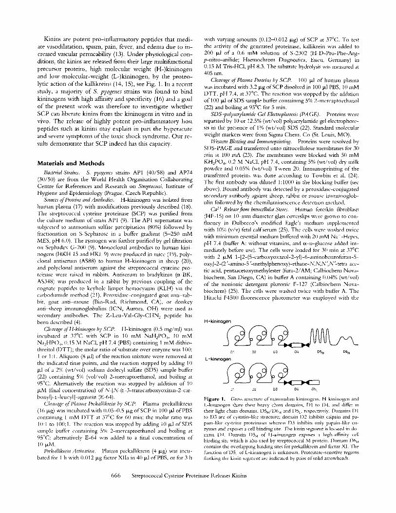
H-kininogen
D1
L-kininogen
D1
D2 D3 D/, D5 H D6 H
D2 D3 D/. D5 L
Figure 1. Gross structure ofmanunalian kininogens. H-kininogen and L-kininogen share their heavy chain domains, l)1 to I)4, and differ m their light chain domains, I)5H/D6 n and DSL, respectively. Domains 1)1 to 1)3 are of cystatm-like structure; domain I)2 inhibits calpam and pa- pain-like cysteme proteinases whereas I)3 inhibits only papain-like en- zymes and exposes a cell binding site. The kinin segment is located in do- main 1)4. Domain I)5~ of H-kininogen exposes a high-affinity cell binding site which is also used by streptococcal M protein, l)omain 1)6 H contains the overlapping binding sites for prekallikrein and factor XI. The fhnction of D5 L of L-kininogen is unknown. Proteinase-sensitive regions flanking the kmin segnnent are indicated by pairs of solid arrowheads.
666 Streptococcal Cysteine Proteinase Releases Kinins
excitation wavelength alternating between 340 nm and 380 nm, and the emission wavelength set at 510 nm. To induce the Ca 2+ release, 2 txg H-kininogen or proteolytic cleavage products thereof in 20 Ixl of reaction buffer was added 60 s after starting the measurement. The release of Ca 2+ from intracellular stores was followed for 300 s; the free intercellular Ca 2+ concentration was calculated from the ratio of 340 nm/380 nm as described (25).
Determination of Kinin Concentrations in Plasma. To measure the SCP-induced kinin release 100 Ixl of plasma was incubated with 3.2 I~g of SCP in 100 ~1 PBS containing 10 mM DTT. Samples (10 Ixl each) were removed after 0, 30, 60, 90, and 120 min. The reaction was stopped by adding E-64 to a final concentration of 10 txM. For control 100 ~l of human plasma was incubated with buffer in the absence of SCP. The samples were diluted 1:100 in distilled water. Aliquots (100 Ixl each) were mixed with 20 txl of 20% (wt/vol) trichloroacetic acid and centrifuged at 1,500 g for 10 rain. The kinin concentrations in the reaction mixtures were quantitated by the Markit-A kit (Dainippon Pharmaceutical Co., Osaka, Japan) as described (26). Briefly, aliquots of the superna- tant (75 ~1 each) were mixed with 75 Ftl of the kit buffer, and ap- plied to the wells (100 ~1 each) of microtiter plates that were coated with capture antibodies to rabbit immunoglobulin fol- lowed by specific anti-bradykinin antibodies. After 1 h of incuba- tion, the peroxidase-labeled bradykinin probe was applied and in- cubated for 1 h. The amount of bound peroxidase was visualized by the substrate solution, 0.1% (wt/vol) diammonium-2,2'-azinobis- (3-ethyl-2,3-dihydrobenzthiazoline)-6-sulfonate (ABTS), 0.012% (vol/vol) H202 in 100 rn/Vl citric acid, 100 mM NaH2PO4, pH 4.5, for 3() min. The change of absorbance was read at 405 nm. The reference standards were prepared according to the manufac- turer's instructions.
Animal Experiments. S. pyogenes of strains AP1 and AP74 were grown in Todd-Hewitt broth (Difco, Detroit, MI) at 37~ for 16 h, and harvested by centrifugation at 3,000 g for 20 rain. The bacteria were washed twice with PBS, and resuspended in PBS to 3 • 10 s cel/s/ml. 1 ml of living bacteria was injected in- traperitoneally into outbred NMRI mice. Plasma samples were taken 10 h after injection. Alternatively, mice were injected with the purified non-activated SCP (0.1-0.5 rag), and plasma samples were taken 60, 150, and 300 rain after injection. For inactivation of SCP, 0.5 mg of the enzyme was mixed with 0.2 mg Z-Leu- Val-Gly-CHN2 prior to injection. To monitor the cleavage of kininogen, l txl of plasma was run on SDS-PAGE followed by Western blotting with antibodies against bradykinin (ec-BK).
Quantification of SCP in Mouse Plasma. One Ixl of plasma sam- ples from mice injected with SCP was run on SDS-PAGE and transferred onto nitrocellulose. The enzyme was visualized by im- nmnostaining using antibodies against SCP. To obtain senti- quantitative estimates of the SCP amounts in plasma samples, pu- rified SCP (3-100 ng) was processed as described above and used as a standard.
Results
Streptococcal Cysteine Proteinase Is Not Inhibited by H-Kinin- ogen. The streptococcal cysteine proteinase (SCP) cleaves surface proteins o f S. pyogenes strain AP1 (9). O n e o f its tar- get structures, the streptococcal M1 protein, specifically binds kininogens (16) the major cysteine proteinase inhibi- tors o f human plasma. These observations prompted the not ion that kininogens bound to the bacterial surface might
regulate the proteolytic activity o f SCP. W e therefore tested the effect o f H-k in inogen on the hydrolysis o f a chromogenic pept ide substrate by SCP. Unexpectedly , H-k in inogen had no inhibi tory effect on the amidolytic ac- tivity o f SCP (not shown), whereas the synthetic cysteine proteinase inhibitor E-64, efficiently b locked the SCP ac- tivity in the same assay. W e therefore asked the question whether H-k in inogen serves as a substra te-- ra ther than an inhibitor for SCP.
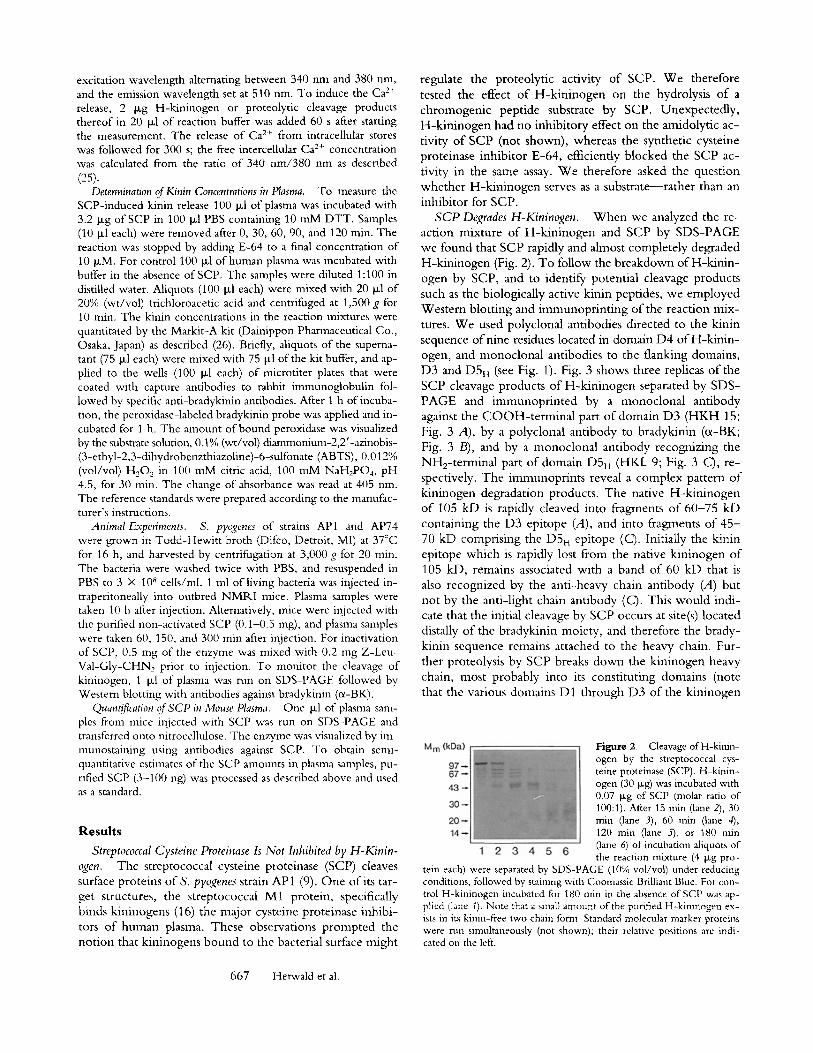
SCP Degrades H-Kininogen. W h e n we analyzed the re- action mixture o f H-k in inogen and SCP by SDS-PAGE we found that SCP rapidly and almost completely degraded H-kininogen (Fig. 2). To follow the breakdown of H-k in in- ogen by SCP, and to identify potential cleavage products such as the biologically active kinin peptides, we employed Western blott ing and immunopr in t ing o f the reaction mix- tures. W e used polyclonal antibodies directed to the kinin sequence o f nine residues located in domain D4 o f H-k in in- ogen, and monoclonal antibodies to the flanking domains, D3 and D5 H (see Fig. 1). Fig. 3 shows three rephcas o f the SCP cleavage products o f H-k in inogen separated by SDS- PAGE and immunopf in ted by a rnonoclonal antibody against the C O O H - t e r m i n a l part o f domain D3 (HKH 15; Fig. 3 A), by a polyclonal antibody to bradykinin (cr Fig. 3 B), and by a monoclonal antibody recognizing the NH2-terminal part of domain D 5 , (HKL 9; Fig. 3 C), re- spectively. The immunoprints reveal a complex pattern o f kininogen degradation products. The native H-k in inogen o f 105 kD is rapidly cleaved into fragments o f 60-75 kD containing the D3 epitope (A), and into fragments o f 4 5 - 70 kD comprising the D5H epitope (C). initially the kinin epitope which is rapidly lost from the native kininogen o f 105 kD, remains associated with a band o f 60 kD that is also recognized by the anti-heavy chain antibody (A) but not by the anti-light chain antibody (C). This would indi- cate that the initial cleavage by SCP occurs at site(s) located distally o f the bradykinin moiety, and therefore the brady- kinin sequence remains attached to the heavy chain. Fur- ther proteolysis by SCP breaks down the kininogen heavy chain, most probably into its constituting domains (note that the various domains D1 through D3 o f the kininogen
Figure 2. Cleavage of H-kinin- ogen by the streptococcal cys- teine proteinase (SCP). H-kinin- ogen (30 Ixg) was incubated with 0.07 ~g of SCP (molar ratio of 100:1). After 15 nfin (lane 2), 30 rain (lane 3), 60 rain (lane 4), 120 min (lane 5), or 180 rain (lane 6) of incubation aliquots of the reacuon mixture (4 Ixg pro-
tein each) were separated by SDS-PAGE (10% vol/vol) under reducing conditions, followed by staining with Coomassie Brilliant Blue. For con- trol H-kininogen incubated for 180 rain in the absence of SCP was ap- plied (lane I). Note that a small amount of the purified H-kininogen ex- ists in its kinin-free two chain form. Standard molecular marker proteins were run simultaneously (not shown); their relative positions are indi- cated on the left.
667 Herwald et al.
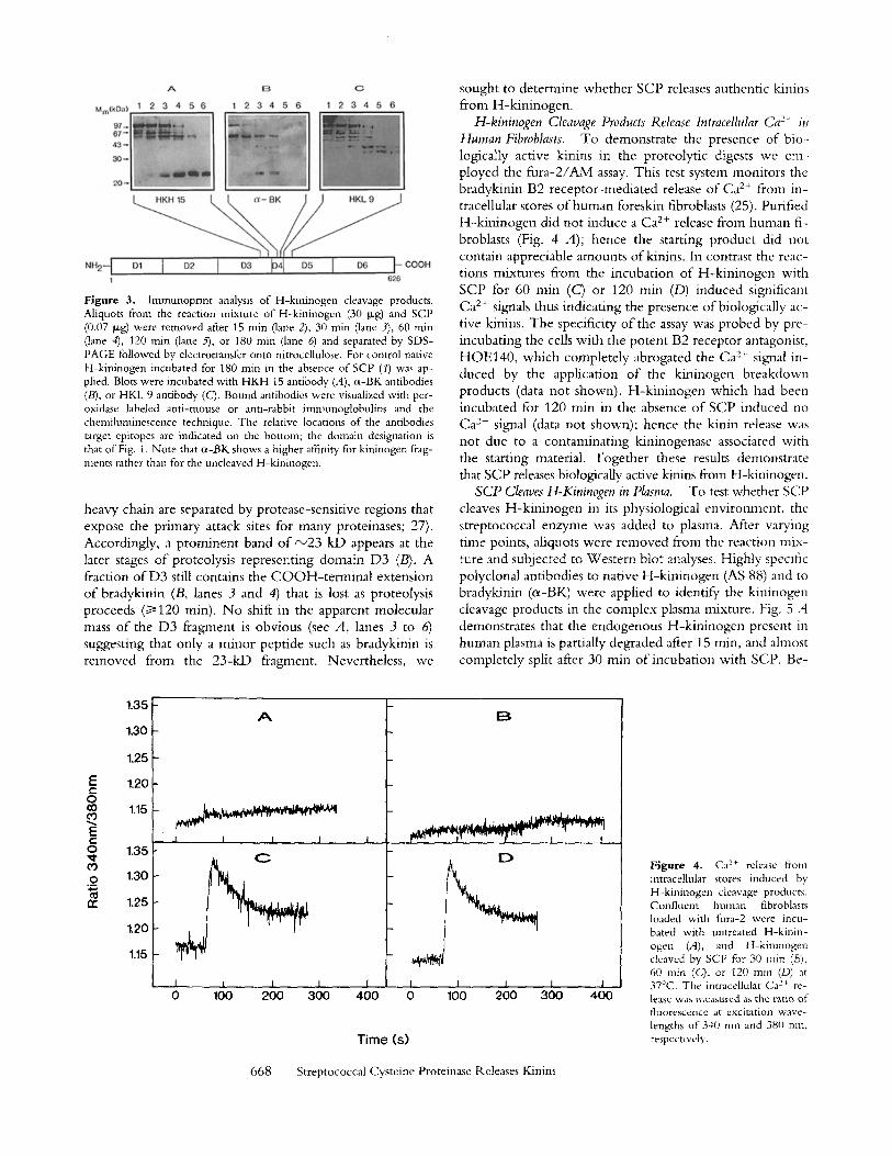
F i g u r e 3. lmmunoprint analysis of H-kininogen cleavage products. Aliquots from the reaction mixture of H-kininogen (30 t, tg) and SCP (0.07 >g) were removed after 15 min (lane 2), 30 rain (lane 3), 60 rain (lane 4), 120 rain (lane ~ , or 180 min (lane 6) and separated by SDS- PAGE followed by electrotransfer onto nitrocellulose. For control nanve H-kininogen incubated for 180 rain in the absence of SCP (1) was ap- plied. Blots were incubated with HKH 15 antibody (A), cl-BK antibodies (B), or HKL 9 antibody (C). Bound antibodies were visualized with per- oxidase labeled anti-mouse or anti-rabbit immunoglobulins and the chemiluminescence technique. The relative locations o f the antibodies target epitopes are indicated on the bottom; the domain designation is that of Fig. 1. Note that cl-BK shows a higher affinity for kininogen frag- ments rather than for the uncleaved H-kininogen.
heavy chain are separated by protease-sensitive regions that expose the primary attack sites for many proteinases; 27). Accordingly, a prominent band of ~23 kD appears at the later stages of proteolysis representing domain D3 (B). A fraction of D3 still contains the COOH-terminal extension of bradykinin (B, lanes 3 and 4) that is lost as proteolysis proceeds (~>120 min). No shift in the apparent molecular mass of the D3 fragment is obvious (see A, lanes 3 to 6) suggesting that only a minor peptide such as bradykinin is removed from the 23-kD fragment. Nevertheless, we
sought to determine whether SCP releases authentic kinins from H-kininogen.
H-kininogen Cleavage Products Release Intracellular Ca 2. i , Human Fibroblasts. To demonstrate the presence of bio- logically active kinins in the proteolytic digests we em- ployed the fura-2/AM assay. This test system monitors the bradykinin B2 receptor-mediated release of Ca 2+ from in- tracellular stores of human foreskin fibroblasts (25). Purified H-kininogen did not induce a Ca 2+ release from human fi- broblasts (Fig. 4 A); hence the starting product did not contain appreciable amounts ofkinins. In contrast the reac- tions mixtures from the incubation of H-kininogen with SCP for 60 min (C) or 120 min (D) induced significant Ca 2+ signals thus indicating the presence of biologically ac- tive kinins. The specificity of the assay was probed by pre- incubating the cells with the potent B2 receptor antagonist, HOE140, which completely abrogated the Ca 2+ signal in- duced by the application of the kininogen breakdown products (data not shown). H-kininogen which had been incubated for 120 min in the absence of SCP induced no Ca 2+ signal (data not shown); hence the kinin release was not due to a contaminating kininogenase associated with the starting material. Together these results demonstrate that SCP releases biologically active kinins from H-kininogen.
SCP Cleaves H-Kininogen in Plasma. To test whether SCP cleaves H-kininogen in its physiological environment, the streptococcal enzyme was added to plasma. After varying time points, aliquots were removed from the reaction mix- ture and subjected to Western blot analyses. Highty specific polyclonal antibodies to native H-kininogen (AS 88) and to bradykinin (oL-BK) were applied to identify the kininogen cleavage products in the complex plasma mixture. Fig. 5 A demonstrates that the endogenous H-kininogen present in human plasma is partially degraded after 15 min, and almost completely split after 30 min of incubation with SCP. Be-
E t -
O cO eo
e ' -
O
o
1.35
1.30
1.25
1.20
1.15
1.35
1.30
1.25
1.20
1.15
A
I I I I
L I I I I 0 100 200 300 400
I g
D
I I I I I 0 100 200 300 400
Time (s)
Figure 4. Ca 2+ release from mtracellular stores induced by H-kminogen cleavage products. Confluent human fibroblasts loaded with fura-2 were incu- bated with untreated H-kinin- ogen (A), and H-kininogen cleaved by SCP for 30 rain (B), 60 rain (C), or 120 mm (D) at 37~ The intracellular Ca 2~ re+ lease was measured as the ratio of fluorescence at excitation wave- lengths os 340 nm and 380 nm+ respectively.
668 Streptococcal Cysteine Proteinase Releases Kinins
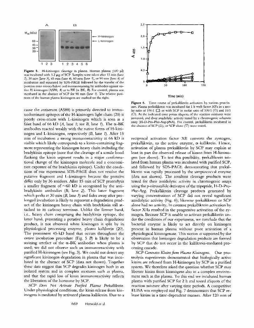
Figure 5. H-kininogen cleavage in plasma. Human plasma (100 I*1) was incubated with 3.2 Dg of SCP. Samples were taken after 15 rain (lane 2), 30 min (lane 3), 45 rain (lane 4), 60 min (lane 5), or 90 rain (lane 6) of incubation and separated by SDS-PAGE followed by the transfer of the proteins onto nitrocellulose and immunostaining by antibodies against na- tive H-kininogen (AS88; A) or to BK (cx-BK;/3). For control, plasma was incubated in the absence of SCP for 90 rain (lane I). The relative posi- tions of the human plasma kininogens are marked on the right.
cause the antiserum (AS88) is primarily directed to immu- nodominant epitopes of the H-kininogen light chain (20) it poorly cross-reacts with L-kininogen which is seen as a faint band of 66 kD (A, lane 1; see B, lane 1). The cx-BK antibodies reacted weakly with the native forms of H-kini- nogen and L-kininogen, respectively (B, lane 1). After 15 rain of incubation a strong immunoreactivity at 66 kD is visible which likely corresponds to a kinin-containing frag- ment representing the kininogen heavy chain including the bradykinin epitope (note that the cleavage of a scissile bond flanking the kinin segment results in a major conforma- tional change of the kininogen molecule and a concomi- tant exposure of the bradykinin epitope), Under the condi- tions of our experiment SDS-PAGE does not resolve the putative fragment and L-kininogen because the proteins differ only by 36 residues. After 15 rain of SCP proteolysis a smaller fragment of ~60 kD is recognized by the anti- bradykinin antibodies (B, lane 2). This latter fragment which peaks at 30 rain (B, lane 3) and fades away after pro- longed incubation is likely to represent a degradation prod- uct of the kininogen heavy chain with bradykinin still at- tached to its carboxy terminus. Unlike the former band, i.e., heavy chain comprising the bradykinin epitope, the latter band, presenting a putative heavy chain degradation product, is not observed when kininogen is split by its physiological processing enzyme, plasma kallikrein (20). The prominent 45-kD band that occurs throughout the entire incubation procedure (Fig. 5 B) is likely to be a staining artefact of the cx-BK antibodies when plasma is used; we did not observe such an immunoreactivity with purified H-kininogen (see Fig. 3). We could not detect any significant kininogen degradation in plasma that was incu- bated in the absence of SCP (data not shown). Together these data suggest that SCP degrades kininogen both in an isolated system and in complex mixtures such as plasma, and that the rapid loss of kinin immunoreactivity reflects the liberation of the hormone by SCP.
SCP Does Not Activate Purified Ptasma Prekallikrein, Under physiological conditions, the kinin release from kin- inogens is mediated by activated plasma kallikrein. Due to a
2,5 2,0 ~ , 1,5 l,O
0'5'~~~L ~ O, 0 50 I00 150
Time (rain)
Figure 6. Time course of prekallikrein activation by various protein- ases. Plasma prekallikrein was incubated for 1 h with factor Xlla in a mo- lar ratio of 100:1 ([~) or with SCP in molar ratio of 100:1 (&) and 10:1 (O), At the indicated time points aliquots of the reaction mixtures were removed, and their amidolytic activity tested by a chromogenic substrate assay (H-D-Pro-Phe-Arg-pNA). For control, prekallikrein incubated in the absence ofSCP (~), or SCP alone (V) were tested.
reciprocal activation factor XII converts the zymogen, prekallikrein, to the active enzyme, o~-kallikrein. Hence, activation of plasma prekallikrein by SCP may explain at least in part the observed release of kinins from H-kinino- gen (see above). To test this possibility, prekallikrein iso- lated from human plasma was incubated with purified SCP, and followed by SDS-PAGE demonstrating that prekal- likrein was rapidly processed by the streptococcal enzyme (data not shown). The resultant cleavage products were tested for their amidolytic activity in chromogenic assays using the p-nitroanilide derivative of the tripeptide, H-D-Pro- Phe-Arg. Prekallikrein cleavage products generated by varying concentrations of SCP did not reveal significant amidolytic activity (Fig. 6); likewise prekallikrein or SCP alone had no activity. In contrast prekallikrein activation by factor XIIa resulted in the progressive activation of the zy- mogen. Because SCP is unable to activate prekallikrein un- der the conditions of our experiment, we conclude that the bacterial enzyme is likely to act directly on kininogen present in human plasma without prior activation of a physiological kininogenase. This notion is supported by the observation that kininogen degradation products are formed by SCP that do not occur in the kallikrein-mediated pro- cessing cascade.
SCP Generates Kinins from Plasma Kininogens. Our pro- teolysis experiments demonstrated that biologically active kinins are released from H-kininogen by SCP in a purified system. We therefore asked the question whether SCP may liberate kinins from kininogens also in a complex environ- ment such as the plasma. To this end we incubated human plasma with purified SCP for 2 h and tested aliquots of the reaction mixture after varying time periods. A competitive ELISA was employed and Fig. 7 demonstrates that SCP re- lease kinins in a time-dependent manner. After 120 min of
669 Herwald et al.
3,0
2,5
2,0 O
0,5
0 I__L
I I 0 30 60 90
I 120
Time (min)
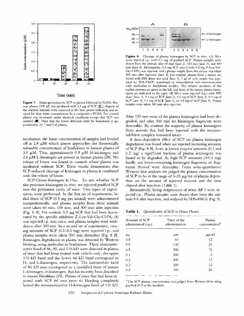
Figure 7. Kinin generation by SCP in plasma followed by ELISA. Hu- man plasma (100 txl) was incubated with 3.2 txg of SCP (IN), aliquots of the reaction mixture were removed at the time points indicated, and as- sayed for their kinin concentration by a competitive ELISA. For control plasma was incubated under identical conditions except that SCP was omitted (n). Note that the lower detection limit for bradykinin is ap- proximately 10 7 mol/l of plasma.
incubation, the kinin concentration o f samples had leveled off at 2.8 #xM which ahnost approaches the theoretically releasable concentrat ion o f bradykinin in human plasma o f 3.5 txM. Thus, approximately 0.9 txM H-k in inogen and 2.6 IxM L-kininogen are present in human plasma (28). N o release of kinins was found in controls where plasma was incubated wi thout SCP. These results demonstrate that SCP- induced cleavage o f kininogen in plasma is combined with the release of kinins.
SCP Cleaves Kininogens In Vivo. To test whether SCP also processes kininogens in vivo, we injected purified SCP into the peritoneal cavity o f mice. Two types o f experi- ments were performed. In the first set of experiments, le- thal doses o f SCP (0.5 mg per animal) were administrated intraperitoneally, and plasma samples from these animals were taken 60 min, 150 rain, and 300 min after injection (Fig. 8 A). For control, 0.5 Ixg SCP that had been inacti- vated by the specific inhibitor Z - L e u - V a l - G l y - C H N 2 (4) was injected i.p. into mice, and plasma samples were wi th- drawn after 300 rain. In a second set of experiments, vary- ing amounts o f SCP (0.1-0.5 mg) were injected i.p., and plasma samples were taken 300 min thereafter (Fig. 8 /3). Kininogen degradation in plasma was detected by Western blotting, using antibodies to bradykinin. Three immunore - active band o f 66, 80, and 110-kD were detected in plasma of mice that had been treated with vehicle only; the upper 110-kD band and the lower 66 -kD band correspond to H- and L-kininogen, respectively. The intermediate band o f 80 kD may cor respond to a modi f ied form o f mouse L-kininogen, i r-kininogen, that has recently been described in mouse fibroblasts (29). Plasma o f mice that had been in- jec ted with SCP 60 nfin prior to bleeding completely lacked the immunoreact ive H-k in inogen band o f 110 kD.
Figure 8. Cleavage of plasma kininogens by SCP in vivo. (A) Mice were injected i.p. with 0.5 mg of purified SCP. Plasma samples were drawn from the animals after 60 rain (lane 2), 150 rain (lane 3), and 300 rain (lane 4). Alternatively, 0.5 mg SCP mixed with 0.2 mg Z-Leu-Val- Gly-CHN 2 was injected, and a plasma sample from this mouse was taken 300 rain after injection (lane 5). For control, plasma from a mouse in- jected with PBS alone was used (lane 1). I Fxl of each sample was sepa- rated by SDS-PAGE, transferred to nitrocellulose and inmmnostained with antibodies to bradykinin (ot-BK). The relative positions of the marker proteins are given to the left, and those of the mouse plasma kinin- ogens are indicated to the fight. (/3) Mice were injected (i.p.) with PBS alone (lane 1), 0.1 nag of SCP (lane 2), 0.2 mg of SCP (lane 3), 0.3 mg of SCP (lane 4), 0.4 mg of SCP (lane 5), or 0.5 mg of SCP (lane 6). Plasma samples were taken 300 rain after injection.
After 150 rain most o f the plasma kininogens had been de- graded, and after 300 min no kininogen fragments were detectable. By contrast the majority o f plasma kininogens from animals that had been injected with the enzyme- inhibitor complex remained intact.
A dose-dependent effect o f SCP on plasma kininogen degradation was found when we injected increasing anaounts o f SCP (Fig. 8 B). Even at lowest enzyme amounts (0.1 and 0.2 nag) a significant fraction o f plasma kininogens was found to be degraded. At high SCP amounts (/>0.4 nag) hardly any kinin-containing kininogen fragments or frag- ments thereof were detectable. From semi-quantitative Western blot analyses we judged the plasma concentration o f SCP to be in the range o f 3-25 Ixg/ml of plasma depen- dent on the anaount of injected enzyme and the time elapsed after injection (Table 1).
Alternatively, living streptococci of strain AP 1 were in- jec ted i.p.. Plasma samples were drawn then from the ani- mals 8 h after injection, and analyzed by SDS-PAGE (Fig. 9).
Table 1. Quant!fication of SCP in Mouse Plasma
Amount of SCP Time of the Plasma administered (i.p.) administration concentration*
mg min lzglml 0.5 6O 12
0.5 15O 2O
0.5 300 25
O. 1 300 2
0.2 3O0 12
0.3 300 12
0.4 300 20
*The SCP plasma concentration was judged from Western blots using purified SCP as the standard.
670 Streptococcal Cysteine Proteinase Releases Kinim
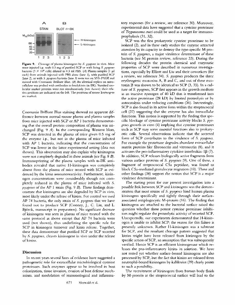
Figure 9. Cleavage of plasma kininogens by S, pyogenes in vivo. Mice were injected i.p. with 0.5 mg of purified SCP or with living S. pyogenes bacteria (3 • l0 s cells) diluted in 0.5 ml PBS. (A) Plasma samples (1 txl each) from animals injected with PBS alone (lane 1), with purified SCP (lane 2), or with S. pyogenes bacteria (lane 3) were run on SDS-PAGE and stained with Coomassie Brilliant Blue. (/3) An identical replica on nitro- cellulose was probed with antibodies to bradykinin (r Standard mo- lecular marker proteins were run simultaneously (not shown); their rela- tive positions are indicated on the left. The positions of mouse kininogens are marked.
Coomassie Brilliant Blue staining showed no apparent dif- ference between normal mouse plasma and plasma samples from mice injected with SCP or AP 1 bacteria demonstrat- ing that the overall protein composition o f plasma was un- changed (Fig. 9 A). In the corresponding Western blots, SCP was detected in the plasma o f mice given 0.5 mg o f the enzyme i.p., but not in the plasma of mice infected with AP 1 bacteria, indicating that the concentration of SCP was lower in the latter experimental setting (data not shown). This observation may also explain why kininogens were not completely degraded in these animals (see Fig. 8 B). Immunopr in t ing o f the plasma samples with oL-BK anti- bodies revealed that native H-kin inogen was completely absent from the plasma o f mice treated with SCP as evi- denced by the kinin immunoreactivity. Furthermore, kinin- ogen concentrations were considerably though not com- pletely reduced in the plasma of mice infected with S. pyogenes of the AP 1 strain (Fig. 9/3). These findings dem- onstrate that kininogens are also degraded by SCP in vivo, most likely under the release of kinins. For control we used AP 74 bacteria, the only strain of S. pyogenes that we have found not to produce SCP (Cooney, J., C. Liu, and L. Bj6rck, manuscript in preparation). No significant decrease ofkininogens was seen in plasma o f mice treated with the same protocol as above except that AP 74 bacteria were used (not shown), thus underlining the specific role for SCP in kininogen turnover and kinin release. Together, these data demonstrate that purified SCP or SCP secreted by S. pyogenes, cleaves kininogens in vivo under the release o f kinins.
Discuss ion
In recent years several lines of evidence have suggested a pathogenetic role for extracellular microbiological cysteine proteinases. Such enzymes appear to be involved in host colonization, tissue invasion, evasion o f host defense mech- anism, and modulation of imnmnological and inflamma-
671 Herwald et al.
tory responses (for a review, see reference 30). Moreover, experimental data have suggested that a cysteine proteinase o f Ttypanosoma cruzi could be used as a target for immuno- prophylaxis (31, 32).
SCP was the first prokaryotic cysteine proteinase to be isolated (2), and in these early studies the enzyme attracted attention by its capacity to destroy the type-specific M pro- tein o f S. pyogenes, a major virulence determinant o f these bacteria (see M protein review, reference 33). During the following decades the protein chemical and enzymatic properties of SCP were described in numerous investiga- tions, especially by Elliott and Liu and their coworkers (for a review, see reference 34). S. pyogenes produces the three erythrogenic exotoxins A, B and C, and out o f these exo- toxin B was shown to be identical to SCP (3, 35). In a cul- ture of S. pyogenes, SCP first appears in the growth medium as an inactive zymogen o f 40 kD that is transformed into the active proteinase (28 kD) by limited proteolysis or by autocatalysis under reducing conditions (36). Interestingly, SCP is also found in its active form within the streptococcal cell (37) suggesting that the enzyme has also intracellular functions. This notion is supported by the finding that spe- cific blockage of cysteine proteinase activity blocks S. pyo- genes growth in vitro (4) implying that cysteine proteinases such as SCP may serve essential functions also in prokary- otic cells. Several observations indicate that the secreted form of SCP contributes to the virulence of S. pyogenes. For example the proteinase degrades abundant extracellular matrix proteins like fibronectin and vitronectin (8), and it activates the pro-inflammatory cytokine interleukin-113 (7). In addition, SCP releases biologically active fragments from various surface proteins of S. pyogenes (9). One of these, a fragment of streptococcal CSa peptidase, was found to block C5a-mediated granulocyte migration (10). These and other findings (38) support the notion that SCP is a major virulence determinant.
The starting point for our present investigation into a possible link between SCP and kininogens was the demon- stration that most strains of S. pyogenes bind human plasma kininogens specifically and tightly through their surface- associated antiphagocytic M-prote in (16). The finding that kininogens are attached to the bacterial surface raised the question whether these potent cysteine proteinase inhibi- tors might regulate the proteolytic activity of secreted SCP. Unexpectedly, our experiments demonstrated that H-kinin- ogen is unable to inhibit SCP; the reason for this failure is presently unknown. Rather H-kininogen was a substrate for SCP, and the resultant cleavage pattern suggested that kinins might have been released from kininogen by the specific action o f SCP, an assumption that was subsequently verified. Hence SCP is an efficient kininogenase which re- leases the pro-inflammatory kinins in solution. W e have not tested yet whether surface-bound kininogens are also processed by SCP, but the fact that kinins are released from neutrophil-bound kininogens by kallikreins (39) clearly points to such a possibility.
The recruitment of kininogens from human body fluids by M-prote in at the streptococcal surface will lead to the
local accumulation of the kinin precursor molecules in in- fected tissues. If SCP secreted by the bacteria cleaves these kininogen molecules, a local burst of kinins will cause in- creased vascular permeability. Such a sequence of events would promote a flow of nutrients into the site o f infection and at the same time enhance the spreading of the infection via facilitated extravasation. This hypothetical scenario is supported by the observation that SCP secretion is depen- dent on environmental factors such as pH (34). Notably the pH is low in the center o f suppurative streptococcal infec- tions (40), and most strains of S. pyogenes produce excessive amounts o f SCP (10-150 mg/L of growth medium) when grown at pH 5.5-6.0.
In severe cases of sepsis, hypovolemic hypotension is a prominent and clinically important finding that is caused by the leakage of plasma into the extravascular space (41). The rapid and efficient cleavage of kininogens to kinins in mouse plasma following the administration o f SCP or liv- ing S. pyogenes bacteria is the major finding of this study. It indicates that a general and massive release o f kinins could take place in severe streptococcal infections, such as sepsis and streptococcal toxic shock syndrome. These conditions
are characterized by raging fever, drop in blood pressure, and multiorgan failure (1, 41). In this context it is notewor- thy that patients with low titers of antibodies to SCP in the acute phase are more likely to die in severe S. pyogenes in- fections (6), and that immunization o f mice with SCP gen- erates partial protection against S. pyogenes administrated i.p. (5). Furthermore, a single dosis o f a tripeptide deriva- tive which blocks the enzymatic activity o f SCP (4) cures mice given an otherwise lethal dosis of this enzyme (Cooney, Liu, and Bj6rck, in preparation). These and other data described above, underline the significance of SCP in the pathogenesis o f streptococcal infections. The major and novel aspect of this work is that we have identified a poten- tial downstream effector of SCP, i.e., kinins. Recrui tment o f host proteins and exploitations o f their intrinsic proper- ties by the parasite is a phenomenon that is probably com- mon to many pathogenic bacteria. Our results also indicate that SCP and/or kinins could be therapeutical targets in hyperacute and severe S. pyogenes infections where treat- ment with antibiotics alone is insufficient. Specific inhibi- tion of these potential mediators of shock could interrupt an otherwise fatal pathologic sequence.
We wish to thank our colleagues Drs. W. Machleidt and I. Assfalg-Machleidt (Ludwig Maximilian Univer- sity, Munich) for experimental help with the inhibitor assays; A. Maidhofand B. Welsch (Johannes Guten- berg University, Mainz) for providing antisera; A. Horstmeyer (Mainz) for experimental help with the fura assay, U. Quitterer (University of Wiirzburg) and P. Akesson (Lund University) for their critical comments on the manuscript, and B. J6nnson (Lund University) for preparing the figures.
This work was supported in part by The Swedish Medical Research Council (grant 7480), the Deutsche Forschungsgemeinschaft (Mu598/5.2), the Fonds der Chemischen Industrie (to W. Mtiller-Esterl), the Foundations of Kock and Osterlund, and High Tech Receptor AB.
Address correspondence to Heiko Herwald, Department of Cell and Molecular Biology, Section for Molec- ular Pathogenesis, Lund University, P.O. Box 94, S-221 00 Lund, Sweden.
Received for publication 12 March I996 and in revised form 28 May 1996.
References
1. Nowak, R. 1994. Flesh-eating bacteria: not new, but still worrisome. Science (Wash. DC). 264:1665.
2. Elliott, S.D. 1945. A proteolytic enzyme produced by group A streptococci with special reference to its effect on the type- specific M antigen.J. Exp. Med. 81:573-592.
3. Gerlach, D., H. Knoll, W. KShler, J.H. Ozegowski, and V. Hrihalova. 1983. Isolation and characterization of erythro- genic toxins. V. Communication: identity of erythrogenic toxin type B and streptococcal proteinase precursor. ZentralbL Bakteriol. Mikrobiol, Hyg. A. 255:221-233.
4. Bj/Srck, L., P. Akesson, M. Bohus, J. Trojnar, M. Abraham- son, I. Olafsson, and A. Grubb. 1989. Bacterial growth blocked by a synthetic peptide based on the structure of a hu- man proteinase inhibitor. Nature (Loud.). 337:385-386.
5. Kapur, V., J.T. Maffei, R.S. Greer, L.L. Li, G.J. Adams, and J.M. Musser. 1994. Vaccination with streptococcal extracel- lular cysteine protease (interleukin-1 beta convertase) protects mice against challenge with heterologous group A strepto-
cocci. Microb. Pathol. 16:443-450. 6. Holm, S.E., A. Norrby, A.-M. Bergholm, and M. Norgren.
1992. Aspects of patogenesis of serious group A streptococcal infections in Sweden 1988-1989.J. Infect, Dis. 166:31-37.
7. Kapur, V., M.W. Majesky, L.L. Li, R.A. Black, and J.M. Musser. 1993. Cleavage ofinterleukin 1 beta (IL-I beta) pre- cursor to produce active IL-1 beta by a conserved extracellu- lar cysteine protease from Streptococcus pyogenes. Proc. Natl. Acad. Sci. USA, 90:7676-7680.
8. Kapur, V,, S. Topouzis, M.W. Majesky, L.L. Li, M.R. Ham- rick, R.J. Hamill, J.M. Patti, and J.M. Musser. 1993. A con- served Streptococcus pyogenes extracellular cysteine protease cleaves human fibronectin and degrades vitronectin. Microb. Pathol. 15:327-346.
9. Berge, A., and L. Bj6rck. 1995. Streptococcal cysteine pro- teinase releases biologically active fragments of streptococcal surface proteins.J. Biol. Chem. 270:9862-9867.
10, Wexler, D.E., D,E. Chenoweth, and P.P. Cleary. 1985.
672 Streptococcal Cysteine Proteinase Releases Kinins
Mechanism of action of the group A streptococcal CSa inac- tivator. Proc. Natl. Acad. Sci. USA. 82:8144-8148.
11. Wolf, B.B., C.A. Gibson, V. Kapur, I.M. Hussaini, J.M. Musser, and S.L. Gonias. 1994. Proteolytically active strepto- coccal pyrogenic exotoxin B cleaves monocytic cell uroki- nase receptor and releases an active fragment of the receptor from the cell surface.J. Biol. Chem. 269:30682-30687.
12. Poon-King, R., J. Bannan, A. Viteri, G. Cu, and J.B. Zabriskie. 1993. Identification of an extracellular plasmin binding protein from nephritogenic streptococci. J. Exp. Mecl. 178:759-763.
13. Hall, J.M. 1992. Bradykinin receptors: pharmacological prop- erties and biological roles. Pharmac. Ther. 56:131-190.
14. Kerbiriou, D.M., and J.H. Griffin. 1979. Human High Mo- lecular Weight Kininogen.J. Biol. Chem. 254:12020-12027.
15. Miiller-Esterl, W., G. Rauth, F. Lottspeich, J. Kellermann, and A. Henschen. 1985. Limited proteolysis of human low- molecular-mass kininogen by tissue kallikrein. Isolation and characterization of the heavy and the light chains. Eur. J. Bio- chem. 149:15-22.
16. Ben Nasr, A.B., H. Herwald, W. Miiller-Esterl, and L. Bjrrck. 1995. Human kininogens interact with M protein, a bacterial surface protein and virulence determinant. Biochem. J. 305:173-180.
17. Salvesen, G., C. Parkes, M. Abrahamson, A. Grubb, and A.J. Barrett. 1986. Human low-Mr kininogen contains three cop- ies of a cystatin sequence that are divergent in structure and in inhibitory activity for cysteine proteinases. Biochem. J. 234: 429-434.
18. Hasan, A.A., D.B. Cines, J. Zhang, and A.H. Schmaier. 1994. The carboxyl terminus ofbradykinin and amino termi- nus of the light chain of kininogens comprise an endothelial cell binding domain.J. Biol. Chem. 269:31822-31830.
19. Kaufmann, J., M. Haasemann, S. Modrow, and W. Miiller- Esterl. 1993. Structural dissection of the multidomain kinino- gens. Fine mapping of the target epitopes of antibodies inter- fering with their functional properties. J. Biol. Chem. 268: 9079-9091.
20. Miiller-Esterl, W., D. Johnson, G. Salvesen, and A.A. Barrett. 1988. Human kininogens. Methods Enzymol. 163:240-256.
21. Herwald, H., A.H.K. Hasan, J. Godovac-Zimmermann, A.H. Schmaier, and W. Miiller-Esterl. 1995. Identification of an endothelial cell binding site on kininogen domain D3. J. Biol. Chem. 270:14634-14642.
22. Laemmli, U.K. 1970. Cleavage of structural proteins during the assembly of the head of bacteriophage T4. Nature (Dnd.). 227:680-685.
23. Khyse-Andersen, J. 1984. Electroblotting of multiple gels: a simple apparatus without buffer tank for rapid transfer of pro- teins from polyacrylamide to nitrocellulose. J. Biochem. Bio- phys. Methods. 10:203-209.
24. Towbin, H., T. Staehelin, and J. Gordon. 1979. Electro- phoretic transfer of proteins from polyacrylamide gels to ni- trocellulose sheets: procedure and some applications. Pro& Natl. Acad. Sci. USA. 76:4350-4354.
25. Quitterer, U., C. Schr/Sder, W. Miiller-Esterl, and H. Rehm. 1995. Effects ofbradykinin and endothelin-I on the calcium homeostasis of mammalian cells. J. Biol. Chem. 270:1992- 1999.
26. Scott, C.F., E.J. Whitaker, B.F. Hammond, and R.W. Col- man. 1993. urification and characterization of a potent 70- kDa thiol lysyl-proteinase (Lys-gingivain) from Porphyromonas gingivalis that cleaves kininogens and fibrinogen. J. Bid. Chem. 268:7935-7942.
27. Vogel, R., I. Assfalg Machleidt, A. Esterl, W. Machleidt, and W. Miiller-Esterl. 1988. Proteinase-sensitive regions in the heavy chain of low molecular weight kininogen map to the inter-domainjunctions.J. Biol. Chem. 263:12661-12668.
28. Miiller-Esterl, W. 1987. Novel functions of kininogens. Semin. Thromb. Hemostas. 13:115-126.
29. Takano, M., K. Yokoyama, K. Yayama, and H. Okamoto. 1995. Murine fibroblasts synthesize and secrete kininogen in response to cyclic-AMP, prostaglandin E2 and tumor necrosis factor. Biochim. Biophys. Acta. 1265:189-195.
30. Travis, J., J. potempa, and H. Maeda. 1995. Are bacterial proteinases pathogenic factors? Trends Microbiol. 3:405--407.
31. Eakin, A.E., M.E. McGrath, J.H. McKerrow, R.J. Fletterick, and C.S. Craik. 1993. Production of crystallizable cruzain, the major cysteine protease from Trypanosoma cruzi. J. Biol. Chem. 268:6115-6118.
32. Martinez, J., O. Campetella, A.C. Frasch, and J.J. Cazzulo. 1991. The major cysteine proteinase (cruzipain) from Trypa- nosoma cruzi is antigenic in human infections. Infect. Immunot. 59:4275-4277.
33. Fischetti, V.A. 1989. Streptococcal M protein: molecular de- sign and biological behavior. Clin. Microbiol. Rev. 2:285-314.
34. Liu, T.-Y., and S.D. Elliott. 1971. The Enzymes Vol. 3. P.D. Boyer, editor. Academic Press, New York. 609-639.
35. Hauser, A.R., and P.M. Schlievert. 1990. Nucleotide se- quence of the streptococcal pyrogenic exotoxin type B gene and relationship between the toxin and the streptococcal pro- teinase precursor.J. Bacteriol. 172:4536-4542.
36. Liu, T.-Y., and S.D. Elliott. 1965. Streptococcal proteinase. the zymogen to enzyme transformation. J. Biol. Chem. 240: 1138-1142.
37. Lo, S.S., S.M. Liang, and T.Y. Liu. 1984. Intracellular form of streptococcal proteinase: a clue to a novel mechanism of secretion. Anal. Biochem. 136:89-92.
38. Kellner, A., and T. Robertson. 1954. Myocardial necrosis produced in animals by means of crystalline streptococcal proteinase.J. Exp. Med. 99:495-504.
39. Henderson, L.M., C.D. Figueroa, W. Miiller-Esterl, and K.D. Bhoola. 1994. Assembly of contact-phase factors on the surface of the human neutrophil membrane. Blood. 84:474-482.
40. Rentzsch, G., and J. Wilke. 1970. Measurements of pH val- ues in vitro and in vivo in chronic tonsillitis. Z. Laryngol. Rhinol. Otol. 49:391-397.
41. Pamllo, J.E. 1993. Pathogenetic mechanisms of septic shock. N. Engl.J. Med. 328:1471-1477.
673 Herwald et al.


![Mass Spectrometric Analysis of l-Cysteine Metabolism: … · tion of [U-13C3, 15N]L-cysteine to the culture, the levels of [13C3,15N]L-cysteine increased, and [13C3, 15N]L-cysteine](https://img.pdfslide.net/doc/110x75/5fe663421198753c202620ce/mass-spectrometric-analysis-of-l-cysteine-metabolism-tion-of-u-13c3-15nl-cysteine.jpg)