Embed Size (px)
Citation preview

LUND UNIVERSITY
PO Box 117221 00 Lund+46 46-222 00 00
Streptococcal M protein and human C4BP
Persson, Jenny J
2006
Link to publication
Citation for published version (APA):Persson, J. J. (2006). Streptococcal M protein and human C4BP. Department of Laboratory Medicine, LundUniversity.
General rightsCopyright and moral rights for the publications made accessible in the public portal are retained by the authorsand/or other copyright owners and it is a condition of accessing publications that users recognise and abide by thelegal requirements associated with these rights.
• Users may download and print one copy of any publication from the public portal for the purpose of private studyor research. • You may not further distribute the material or use it for any profit-making activity or commercial gain • You may freely distribute the URL identifying the publication in the public portalTake down policyIf you believe that this document breaches copyright please contact us providing details, and we will removeaccess to the work immediately and investigate your claim.

Streptococcal M protein and human C4BP
Jenny Persson
Division of Medical Microbiology
Department of Laboratory Medicine
Lund University, Sweden
LUND
2006
Doktorsexamen i Medicisk Mikrobiologi
som med vederbörligt tillstånd från Medicinska Fakulteten vid Lunds Universitet för avläggande
doktorsexamen i medicinsk vetenskap kommer att offentligen försvaras i Segerfalksalen,
Wallenbergs Neurocentrum, Sölvegatan 17, Lund, lördagen den 29 april 2006, kl. 09.30
Fakultetsopponent
Professor Brian G. Spratt, Imperial College London, London, Storbritannien


Streptococcal M protein and human C4BP
Jenny Persson
Division of Medical Microbiology
Department of Laboratory Medicine
Lund University, Sweden
Academic dissertation
LUND
2006
4
CONTENTS
CONTENTS 4
LIST OF PAPERS 6
ABBREVIATIONS 7
SUMMARY 8
INTRODUCTION 9THE COMPLEMENT SYSTEM 10
Activation of complement 10 Classical pathway 10 Lectin pathway 12 Alternative pathway 13
Functions of complement 14 Opsonization and phagocytosis 14 Membrane attack complex 14Anaphylatoxins 15 Additional roles for complement 15
Regulation of complement activation 16 Regulators in plasma 16 C4BP 18 Cell-bound regulators 20
STREPTOCOCCUS PYOGENES 21 Infections and sequelae 22 Classification 24 Genomics 25 Virulence factors 25 The M protein family 27
The Mga regulon and the emm locus 28 Mrp, M and Enn proteins 30 The coiled-coil structure and M protein 31 Binding of human plasma proteins to M proteins 33 Binding of human plasma proteins to Mrp and Enn proteins 36M protein and human C4BP 36
VARIABILITY IN MICROBIAL SURFACE PROTEINS 39 Antigenic variation 39 The hypervariable region in streptococcal M proteins 41
BIOPHYSICAL METHODOLOGY – IN BRIEF 42 Circular Dichroism 42 Nuclear Magnetic Resonance Spectroscopy 44

5
PRESENT INVESTIGATION 45PAPER I: Isolated hypervariable regions derived from streptococcal M proteins specifically bind human C4b-binding protein: implications for antigenic variation 45
PAPER II: Single-step purification of human C4b-binding protein (C4BP) by affinity chromatography on a peptide derived from a streptococcal surface protein 46
PAPER III: Extreme sequence divergence but conserved ligand-binding specificity in Streptococcus pyogenes M protein 47
PAPER IV: Streptococcal M protein: structural studies of thehypervariable region, free and bound to human C4BP 50
CONCLUSIONS 53
SAMMANFATTNING PÅ SVENSKA 54
ACKNOWLEDGMENTS 56
REFERENCES 58
APPENDICES: papers I-IV 77

6
LIST OF PAPERS
This thesis is based on the following papers, which are referred to in the text as papers I-IV.
I. Morfeldt E., Berggård K., Persson J., Drakenberg T., Johnsson E., Lindahl E., Linse S. and Lindahl G. (2001) Isolated hypervariable regions derived from streptococcal M proteins specifically bind human C4b-binding protein: implications for antigenic variation. J Immunol 167: 3870-38771
II. Persson J. and Lindahl G. (2005) Single-step purification of human C4b-binding protein (C4BP) by affinity chromatography on a peptide derived from a streptococcal surface protein. J Immunol Methods 297: 83-952
III. Persson J., Beall B., Linse S., and Lindahl G. (2006) Extreme sequence divergence but conserved ligand-binding specificity in Streptococcus pyogenes M protein. PLoS
Pathogens, under revision.
IV. André I., Persson J., Blom A. M., Nilsson H., Drakenberg T., Lindahl G. and Linse S.(2006) Streptococcal M protein: structural studies of the hypervariable region, free and bound to human C4BP. Biochemistry. In press.3
Printed papers are reproduced with permission from 1Copyright 2001. The American
Association of Immunologists, Inc., 2Copyright 2005, Elsevier Press B.V. and 3Biochemistry,
in press, unpublished work copyright 2006, American Chemical Society.

7
ABBREVIATIONS
aa amino acid
ARF Acute rheumatic fever
C1-inh C1-inhibitor
C4BP C4b-binding protein
CCP Complement control protein
CD(followed by number) Cluster of differentiation
CD Circular dichroism
CR Complement receptor
DAF Decay accelerating factor
FH Factor H
FHL-1 Factor H-like protein 1
fI Factor I
HVR Hypervariable region
Ig Immunoglobulin
kDa kiloDalton
MAC Membrane attack complex
MASP MBL-associated serine protease
MBL Mannose-binding lectin
MCP Membrane cofactor protein
Mga Multigene regulator of group A streptococcus
NMR Nuclear magnetic resonance
OF Opacity factor
PSGN Post-streptococcal glomerulonephritis
RCA Regulators of complement activation
SOF Serum opacity factor
SVR Semivariable region
8
SUMMARY
Antigenic variation of surface proteins allows microorganisms to evade the immune system of
the infected host. This phenomenon represents an apparent paradox, because the variable
protein must retain an important function, while its antigenic properties vary extensively. The
surface associated M protein of Streptococcus pyogenes, a common human pathogen, exhibits
antigenic variation due to an N-terminal hypervariable region (HVR). The HVRs of many M
proteins bind the human complement regulator C4b-binding protein (C4BP), which down-
regulates deposition of complement on the bacterial surface and thereby protects the bacteria
against complement-mediated phagocytosis. Different immunological, biochemical and
structural aspects of this biologically important interaction is the focus of the four papers
included in this thesis.
C4BP-binding HVRs exhibit remarkable sequence divergence, yet bind the same ligand.
In the first study, we found that such HVRs can be studied in isolated form, as synthetic
peptides, thus allowing us to directly characterize the HVRs and their interaction with C4BP.
Our data indicate that the peptides bind to the same region in C4BP and assume similar folds,
although they are antigenically unrelated. In the second study, we show that such a synthetic
peptide can be used to purify human C4BP by a simple one-step affinity-chromatography
method.
The third study was focused on the sequence divergence among C4BP-binding HVRs.
Remarkably, analysis of seven HVRs demonstrated that they completely lack residue
identities. However, use of site-specific mutagenesis to substitute relatively conserved
residues in the M22 protein indicated that the predicted coiled-coil structure of the HVR is
crucial for ability to bind C4BP. Interestingly, change of single residues that do not affect
C4BP-binding induced major immunological changes. Together, the data in paper III indicate
that HVRs of C4BP-binding M proteins have an extraordinary capacity for sequence change
and antigenic variability, while retaining a specific ligand-binding function.
In paper IV, we studied the three-dimensional structure of C4BP-binding HVRs, using
peptides derived from the M4 and M22 proteins. No structure could be obtained, but the data
clearly indicate that the central parts of the HVRs are folded as coiled-coils, both in solution
and in complex with C4BP, while the termini are flexible. Remarkably, the peptides derived
from M4 and M22 appear to adopt similar structures, in spite of a limited number of residue
identities.

9
INTRODUCTION
Throughout history, infectious diseases have caused a number of infamous pandemics, each in
its own time killing vast numbers of people. In the 14th century, the Black Death killed 30-
40% of the European population, and smallpox and measles practically eradicated the Aztec
and Inca populations during the 16th century. More people were killed by the post-World War
I influenza pandemic, the Spanish flu, than by the war itself, and in modern times millions are
dying from AIDS each year (Morens et al., 2004; Harrison, 2006). With the awareness that
arose, in the second part of the 19th century, of the existence of contagious matter (the germ
theory) and the introduction and soon widespread use of penicillin in the early 20th century,
the common expectancy was that the era of infectious diseases would soon come to an end. In
the 1960’s, some American scientists therefore declared that “the war against the microbes
has been won”. Today, with newly emerging and re-emerging diseases, and the development
of resistance against antibiotics and other substances in many microbes, it is clear that this
battle is far from being won. At present, >25% of yearly deaths worldwide can be ascribed to
infections, and twice as many people are killed by viruses, parasites and bacteria as by
neoplastic diseases (Morens et al., 2004). Therefore, the necessity of increased knowledge
about microbes and their interplay with the human host is more evident than ever.
In the combat against invading pathogenic microorganisms, the human being has two
defense systems, innate and acquired immunity. The acquired, or adaptive, immune system
evolves throughout life, and adapts to every new microbial encounter. It is composed of B
cells and T cells and is responsible for production of specific antibody responses and
development of immunological memory. The innate immune system is unspecific in its nature
and does not adapt as a result of infection. This system consists of physical barriers such as
skin and mucosal membranes, phagocytic cells such as neutrophils and macrophages, and a
vast number of bactericidal molecules present in blood, mucosa, tears and saliva. An
important part of the innate immune system is the complement system, which plays a key role
in the defense against microbes.
This thesis is focused on the interaction between the surface exposed M protein of the
bacterium Streptococcus pyogenes, which causes disease in humans, and the human plasma
protein C4b-binding protein (C4BP), which is involved in regulation of the complement
system. The binding of C4BP to the surface of S. pyogenes protects the bacterium against
complement-mediated killing and thereby aids the bacterium in the fight for survival and
multiplication in the human host.
10
THE COMPLEMENT SYSTEM
The complement system is a major part of innate immunity and is essential as an early
participator in the defense against invading microbes. Moreover, the complement system is
implicated in diverse tasks such as clearance of immune-complexes and apoptotic cells, and
stimulation of acquired immune responses. The importance of complement in human
immunity is demonstrated by individuals with certain complement-deficiencies, who suffer
from recurring microbial infections, immune-complex diseases and autoimmune disorders
(Walport, 2001a, b; Verschoor and Carroll, 2004).
At the end of the 19th century, the human complement system was discovered and
described as a “complement” to antibody-mediated killing of microbes and was also found to
have the ability to lyse red blood cells of other species (Morgan and Harris, 1999). Many of
the individual players in the complement system were isolated and characterized in the 1950’s
and 1960’s, and were designated C1-C9, in order of discovery. Notably, the order of action of
these proteins is C1-C4-C2-C3-C5-C6-C7-C8-C9. However, complement also comprises
several other components and is a cascade system of >35 soluble and surface-bound proteins,
where one enzymatically cleaved molecule cleaves and activates the next (Morgan and Harris,
1999; Prodinger et al., 2003). As discussed below, complement can be activated through one
of three pathways, the classical, lectin or alternative pathway, which result in formation of so
called C3-convertases. As signified by their names, the C3-convertases cleave and activate
C3, the key molecule of complement. At the C3 conversion step, the three pathways converge
into one (Figure 1).
Activation of complement
Classical pathway
The classical activation pathway was the first complement pathway described (Morgan and
Harris, 1999). The traditional belief has been that activation of the classical pathway (CP) is
completely dependent on specific antibodies, but it is now accepted that a number of non-
immune activators exist, such as C reactive protein (CRP) or bacterial lipopolysaccaride
(LPS) (Gewurz et al., 1993). Moreover, there is evidence that broadly reacting so called
natural IgM antibodies play an important role in triggering of the CP through spontaneous
low-level activation (Ochsenbein and Zinkernagel, 2000; Manderson et al., 2001). The first

11
Figure 1. Schematic outline of the human complement system and its activation. Modified from
Carlsson (2005) and Sandin (2005).
molecule in the CP is C1, a large hetero-oligomer composed of three types of subunits, C1q,
C1r and C1s, in a 1:2:2 complex. C1q has a collagen-like tail and six globular heads, and the
elongated C1r2:C1s2 component binds between these parts in C1q. The C1r2:C1s2 component,
which is formed only in the presence of Ca2+, is responsible for the enzymatic activity of the
intact C1-complex. The globular heads of the C1q molecule bind to the Fc-part of IgG and
IgM, but importantly, simultaneous engagement of multiple heads must occur to initiate
activation. Such multiple-point interaction can only be caused by aggregated Ig, i.e. IgG
bound to an antigen, preventing constant triggering of C1 by fluid-phase IgG, or by
insufficient amounts of surface-bound Ig. Upon multiple head engagement, a conformational
change is induced within C1q, inducing the auto-activation of the C1r proenzyme, which in
turn cleaves and activates C1s. C1s then exerts its enzymatic activity on the next molecule of
the cascade, C4. When C4 is cleaved, a short peptide, C4a, is released and a highly reactive

12
thioester is exposed in the remaining C4b molecule. The thioester anchors the C4b molecule
to amino or hydroxyl groups on the activating surface, or is rapidly hydrolyzed, if no surface
is present in the immediate vicinity. In an Mg2+-dependent manner, plasma C2 may now bind
to the membrane-attached C4b and may then be cleaved by the C1-complex. The cleavage of
C2 releases the fragment C2b and leaves the larger C2a bound to C4b, causing formation of
the CP C3-convertase C4b2a, which can cleave and activate the key molecule of the
complement system, C3. In plasma, C3 is the most abundant of the complement proteins (1-2
mg/ml), and it is essential for full function of complement, regardless of activation pathway.
Cleavage of C3 by C4b2a causes formation of the anaphylatoxin C3a (see below) and C3b,
which contains a highly reactive thioester and may bind to a neighboring surface, like C4b. At
this step, the CP converges with the lectin and alternative pathways (Morgan and Harris,
1999; Prodinger et al., 2003; Verschoor and Carroll, 2004).
Lectin pathway
The most recently discovered, but probably the most evolutionarily ancient, pathway is the
lectin pathway (LP) (Ikeda et al., 1987; Fujita, 2002). Activators of the LP are molecules of
the innate immune system: mannose-binding lectin (MBL, a so called collectin) and a group
of proteins known as ficolins (Gadjeva et al., 2001; Holmskov et al., 2003). These proteins
are present in plasma and on mucosal surfaces, and may directly recognize certain bacterial
surface carbohydrates, such as mannose and glucose groups. The activators of the LP all
contain a collagenous region and a carbohydrate-binding part composed of either a C-type
lectin (the collectins) or fibrinogen-like domains (the ficolins). When MBL or a ficolin binds
to a bacterium, the LP is activated by the MBL-associated serine proteases (MASPs). The
MBL/ficolin-MASP complexes share several similarities with the C1-complex, where MBL
and the ficolins structurally resemble C1q, and MASP-2 is a functional equivalent of C1s,
cleaving C4 and C2, thus causing the formation of a classical pathway C3-convertase, C4b2a.
In addition, a MBL-MASP-1 complex may directly cleave C3, but the physiological relevance
of this activity is unclear because of its relative inefficiency (Holmskov et al., 2003). Because
activation of the LP results in formation of the CP C3-convertase, the LP may be considered a
branch of the CP. Interestingly, MBL has been shown to bind to several important pathogens,
among them S. pyogenes (Neth et al., 2000), and more recently, one of the ficolins was
reported to specifically interact with LTA, a constituent of the Gram-positive cell wall (Lynch
et al., 2004), suggesting that the LP may be active in the early defense against invading
bacteria.

13
Alternative pathway
In plasma, spontaneous hydrolysis of the internal thioester in C3 to form C3(H2O) occurs
continuously (Pangburn et al., 1981). The C3(H2O) molecule has many features of C3b, but
does not have the ability to attach to adjacent surfaces, thus resides in the fluid phase. Factor
B (fB), a protein with structural and functional similarities to the CP component C2, binds
C3(H2O) in an Mg2+-dependent manner and may subsequently become cleaved by factor D, a
serine protease present in plasma in minute amounts. The cleavage product Bb remains
attached to C3(H2O), resulting in formation of C3(H2O)Bb, a fluid-phase C3-convertase
which probably is continuously formed but rapidly degraded. This soluble C3-convertase may
cleave additional C3, in the same manner as the CP convertase, into C3a and C3b. Via the
exposed internal thioester, C3b may attach to a surface and form the AP C3-convertase,
C3bBb, which then initiates a positive feed-back loop, generating C3b that can deposit onto
the same surface. The formation of this amplification loop is a key property of the AP and
may be its most important function. Notably, C3b does not discriminate between self and non-
self surfaces, but on human cells the amplification loop will normally not arise and cause
complement deposition, due to the presence of surface-bound complement regulators.
Moreover, surface-exposed sialic acid increases the affinity of the soluble inhibitor factor H
(FH, see below) for surface-deposited C3b. Such inert surfaces are referred to as “non-
activating”, while e.g. bacterial surfaces, not containing sialic acid or complement inhibitors,
are referred to as activating surfaces (Morgan and Harris, 1999; Prodinger et al., 2003;
Verschoor and Carroll, 2004).
The constant low-level hydrolysis of C3, or C3 “tick-over”, and subsequent deposition
of C3b on a foreign surface is one mechanism by which the AP and its amplification loop may
be activated. However, it is likely that a more important manner of activation in vivo is
through generation of the first C3b via the classical or lectin pathways, followed by
accelerated C3b deposition through the AP loop (Morgan and Harris, 1999). In agreement
with this proposition, a number of recent studies indicate that initiation of complement
deposition on several important human pathogens occur via the CP (Brown et al., 2002;
Barnes and Weiss, 2003; Carlsson et al., 2003; Ferguson et al., 2004; Ren et al., 2004;
Carlsson et al., 2005).

14
Functions of complement
Opsonization and phagocytosis
The most important function of the complement system is probably to cause opsonization, i.e.
to promote recognition and killing of invading microorganisms by phagocytic cells.
Phagocytosis is promoted by an activated complement cascade through the deposition of C3b
and iC3b (so called inactivated C3b, see “Regulators in plasma”), and to some extent C4b, on
the surface of the foreign particle. A microbe opsonized by complement is visible to
neutrophils and macrophages that display specific complement receptors (CR). There are a
number of receptors specific for different complement components or fragments, where the
main receptor involved in phagocytosis is CR3, which binds iC3b. CR4, another receptor for
iC3b, and receptors for C3b, C4b, C1q and MBL have also been implicated in phagocytosis,
but the function of these receptors in phagocytosis is not as extensively documented as that of
CR3. Invading pathogens may also be opsonized by antibodies, primarily of the IgG class,
and for optimal phagocytosis to occur, complement and CRs act in concert with antibodies
and Fc-receptors (Brown and Gresham, 2003; Prodinger et al., 2003; Verschoor and Carroll,
2004).
Membrane attack complex
The final product of the complement system is the membrane attack complex (MAC), which
is formed through the terminal pathway. The first actors in the terminal pathway are the C5-
convertases, which bind to and cleave component C5. These convertases are formed when an
additional C3b molecule associates with a preexisting C3-convertase, produced via the
classical or the alternative pathway, resulting in formation of C4b2a3b or C3bBb3b,
respectively, and shifting the convertase specificity from C3 to C5. The C2a and Bb units of
the convertases cleave C5 in the last enzymatic step of the complement cascade, generating
the very potent anaphylatoxin C5a and the larger fragment C5b, which remains bound to C3b
in the convertase. Complement components C6 and C7 then associate with C5b. The C5b67
trimer may be inserted into adjacent membranes, where it recruits C8 and C9, causing the
formation of a pore in the affected membrane and lysis of the cell through osmotic shock.
Oligomerization of C9 molecules in the complex widens the pore and is necessary for
efficient lysis. Importantly, only some gram-negative bacteria are sensitive to lytic killing by
MAC. The thick peptidoglycan cell wall of gram-positive bacteria, such as S. pyogenes,
prevents insertion of MAC into the membrane. Thus, gram-positive bacteria are resistant to
lysis mediated by the terminal pathway of complement. Moreover, many gram-negative

15
bacteria have surface structures that make them resistant to the MAC. Therefore, the MAC is
much less important than the C3b opsonin for the biological function of the complement
system.
Anaphylatoxins
During the progression of the complement cascade, a number of small fragments are released
upon cleavage of certain components. The C3a and C5a fragments are potent pro-
inflammatory mediators and are known as anaphylatoxins, while no function for C4a has been
found in humans. The anaphylatoxins exert important biological functions, such as
recruitment and activation of leukocytes, release of inflammatory cytokines, contraction of
smooth muscle and upregulation of Fc-receptors on phagocytes (Kumar et al., 2006).
Receptors for C3a and C5a are present on a variety of cells, mainly on myeloid lineage cells
such as neutrophils and macrophages, but also on epithelial and endothelial cells and possibly
also B and T cells (Morgan and Harris, 1999; Prodinger et al., 2003; Verschoor and Carroll,
2004).
Additional roles for complement
Immune complexes (IC) are antigen-antibody aggregates that may be deposited in capillaries
and cause inflammation. ICs with deposited C3- or C4-derived fragments are recognized by
complement receptor 1 (CR1) on erythrocytes and are directed to the liver and spleen for
removal (Morgan and Harris, 1999; Prodinger et al., 2003; Verschoor and Carroll, 2004).
Complement has also been implicated in clearance of cells undergoing apoptosis
(programmed cell death). The C1q, MBL and iC3b components have been reported to play a
role in the uptake of apoptotic cells by macrophages, and the complement regulator C4b-
binding protein, which is studied in this thesis, is recruited to the surface of apoptotic cells,
and may inhibit inflammation by down-regulation of complement activation (Webb et al.,
2002; Kask et al., 2004).
Complement also provides a link between innate and acquired immunity. The surface-
localized protein CR2 (CD21) is a member of the B cell coreceptor complex and may interact
with iC3b, C3dg and C3d (see below). Any of these complement products may, when
attached to an antigen that is recognized by the B cell receptor, cross-link the B cell receptor
with the coreceptor complex, which may lead to a lowered threshold of B cell activation and
subsequent antibody production (Carroll, 2004). It has also been suggested that complement
plays a role in the T cell response to infection, but the mechanism remains obscure (Carroll,

16
2004). Interestingly, complement may be involved in the induction of regulatory T cells,
because costimulation of the T cell receptor and the cell-bound complement regulator CD46
induces a regulatory phenotype in T cells (Kemper et al., 2003; Price et al., 2005).
Finally, there is evidence that complement has important functions in bone and organ
regeneration. Moreover, complement regulators serve to protect the reproductive system and
the fetus, and possibly have a direct role in fertilization (Morgan and Harris, 1999; Mastellos
and Lambris, 2002).
Regulation of complement activation
The complement system is an extremely potent system, capable of causing considerable
damage to human tissue. Two crucial constraints prevent unlimited complement activation.
First, the short half-life of the activated components C3b and C4b and the instability of the
convertases confines complement deposition to the close proximity of the site of activation.
Second, the presence of numerous plasma and cell-bound regulators, that are active at
different steps in the cascade, effectively controls activation of the system (Figure 2).
Regulators in plasma
The C1-inhibitor (C1-inh), a representative of the serine protease inhibitors (serpins), is the
only known regulator of the first step of activation via the CP. An assembled C1-complex
may undergo spontaneous activation at low levels, in the absence of antibodies. The C1-inh
may reversibly bind to an assembled C1-complex and inhibit such autoactivation. Moreover,
the C1-inh may irreversibly bind to, and dissociate C1r and C1s from C1q in an activated C1-
complex, thereby limiting CP activation. C1-inh may also inhibit the LP by binding to active
MASPs in an MBL/ficolin-MASP complex.
At the stage of the C3-convertases, C4b-binding protein (C4BP) and factor H (FH), or
the FH splice variant factor H-like protein-1 (FHL-1), may inhibit the formation and
accelerate the decay of CP and AP convertases through binding to C4b and C3b, respectively.
Moreover, C4BP and FH/FHL-1 act as cofactors to the serine protease factor I (fI) in the
degradation of C4b and C3b. Because C4BP is central to the work described in this thesis, this
particular regulator will be more thoroughly described in a separate section. When bound to
FH, C3b is cleaved by fI at two sites, generating the large, surface-attached iC3b and
releasing the smaller C3f. Of note, iC3b can not form a C3-convertase and a fI-cleaved C3b
molecule is thus excluded from the AP amplification loop. However, iC3b is an important

17
Figure 2. Schematic representation of the regulation of the complement system. Complement
regulators (grey boxes) are either plasma proteins (bold) or cell membrane proteins. All regulators
except properdin (which stabilizes the AP C3-convertase) inhibit activation at the step indicated.
Modified from Johnsson (1999) and Sandin (2005).
opsonin and interacts with CR3, the main complement receptor active in stimulation of
phagocytosis. Thus, iC3b is not “inactive”, as suggested by its (unfortunate) name.
The only positive regulator of complement, properdin, is a stabilizer of the AP
convertase C3bBb. It binds to an existing convertase and considerably prolongs its half-life in
serum. In addition, properdin may inhibit fI-mediated inactivation of C3b.
S-protein (vitronectin) is one of several molecules with the ability to bind to the soluble
precursor of MAC, C5b67, inhibiting its insertion into a cell membrane, thereby limiting lysis
mediated by the terminal pathway of complement.
The anaphylatoxins are regulated by carboxypeptidase-N which removes a C-terminal
Arg residue from C3a and C5a, converting them into C3adesArg and C5adesArg. C3adesArg is
unable to interact with the C3a-receptor, and is considered biologically inactive, while
C5adesArg still possesses limited proinflammatory capacity (Morgan and Harris, 1999;
Prodinger et al., 2003; Verschoor and Carroll, 2004).

18
C4BP
C4BP is a large (~570 kDa), heavily glycosylated protein, present in normal human plasma in
concentrations of 150-300 mg/l (Scharfstein et al., 1978; Dahlbäck, 1991). However, during
the acute phase response the concentration of C4BP may be increased significantly (Barnum
and Dahlbäck, 1990). C4BP is a major fluid-phase regulator of the CP of complement. By
binding to C4b, C4BP inhibits the formation of the CP C3-convertase and may also accelerate
the decay of an existing convertase. Moreover, C4BP acts as a cofactor in the degradation of
C4b by fI, which cleaves C4b at two sites, releasing the larger fragment C4c, while the
smaller C4d fragment remains attached to the surface via the thioester. C4BP has also been
suggested to act as a cofactor in the degradation of C3b, but the physiological relevance of
this interaction is uncertain (Blom et al., 2003).
Figure 3. Representation of a CCP domain (left) (Modified from Morgan and Harris, 1999) and
human C4BP (right). A CCP domain contains ~60 aa held together by two disulphide bridges. The
major C4BP isoform in human plasma contains seven -chains and one -chain. These subunits are
composed of eight and three CCP domains, respectively. The natural ligand C4b and streptococcal M
protein bind at the CCP1-2 interface, as indicated.
C4BP belongs to a family of proteins known as regulators of complement activation
(RCA). The members of the RCA-family are proteins composed of domains referred to as
short consensus repeats (SCRs) or complement control protein domains (CCPs) (Liszewski et
al., 1996; Morgan and Harris, 1999). Such domains are ~60 aa long and are stabilized by two
disulphide bridges (Figure 3). The C4BP molecule is normally composed of seven 70 kDa -
chains and one 45 kDa -chain, containing eight and three CCPs, respectively. In addition to
this 7 :1 form, C4BP also exists in the isoforms 7 :0 and 6 :1 (Hillarp et al., 1989;
Criado García et al., 1995). The chains of C4BP are held together by disulphide bonds in a

19
central core, via their C-terminal -helical regions, giving the molecule an octopus-like shape
(Dahlbäck et al., 1983; Perkins et al., 1986) (Figure 3). Each -chain has a C4b-binding site
in CCP1-2 (Dahlbäck et al., 1983; Blom et al., 1999), but under physiological conditions only
four C4b molecules can bind to C4BP, apparently due to sterical hindrance (Scharfstein et al.,
1978; Ziccardi et al., 1984).
The C4BP -chain binds the anticoagulant plasma component protein S (PS), and in
humans virtually all -chain-containing C4BP circulates in complex with this protein
(Dahlbäck and Stenflo, 1981; Dahlbäck, 1991). PS bound to C4BP cannot exert its inhibitory
function in the coagulation cascade, and in agreement with this observation patients that
express high levels of C4BP -chain seem to suffer an increased risk of thrombic disease
(Esparza-Gordillo et al., 2004). Interestingly, in an acute phase response, expression of the
non- C4BP isoform is elevated to a greater extent than the -chain-containing isoforms
(Garcia de Frutos et al., 1994; Criado García et al., 1995). Because of the high-affinity
association of the -chain with PS, and the molar excess of PS as compared to C4BP in
plasma, the elevated synthesis of the non- forms may reflect a mechanism to maintain
appropriate levels of free PS (Griffin et al., 1992). Moreover, C4BP is recruited to apoptotic
cells as a result of the interaction between C4BP-bound PS and membrane structures exposed
specifically on the apoptotic cell surface (Webb et al., 2002, 2003). Because of the necessity
to not induce inflammation upon apoptosis, the engagement of C4BP may be of importance in
down-regulating complement at the surface of apoptotic cells.
C4BP also interacts with heparin and serum amyloid P component (SAP) (Hessing et
al., 1990; Schwalbe et al., 1990). Neither of these interactions is fully understood, but a
possible function of heparin-binding is to localize C4BP to host cell surfaces, thus protecting
self tissue from complement deposition and damage. The interaction with SAP is even less
understood and may represent an in vitro artifact, because SAP was not found to circulate
with C4BP under physiological conditions (Sen and Heegaard, 2002). Recently, a novel C4BP
ligand was described. Brodeur et al. (2003) reported that C4BP binds to CD40 on B cells and
may induce activation of these cells independently of the standard activator CD40L. The
authors speculate that because C4BP is found associated with B cells in locales with virtually
undetectable levels of CD40L, the CD40-C4BP interaction may be a redundancy mechanism
playing important roles for B cell survival, proliferation and differentiation when CD40L is
not present (Brodeur et al., 2003).
Many pathogenic microbes have developed mechanisms for protection against
complement deposition. One such mechanism is the recruitment of soluble complement

20
regulators which down-regulate complement deposition on the microbial surface (Lindahl et
al., 2000). A number of bacterial species have been shown to bind human C4BP, of which the
first described and most extensively studied interaction is that with Streptococcus pyogenes
(Thern et al., 1995; Jenkins et al., 2006). Different aspects of this interaction are the focus of
this thesis, and it will be discussed in greater detail below. C4BP has also been shown to bind
some other pathogens. All tested strains of the causative agent of whooping cough, Bordetella
pertussis, bind C4BP, and the binding is significantly reduced in bacterial mutants lacking the
virulence factor filamentous hemagglutinin (Berggård et al., 1997). Moreover, all strains of
Neisseria gonorrhoeae (gonococcus), a Gram negative bacterium that causes the sexually
transmitted disease gonorrhea, bind C4BP to the major neisserial surface proteins Por1A and
Por1B, and contributes to serum resistance (complement resistance) in these strains (Ram et
al., 2001). Interestingly, gonorrheal disease is found only in humans, and a recent report
indicates that the species specificity of gonococci may be due to the interaction with human
C4BP, but not with C4BP from other species (Ngampasutadol et al., 2005). In control
experiments in the same study, the plague bacterium Yersinia pestis also bound C4BP. In
addition, C4BP has been demonstrated to bind to gonococcal type IV pili (Blom et al., 2001),
to the outer membrane protein A (OmpA) of Escherichia coli K1 (Prasadarao et al., 2002), to
the ubiquitous surface proteins A1 and A2 (UspA1 and UspA2) of the mucosal pathogen
Moraxella catarrhalis (Nordström et al., 2004) and to the surface of N. meningitidis, possibly
via the outer membrane protein PorA and/or the sialic acid capsule (Jarva et al., 2005). In all
cases analyzed, bacteria-bound C4BP retains its cofactor activity, suggesting that the
interaction may be of importance in escaping complement attack, but conclusive evidence for
a role in pathogenesis has only been provided for S. pyogenes M protein (Carlsson et al.,
2003).
Cell-bound regulators
Decay accelerating factor (DAF, CD55) binds to and accelerates the decay of C3- and
C5-convertases formed on host cell membranes. CD55 is a GPI-anchored protein that is
present on erythrocytes, platelets and all nucleated cells except natural killer (NK) cells and
some subsets of T cells.
Membrane cofactor protein (MCP, CD46) is expressed by all nucleated human cells, but
not by erythrocytes. In contrast to CD55, CD46 is not active in decay of the convertases, but
acts as a cofactor to fI in the degradation of C3b and C4b. Like CD55, CD46 is functional
only if the substrate and CD46 are bound to the same cell membrane.

21
Complement receptor 1 (CR1, CD35) is a transmembrane glycoprotein mainly found on
circulating cells. CR1 acts as a cofactor to fI in the degradation of C3b and C4b, and
accelerates decay of the CP and AP C3-convertases. As previously mentioned, CR1 is
abundant on erythrocytes and mediates clearance of ICs, which probably represents the most
important physiological function of CR1.
CR1, CD55 and CD46 are all members of the RCA-family of proteins and they regulate
both the CP and AP of activation. Interestingly, CD55 and CD46 are exploited as cellular
receptors by many pathogens (Lindahl et al., 2000). For the subject described in this thesis, it
is of particular interest that streptococcal M proteins have the ability to bind CD46. This
interaction does not seem to be directly concerned with deposition or regulation of
complement, but may contribute to adhesion of the bacteria to host tissue (Okada et al., 1995;
Rezcallah et al., 2005). Moreover, a recent study suggests a new function for the M protein-
CD46 interaction, the induction of a regulatory phenotype in naïve CD4+ T cells (Price et al.,
2005).
The major regulator of the terminal pathway of complement is the GPI-anchored
glycoprotein CD59. This protein is expressed at high levels by virtually all cells in the human
body and binds to C8 in the C5b678 complex, thereby restricting C9 aggregation and lytic
pore formation.
STREPTOCOCCUS PYOGENES
The Greek words strepto and coccus mean “string of” and
“round”, and illustrate the typical appearance of bacteria in the
Streptococcus genus. This genus comprises Gram-positive bacteria
that are spherical in shape, typically growing in pairs or chains.
Most species of streptococci are facultative anaerobes, i.e. they are
able to grow both in absence and presence of oxygen. The first
attempts to classify streptococci were made ~100 years ago and
were based on the ability of the bacteria to cause lysis of red blood
Figure 4. Electron micrograph of Streptococcus pyogenes. The surface
exposed M protein, a major virulence factor, is visible as a tuft-like layer
surrounding each bacterium (Swanson et al., 1969). (Electron
micrograph, courtesy of Eric Carlemalm.)

22
cells, which is observed on blood-agar plates. Shortly thereafter, these different hemolytic
reactions were more extensively described as alpha ( , incomplete or green hemolysis), beta
( , complete or clear hemolysis) and gamma ( , no hemolysis) hemolysis (Stevens and
Kaplan, 2000). The first serological classifications of streptococci were based on group-
specific carbohydrates (groups A - O) present in the cell-wall of different species in this genus
(Lancefield, 1933, 1962).
Streptococcus pyogenes (Figure 4) is a -hemolytic, chain-forming coccus and is often
referred to as Lancefield group A Streptococcus (GAS) because of its cell-wall group A
carbohydrate.
Infections and sequelae
According to a recent estimate, >500,000 deaths each year can be attributed to acute S.
pyogenes infections or consequences thereof, making this bacterium one of the most
important human pathogens globally. Moreover, the number of individuals with current
infections was estimated to ~120 million, and the number of new cases each year was
estimated to ~620 million. Most of these infections are superficial, with low risk of death, but
with enormous socio-economical consequences (Carapetis et al., 2005b).
Importantly, S. pyogenes is a strict human pathogen, i.e. it only causes disease in
humans. Although S. pyogenes is usually not considered part of the normal flora,
asymptomatic carriage of the pathogen is not uncommon, particularly in school children and
younger children attending day-care centers (Cunningham, 2000; Courtney et al., 2002; Bisno
et al., 2003).
The diseases caused by S. pyogenes cover a wide spectrum, from mild infections of the
throat and skin to life-threatening deep-tissue infections. The main routes of infection are
through the upper respiratory tract or abrasions in the skin (Courtney et al., 2002). The most
common S. pyogenes disease is acute pharyngitis, or “strep throat”, which mainly affects
children (Carapetis et al., 2005b). The main symptoms of pharyngitis are sore throat, pain
upon swallowing and fever. Moreover, general swelling of the pharynx and pus-containing,
white patches on the tonsils are common visible characteristics of this disease (Bisno, 2001).
Before the introduction of antibiotics, scarlet fever was a common complication of pharyngitis
and often had a deadly outcome due to septic shock. The sporadic cases of scarlet fever that
now are seen in the western world are generally mild and can be described as streptococcal
pharyngitis with skin rash (Stevens and Kaplan, 2000).
23
Impetigo, or pyoderma, is a purulent infection of the skin and is the second most
common manifestation of S. pyogenes disease. Impetigo is characterized by superficial skin
lesions, which develop into rupturing blisters that become covered by a thick crust, generally
appearing on the extremities, trunk or face of young children (Bisno and Stevens, 1996;
Stevens and Kaplan, 2000). Pharyngitis and impetigo are both mild, generally self-limiting
diseases. However, if left untreated, infection may spread to deeper tissues or, from
pharyngitis in particular, acute rheumatic fever may develop (see below).
Erysipelas and cellulitis are skin infections that, in contrast to pharyngitis and impetigo,
mainly occur in adults and the elderly (Stevens and Kaplan, 2000). Both infections present
with local signs of inflammation, fever and at times inflammation of lymphoid tissue.
However, while erysipelas develops in the superficial layers of the skin and cutaneous
lymphatic vessels, cellulitis involves deeper subcutaneous tissues (Bisno and Stevens, 1996).
In earlier centuries, puerperal sepsis (childbed fever) was a common cause of death
among postpartum women. These infections were caused by S. pyogenes (Lancefield and
Hare, 1935). In the 1840’s, when it was recognized by Semmelweis that the majority of these
women were contaminated by their physicians, and that the infection could be avoided by
careful hand hygiene, the incidence of the disease was much reduced and it is rare today
(Semmelweis, 1861; Stevens and Kaplan, 2000). Other invasive and life-threatening
syndromes caused by S. pyogenes are necrotizing fasciitis and streptococcal toxic shock
syndrome, disorders that unfortunately have increased dramatically over the last decades and
have awarded S. pyogenes massive medial attention as “flesh-eating bacteria” (Bisno and
Stevens, 1996; Stevens, 1999).
Two non-suppurative sequelae are associated with S. pyogenes infection, acute
rheumatic fever (ARF) and post-streptococcal glomerulonephritis (PSGN). ARF is believed to
be an autoimmune disease that is caused by cross-reactivity of antibodies directed against
streptococcal surface proteins with human tissue. The major manifestations of ARF are
inflammation of the heart valves, skin, joints and CNS (Cunningham, 2000; Stevens and
Kaplan, 2000). In PSGN, the most common clinical signs are hematuria, back pain and
hypertension. The pathogenesis behind PSGN is not fully understood, but theories include
deposition of bacterial proteins or immune complexes, or both, in the glomeruli of the kidneys
(Stevens and Kaplan, 2000). It is a widely accepted concept that S. pyogenes strains can be
either rheumatogenic or nephritogenic, and that PSGN may follow either pharyngitis or
impetigo, while ARF almost invariably is a consequence of respiratory tract infection (Bisno
and Stevens, 1996; Stevens and Kaplan, 2000). However, in populations with high

24
frequencies of mild S. pyogenes infections, the concept that strains cause either ARF or PSGN
is distorted (Carapetis et al., 2005a). This distortion is highlighted by the high incidence of
ARF in the aboriginal population in Australia, a population where impetigo cases significantly
outnumber respiratory tract infections (Bessen et al., 2000; McGregor et al., 2004a; Carapetis
et al., 2005a).
S. pyogenes infection can most often be effectively treated with antibiotics, and
penicillin is usually the drug of choice. Fortunately, all strains are still susceptible to penicillin
and similar antibiotics, but strains resistant to other classes of antibiotics, e.g. erythromycin,
occur with relatively high frequency (Stevens and Kaplan, 2000). In cases of severe necrotic
S. pyogenes disease, treatment with antibiotics may be insufficient and surgical removal of
involved tissue is often required.
Classification
The cell-wall associated M protein is a highly diverse surface protein that allows for
subdivision of S. pyogenes strains into M types. Importantly, all clinical isolates express an M
protein (Kehoe, 1994), which gives rise to protective antibodies specific for the given M type.
Initially, M typing was achieved serologically by demonstration of a reaction with type-
specific antibodies raised in rabbits (Lancefield, 1962). Today, new types are identified
through sequencing of the variable 5’ end of the emm gene (Beall et al., 1996). This sequence
encodes the variable N-terminal region of M protein, which elicits the type-specific antibodies
mentioned above. At present, >100 M types have been recognized, where types 1-81 were
defined using the original serological method, types 82-93 were first determined by sequence
and subsequently by serology (Facklam et al., 1999), while types 94-124 were defined by
sequence only (Facklam et al., 2000; Facklam et al., 2002). To separate types that are
characterized serologically from those defined solely by sequence, actual serotypes are
referred to as M types, while sequence types are denoted emm types (Facklam et al., 2002).
However, for simplicity, serological M types and sequence determined emm types will not be
distinguished in this thesis. Thus, all types will be referred to as M types.
Classification of isolates based on the expression of the so called serum opacity factor
(OF or SOF) is commonly used and separates strains into two main lineages, OF+ and OF-.
For unknown reasons, the OF type of a particular strain is strongly correlated with its M type
(Gooder, 1961; Widdowson et al., 1970). In some studies, throat isolates were almost

25
invariably OF-, while skin strains were both OF+ and OF- (Cunningham, 2000; McGregor et
al., 2004b).
Additionally, strains can be categorized with regard to the so called T antigens. These
antigens are trypsin resistant surface proteins and ~25 distinct serotypes have been defined
(Kehoe, 1994; Johnson et al., 2006). Certain T types are often associated with certain M
types, although a single M type can express more than one T antigen and a particular T
antigen can be expressed by different M types (Kehoe, 1994; Beall et al., 1997). For many
years, the function of the T antigens remained unclear. However, in a recent report light was
shed on the role of the T antigens, when the T6 antigen was shown to form pilus-like
structures on the bacterial surface. Interestingly, mice immunized with recombinant pilus
proteins were protected against infection with S. pyogenes (Mora et al., 2005).
Genomics
The genome sequence of an S. pyogenes strain was first determined for an M1 strain (Ferretti
et al., 2001), and subsequently the genome sequence of a second M1 strain (Sumby et al.,
2005), two strains of type M3 (Beres et al., 2002; Nakagawa et al., 2003), and one each for
strains of type M6 (Banks et al., 2004), M18 (Smoot et al., 2002) and M28 (Green et al.,
2005) have been published. Unpublished sequence data are also available for an M5 strain
(http://www.sanger.ac.uk/Projects/S_pyogenes). These genomes contain ~1.9 Mbp, and are
predicted to encode 1700-1900 proteins. The genomic variability observed between these
strains is mainly due to prophages or other foreign elements, while the core chromosome is
very similar in all sequenced genomes (Green et al., 2005). Of note, 7 of the 8 genomes
sequenced so far represent OF- strains. The M28 strain is the only OF+ strain sequenced to
date, but work is in progress on an OF+ strain of type M49 (http://www.uni-
rostock.de/fakult/medfak/mibi/page/forschi/research.html).
Virulence factors
The ability of a pathogen to cause disease is dependent on the expression of a number of
virulence factors, which may be defined as microbial products essential for initiation and
maintenance of an infection but not for growth in vitro. As stated above, S. pyogenes causes
many different diseases, both mild and lethal. The ability to cause such a wide variety of
diseases may be due to the production of a large number of virulence factors (Cunningham,

26
2000; Ferretti et al., 2001). The first genome sequence of an S. pyogenes strain revealed >40
putative virulence associated genes (Ferretti et al., 2001). It is important to realize that not all
strains express all virulence factors, and that different strains may employ different modes of
infecting humans. In addition, local environmental factors at separate sites of infection may
influence the virulence factors expressed (Cunningham, 2000; Stevens and Kaplan, 2000;
Bisno et al., 2003).
The putative and known virulence factors of S. pyogenes may be divided into those
associated with the bacterial surface and those secreted into the surrounding medium. The
surface localized M protein is commonly regarded as the most important virulence factor of S.
pyogenes, and its ability to allow the bacterium to resist phagocytosis and multiply in human
blood has long been recognized (Lancefield, 1962; Fischetti, 1989). This antiphagocytic
property of M protein can at least partly be explained by its ability to interfere with
complement deposition on the bacterial surface (Jacks-Weis et al., 1982; Whitnack and
Beachey, 1982; Carlsson et al., 2003; Carlsson et al., 2005). Some M proteins may also
contribute to virulence by acting as adhesins (Okada et al., 1995; Courtney et al., 2002). The
hyaluronic acid capsule of S. pyogenes also confers phagocytosis resistance (Wessels et al.,
1991) and may contribute to adhesion (Cywes and Wessels, 2001). In a primate model of S.
pyogenes colonization, the M protein and the hyaluronic acid capsule were both found to be
important for throat colonization and persistence, a finding attributed to their ability to confer
phagocytosis resistance, adhesion, or both (Ashbaugh et al., 2000).
Several surface components, other than M protein and capsule, have been implicated in
adhesion to human cells (Courtney et al., 2002). Among them are SOF (which may be cell-
bound or secreted), lipoteichoic acid (LTA) and protein F (Courtney et al., 1992; Hanski and
Caparon, 1992; Oehmcke et al., 2004), which all bind fibronectin. Moreover, a collection of
surface proteins interacting with e.g. fibrinogen, vitronectin and collagen have been
implicated in adhesion (Cunningham, 2000; Lukomski et al., 2001; Rasmussen and Björck,
2001; Bisno et al., 2003). As mentioned above, surface-localized pili have recently been
identified in S. pyogenes, and it seems possible that they play an important role in adhesion
(Mora et al., 2005). An extensively studied surface protein is the C5a-peptidase, which is
expressed by both S. pyogenes and S. agalactiae and has proteolytic activity specific for the
complement-derived chemoattractant C5a, a potent recruiter and activator of neutrophils.
Accordingly, the C5a-peptidase may reduce the number of infiltrating neutrophils and thereby
promote the establishment of an infection (Wexler et al., 1985; Lindahl et al., 2000). Another

27
surface protein, designated GRAB, binds human 2-macrogrobulin, a property that may
contribute to virulence (Rasmussen et al., 1999; Nyberg et al., 2004).
S. pyogenes isolates also produce a number of putative virulence factors secreted into
the surrounding medium. Two hemolysins are expressed by virtually all strains, the pore-
forming streptolysin O (SLO) and the peptide streptolysin S (SLS). Both hemolysins are
potent cytolysins and are toxic to a number of host cells in addition to erythrocytes (Bisno et
al., 2003; Wessels, 2005). Most S. pyogenes strains also express streptokinase, a protein that
binds to plasminogen and thereby induces plasmin activity, which promotes fibrinolysis and
clot dissolution (Bisno et al., 2003). The pyrogenic exotoxins (Spe proteins) are often
associated with severe streptococcal disease and belong to a group of proteins referred to as
superantigens. These proteins may non-specifically activate certain sets of T cells, leading to
production of massive amounts of proinflammatory factors that trigger several signal cascades
and the complement, coagulation and fibrinolysis systems, a recognized hallmark of the
streptococcal toxic shock syndrome (Bisno et al., 2003). Indeed, there is considerable
evidence that Spe-like toxins play a key role in the pathogenesis of the streptococcal toxic
shock syndrome (Kotb et al., 2002). A number of Spe:s have been described to date, with
SpeA, B and C being the most thoroughly characterized. Other factors with superantigen
activity are SSA (streptococcal superantigen), SMEZ (streptococcal mitogenic exotoxin Z)
and SMEZ-2 (Bisno et al., 2003). SpeB has been classified among the superantigens but is
probably more important in its role as a cysteine proteinase that may cleave various host or
bacterial proteins (Collin and Olsen, 2001a). Two other secreted enzymes implicated in
virulence are EndoS, which deglycosylates IgG (Collin and Olsen, 2001b) and IdeS (Mac-1),
which cleaves IgG in the hinge region (Lei et al., 2001; von Pawel-Rammingen et al., 2002).
The M protein family
In the 1920’s, Rebecca Lancefield first identified the M proteins as serologically distinct
surface antigens (Lancefield, 1928). The M protein is a wall-attached coiled-coil protein that
gives rise to protective antibodies and has antiphagocytic properties (Lancefield, 1962;
Fischetti, 1989). It is a key virulence determinant, and is the most extensively characterized
virulence factor of S. pyogenes. Initially, S. pyogenes strains were thought to express only one
M protein, but it is now recognized that a strain may express one, two or three members of the
M protein family (Kehoe, 1994), now known as the M (Emm), Mrp and Enn proteins
(encoded by genes emm, mrp and enn, respectively). Although all three of these proteins may
28
have similar function(s) and properties, the designation “M protein” is usually used only for
the originally described antiphagocytic M protein, while the Mrp and Enn proteins are
designated “M-like proteins”. Of note, the designation Emm has also been used for the M
protein expressed by OF+ strains (Thern et al., 1998; Courtney et al., 2006). It should be
noted that the classical property of M proteins, the ability to confer resistance to phagocytosis,
is a property not only of “true” M proteins but also of Mrp proteins (Podbielski et al., 1996;
Thern et al., 1998; Courtney et al., 2006) and possibly of some Enn proteins.
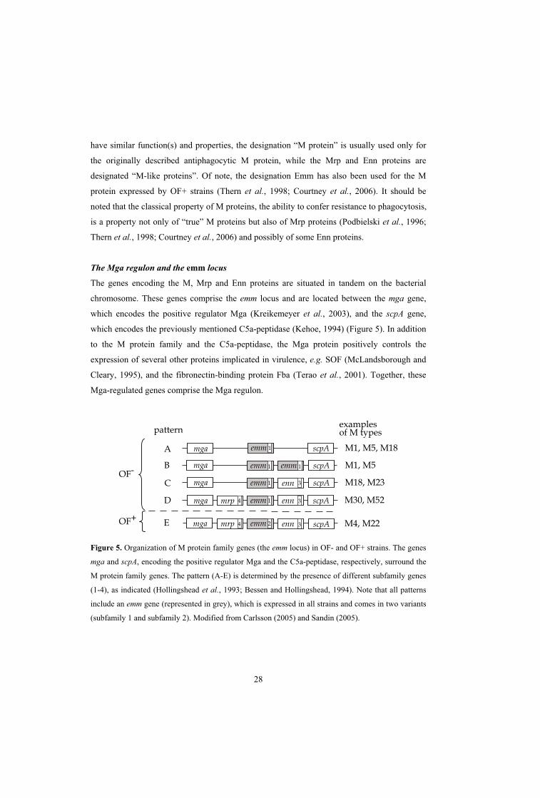
The Mga regulon and the emm locus
The genes encoding the M, Mrp and Enn proteins are situated in tandem on the bacterial
chromosome. These genes comprise the emm locus and are located between the mga gene,
which encodes the positive regulator Mga (Kreikemeyer et al., 2003), and the scpA gene,
which encodes the previously mentioned C5a-peptidase (Kehoe, 1994) (Figure 5). In addition
to the M protein family and the C5a-peptidase, the Mga protein positively controls the
expression of several other proteins implicated in virulence, e.g. SOF (McLandsborough and
Cleary, 1995), and the fibronectin-binding protein Fba (Terao et al., 2001). Together, these
Mga-regulated genes comprise the Mga regulon.
OF-
OF+
A
B
C
D
patternexamplesof M types
mga
mga
mga
mga mrp 4 enn 3
enn 3
scpA
scpA
scpA
scpA M1, M5, M18
M1, M5
M18, M23
M30, M52
1emm
1emm
1emm
1emm
1emm
E mga mrp 4 enn 3 scpA M4, M222emm
Figure 5. Organization of M protein family genes (the emm locus) in OF- and OF+ strains. The genes
mga and scpA, encoding the positive regulator Mga and the C5a-peptidase, respectively, surround the
M protein family genes. The pattern (A-E) is determined by the presence of different subfamily genes
(1-4), as indicated (Hollingshead et al., 1993; Bessen and Hollingshead, 1994). Note that all patterns
include an emm gene (represented in grey), which is expressed in all strains and comes in two variants
(subfamily 1 and subfamily 2). Modified from Carlsson (2005) and Sandin (2005).

29
All strains of S. pyogenes have an Mga regulon, but the genes constituting the regulon
differ between strains. The Mga protein was initially believed to be the response regulator of a
two-component system. However, because a sensory protein has not been found, and
phosphorylation of Mga has not been demonstrated, this is still an open question. It is known
that Mga responds to elevated CO2, iron limitation and increased temperature, and one
possibility is that Mga is directly controlled by these environmental factors (Kreikemeyer et
al., 2003).
As mentioned above, the emm locus of an S. pyogenes strain may include one, two or
three genes encoding members of the M protein family. Differences in the 3’ region separate
these genes into four distinct subfamilies (SF1-4) (Hollingshead et al., 1993), and based on
the combination of SF genes present, the emm locus of a strain may come in one of five
possible patterns (A-E) (Bessen and Hollingshead, 1994) (Figure 5). Of note, the emm locus
of OF+ strains invariably contains three genes, while the emm locus of OF- strains are more
diverse and may contain 1-3 genes. With few exceptions, strains of patterns A-C are
associated with throat infections and pattern D strains with skin infections, while pattern E
strains are found at either infection site (Bessen et al., 1996; McGregor et al., 2004b).
However, as stated above, this association is not clear-cut in areas with very high infection
rates.
Among clinical isolates of S. pyogenes, pattern A and pattern E strains are probably the
most important, reflected in the fact that 7 of the 8 genome sequences were determined for
OF- pattern A strains, while one was an OF+ pattern E strain. The combined data of Li et al.
(2003) and McGregor et al. (2004) indicate that the vast majority of isolates from invasive
disease are pattern A-C or pattern E strains and the isolates were almost evenly distributed
between these two groups of strains. Notably, among the pattern A-C strains, strains of pattern
A were most prevalent.
Strains of different patterns are believed to have arisen, during evolution, from an
ancestral strain of pattern A, i.e. the different genes in the emm locus may have evolved from
an SF1 emm gene (Figure 5) (Bessen and Hollingshead, 1994). Recombination events such as
gene duplication and subsequent divergence are proposed to explain the generation of the
other types of genes in this locus, and strains of patterns D and E are considered the most
recently emerged (Bessen and Hollingshead, 1994; Kehoe et al., 1996). Interestingly, SF1 and
SF2 genes, encoding two distinct types of M proteins, are mutually exclusive and define two
separate lineages of strains (Hollingshead et al., 1993).

30
There are several lines of evidence for horizontal gene transfer between S. pyogenes
strains (Kehoe et al., 1996). Examples of M proteins with a mosaic structure that may have
arisen through horizontal gene transfer and recombination are found e.g. in a pattern B strain
of serotype M5 (Whatmore and Kehoe, 1994), and in pattern E strains of serotypes M4 and
M60 (Hedén and Lindahl, 1993).
Mrp, M and Enn proteins
All proteins in the M protein family have similar overall organization and are anchored to the
cell wall peptidoglycan via an LPXTG-motif in the C-terminal region (Navarre and
Schneewind, 1999; Barnett and Scott, 2002), with the N-terminal protruding into the
environment (Figure 6). The C-terminal region is relatively conserved between M proteins,
whereas the N-terminal part is more variable. The C-terminal half of an M protein includes a
repeat region, which may contain repeats of type A or type C (Hollingshead et al., 1986;
Heath and Cleary, 1989; O'Toole et al., 1992). The variability increases towards the N-
terminus and the variable half of the protein can be divided into a semivariable region (SVR)
and a hypervariable region (HVR). A signal sequence, which has similar sequence in all M
proteins, is cleaved off upon secretion. Thus, in a surface exposed M protein, the HVR is the
protein region most distal from the bacterial surface (Kehoe, 1994).
signalsequence repeat region
(A or C repeats)
variable
conserved
cell wallanchoring region
N C
HVR SVR
Figure 6. Schematic representation of an M protein. The C-terminal half of the M protein is relatively
conserved while the N-terminal half is more variable and can be separated in a hypervariable region
(HVR) and a semivariable region (SVR). The conserved region contains repeats of type A or C. Note
that in the surface-expressed protein, the signal sequence (grey) has been cleaved off.
Mrp proteins, encoded by mrp genes, have conserved repeat region with three so called
A-repeats. The variability in the N-terminal part is more limited than among M proteins
(encoded by emm genes) (Bessen and Hollingshead, 1994). Mrp proteins have been shown to

31
confer phagocytosis resistance (Podbielski et al., 1996; Thern et al., 1998), and a recent study
showed that antibodies against Mrp4 were opsonizing for phagocytosis (Courtney et al.,
2006).
M proteins are antiphagocytic proteins that give rise to protective immunity and are
found in all S. pyogenes strains (Fischetti, 1989). The ability of M proteins to confer
resistance to phagocytosis is usually analyzed in the so called Lancefield test, which employs
fresh human blood and a small bacterial inoculum. After incubation (37°C, usually for 3 h)
bacterial growth is determined. Presence of M protein inhibits recognition by neutrophils
(PMNs) and allows rapid bacterial growth (Schnitzler et al., 1995; Schnitzler et al., 1999),
while strains lacking M protein are phagocytosed. Because of their general importance and
because they are the subject of this thesis work, M proteins are described in more detail
below.
The conserved repeat region in M proteins has so called C-repeats. Based on antigenic
differences within the C-repeat region, M proteins may be divided into two subclasses (I and
II), where class I proteins are generally expressed by OF- strains and class II proteins by OF+
strains (Bessen et al., 1989). The M proteins contain a ~50-100 residue N-terminal
hypervariable region (HVR). As mentioned above, the HVR is stable within a strain, allowing
the identification of >100 different M types (Facklam et al., 2002). Thus, the number of
known M types is small, compared to the large number of possible sequence variants,
suggesting that these M types have been selected because of their superior fitness.
Enn proteins, like M proteins, have a C-repeat region in the conserved half of the
protein. The enn gene is present in many strains, but is often silent, or expressed at very low
levels during in vitro growth (Haanes and Cleary, 1989; Jeppson et al., 1992). The function of
Enn proteins is not clear. They are not known to elicit protective antibodies and exhibit
limited sequence variation in the N-terminal region. Even so, the variability of Enn proteins
appears to be intermediate between that of M and Mrp proteins (M > Enn > Mrp) (Bessen and
Hollingshead, 1994).
The coiled-coil structure and M protein
Streptococcal M proteins are homodimeric, 50-60 nm long proteins that largely adopt a so
called coiled-coil structure, one of the most commonly found tertiary structures in proteins
(Figure 7A) (Lupas and Gruber, 2005). In a coiled-coil, two or more -helices twist around
each other to form a rope-like fibrillar structure. A coiled-coil dimer, a very common type of
coiled-coil, may be composed of two identical or two different helices, thus forming a homo-

32
or a heterodimer. Moreover, the helices in a coiled-coil can be arranged in a parallel or an
anti-parallel fashion, i.e. the N- and C-termini of the two helices can be oriented in the same
or opposite directions.
The amino acid sequence of coiled-coil proteins exhibits a characteristic seven-residue
repeat pattern, with the residue positions designated a-g (Figure 7) (Crick, 1953). In Figure
Figure 7. Schematic representations of a dimeric coiled-coil protein. A. Representation of the rope-
like structure of the coiled-coil. The protein is characterized by a heptadic sequence pattern where the
residues are designated a-g, as indicated. B. Helical wheel diagram of a cross-section of the dimeric
coiled-coil. The arrows represent the main interactions between residues in the two chains:
hydrophobic interactions involving a and d residues, and electrostatic interactions between e and g
residues
7B, a cross-section of a dimeric coiled-coil protein is depicted in a so called helical wheel
diagram (Mason and Arndt, 2004). This representation shows a single heptad repeat of each
helix and the arrangement of the a-g residues in the coiled-coil. The main inter-helix
interactions are also illustrated. These residues are often denoted a-g in one helix of the coil
and a -g in the other. Positions a and d (or a and d ) are found on the same face of a helix,
and compose the inner core between the two helices of the coiled-coil. The side chain of
residue a mainly contacts the side chain of an a residue in the other helix, and the same is
true for d and d residues. The a/a and d/d positions are usually occupied by hydrophobic
residues and the stability of the coiled-coil is largely determined by interactions between these
hydrophobic residues in the structural core, where improved stability follows from increased
hydrophobicity (Hu et al., 1990; Mason and Arndt, 2004). Residues in positions e and g are

33
solvent exposed, and they are often polar or charged, accounting for electrostatic interactions
between and/or within the helices. The e and g residues are positioned so that e in one helix
interacts with g in the other. Residues in positions e and g are less important for structural
stability than those in positions a and d, but may be important for the local conformation and
parallel/anti-parallel helix orientation of the coiled-coil (Hu et al., 1993; Zeng et al., 1997;
Mason and Arndt, 2004). The b, c and f residues are often hydrophilic residues, located
opposite to the a and d residues in the helix, and on the outer face of the coiled-coil (Figure
7B). Accordingly, these residues are exposed to the surroundings and may be important for
inter-protein interactions rather than for the formation or stability of the coiled-coil structure
as such. In an undistorted -helix, 3.6 residues make up each turn. However, because of a
slight twist to the helix in a coiled-coil, 3.5 residues compose a full turn and consequently one
heptad repeat completes two turns (Lupas and Gruber, 2005). Many coiled-coils are dimeric
proteins, but different oligomerization states are common and coiled-coils containing up to
five helices exist (Mason and Arndt, 2004).
The distribution of residues in the heptadic pattern of M proteins is often non-optimal,
with Asn residues frequently occupying position a (Manjula and Fischetti, 1980; Nilson et al.,
1995; Cedervall et al., 1997). Such deviation from the most favorable pattern often reflects
the individual conformation of the coiled-coil, at the expense of structural stability (Mason
and Arndt, 2004). For example, the Mrp4 protein deviates less from the optimal heptadic
pattern than does the M4 protein expressed by the same strain, and consequently has higher
stability (Cedervall et al., 1997).
Binding of human plasma proteins to M proteins
M proteins interact with a range of host plasma proteins, but the plasma proteins bound may
vary between different M proteins. These interactions may confer different properties, such as
phagocytosis resistance, ability to adhere or ability to invade tissues.
The available evidence suggests that M proteins can be divided into two major groups,
depending on their ability to bind either fibrinogen or C4BP (Carlsson et al., 2005). These
two groups apparently correspond to class I and class II M proteins, which show slight
variations in the sequence of the C-repeats (Bessen et al., 1989). Because fibrinogen and
C4BP play important roles for the function of M proteins, the interaction with these plasma
proteins will be considered first in this section. The interaction with C4BP will also be
described in a separate section, due to its importance for this thesis work. Several other
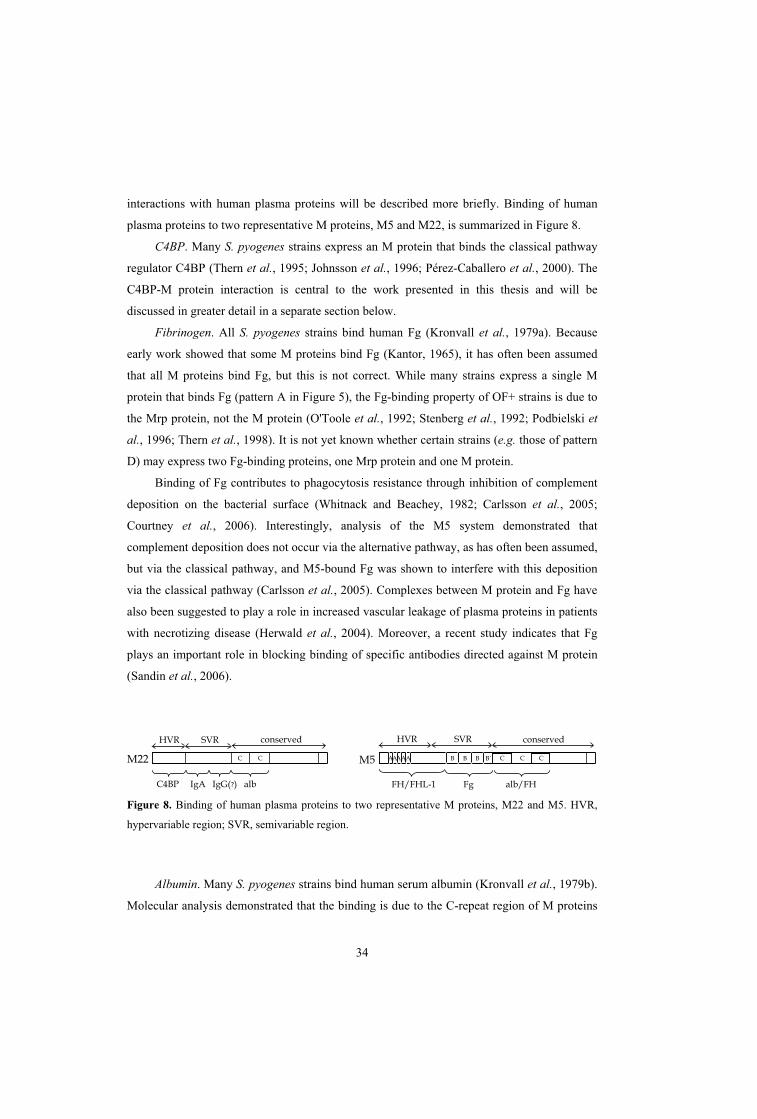
34
interactions with human plasma proteins will be described more briefly. Binding of human
plasma proteins to two representative M proteins, M5 and M22, is summarized in Figure 8.
C4BP. Many S. pyogenes strains express an M protein that binds the classical pathway
regulator C4BP (Thern et al., 1995; Johnsson et al., 1996; Pérez-Caballero et al., 2000). The
C4BP-M protein interaction is central to the work presented in this thesis and will be
discussed in greater detail in a separate section below.
Fibrinogen. All S. pyogenes strains bind human Fg (Kronvall et al., 1979a). Because
early work showed that some M proteins bind Fg (Kantor, 1965), it has often been assumed
that all M proteins bind Fg, but this is not correct. While many strains express a single M
protein that binds Fg (pattern A in Figure 5), the Fg-binding property of OF+ strains is due to
the Mrp protein, not the M protein (O'Toole et al., 1992; Stenberg et al., 1992; Podbielski et
al., 1996; Thern et al., 1998). It is not yet known whether certain strains (e.g. those of pattern
D) may express two Fg-binding proteins, one Mrp protein and one M protein.
Binding of Fg contributes to phagocytosis resistance through inhibition of complement
deposition on the bacterial surface (Whitnack and Beachey, 1982; Carlsson et al., 2005;
Courtney et al., 2006). Interestingly, analysis of the M5 system demonstrated that
complement deposition does not occur via the alternative pathway, as has often been assumed,
but via the classical pathway, and M5-bound Fg was shown to interfere with this deposition
via the classical pathway (Carlsson et al., 2005). Complexes between M protein and Fg have
also been suggested to play a role in increased vascular leakage of plasma proteins in patients
with necrotizing disease (Herwald et al., 2004). Moreover, a recent study indicates that Fg
plays an important role in blocking binding of specific antibodies directed against M protein
(Sandin et al., 2006).
C C
HVR SVR conserved
M22 C C C
HVR conserved
M5
SVR
C4BP IgA IgG(?) alb FH/FHL-1 Fg alb/FH
B B B B´A AAAA
Figure 8. Binding of human plasma proteins to two representative M proteins, M22 and M5. HVR,
hypervariable region; SVR, semivariable region.
Albumin. Many S. pyogenes strains bind human serum albumin (Kronvall et al., 1979b).
Molecular analysis demonstrated that the binding is due to the C-repeat region of M proteins

35
(Åkesson et al., 1994; Retnoningrum and Cleary, 1994). The function of this interaction
remains unclear. However, studies by Sandin et al. (2006) demonstrated that albumin bound
to the C-repeat region of an M protein may play an important role in evasion of specific
antibodies directed against that region.
Factor H/FHL-1. Many M proteins interact with different regulators of the human
complement system (other than C4BP) (Horstmann et al., 1988; Johnsson et al., 1998;
Lindahl et al., 2000). Two regulators of the alternative pathway of complement, factor H (FH)
and its splice variant factor H-like protein 1 (FHL-1) have been suggested to contribute to the
phagocytosis resistance of S. pyogenes by protecting the bacteria from alternative pathway
complement deposition (Horstmann et al., 1988; Johnsson et al., 1998). However, this
hypothesis has not been confirmed; on the contrary, emerging data suggest that neither of
these proteins (FH and FHL-1) is implied in M protein-mediated phagocytosis resistance
(Perez-Casal et al., 1995; Kotarsky et al., 2001; Sandin et al., 2006). Thus, the role of these
two complement regulators in S. pyogenes infections remains unclear.
Fibronectin. Some M proteins bind human fibronectin (Frick et al., 1995; Cue et al.,
2001), an interaction that has been implicated in invasion of epithelial cells by S. pyogenes
(Cue et al., 2001).
IgA and IgG. It was reported in 1973 that many S. pyogenes strains bind the Fc-part of
human IgG (Kronvall, 1973), and shortly thereafter this was found to be the case also for IgA-
Fc (Christensen and Oxelius, 1975). In 1989, two separate studies found these interactions to
be M protein dependent (Frithz et al., 1989; Heath and Cleary, 1989).
The binding of IgA contributes to phagocytosis resistance (Carlsson et al., 2003),
possibly by interfering with the binding of IgA-Fc to its cellular receptor CD89 (Pleass et al.,
2001). The IgA-binding region is a well-defined protein domain that can be studied in isolated
form, as a synthetic peptide (Johnsson et al., 1999; Sandin et al., 2002).
Many M and M-like proteins are also known to bind IgG-Fc (Heath and Cleary, 1989;
Lindahl and Åkerström, 1989; Gomi et al., 1990; Stenberg et al., 1992; Stenberg et al., 1994;
Åkesson et al., 1994), but the function of this interaction is not clear.
Kininogen. Some M proteins were reported to bind human kininogen (Ben Nasr et al.,
1995), but the binding site has not been identified. Analysis of this interaction suggested that
streptococcal cysteine protease may cleave the M protein-bound kininogen, thereby causing
release of the pro-inflammatory peptide kinin, possibly inducing increased vascular
permeability (Ben Nasr et al., 1995; Herwald et al., 1996).

36
Plasminogen. Certain strains of the OF+ lineage have been shown to express M proteins
that bind human plasminogen (Berge and Sjöbring, 1993; Wistedt et al., 1995). Plasminogen
bound to the streptococcal surface may be activated by host activators or streptokinase to
generate plasmin activity, causing degradation of extracellular matrix proteins and facilitating
tissue invasion (Boyle and Lottenberg, 1997; Ringdahl et al., 1998). This interaction causes
an important increase in bacterial virulence (Khil et al., 2003; Sun et al., 2004).
Binding of human plasma proteins to Mrp and Enn proteins
Description of the Mrp and Enn proteins is beyond the scope of this thesis. Moreover, much
less is known about these proteins than about the extensively studied M proteins. However,
the ability of the Mrp and Enn proteins to bind plasma proteins will be briefly summarized
here, since this information underlines the similarity between these proteins and the classical
M proteins and also underlines the remarkable finding that all members of the M protein
family bind at least one human plasma protein.
Mrp. Mrp proteins have been shown to bind IgG-Fc and Fg (Heath et al., 1990;
Stenberg et al., 1992), and to confer resistance to phagocytosis (Podbielski et al., 1996; Thern
et al., 1998). Recently a mechanism by which Mrp proteins may confer phagocytosis-
resistance was suggested. As in the case of Fg bound to M proteins (discussed above), Fg
bound to the Mrp protein of a type 4 strain was shown to protect the bacterium against
complement-deposition via the classical pathway (Courtney et al., 2006).
Enn. When expressed in E. coli, most or all Enn proteins bind IgA-Fc (Bessen, 1994;
Thern et al., 1998). However, because of the low, or even absent, expression of many Enn
proteins in streptococci, it is unclear whether this interaction plays a role in vivo. Recently, the
Enn protein in a serotype 18 strain was shown to bind C4BP (Pérez-Caballero et al., 2004),
suggesting a possible role for that Enn protein in phagocytosis resistance. However, such a
role remains to be demonstrated. Moreover, protein H, a mosaic protein expressed by some
M1 strains (Åkesson et al., 1994), has the same genetic location as the Enn proteins, and also
binds C4BP (Johnsson et al., 1996).
M protein and human C4BP
The binding of human C4BP to S. pyogenes was first described by Thern et al. (1995), who
tested a large number of strains, and found that most of them (~83%) could bind C4BP. For
two strains of different M types, M4 and M22, this property was analyzed at the molecular
level and found to be due to the M protein. In addition, C4BP bound to the bacteria was

37
shown to retain its function as a cofactor to factor I (fI) in the degradation of C4b. Subsequent
studies of the interaction between C4BP and M protein demonstrated that C4BP binds to the
most N-terminal ~50 aa HVR (Johnsson et al., 1996). Moreover, the binding site in C4BP was
found to overlap with the binding site for C4b, the natural ligand of C4BP (Figure 2)
(Accardo et al., 1996; Blom et al., 2000), but because of the polymeric structure of C4BP,
different -chains may either bind C4b or the bacterium, probably explaining why bacteria-
bound C4BP retains its cofactor function.
The HVRs of C4BP-binding M proteins exhibit extensive sequence and antigenic
variability, yet molecular studies indicate that they bind to the same site in C4BP (Johnsson et
al., 1996; Blom et al., 2000; paper I; paper III). A recent paper analyzed the structural basis
for this interaction between M protein and human C4BP (Jenkins et al., 2006). Previous
studies had shown that CCP1-2 of the C4BP -chain were important for binding of M protein
(Accardo et al., 1996; Blom et al., 2000) and the study by Jenkins et al. (2006) shows that
these two CCPs are sufficient for binding. Employing heteronuclear NMR spectroscopy, the
structure of CCP1-2 was solved, and the interaction with the M4 protein was studied using a
peptide derived from the C4BP-binding HVR of this M protein. Interestingly, a four-residue
linker connecting the CCP1-2 domains also plays an important role in binding, because the
insertion of two alanine residues into this linker completely abrogated the interaction with M
protein. The spectroscopic data suggested that residues in both CCPs are affected upon
binding of the M protein-derived peptide. However, because these affected residues are not
located on the same face of the -chain, it seemed unlikely that they were all directly involved
in the binding. Rather, the data were interpreted to reflect a conformational change in C4BP,
induced upon interaction with the peptide (Jenkins et al., 2006). Previously, a cluster of
positive residues on the C4BP CCP1-2 interface had been suggested to be important for the
C4BP-M protein binding (Blom et al., 2000). However, due to the salt-resistant nature of the
interaction, hydrophobic interactions and hydrogen bonds are likely to contribute (Blom et al.,
2000; Jenkins et al., 2006). Possibly, the positive cluster in C4BP participates in an important
“first-step” electrostatic interaction, guiding the M protein to the right location on C4BP,
while this cluster may not be involved in the binding per se (Jenkins et al., 2006).
The fact that bacteria-bound C4BP is still functional as a cofactor in C4b degradation,
suggested that C4BP-binding might play an important role in protection of the bacteria against
phagocytosis, by interfering with complement deposition on the bacterial surface (Thern et
al., 1995). To analyze this hypothesis, Berggård et al. (2001) constructed a strain expressing
an M22 protein with a seven residue deletion in the HVR. This strain completely lacked

38
C4BP-binding, and was partially deficient in its ability to grow in human blood. This result
suggested that the C4BP-binding indeed contributed to M protein-mediated resistance to
phagocytosis, but binding of C4BP could not alone account for the anti-phagocytic property
of the M22 protein (Berggård et al., 2001). However, Carlsson et al. (2003) demonstrated that
the anti-phagocytic property of the M22 protein can be fully explained as a cooperation
between two distinct ligand-binding regions in this M protein, the C4BP-binding region and
the IgA-binding region. While the mechanism by which bound IgA protects against
phagocytosis remains unclear, this study confirmed that bound C4BP plays a key role in
protecting S. pyogenes against phagocytosis.
The finding that S. pyogenes recruits C4BP and that this interaction is important for the
ability of the bacterium to resist phagocytosis was puzzling because C4BP is a regulator of the
classical pathway of complement, whereas the general assumption has been that bacteria
mainly activate the alternative pathway. However, analysis of complement deposition on an
M-negative S. pyogenes strain demonstrated that the complement system was activated via the
classical pathway, explaining the value of recruiting a classical pathway regulator (Carlsson et
al. 2003). Thus, C4BP bound to S. pyogenes M protein protects the bacteria against attack via
the classical pathway (Figure 9, right part). Even if this biological function of M protein-
bound C4BP has been demonstrated only in the M22 system, it is reasonable to assume that
the role of surface-bound C4BP is similar in other C4BP-binding S. pyogenes strains.
C4BP
M22
protein
IgA
M5
protein
Fg
Complement activation via classical pathway
C3b C3b
Inhib. Inhib.
Figure 9. Unifying model for the inhibition of complement deposition by M proteins. Complement is
activated via the classical pathway by S. pyogenes, resulting in surface deposition of C3b. However, M
protein inhibits the C3b deposition by recruiting a human plasma protein. Some M proteins, such as
M5, bind Fg, while other M proteins, such as M22, bind C4BP. As indicated, the M22 protein also
binds human IgA, which contributes to phagocytosis resistance by an unknown mechanism (Carlsson
et al., 2003). Modified from Carlsson et al. (2005).

39
As mentioned above, human Fg bound to S. pyogenes M protein also inhibits
complement deposition via the classical pathway. Because all M proteins appear to bind either
C4BP or Fg, these findings have allowed the construction of a unifying model for the
protection of S. pyogenes against complement deposition and subsequent phagocytosis
(Carlsson et al., 2005) (Figure 9).
Because M proteins bind multiple ligands and cross-react immunologically due to the
more conserved C-terminal part, the binding properties or immunological properties of a
ligand-binding domain are not easily analyzed using whole M proteins. Characterization of
the interaction between C4BP and M proteins was much facilitated by the finding that the
C4BP-binding HVRs of M proteins can be studied in isolated form as synthetic peptides
(paper I). An important property of these peptides is the presence of a C-terminal cysteine
residue, not present in the intact M proteins, which allowed for dimerization via a disulphide
bridge. This cysteine residue was included because intact M proteins are dimeric coiled-coils
and because dimerization is known to be critical for the ability of M proteins to bind ligands
(Åkerström et al., 1992). A peptide not containing a C-terminal cysteine showed very limited
binding to C4BP, as compared to the binding of the dimerized peptides, indicating that the
dimeric nature of the M protein is indeed essential for ligand binding (paper I and paper II).
Remarkably, out of all proteins in human serum, C4BP is the only one that binds to
HVR-derived peptides, demonstrating that the HVRs are well-defined ligand-binding protein
domains with highly specific ligand-binding properties (paper I). Because of this binding
specificity, the HVR of C4BP-binding M proteins can be used to purify C4BP by a simple
single-step procedure (paper II). The finding that HVRs of C4BP-binding M proteins are
well-defined ligand-binding domains that can be studied in isolated form, as synthetic
peptides, was an important finding that enabled much of the work presented in this thesis.
VARIABILITY IN MICROBIAL SURFACE PROTEINS
Microorganisms constantly evolve to adapt to a changing environment. For pathogens,
sequence variability in surface proteins is a common mechanism by which they adjust to
survive better in their hosts. The effects of sequence variability may be diverse. A change in
receptor specificity may provide the microbe with new tools for infection of a specific cell
type (Baranowski et al., 2001; Mayer et al., 2002), or may allow the microbe to change tissue
tropism (Weissman et al., 2003). In some pathogens, sequence variability may even permit
40
the organism to switch between different species (Strömberg et al., 1990; van Luenen et al.,
2005). However, most cases of variability probably represent so called antigenic variation, an
important phenomenon that will be discussed in this section.
Antigenic variation
In pathogenic microorganisms, variability in a surface structure targeted by protective
immunity is commonly referred to as antigenic variation. Importantly, the term antigenic
variation implies that the variability confers a selective advantage and promotes virulence.
The variable surface structure is most often a protein, but in the literature the term antigenic
variation is sometimes also used for changes in e.g. carbohydrates (Deitsch et al., 1997).
Here, I will only consider antigenic variation in proteins. An important aspect of antigenic
variation is that the variable protein must retain an important function, while varying to avoid
host immunity, otherwise this structure would probably be lost by mutation.
The significance of antigenic variation can hardly be overestimated, because it is the
major obstacle in the development of new vaccines. Because of antigenic variation, efficient
vaccines are not available against pathogens such as HIV, Plasmodium falciparum (the
causative agent of malaria) or influenza A virus. Importantly, antigenic variation may arise
through one of two general mechanisms, antigenic shift or antigenic drift (Borst, 1991).
Antigenic shift represents a drastic change in protein expression. For example, the
expression of the protein may be switched on or off (phase variation), or one sequence may be
replaced with another (gene conversion). In gene conversion mechanisms a gene from a silent
part of the genome is copied into an expression site (Borst, 1991; Deitsch et al., 1997). A
classical example of a pathogen exhibiting antigenic shift is influenza A virus, in which the
genes, including those encoding the major surface proteins hemagglutinin (HA) and
neuraminidase (NA), may be reassembled to create a virus with new immunologic properties,
which may cause a rapidly spreading influenza epidemic (Steinhauer and Skehel, 2002;
Stevens et al., 2004).
Antigenic drift represents the accumulation of point mutations or small
insertions/deletions that gradually change the antigenic properties of a protein. In bacteria,
antigenic variation through antigenic drift is most likely a slower process than antigenic shift,
because a single small genetic change will not cause the appearance of an immunologically
completely different variant. However, due to the lack of proof-reading systems in RNA
viruses, and their rapid replication, viruses such as HIV may mutate very rapidly (Wilson and

41
Cox, 1990; Malim and Emerman, 2001; Steinhauer and Skehel, 2002). In the M protein of S.
pyogenes, the N-terminal hypervariable region (HVR) exhibits antigenic variation that most
likely has arisen through antigenic drift.
The hypervariable region in streptococcal M protein
The ~50-100 aa HVR of M proteins varies extensively in sequence. The HVR is stable
within a strain of S. pyogenes, allowing the identification of >100 different M types (Facklam
et al., 2002). However, the number of known M types is small, compared to the large number
of possible sequence variants, suggesting that these M types have been selected because of
their superior fitness. The M protein HVRs are targets for protective antibodies and exhibit
antigenic variation (Lancefield, 1962; Ashbaugh et al., 2000). The variability in the HVRs
arises through antigenic drift, i.e. point mutations and small insertions or deletions, and in
certain M proteins it may also arise through variation in the size of a repeat region (Fischetti,
1989). Small changes in the gene segment encoding the HVR will give rise to a new allele
(subtype) of an existing M type. Definition of a completely new type requires that the first 90
bases encoding mature protein share less than 92% similarity to any previously determined
M/emm type (http://www.cdc.gov/ncidod/ biotech/strep/doc.htm).
Because of the extensive sequence variability it was initially suggested that the HVRs
do not have a specific ligand-binding function, but inhibit phagocytosis by having a negative
charge, causing repulsion of the phagocyte cell surface or IgG antibodies (Fischetti, 1983;
Fischetti, 1989). However, it is now known that many HVRs do have a specific ligand-
binding function, in spite of the extensive sequence variability. In this regard, M proteins may
be divided into two major groups. In one group of M proteins, the HVR binds the complement
regulator C4BP (Thern et al., 1995; Accardo et al., 1996; Johnsson et al., 1996), an
interaction that is important for phagocytosis resistance because M protein-bound C4BP
inhibits surface-deposition of complement via the classical pathway (Berggård et al., 2001;
Carlsson et al., 2003). The second group of M proteins do not bind C4BP. In some of these
non-C4BP-binding proteins, the HVR has been found to bind the complement regulator FHL-
1 (Johnsson et al., 1998), but the physiological relevance of this interaction is unknown
(Kotarsky et al., 2001; Sandin et al., 2006). It seems possible that these HVRs also interact
with other, unknown ligands, explaining the predicted structural constraints on these HVRs
(Li et al., 2003).

42
Interestingly, it has been observed that mutations that occur in the gene segments
encoding HVRs are almost exclusively non-synonymous, which is indicative of a strong
immune pressure (Kehoe et al., 1996; Li et al., 2003). The selective pressure to retain a
particular structure and function in the M protein HVR, which changes to avoid recognition
by specific antibodies, therefore gives rise to a Darwinian evolution in which only the most fit
M protein variants have survived during evolution (Phillips, 2002). However, new M types
may slowly appear through a similar process.
In addition to their medical importance, M proteins are attractive model systems for
studies of antigenic variation. In contrast to e.g. gp120 of HIV, which varies also under
experimental conditions, the M protein HVR is stable within a strain and is therefore a system
where the variability can be studied under more controlled conditions. Moreover, in spite of
the extreme sequence variability, many HVRs bind a common ligand, C4BP. These HVRs are
well-defined ligand-binding domains that can be studied in isolated form, as synthetic
peptides, allowing detailed biochemical and immunological characterization.
BIOPHYSICAL METHODS - IN BRIEF
In the last paper included in this thesis, an attempt to solve the three-dimensional structure of
C4BP-binding HVRs is reported (paper IV). This work, performed in collaboration with
biophysical chemists, gave me the opportunity to learn the basic principles of two methods
that are commonly used in studies of protein structure, circular dichroism (CD) and nuclear
magnetic resonance spectroscopy (NMR). In this section, brief and very simplified
descriptions of these methods will be given.
Circular Dichroism
Objects that are not identical to their mirror image counterpart are referred to as being chiral.
For example, left and right hand gloves are mirror images that differ from each other, i.e. the
left hand glove will only fit your left hand. Such objects, as the two gloves in a pair, are
referred to as enantiomers.
Plane polarized (or linearly polarized) light may be explained as the sum of two
circularly polarized light waves, one to the left and one to the right. If plane polarized light is
passed through a medium, and the effect by the medium on the left and right circular light
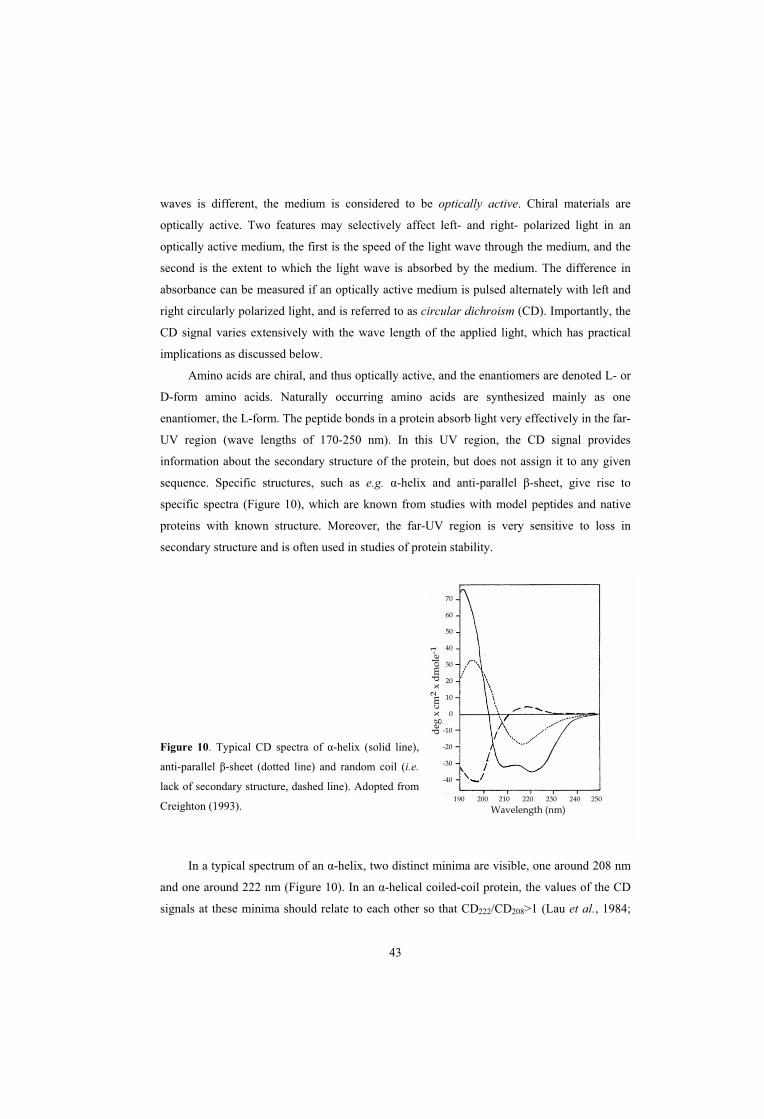
43
190 200 210 220 230 240 250
Wavelength (nm)
70
60
50
40
30
20
10
0
-10
-20
-30
-40
deg
x c
m2
x d
mo
le-1
waves is different, the medium is considered to be optically active. Chiral materials are
optically active. Two features may selectively affect left- and right- polarized light in an
optically active medium, the first is the speed of the light wave through the medium, and the
second is the extent to which the light wave is absorbed by the medium. The difference in
absorbance can be measured if an optically active medium is pulsed alternately with left and
right circularly polarized light, and is referred to as circular dichroism (CD). Importantly, the
CD signal varies extensively with the wave length of the applied light, which has practical
implications as discussed below.
Amino acids are chiral, and thus optically active, and the enantiomers are denoted L- or
D-form amino acids. Naturally occurring amino acids are synthesized mainly as one
enantiomer, the L-form. The peptide bonds in a protein absorb light very effectively in the far-
UV region (wave lengths of 170-250 nm). In this UV region, the CD signal provides
information about the secondary structure of the protein, but does not assign it to any given
sequence. Specific structures, such as e.g. -helix and anti-parallel -sheet, give rise to
specific spectra (Figure 10), which are known from studies with model peptides and native
proteins with known structure. Moreover, the far-UV region is very sensitive to loss in
secondary structure and is often used in studies of protein stability.
Figure 10. Typical CD spectra of -helix (solid line),
anti-parallel -sheet (dotted line) and random coil (i.e.
lack of secondary structure, dashed line). Adopted from
Creighton (1993).
In a typical spectrum of an -helix, two distinct minima are visible, one around 208 nm
and one around 222 nm (Figure 10). In an -helical coiled-coil protein, the values of the CD
signals at these minima should relate to each other so that CD222/CD208>1 (Lau et al., 1984;

44
Engel et al., 1991). Thus, CD analysis may be used to analyze the secondary structure of a
protein, and may also give some insights about tertiary structure, such as coiled-coil formation
(Cantor and Schimmel, 1980; Creighton, 1993).
Nuclear Magnetic Resonance Spectroscopy
The NMR technique has been awarded several Nobel prizes, one in physics (1952), two in
chemistry (1991 and 2002), and one in medicine (2003). NMR is a powerful and theoretically
very complex method, which is not readily explained without involving mathematical models
and quantum mechanics. However, it is fully possible to appreciate some general principles of
NMR without a deeper understanding of the underlying physics of the actual NMR
phenomenon. NMR is a method to study nuclei of atoms in a molecule, and is the only
technique available to date that can resolve the three-dimensional structure of a molecule in
solution. The nuclei most commonly studied are 1H, 13C and 15N, where the easiest nucleus to
use is the proton (1H), because of its abundance in nature.
For simplicity, the nucleus of an atom may be envisaged as a small magnet, with a north
and a south pole. In an NMR experiment, these polarized nuclei are placed under the
influence of a strong magnet, where they align with the applied magnetic field, and are
subsequently pulsed with radio waves. The nuclei absorb energy and together change their
orientation within the magnetic field, thereby creating a radiofrequency that may be
measured. To assure that measurements performed on different NMR spectrometers provide
the same information, the emitted frequency of a sample is compared to a reference substance.
This measured difference from the reference substance is referred to as the chemical shift of a
sample nucleus. The chemical shift is specific for a nucleus in a given environment, such that
e.g. a proton in a methylene group (-CH2) next to a hydroxyl group (-OH) has a different
chemical shift than a proton in a methyl group (-CH3) next to a hydroxyl group, thereby
providing information about the positions of the nuclei in relation to each other, which allows
for the determination of a three-dimensional structure. In protein NMR, knowledge about the
primary structure of the protein is necessary for an accurate model of the three-dimensional
structure. The study of a single type of nucleus is known as one-dimensional (1D) NMR.
However, several types of nuclei can be studied simultaneously, in 2D or multidimensional
NMR (Van Holde et al., 1988; Rattle, 1995; http://www.wikipedia.org; S. Linse and I. André,
personal communication).

45
PRESENT INVESTIGATION
PAPER I: Isolated hypervariable regions derived from streptococcal M proteins
specifically bind human C4b-binding protein: implications for antigenic variation
Pathogenic microorganisms have evolved a number of mechanisms to escape the immune
system of the host. One such mechanism is antigenic variation, i.e. alterations in a surface
structure to avoid immune recognition. The ~50-100 aa HVR of M proteins exhibit antigenic
variation and has long been recognized to be targeted by specific antibodies (Fischetti, 1989).
However, the function of this region in pathogenesis remained unclear. In 1995, Thern et al.
found that many M proteins bind human C4BP, and Johnsson et al. (1996) attributed the
interaction to the HVR in these M proteins. Subsequent studies of the biological function of
this binding demonstrated that C4BP plays an important role in phagocytosis resistance
(Berggård et al., 2001; Carlsson et al., 2003), a situation that made it of interest to further
characterize this interaction. However, because streptococcal M proteins interact with
multiple human plasma proteins, the nature of the M protein-C4BP interaction is not easily
studied with intact M proteins. To circumvent this problem, peptides corresponding to the
HVRs of three C4BP-binding proteins (M2, M4 and M22) were synthesized, in the hope that
it would be possible to study the C4BP-binding HVRs in isolated form.
It had been demonstrated previously that the ability of M proteins to bind ligands
requires that the M proteins have dimeric structure (Åkerström et al., 1992). Therefore, the
synthetic peptides contained a C-terminal cysteine not present in the native protein, to allow
dimerization via a disulphide bridge. Interestingly, the dimerized peptides were able to bind
C4BP, while a peptide without the C-terminal cysteine residue showed no or little binding to
C4BP. Affinity chromatography experiments, in which whole human serum was applied to
dimerized peptide immobilized in a column, showed that the M protein-C4BP interaction is
highly specific, because out of all proteins present in serum, only C4BP was retained.
Analysis of the sequence of different C4BP-binding HVRs identified only very limited
residue identity. Indeed, only three conserved residues could be identified, possibly
representing a C4BP-binding motif. However, this putative motif was also found in non-
C4BP-binding M proteins, indicating that the three residues were not sufficient to confer
ability to bind C4BP. Further analysis of the C4BP-binding peptides suggested that, in spite of
the remarkable sequence variability, these peptides bound to the same region in C4BP, at the
CCP1-2 interface. Moreover, secondary structure analysis using CD, as well as computational
analysis, suggested that the peptides assume similar folds.

46
Although the C4BP-binding peptides/HVRs display extensive sequence variability, the
apparent similarities in folding and C4BP-binding properties suggested that they might
possess at least some immunological cross-reactivity. However, analysis exploiting rabbit
antiserum against each of the peptides demonstrated that each antiserum recognized only the
corresponding peptide, indicating that the HVRs are immunologically unrelated.
In summary, C4BP-binding HVRs represent distinct ligand-binding domains that can be
studied as synthetic peptides. Such peptides exhibit extensive sequence variability but have
similar ligand-binding properties, suggesting that they have similar three-dimensional
structure. Nevertheless several C4BP-binding peptides did not cross-react immunologically.
PAPER II: Single-step purification of human C4b-binding protein (C4BP) by affinity
chromatography on a peptide derived from a streptococcal surface protein
In recent years, the interest in C4BP has increased, emphasizing the value of a simple method
to purify this protein (Lindahl et al., 2000 ; Nonaka et al., 2001; Webb et al., 2002; Brodeur et
al., 2003; Carlsson et al., 2003; Blom et al., 2004). Because previously described techniques
to purify C4BP are based on precipitation of human plasma and one or several subsequent
chromatographic steps (Villiers et al., 1981; Dahlbäck, 1983), we tried to develop a simple
purification technique based on the specific interaction between C4BP and peptides derived
from streptococcal M proteins.
A dimerized peptide (M22-N) corresponding to the HVR of the M22 protein was
previously shown to bind human C4BP with high specificity (paper I), indicating that this
peptide could be used to develop a simple affinity chromatography method to purify C4BP.
Dimerized M22-N was immobilized in a column, and whole human serum was applied. After
extensive washing, bound proteins were eluted and subjected to dialysis against a neutral
buffer. The column, containing 5 mg immobilized streptococcal peptide, had a binding
capacity of ~3.5 mg C4BP, and could be used repeatedly during a 1-year period with no
apparent loss of function.
The purified C4BP was shown to have high molecular weight, indicating that it had the
multimeric structure typical of C4BP, and was demonstrated to be more pure than a
commercially available preparation of C4BP. Functional analysis demonstrated that the
purified C4BP retained its activity as a cofactor to factor I in C4b-degradation. Moreover,
binding assays using C4b, the natural ligand for C4BP, and streptococcal M protein, pure or
expressed on the surface of whole streptococci, indicated that the binding properties of the
47
C4BP purified by the single-step method exceeded those of commercially available C4BP. In
addition, this affinity chromatography method could be used to deplete serum of C4BP.
Indeed, >99.5% of the C4BP present in a serum sample was retained on a column.
In conclusion, a synthetic peptide corresponding to the C4BP-binding region of the M22
protein can be used to purify human C4BP, and to deplete serum of C4BP, by a single-step
affinity chromatography method,. In addition to its interest for work on C4BP, this result
emphasizes the potential of bacterial proteins/peptides for the purification of human proteins.
PAPER III: Extreme sequence divergence but conserved ligand-binding specificity in
Streptococcus pyogenes M protein
Most cases of sequence variability in surface proteins of pathogenic microorganisms,
probably represent antigenic variation. An important aspect of antigenic variation is the
requirement on the variable protein to retain a specific function, such as ligand-binding, while
varying in sequence to escape immune recognition. It is generally assumed that ligand-
binding is dependent on a specific conserved sequence motif (Shakhnovich et al., 1996). Even
the extremely variable surface proteins gp120 of HIV and hemagglutinin of influenza virus
contain ligand-binding sites including a few highly conserved amino acid residues (Wilson
and Cox, 1990; Kwong et al., 1998; Skehel and Wiley, 2000).
We studied the limits of variability in C4BP-binding HVRs of M proteins. Previous
analysis of five C4BP-binding HVRs revealed only three conserved residues (paper I),
corresponding to residues L28, E31 and D40 in the extensively studied M22 protein (Figure
1B in paper III). Because of the limited residue identity between these five HVRs, we
hypothesized that not even the three conserved residues would be essential for ability to bind
C4BP.
Initial analysis was focused on the M22 protein. Studies using fusion proteins showed
that residues 1-39 of M22 are sufficient for C4BP-binding. Consequently, residue D40 of
M22 is not necessary for C4BP-binding, implying that only two of the three residues
mentioned above might be required for C4BP-binding. To analyze whether these two residues
are conserved in C4BP-binding HVRs, a large number of S. pyogenes strains were screened
for ability to bind C4BP. Based on sequence analysis of the M protein HVRs of C4BP-
binding strains, two strains, expressing the M4.1 and M114 proteins, were chosen for further
studies. The HVRs of these proteins were demonstrated to represent distinct C4BP-binding

48
domains, and the addition of the M4.1 and M114 HVR sequences to the alignment performed
in paper I showed that C4BP-binding HVRs completely lack residue identities.
Although different C4BP-binding HVRs lack residue identities, a sequence logo of the
seven known C4BP-binding HVRs indicated that the C-terminal half is less variable (Figure
11A). This finding made it of interest to study the possible role in binding of relatively
conserved residues. Four of these residues, L21, E24, L28 and E31, were substituted for
alanines and the mutant proteins, containing single aa substitutions, were expressed on the
surface of streptococci. Interestingly, substitution of either L21 or L28 completely abolished
the ability of the M22 protein to bind C4BP. A computational prediction, and the distribution
of residues in the sequence logo, suggested that the central part of the M22 HVR forms a
coiled-coil and the L21 and L28 residues were predicted to represent core residues essential
for the formation of this coiled-coil. Although it cannot be ruled out that these two Leu
residues are directly involved in the binding of C4BP, our mutagenesis data support the
conclusion that the centre of the HVR is folded as a coiled-coil (see paper IV). In contrast,
substitution of residues E24 and E31, which are predicted to be solvent exposed in a coiled-
coil, had no effect on C4BP-binding. Thus, it is not clear why these residues are relatively
conserved among the C4BP-binding HVRs studied.
It was of interest to know whether the sequence logo of C4BP-binding HVRs would
have similar appearance if many more HVRs were included, and whether it is different from a
logo derived from non-C4BP-binding HVRs. To analyze additional C4BP-binding HVRs, we
compared M protein HVRs in OF+ strains of different M types. It seemed likely that these
HVRs promote binding of C4BP, because all OF+ strains bind C4BP, and because binding of
C4BP has been attributed to the M protein in all OF+ strains studied at the molecular level
(Thern et al., 1995; Johnsson et al., 1996; paper I; paper III). A logo of the M protein HVRs
of 47 OF+ strains tested in paper III was therefore created (Figure 11B). This logo is very
similar to that shown in Figure 11A. As in Figure 11A, the logo in Figure 11B is dominated
by two Leu residues (corresponding to L21 and L28 in M22). In addition, Glu residues
corresponding to E24 and E31 in M22 are prominent, although the conservation of these
residues is less prominent than in the logo in Figure 11A. Thus, the logos in Figures 11A and
11B have similar overall appearance, with two prominent Leu residues and an abundance of
Glu residues, suggesting that these features are important for C4BP-binding.
A logo derived from the HVRs of M proteins in 11 non-C4BP-binding strains (all OF-)
is shown in Figure 11C. The M protein-expressing strains used to construct this logo were
analyzed in paper III. The lack of C4BP-binding in these strains was not due to lack of M
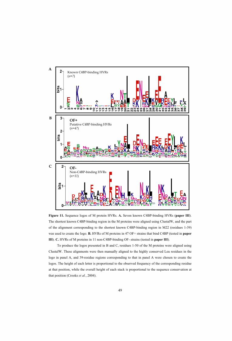
49
A
B
C OF-Non-C4BP-binding HVRs(n=11)
OF+Putative C4BP-binding HVRs(n=47)
Known C4BP-binding HVRs(n=7)
Figure 11. Sequence logos of M protein HVRs. A. Seven known C4BP-binding HVRs (paper III).
The shortest known C4BP-binding region in the M proteins were aligned using ClustalW, and the part
of the alignment corresponding to the shortest known C4BP-binding region in M22 (residues 1-39)
was used to create the logo. B. HVRs of M proteins in 47 OF+ strains that bind C4BP (tested in paper
III). C. HVRs of M proteins in 11 non-C4BP-binding OF- strains (tested in paper III).
To produce the logos presented in B and C, residues 1-50 of the M proteins were aligned using
ClustalW. These alignments were then manually aligned to the highly conserved Leu residues in the
logo in panel A, and 39-residue regions corresponding to that in panel A were chosen to create the
logos. The height of each letter is proportional to the observed frequency of the corresponding residue
at that position, while the overall height of each stack is proportional to the sequence conservation at
that position (Crooks et al., 2004).

50
protein expression, because all strains bound human Fg, a feature known to be due to
members of the M protein family. Therefore, in contrast to the hypothetical logo in panel B,
the logo of non-C4BP-binding HVRs is based on reliable experimental data. Interestingly, this
logo also displays two dominating Leu residues, strengthening the hypothesis that these
residues are not involved in C4BP-binding but may be important for formation of a coiled-
coil. However, the relatively conserved Glu residues in the C4BP-binding HVRs were not
prominent in the non-C4BP-binding HVRs. This analysis suggest that alignments of C4BP-
binding and non-C4BP-binding HVRs indeed gives rise to different logos. In particular,
comparison of the different logos indicates that the C-terminal half of a C4BP-binding HVR
is more negatively charged than the corresponding region in a non-binding HVR.
As mentioned above, substitution of L21 or L28 completely abolished the C4BP-
binding ability of the M22 protein, while substitution of E24 or E31 did not affect C4BP-
binding. This finding made it of interest to analyze whether the single residue changes, not
affecting C4BP-binding, induced antigenic alterations. Remarkably, although substitution of
E24 or E31 with Ala did not affect the ability of M22 to bind C4BP, these changes strongly
decreased the ability of a polyclonal antiserum, directed against the HVR, to recognize the
mutant proteins. This finding implies that single amino acid changes in an HVR may give rise
to antigenic variation and that accumulation of such changes may eventually give rise to a
new M type.
To conclude, C4BP-binding HVRs represent a unique example of sequence divergence,
because they completely lack residue identities but bind the same ligand. Moreover, single
residue changes in an HVR, not affecting the ability to bind C4BP, may considerably change
the immunological properties of the HVR, supporting classical models for the appearance of
antigenic drift through immunological selection.
PAPER IV: Streptococcal M protein: structural studies of the hypervariable region, free
and bound to human C4BP
Streptococcal M proteins are known to largely assume a coiled-coil structure (Fischetti, 1989;
Kehoe, 1994; Navarre and Schneewind, 1999). However, it is not known whether the coiled-
coil extends through the N-terminal HVR, which has been depicted as non-structured in the
literature. Because computational data and studies of secondary structure had indicated that
C4BP-binding HVRs adopt similar folds, although their sequences are highly divergent
(paper I), it was of interest to more accurately study the three-dimensional structure of these

51
M protein HVRs. The aim of this study was to identify the three-dimensional structure for
C4BP-binding HVRs, using heteronuclear NMR spectroscopy.
Three streptococcal peptides expressed in E. coli. Two peptides, M4-N and M22-N,
derived from the HVRs of the M4 and M22 proteins, respectively, correspond to the C4BP-
binding regions in these proteins. One peptide, M22-NL, contained the HVR of M22 but also
included an adjacent semivariable region that binds IgA-Fc (Stenberg et al., 1994; Sandin et
al., 2002). Because of certain properties of the peptides, such as a tendency to aggregate at
high concentrations, a three-dimensional structure could not be determined. However, using a
range of techniques, we could obtain clear indications concerning the structural properties of
the peptides.
Circular dichroism (CD) spectra indicated that the peptides assume a helical structure.
Typically, a coiled-coil structure produces a CD spectrum with minima at 208 and 221 nm,
where the ratio between the signals at these wave lengths should relate to each other as:
208/221>1 (Lau et al., 1984; Engel et al., 1991). This result was indeed obtained for the M22-
NL peptide. However, this ratio is probably not as useful when a significant part of the
molecule is in an unstructured state, as appeared to be the case for the M4-N and M22-N
peptides.
For NMR analysis, the peptides were labelled with 15N, or 13C and 15N. These studies
were focused mainly on the M4-N peptide, because preliminary NMR analysis of the peptides
indicated that this peptide gave rise to superior spectral data. Results obtained from analysis
of the M22-N peptide were less clear than for M4-N, but pointed in the same direction.
Through the use of different NMR techniques, permitting analysis of nuclei in either the
peptide backbone or the amino acid side chains, it was possible to conclude that the M4-N
peptide has a structured central part of ~4 heptads, with flexible termini. The peptides were
also studied in complex with CCP1-2 of the C4BP -chain. Because the signals arising from
the peptide backbone are lost for interacting residues, it was possible to map the C4BP-
binding region in the streptococcal peptide. The data indicated that the C4BP-binding region,
possibly as many as 28 residues, is contained within the structured centre of the peptide. It
should be noted that if the peptide binds as a coiled-coil, only about half of the ~28 residues
will be on the binding interface.
A recent NMR study of CCP1-2 of the C4BP -chain, in complex with the unlabelled
M4-N peptide indicates that the peptide mainly binds to CCP2, but that the four-residue linker
connecting CCP1-2 is required for binding, and that residues in CCP1 are affected by binding
of the peptide (Jenkins et al., 2006). Together with the NMR analysis of M4-N reported in

52
paper IV, these data suggest that the elongated M4-N peptide may interact with both CCP1
and CCP2 of the C4BP -chain.
In conclusion, the data in paper IV indicate that M4-N has a folded central region of ~4
heptads with flexible termini and that the C4BP-binding surface is contained within these four
heptads. Moreover, in spite of the very limited identity between M4-N and M22-N, these
peptides probably adopt similar folds, both in solution and in complex with C4BP.
Intriguingly, these C4BP-binding HVRs completely lack immunological cross-reactivity,
although they specifically bind the same ligand and have similar structure. This apparent
contradiction may be explained if C4BP mainly interacts with the backbone of the HVRs,
while antibodies recognize the amino acid side chains, which vary extensively between
HVRs.

53
CONCLUSIONS
The ~50 residue hypervariable regions (HVRs) of C4BP-binding streptococcal M proteins
can be studied as synthetic peptides that retain their C4BP-binding ability. Different
C4BP-binding HVRs show limited sequence identity, yet bind with high specificity to the
same region in C4BP and seem to assume similar folds. Comparison of three such HVRs
showed that they completely lacked immunological cross-reactivity, in spite of their
similar properties.
A synthetic peptide corresponding to the HVR of the M22 protein can be used to isolate
C4BP from whole human serum, in a simple one-step affinity chromatography
purification method. This highly purified C4BP retains its normal biological functions,
including cofactor activity and ligand-binding. Immobilized peptide can also be used to
efficiently deplete human serum of C4BP.
Extended studies of C4BP-binding HVRs showed that they are extremely variable and
lack any residue identities. Thus, in contrast to common assumptions that variable proteins
must have a limited number of conserved residues to retain a specific function,
Streptococcus pyogenes M protein exhibits extreme sequence divergence but retains a
highly specific ligand-binding property.
The introduction of single amino acid substitutions in the HVR of the M22 protein
showed that mutation of relatively conserved residues, likely to be important for the
coiled-coil structure of the region, completely abolish the ability to bind C4BP. In
contrast, mutation of either of two other relatively conserved residues, which probably are
not implicated in structural stability, showed no effect on C4BP-binding. Interestingly,
proteins containing these single aa changes were not as effectively recognized as the wild
type protein by a polyclonal antiserum. Together, these findings show that HVRs of M
proteins have an extraordinary capacity for sequence change and antigenic variability,
while retaining a specific ligand-binding function.
Peptides corresponding to the HVRs of the M4 and M22 proteins are folded as coiled-
coils both in solution and when bound to human C4BP. The C4BP-binding surface is
elongated and may cover as much as four coiled-coil heptads in the centre of the peptides.
The analysis indicated that the two peptides have similar folds, in spite of their very
limited sequence identity.

54
SAMMANFATTNING PÅ SVENSKA
Mikroorganismer som orsakar sjukdom har ofta väl utvecklade mekanismer för att undvika att
upptäckas och avdödas av människans immunförsvar. En av dessa mekanismer kallas antigen
variation och är ett vanligt förekommande fenomen i den mikrobiella världen. Vid en
infektion bildar människans immunsystem antikroppar som känner igen strukturer, t.ex. ett
protein, på mikroorganismens yta. Antigen variation innebär att ett sådant protein varierar i
struktur så att det inte längre känns igen av de bildade antikropparna. En viktig aspekt
gällande antigen variation är dock att det varierande proteinet samtidigt måste bibehålla en
viss funktion, d.v.s. variationen begränsas av proteinets nödvändiga egenskaper. Inom den
medicinska mikrobiologin är antigen variation ett problematiskt fenomen som medför att det
saknas effektiva vaccin mot globalt mycket viktiga sjukdomar som t. ex. AIDS, malaria och
influensa.
Streptococcus pyogenes, även kallade grupp A streptokocker, är en vanligt
förekommande sjukdomsalstrande bakterie. S. pyogenes ger vanligen upphov till lindriga
sjukdomar såsom halsfluss och svinkoppor, men kan också verka som ”mördarbakterier” och
ge upphov till livshotande infektioner och allvarliga följdsjukdomar. I en nyligen utkommen
studie uppskattas att ca 500.000 människor om året avlider i, eller i sviterna av, infektioner
med S. pyogenes, vilket gör att denna bakterie kan räknas till en av de tio mikroorganismer
som årligen orsakar flest dödsfall i världen.
S. pyogenes har på sin yta det s.k. M-proteinet, vilket är viktigt för bakteriens förmåga
att ge upphov till sjukdom. M-proteiner finns i mer än 100 olika varianter (M-typer; M1, M2,
M3 o.s.v.), vilka definieras av en specifik del av M-proteinet, den s.k. hypervariabla regionen
(HVR). Vid en infektion med S. pyogenes bildar människans immunförsvar antikroppar
riktade mot M-proteinets HVR. Dessa antikroppar skyddar individen mot återkommande
infektioner med streptokocker som bär denna typ av M-protein, men inte mot infektion med
en streptokock som bär en annan typ av M-protein. Sålunda uppvisar HVRerna hos M-
proteiner antigen variation. Trots denna variation binder många M-proteiners HVRer det
humana proteinet C4BP, ett protein i människans blod vars funktion är att hämma det s.k.
komplementsystemet. Komplementsystemet utgör en viktig del av vårt immunförsvar och har
förmågan att leda till avdödning av invaderande mikroorganismer. C4BP bundet till
bakterieytan via M-proteinet skyddar S. pyogenes mot angrepp från komplementsystemet.
Bindningen av C4BP är därför viktig för bakteriens förmåga att överleva och orsaka sjukdom

55
i människa. I de fyra delarbeten som ingår i denna avhandling studeras denna för bakterien
viktiga interaktion ur biokemiska, immunkemiska och strukturella perspektiv.
I två delarbeten studeras C4BP-bindande HVRers egenskaper, bl.a. med avseende på
variabilitet och bindning till C4BP (delarbete I och III). I delarbete I visar vi att C4BP-
bindande HVRer kan studeras i form av syntetiska peptider (d.v.s. konstgjorda kortare
proteiner) som binder C4BP, vilket möjliggör specifika studier av denna interaktion. Studier
av tre sådana peptider visade att dessa binder till samma yta i C4BP och att de troligen veckas
likartat. Trots detta uppvisar HVRerna/peptiderna en betydande skillnad i aminosyrasekvens
(aminosyror är byggstenar med vilka proteiner är sammansatta) och korsreagerar inte
immunologiskt (d.v.s. antikroppar som skapats mot en HVR känner inte igen de övriga).
Många mikroorganismer uttrycker proteiner som uppvisar mycket stor variabilitet men
bibehåller en specifik bindningsförmåga. Trots denna variabilitet återfinns oftast ett mönster
av ett litet antal aminosyror som finns kvar i alla varianter av det varierande proteinet. Dessa
konserverade aminosyror utgör sålunda ett ”motiv”, något som ofta antas vara nödvändigt för
bibehållen bindning. I delarbete III studerade vi ytterligare C4BP-bindande HVRer och visade
att dessa fullständigt saknar sådana konserverade aminosyror. Detta fynd innebär att S.
pyogenes M proteiner utgör ett unikt exempel med avseende på variabilitet. Vidare visar vi att
förändring av enstaka aminosyror, vilka sannolikt påverkar proteinets tredimensionella
utseende, helt eliminerar bindningen till C4BP. Förändring av vissa andra aminosyror
påverkade däremot inte C4BP-bindningen men hade ändå stor effekt på proteinets
immunologiska egenskaper. Således kan en mikroorganism, genom en mycket begränsad
förändring av en ytstruktur, undkomma de specifika antikroppar människans immunförsvar
skapar.
Försök som utfördes i delarbete I antydde att en peptid som motsvarar en C4BP-
bindande HVR kan användas för att enkelt rena C4BP från serum. Tidigare använda metoder
för rening av C4BP är tidskrävande, och en mer lättillgänglig reningsmetod skulle vara
värdefull. En sådan metod beskrivs i delarbete II. Med hjälp av en peptid motsvarande en
C4BP-bindande HVR kunde funktionellt C4BP renas ur serum i ett enda steg.
I delarbete IV gjordes ett försök att bestämma konformationen hos två peptider,
motsvarande HVRerna i de C4BP-bindande M proteinerna M4 och M22. En exakt
konformation kunde inte fastställas, men resultaten indikerade tydligt att dessa två,
sekvensmässigt skilda HVRer, antar samma tredimensionella utseende, både i lösning och
bundna till C4BP. Detta fynd stöder den ovan nämnda observationen att C4BP-bindande
HVRer kan veckas likartat, trots betydande skillnad i aminosyrasekvens.

56
ACKNOWLEDGMENTS
I could not have done any of this without the help of a large number of people. There is no
way I can mention all of you here, but I hope you all know who you are and that I am very
grateful. Nevertheless, I would particularly like to thank:
Gunnar Lindahl, my supervisor. For excellent scientific guidance and never-ending support
and encouragement. Thanks to you I feel confident and comfortable, knowing that I will
always be well off bringing your ways of science with me. I would also like to thank you for
giving me the time and space I much needed during the roughest of times.
The Lindahl group: Margaretha Stålhammar-Carlemalm, if it wasn’t for you we would all
spend most of our time trying to find stuff (thank you also for nice days in Vrigstad!), Lotta
Sandin, my personal loan-shark (without you I would starve) and Johan Waldemarsson, the
Master of Tråkiga Ordvitsar. You all make the GL lab a great place to work at.
The former members of the GL group, Eskil Johnsson, Eva Morfeldt, Jessica Schaumburg,
Karin Berggård, Thomas Areschoug and Ulla Regnér. Especially, I would like to thank Eva
for supervising me during my first period in the lab, and Ulla for the outrageous number of
binding-tests you helped me run. Thank you!
My co-authors and co-workers Sara Linse and Ingemar André, who taught me just about
everything I know about protein chemistry and biophysics. Thank you for hours of
explanations (I’m quite sure I still don’t really get all of it…) and for being so very patient
with me (that, of course, does not include you, Ingemar ).
Susanna Cardell, my mentor. I don’t know if we ever figured what the mentor-system was (or
is), but at least we tried. Thank you for lots of good advice and many laughs.
Martin Stenström, for being there when I needed it most. I am deeply in debt to you and I can
only hope that you know how much you mean to me.

57
My partners in crime…The PK! Anna Tranberg, Ellen Hägerdal, Emma Johansson-Frigyesi,
Karin Gustavsson and Malin Späth. For being extraordinarily good friends with which I am
privileged to have shared years of tea-drinking (among many other things), and for plain and
simple being there through everything. I love you! And thank you Attila, Robbie, Seb, Stu and
Felix for being such great guys and for making my friends happy.
Anja Morell, Anna Kristoffersson and Ulrika Emanuelsson for all the hard work you do (or
did…) to keeping the choir running, and for being great friends! Anja, thanks to you, choir
music and singing is an important part of my life, how can I ever repay you!?!
My old stable-pals, Catti Ogén and Jenny Mattisson. We have shared many, many bizarre
moments, on and off the horseback. Even though we don’t keep in such close touch as before,
you are always on my mind and you mean a whole lot to me!
The people closest to me, my darling nutcase of a father Bosse (the best dad in the world!) and
my “step-mom” – the world’s sweetest Cilla, my beloved siblings Johan, Stefi and Joachim
(my biggest idols) and the rest of the Östlund families, Bengt, Malin, Axel, Cecilia, Marika,
Mikaela och Tove. Your unconditional love and support carries me. I am proud to call you all
family!
Lilian Larsson, my mother and the bravest woman I ever knew. I wish you could have been
here to share this with me.
Fredric. Baby. You have my heart.

58
REFERENCES
Accardo, P., Sánchez-Corral, P., Criado, O., García, E., and Rodríguez de Córdoba, S. (1996)
Binding of human complement component C4b-binding protein (C4BP) to
Streptococcus pyogenes involves the C4b-binding site. J Immunol 157: 4935-4939.
Åkerström, B., Lindahl, G., Björck, L., and Lindqvist, A. (1992) Protein Arp and protein H
from group A streptococci. Ig binding and dimerization are regulated by temperature. J
Immunol 148: 3238-3243.
Åkesson, P., Schmidt, K.H., Cooney, J., and Björck, L. (1994) M1 protein and protein H:
IgGFc- and albumin-binding streptococcal surface proteins encoded by adjacent genes.
Biochem J 300: 877-886.
Ashbaugh, C.D., Moser, T.J., Shearer, M.H., White, G.L., Kennedy, R.C., and Wessels, M.R.
(2000) Bacterial determinants of persistent throat colonization and the associated
immune response in a primate model of human group A streptococcal pharyngeal
infection. Cell Microbiol 2: 283-292.
Banks, D.J., Porcella, S.F., Barbian, K.D., Beres, S.B., Philips, L.E., Voyich, J.M., DeLeo,
F.R., Martin, J.M., Somerville, G.A., and Musser, J.M. (2004) Progress toward
characterization of the group A Streptococcus metagenome: complete genome sequence
of a macrolide-resistant serotype M6 strain. J Infect Dis 190: 727-738.
Baranowski, E., Ruiz-Jarabo, C.M., and Domingo, E. (2001) Evolution of cell recognition by
viruses. Science 292: 1102-1105.
Barnes, M.G., and Weiss, A.A. (2003) Activation of the complement cascade by Bordetella
pertussis. FEMS Microbiol Lett 220: 271-275.
Barnett, T.C., and Scott, J.R. (2002) Differential recognition of surface proteins in
Streptococcus pyogenes by two sortase gene homologs. J Bacteriol 184: 2181-2191.
Barnum, S.R., and Dahlbäck, B. (1990) C4b-binding protein, a regulatory component of the
classical pathway of complement, is an acute-phase protein and is elevated in systemic
lupus erythematosus. Complement Inflamm 7: 71-77.
Beall, B., Facklam, R., and Thompson, T. (1996) Sequencing emm-specific PCR products for
routine and accurate typing of group A streptococci. J Clin Microbiol 34: 953-958.
Beall, B., Facklam, R., Hoenes, T., and Schwartz, B. (1997) Survey of emm gene sequences
and T-antigen types from systemic Streptococcus pyogenes infection isolates collected
in San Francisco, California; Atlanta, Georgia; and Connecticut in 1994 and 1995. J
Clin Microbiol 35: 1231-1235.

59
Ben Nasr, A., Herwald, H., Müller-Esterl, W., and Björck, L. (1995) Human kininogens
interact with M protein, a bacterial surface protein and virulence determinant. Biochem J
305: 173-180.
Beres, S.B., Sylva, G.L., Barbian, K.D., Lei, B., Hoff, J.S., Mammarella, N.D., Liu, M.-Y.,
Smoot, J.C., Porcella, S.F., Parkins, L.D., Campbell, D.S., Smith, T.M., McCormick,
J.K., Leung, D.Y.M., Schlievert, P.M., and Musser, J.M. (2002) Genome sequence of a
serotype M3 strain of group A Streptococcus: Phage-encoded toxins, the high-virulence
phenotype, and clone emergence. PNAS 99: 10078-10083.
Berge, A., and Sjöbring, U. (1993) PAM, a novel plasminogen-binding protein from
Streptococcus pyogenes. J Biol Chem 268: 25417-25424.
Berggård, K., Johnsson, E., Mooi, F.R., and Lindahl, G. (1997) Bordetella pertussis binds the
human complement regulator C4BP: role of filamentous hemagglutinin. Infect Immun
65: 3638-3643.
Berggård, K., Johnsson, E., Morfeldt, E., Persson, J., Stålhammar-Carlemalm, M., and
Lindahl, G. (2001) Binding of human C4BP to the hypervariable region of M protein: a
molecular mechanism of phagocytosis resistance in Streptococcus pyogenes. Mol
Microbiol 42: 539-551.
Bessen, D., Jones, K.F., and Fischetti, V.A. (1989) Evidence for two distinct classes of
streptococcal M protein and their relationship to rheumatic fever. J Exp Med 169: 269-
283.
Bessen, D.E. (1994) Localization of immunoglobulin A-binding sites within M or M-like
proteins of group A streptococci. Infect Immun 62: 1968-1974.
Bessen, D.E., and Hollingshead, S.K. (1994) Allelic polymorphism of emm loci provides
evidence for horizontal gene spread in group A streptococci. Proc Natl Acad Sci USA
91: 3280-3284.
Bessen, D.E., Sotir, C.M., Readdy, T.L., and Hollingshead, S.K. (1996) Genetic correlates of
throat and skin isolates of group A streptococci. J Infect Dis 173: 896-900.
Bessen, D.E., Carapetis, J.R., Beall, B., Katz, R., Hibble, M., Currie, B.J., Collingridge, T.,
Izzo, M.W., Scaramuzzino, D.A., and Sriprakash, K.S. (2000) Contrasting molecular
epidemiology of group A streptococci causing tropical and nontropical infections of the
skin and throat. J Infect Dis 182: 1109-1116.
Bisno, A.L., and Stevens, D.L. (1996) Streptococcal infections of skin and soft tissues. N Engl
J Med 334: 240-245.
Bisno, A.L. (2001) Acute pharyngitis. N Engl J Med 344: 205-211.
60
Bisno, A.L., Brito, M.O., and Collins, C.M. (2003) Molecular basis of group A streptococcal
virulence. Lancet Infect Dis 3: 191-200.
Blom, A.M., Webb, J., Villoutreix, B.O., and Dahlbäck, B. (1999) A cluster of positively
charged amino acids in the C4BP -chain is crucial for C4b binding and factor I
cofactor function. J Biol Chem 274: 19237-19245.
Blom, A.M., Berggård, K., Webb, J.H., Lindahl, G., Villoutreix, B.O., and Dahlbäck, B.
(2000) Human C4b-binding protein has overlapping, but not identical, binding sites for
C4b and streptococcal M proteins. J Immunol 164: 5328-5336.
Blom, A.M., Rytkönen, A., Vasquez, P., Lindahl, G., Dahlbäck, B., and Jonsson, A.-B. (2001)
A novel interaction between type IV pili of Neisseria gonorrhoeae and the human
complement regulator C4b-binding protein. J Immunol 166: 6764-6770.
Blom, A.M., Kask, L., and Dahlback, B. (2003) CCP1-4 of the C4b-binding protein alpha-
chain are required for factor I mediated cleavage of complement factor C3b. Mol
Immunol 39: 547-556.
Blom, A.M., Villoutreix, B.O., and Dahlbäck, B. (2004) Complement inhibitor C4b-binding
protein - friend or foe in the innate immune system? Mol Immunol 40: 1333-1346.
Borst, P. (1991) Molecular genetics of antigenic variation. Immunol Today 12: A29-A33.
Boyle, M.D., and Lottenberg, R. (1997) Plasminogen activation by invasive human
pathogens. Thromb Haemost 77: 1-10.
Brodeur, S.R., Angelini, F., Bacharier, L.B., Blom, A.M., Mizoguchi, E., Fujiwara, H.,
Plebani, A., Notarangelo, L.D., Dahlbäck, B., Tsitsikov, E., and Geha, R.S. (2003) C4b-
binding protein (C4BP) activates B cells through the CD40 receptor. Immunity 18: 837-
848.
Brown, E.J., and Gresham, H.D. (2003) Phagocytosis. In Fundamental immunology. Paul,
W.E. (ed). New York: Lippincott Williams and Wilkins, pp. 1105-1150.
Brown, J.S., Hussell, T., Gilliland, S.M., Holden, D.W., Paton, J.C., Ehrenstein, M.R.,
Walport, M.J., and Botto, M. (2002) The classical pathway is the dominant complement
pathway required for innate immunity to Streptococcus pneumoniae infection in mice.
Proc Natl Acad Sci U S A 99: 16969-16974.
Cantor, C.R., and Schimmel, P.R. (1980) Biophysical Chemistry; Part II. San Fransisco:
Freeman & Co.
Carapetis, J.R., McDonald, M., and Wilson, N.J. (2005a) Acute rheumatic fever. Lancet 366:
155-168.

61
Carapetis, J.R., Steer, A.C., Mulholland, E.K., and Weber, M. (2005b) The global burden of
group A streptococcal diseases. Lancet Infect Dis 5: 685-694.
Carlsson, F., Berggård, K., Stålhammar-Carlemalm, M., and Lindahl, G. (2003) Evasion of
phagocytosis through cooperation between two ligand-binding regions in Streptococcus
pyogenes M protein. J Exp Med 198: 1057-1068.
Carlsson, F. (2005) Resistance to phagocytosis in Streptococcus pyogenes. Role of surface M
protein. Doctoral thesis. Lund University, pp. 48.
Carlsson, F., Sandin, C., and Lindahl, G. (2005) Human fibrinogen bound to Streptococcus
pyogenes M protein inhibits complement deposition via the classical pathway. Mol
Microbiol 56: 28-39.
Carroll, M.C. (2004) The complement system in regulation of adaptive immunity. Nat
Immunol 5: 981-986.
Cedervall, T., Johansson, M.U., and Åkerström, B. (1997) Coiled-coil structure of group A
streptococcal M proteins. Different temperature stability of class A and C proteins by
hydrophobic- nonhydrophobic amino acid substitutions at heptad positions a and d.
Biochemistry 36: 4987-4994.
Christensen, P., and Oxelius, V.-A. (1975) A reaction between some streptococci and IgA
myeloma proteins. APMIS 83: 184-188.
Collin, M., and Olsen, A. (2001a) Effect of SpeB and EndoS from Streptococcus pyogenes on
human immunoglobulins. Infect Immun 69: 7187-7189.
Collin, M., and Olsen, A. (2001b) EndoS, a novel secreted protein from Streptococcus
pyogenes with endoglycosidase activity on human IgG. EMBO J 20: 3046-3055.
Courtney, H.S., von Hunolstein, C., Dale, J.B., Bronze, M.S., Beachey, E.H., and Hasty, D.L.
(1992) Lipoteichoic acid and M protein: dual adhesins of group A streptococci. Microb
Pathog 12: 199-208.
Courtney, H.S., Hasty, D.L., and Dale, J.B. (2002) Molecular mechanisms of adhesion,
colonization, and invasion of group A streptococci. Ann Med 34: 77-87.
Courtney, H.S., Hasty, D.L., and Dale, J.B. (2006) Anti-phagocytic mechanisms of
Streptococcus pyogenes: binding of fibrinogen to M-related protein. Mol Microbiol 59:
936-947.
Creighton, T.E. (1993) Proteins: structures and molecular properties. New York: W. H.
Freeman and Company.

62
Criado García, O., Sánchez-Corral, P., and Rodríguez de Córdoba, S. (1995) Isoforms of
human C4b-binding protein. II. Differential modulation of the C4BPA and C4BPB genes
by acute phase cytokines. J Immunol 155: 4037-4043.
Crick, F.H.C. (1953) The packing of -helices: Simple coiled-coils. Acta Crystallogr 6: 689-
697.
Crooks, G.E., Hon, G., Chandonia, J.M., and Brenner, S.E. (2004) WebLogo: a sequence logo
generator. Genome Res 14: 1188-1190.
Cue, D., Lam, H., and Cleary, P.P. (2001) Genetic dissection of the Streptococcus pyogenes
M1 protein: regions involved in fibronectin binding and intracellular invasion. Microb
Pathog 31: 231-242.
Cunningham, M.W. (2000) Pathogenesis of group A streptococcal infections. Clin Microbiol
Rev 13: 470-511.
Cywes, C., and Wessels, M.R. (2001) Group A Streptococcus tissue invasion by CD44-
mediated cell signalling. Nature 414: 648-652.
Dahlbäck, B., and Stenflo, J. (1981) High molecular weight complex in human plasma
between vitamin K-dependent protein S and complement component C4b-binding
protein. Proc Natl Acad Sci USA 78: 2512-2516.
Dahlbäck, B. (1983) Purification of human C4b-binding protein and formation of its complex
with vitamin K-dependent protein S. Biochem J 209: 847-856.
Dahlbäck, B., Smith, C.A., and Müller-Eberhard, H.J. (1983) Visualization of human C4b-
binding protein and its complexes with vitamin K-dependent protein S and complement
protein C4b. Proc Natl Acad Sci USA 80: 3461-3465.
Dahlbäck, B. (1991) Protein S and C4b-binding protein: components involved in the
regulation of the protein C anticoagulant system. Thromb Haemost 66: 49-61.
Deitsch, K.W., Moxon, E.R., and Wellems, T.E. (1997) Shared themes of antigenic variation
and virulence in bacterial, protozoal, and fungal infections. Microbiol Mol Biol Rev 61:
281-293.
Engel, M., Williams, R.W., and Erickson, B.W. (1991) Designed coiled-coil proteins:
synthesis and spectroscopy of two 78-residue alpha-helical dimers. Biochemistry 30:
3161-3169.
Esparza-Gordillo, J., Soria, J.M., Buil, A., Souto, J.C., Almasy, L., Blangero, J., de Córdoba,
S.R., and Fontcuberta, J. (2004) Genetic correlation between plasma levels of C4BP
isoforms containing chains and susceptibility to thrombosis. J Med Genet 41: e5.

63
Facklam, R., Beall, B., Efstratiou, A., Fischetti, V., Johnson, D., Kaplan, E., Kriz, P.,
Lovgren, M., Martin, D., Schwartz, B., Totolian, A., Bessen, D., Hollingshead, S.,
Rubin, F., Scott, J., and Tyrrell, G. (1999) emm typing and validation of provisional M
types for group A streptococci. Emerg Infect Dis 5: 247-253.
Facklam, R., Beall, B., Efstratiou, A., Kriz, P., Tyrrell, G., Lovgren, M., Martin, D., and
Kaplan, E. (2000) Nomenclature of new emm types of group A streptococci. In
Streptococci and streptococcal diseases. Martin, D.R. and Tagg, J.R. (eds). Securacopy,
Porirua, New Zealand.: Proc. XIV Lancefield Intl. Symp., pp. 797-803.
Facklam, R.F., Martin, D.R., Lovgren, M., Johnson, D.R., Efstratiou, A., Thompson, T.A.,
Gowan, S., Kriz, P., Tyrrell, G.J., Kaplan, E., and Beall, B. (2002) Extension of the
Lancefield classification for group A streptococci by addition of 22 new M protein gene
sequence types from clinical isolates: emm103 to emm124. Clin Infect Dis 34: 28-38.
Ferguson, J.S., Weis, J.J., Martin, J.L., and Schlesinger, L.S. (2004) Complement protein C3
binding to Mycobacterium tuberculosis is initiated by the classical pathway in human
bronchoalveolar lavage fluid. Infect Immun 72: 2564-2573.
Ferretti, J.J., McShan, W.M., Ajdic, D., Savic, D.J., Savic, G., Lyon, K., Primeaux, C.,
Sezate, S., Suvorov, A.N., Kenton, S., Lai, H.S., Lin, S.P., Qian, Y., Jia, H.G., Najar,
F.Z., Ren, Q., Zhu, H., Song, L., White, J., Yuan, X., Clifton, S.W., Roe, B.A., and
McLaughlin, R. (2001) Complete genome sequence of an M1 strain of Streptococcus
pyogenes. Proc Natl Acad Sci U S A 98: 4658-4663.
Fischetti, V. (1983) Requirements for the opsonic activity of human IgG directed to type 6
group A streptococci: net basic charge and intact Fc region. J Immunol 130: 896-902.
Fischetti, V.A. (1989) Streptococcal M protein: molecular design and biological behavior.
Clin Microbiol Rev 2: 285-314.
Frick, I.M., Crossin, K.L., Edelman, G.M., and Björck, L. (1995) Protein H - a bacterial
surface protein with affinity for both immunoglobulin and fibronectin type III domains.
EMBO J 14: 1674-1679.
Frithz, E., Hedén, L.-O., and Lindahl, G. (1989) Extensive sequence homology between IgA
receptor and M proteins in Streptococcus pyogenes. Mol Microbiol 3: 1111-1119.
Fujita, T. (2002) Evolution of the lectin-complement pathway and its role in innate immunity.
Nat Rev Immunol 2: 346-353.
Gadjeva, M., Thiel, S., and Jensenius, J.C. (2001) The mannan-binding-lectin pathway of the
innate immune response. Curr Opin Immunol 13: 74-78.

64
Garcia de Frutos, P., Alim, R., Härdig, Y., Zoller, B., and Dahlbäck, B. (1994) Differential
regulation of alpha and beta chains of C4b-binding protein during acute-phase response
resulting in stable plasma levels of free anticoagulant protein S. Blood 84: 815-822.
Gewurz, H., Ying, S.-C., Jiang, H., and Lint, T.F. (1993) Nonimmune activation of the
classical complement pathway. Behring Inst Mitt 93: 138-147.
Gomi, H., Hozumi, T., Hattori, S., Tagawa, C., Kishimoto, F., and Bjorck, L. (1990) The gene
sequence and some properties of protein H. A novel IgG-binding protein. J Immunol
144: 4046-4052.
Gooder, H. (1961) Association of a serum opacity reaction with serological type in
Streptococcus pyogenes. J Gen Microbiol 25: 347-352.
Green, N.M., Zhang, S., Porcella, S.F., Nagiec, M.J., Barbian, K.D., Beres, S.B., LeFebvre,
R.B., and Musser, J.M. (2005) Genome sequence of a serotype M28 strain of group A
streptococcus: potential new insights into puerperal sepsis and bacterial disease
specificity. J Infect Dis 192: 760-770.
Griffin, J.H., Gruber, A., and Fernandez, J.A. (1992) Reevaluation of total, free, and bound
protein S and C4b-binding protein levels in plasma anticoagulated with citrate or
hirudin. Blood 79: 3203-3211.
Haanes, E.J., and Cleary, P.P. (1989) Identification of a divergent M protein gene and an M
protein-related gene family in Streptococcus pyogenes serotype 49. J Bacteriol 171:
6397-6408.
Hanski, E., and Caparon, M. (1992) Protein F, a fibronectin-binding protein, is an adhesin of
the group A streptococcus Streptococcus pyogenes. Proc Natl Acad Sci USA 89: 6172-
6176.
Harrison, D. (2006) Smittorna som dödat miljoner. Populär Historia 1: 25-28.
Heath, D.G., and Cleary, P.P. (1989) Fc-receptor and M-protein genes of group A
streptococci are products of gene duplication. Proc Natl Acad Sci USA 86: 4741-4745.
Heath, D.G., Boyle, M.D.P., and Cleary, P.P. (1990) Isolated DNA repeat region from fcrA76,
the Fc-binding protein gene from an M-type 76 strain of group A streptococci, encodes a
protein with Fc-binding activity. Mol Microbiol 4: 2071-2079.
Hedén, L.-O., and Lindahl, G. (1993) Conserved and variable regions in protein Arp, the IgA
receptor of Streptococcus pyogenes. J Gen Microbiol 139: 2067-2074.
Herwald, H., Collin, M., Müller-Esterl, W., and Björck, L. (1996) Streptococcal cysteine
proteinase releases kinins: a novel virulence mechanism. J Exp Med 184: 665-673.

65
Herwald, H., Cramer, H., Mörgelin, M., Russell, W., Sollenberg, U., Norrby-Teglund, A.,
Flodgaard, H., Lindbom, L., and Björck, L. (2004) M protein, a classical bacterial
virulence determinant, forms complexes with fibrinogen that induce vascular leakage.
Cell 116: 367-379.
Hessing, M., Vlooswijk, R.A.A., Hackeng, T.M., Kanters, D., and Bouma, B.N. (1990) The
localization of heparin-binding fragments on human C4b-binding protein. J Immunol
144: 204-208.
Hillarp, A., Hessing, M., and Dahlbäck, B. (1989) Protein S binding in relation to the subunit
composition of human C4b-binding protein. FEBS Lett 259: 53-56.
Hollingshead, S.K., Fischetti, V.A., and Scott, J.R. (1986) Complete nucleotide sequence of
type 6 M protein of the group A streptococcus. Repetitive structure and membrane
anchor. J Biol Chem 261: 1677-1686.
Hollingshead, S.K., Readdy, T.L., Yung, D.L., and Bessen, D.E. (1993) Structural
heterogeneity of the emm gene cluster in group A streptococci. Mol Microbiol 8: 707-
717.
Holmskov, U., Thiel, S., and Jensenius, J.C. (2003) Collections and ficolins: humoral lectins
of the innate immune defense. Annu Rev Immunol 21: 547-578.
Horstmann, R.D., Sievertsen, H.J., Knobloch, J., and Fischetti, V.A. (1988) Antiphagocytic
activity of streptococcal M protein: selective binding of complement control protein
factor H. Proc Natl Acad Sci USA 85: 1657-1661.
Hu, J.C., O'Shea, E.K., Kim, P.S., and Sauer, R.T. (1990) Sequence requirements for coiled-
coils: analysis with lambda repressor-GCN4 leucine zipper fusions. Science 250: 1400-
1403.
Hu, J.C., Newell, N.E., Tidor, B., and Sauer, R.T. (1993) Probing the roles of residues at the e
and g positions of the GCN4 leucine zipper by combinatorial mutagenesis. Protein Sci
2: 1072-1084.
Ikeda, K., Sannoh, T., Kawasaki, N., Kawasaki, T., and Yamashina, I. (1987) Serum lectin
with known structure activates complement through the classical pathway. J Biol Chem
262: 7451-7454.
Jacks-Weis, J., Kim, Y., and Cleary, P.P. (1982) Restricted deposition of C3 on M+ group A
streptococci: correlation with resistance to phagocytosis. J Immunol 128: 1897-1902.
Jarva, H., Ram, S., Vogel, U., Blom, A.M., and Meri, S. (2005) Binding of the complement
inhibitor C4bp to serogroup B Neisseria meningitidis. J Immunol 174: 6299-6307.

66
Jenkins, H.T., Mark, L., Ball, G., Persson, J., Lindahl, G., Uhrin, D., Blom, A.M., and
Barlow, P.N. (2006) Human C4b-binding protein, structural basis for interaction with
streptococcal M protein, a major bacterial virulence factor. J Biol Chem 281: 3690-
3697.
Jeppson, H., Frithz, E., and Hedén, L.-O. (1992) Duplication of a DNA sequence homologous
to genes for immunoglobulin receptors and M proteins in Streptococcus pyogenes.
FEMS Microbiol Lett 71: 139-145.
Johnson, D.R., Kaplan, E.L., VanGheem, A., Facklam, R.R., and Beall, B. (2006)
Characterization of group A streptococci (Streptococcus pyogenes): correlation of M-
protein and emm-gene type with T-protein agglutination pattern and serum opacity
factor. J Med Microbiol 55: 157-164.
Johnsson, E., Thern, A., Dahlbäck, B., Hedén, L.O., Wikström, M., and Lindahl, G. (1996) A
highly variable region in members of the streptococcal M protein family binds the
human complement regulator C4BP. J Immunol 157: 3021-3029.
Johnsson, E., Berggård, K., Kotarsky, H., Hellwage, J., Zipfel, P.F., Sjöbring, U., and
Lindahl, G. (1998) Role of the hypervariable region in streptococcal M proteins: binding
of a human complement inhibitor. J Immunol 161: 4894-4901.
Johnsson, E. (1999) Studies on streptococcal M proteins. Interactions with IgA and human
complement regulators. Doctoral thesis. Lund University, pp. 128.
Johnsson, E., Areschoug, T., Mestecky, J., and Lindahl, G. (1999) An IgA-binding peptide
derived from a streptococcal surface protein. J Biol Chem 274: 14521-14524.
Kantor, F.S. (1965) Fibrinogen precipitation by streptococcal M protein. I. Identity of the
reactants, and stoichiometry of the reaction. J Exp Med 121: 849-859.
Kask, L., Trouw, L.A., Dahlbäck, B., and Blom, A.M. (2004) The C4b-binding protein-
protein S complex inhibits the phagocytosis of apoptotic cells. J Biol Chem 279: 23869-
23873.
Kehoe, M.A. (1994) Cell-wall-associated proteins in Gram-positive bacteria. In Bacterial cell
wall. New Comp Biochem. Vol. 27. Ghuysen, J.-M. and Hakenbeck, R. (eds).
Amsterdam: Elsevier Science B.V., pp. 217.
Kehoe, M.A., Kapur, V., Whatmore, A.M., and Musser, J.M. (1996) Horizontal gene transfer
among group A streptococci: implications for pathogenesis and epidemiology. Trends
Microbiol 4: 436-443.

67
Kemper, C., Chan, A.C., Green, J.M., Brett, K.A., Murphy, K.M., and Atkinson, J.P. (2003)
Activation of human CD4+ cells with CD3 and CD46 induces a T-regulatory cell 1
phenotype. Nature 421: 388-392.
Khil, J., Im, M., Heath, A., Ringdahl, U., Mundada, L., Cary Engleberg, N., and Fay, W.P.
(2003) Plasminogen enhances virulence of group A streptococci by streptokinase-
dependent and streptokinase-independent mechanisms. J Infect Dis 188: 497-505.
Kotarsky, H., Gustafsson, M., Svensson, H.G., Zipfel, P.F., Truedsson, L., and Sjöbring, U.
(2001) Group A streptococcal phagocytosis resistance is independent of complement
factor H and factor H-like protein 1 binding. Mol Microbiol 41: 817-826.
Kotb, M., Norrby-Teglund, A., McGeer, A., El-Sherbini, H., Dorak, M.T., Khurshid, A.,
Green, K., Peeples, J., Wade, J., Thomson, G., Schwartz, B., and Low, D.E. (2002) An
immunogenetic and molecular basis for differences in outcomes of invasive group A
streptococcal infections. Nat Med 8: 1398-1404.
Kreikemeyer, B., McIver, K.S., and Podbielski, A. (2003) Virulence factor regulation and
regulatory networks in Streptococcus pyogenes and their impact on pathogen-host
interactions. Trends in Microbiology 11: 224-232.
Kronvall, G. (1973) A surface component in group A, C, and G streptococci with non-
immune reactivity for immunoglobulin G. J Immunol 111: 1401-1406.
Kronvall, G., Schönbeck, C., and Myhre, E. (1979a) Fibrinogen binding structures in beta-
hemolytic streptococci group A, C, and G. Comparisons with receptors for IgG and
aggregated beta 2-microglobulin. Acta Pathol Microbiol Scand 87: 303-310.
Kronvall, G., Simmons, A., Myhre, E.B., and Jonsson, S. (1979b) Specific absorption of
human serum albumin, immunoglobulin A, and immunoglobulin G with selected strains
of group A and G streptococci. Infect Immun 25: 1-10.
Kumar, V., Ali, S.R., Konrad, S., Zwirner, J., Verbeek, J.S., Schmidt, R.E., and Gessner, J.E.
(2006) Cell-derived anaphylatoxins as key mediators of antibody-dependent type II
autoimmunity in mice. J Clin Invest 116: 512-520.
Kwong, P.D., Wyatt, R., Robinson, J., Sweet, R.W., Sodroski, J., and Hendrickson, W.A.
(1998) Structure of an HIV gp120 envelope glycoprotein in complex with the CD4
receptor and a neutralizing human antibody. Nature 393: 648-659.
Lancefield, R.C. (1928) The antigenic complex of Streptococcus haemolyticus. I.
Demonstration of a type-specific substance in extracts of Streptococcus haemolyticus. J
Exp Med 47: 91-103.

68
Lancefield, R.C. (1933) A serological differentiation of human and other groups of hemolytic
streptococci. J Exp Med 57: 571-595.
Lancefield, R.C., and Hare, R. (1935) The serological differentiation of pathogenic and non-
pathogenic strains of hemolytic streptococci of parturient women. J Exp Med 61: 335-
349.
Lancefield, R.C. (1962) Current knowledge of type-specific M antigens of group A
streptococci. J Immunol 89: 307-313.
Lau, S.Y.M., Taneja, A.K., and Hodges, R.S. (1984) Synthesis of a model protein of defined
secondary and quaternary structure - effect of chain-length on the stabilization and
formation of 2-stranded alpha-helical coiled-coils. J Biol Chem 259: 3253-3261.
Lei, B., DeLeo, F.R., Hoe, N.P., Graham, M.R., Mackie, S.M., Cole, R.L., Liu, M., Hill, H.R.,
Low, D.E., Federle, M.J., Scott, J.R., and Musser, J.M. (2001) Evasion of human innate
and acquired immunity by a bacterial homolog of CD11b that inhibits
opsonophagocytosis. Nat Med 7: 1298-1305.
Li, Z., Sakota, V., Jackson, D., Franklin, A.R., and Beall, B. (2003) Array of M protein gene
subtypes in 1064 recent invasive group A streptococcus isolates recovered from the
active bacterial core surveillance. J Infect Dis 188: 1587-1592.
Lindahl, G., and Åkerström, B. (1989) Receptor for IgA in group A streptococci: cloning of
the gene and characterization of the protein expressed in Escherichia coli. Mol
Microbiol 3: 239-247.
Lindahl, G., Sjöbring, U., and Johnsson, E. (2000) Human complement regulators: a major
target for pathogenic microorganisms. Curr Opin Immunol 12: 44-51.
Liszewski, M.K., Farries, T.C., Lublin, D.M., Rooney, I.A., and Atkinson, J.P. (1996) Control
of the complement system. Adv Immunol 61: 201-283.
Lukomski, S., Nakashima, K., Abdi, I., Cipriano, V.J., Shelvin, B.J., Graviss, E.A., and
Musser, J.M. (2001) Identification and characterization of a second extracellular
collagen-like protein made by group A streptococcus: control of production at the level
of translation. Infect Immun 69: 1729-1738.
Lupas, A.N., and Gruber, M. (2005) The structure of alpha-helical coiled coils. Adv Protein
Chem 70: 37-78.
Lynch, N.J., Roscher, S., Hartung, T., Morath, S., Matsushita, M., Maennel, D.N., Kuraya,
M., Fujita, T., and Schwaeble, W.J. (2004) L-ficolin specifically binds to lipoteichoic
acid, a cell wall constituent of Gram-positive bacteria, and activates the lectin pathway
of complement. J Immunol 172: 1198-1202.
69
Malim, M.H., and Emerman, M. (2001) HIV-1 sequence variation: drift, shift, and
attenuation. Cell 104: 469-472.
Manderson, A.P., Pickering, M.C., Botto, M., Walport, M.J., and Parish, C.R. (2001)
Continual low-level activation of the classical complement pathway. J Exp Med 194:
747-756.
Manjula, B.N., and Fischetti, V.A. (1980) Tropomyosin-like seven residue periodicity in three
immunologically distinct streptococcal M proteins and its implications for the
antiphagocytic property of the molecule. J Exp Med 151: 695-708.
Mason, J.M., and Arndt, K.M. (2004) Coiled coil domains: stability, specificity, and
biological implications. Chembiochem 5: 170-176.
Mastellos, D., and Lambris, J.D. (2002) Complement: more than a 'guard' against invading
pathogens? Trends Immunol 23: 485-491.
Mayer, D.C.G., Mu, J.-B., Feng, X., Su, X.-z., and Miller, L.H. (2002) Polymorphism in a
Plasmodium falciparum erythrocyte-binding ligand changes its receptor specificity. J
Exp Med 196: 1523-1528.
McGregor, K.F., Bilek, N., Bennett, A., Kalia, A., Beall, B., Carapetis, J.R., Currie, B.J.,
Sriprakash, K.S., Spratt, B.G., and Bessen, D.E. (2004a) Group A streptococci from a
remote community have novel multilocus genotypes but share emm types and
housekeeping alleles with isolates from worldwide sources. J Infect Dis 189: 717-723.
McGregor, K.F., Spratt, B.G., Kalia, A., Bennett, A., Bilek, N., Beall, B., and Bessen, D.E.
(2004b) Multilocus sequence typing of Streptococcus pyogenes representing most
known emm types and distinctions among subpopulation genetic structures. J Bacteriol
186: 4285-4294.
McLandsborough, L.A., and Cleary, P.P. (1995) Insertional inactivation of virR in
Streptococcus pyogenes M49 demonstrates that VirR functions as a positive regulator of
ScpA, FcRA, OF, and M protein. FEMS Microbiol Lett 128: 45-52.
Mora, M., Bensi, G., Capo, S., Falugi, F., Zingaretti, C., Manetti, A.G.O., Maggi, T., Taddei,
A.R., Grandi, G., and Telford, J.L. (2005) Group A streptococcus produce pilus-like
structures containing protective antigens and Lancefield T antigens. PNAS 102: 15641-
15646.
Morens, D.M., Folkers, G.K., and Fauci, A.S. (2004) The challenge of emerging and re-
emerging infectious diseases. Nature 430: 242-249.
Morgan, B.P., and Harris, C.L. (1999) Complement regulatory proteins. San Diego:
Academic Press.

70
Nakagawa, I., Kurokawa, K., Yamashita, A., Nakata, M., Tomiyasu, Y., Okahashi, N.,
Kawabata, S., Yamazaki, K., Shiba, T., Yasunaga, T., Hayashi, H., Hattori, M., and
Hamada, S. (2003) Genome sequence of an M3 strain of Streptococcus pyogenes reveals
a large-scale genomic rearrangement in invasive strains and new insights into phage
evolution. Genome Res 13: 1042-1055.
Navarre, W.W., and Schneewind, O. (1999) Surface proteins of gram-positive bacteria and
mechanisms of their targeting to the cell wall envelope. Microbiol Mol Biol Rev 63:
174-229.
Neth, O., Jack, D.L., Dodds, A.W., Holzel, H., Klein, N.J., and Turner, M.W. (2000)
Mannose-binding lectin binds to a range of clinically relevant microorganisms and
promotes complement deposition. Infect Immun 68: 688-693.
Ngampasutadol, J., Ram, S., Blom, A.M., Jarva, H., Jerse, A.E., Lien, E., Goguen, J., Gulati,
S., and Rice, P.A. (2005) Human C4b-binding protein selectively interacts with
Neisseria gonorrhoeae and results in species-specific infection. Proc Natl Acad Sci U S
A 102: 17142-17147.
Nilson, B.H.K., Frick, I.M., Åkesson, P., Forsén, S., Björck, L., Åkerström, B., and
Wikström, M. (1995) Structure and stability of protein H and the M1 protein from
Streptococcus pyogenes. Implications for other surface proteins of gram-positive
bacteria. Biochemistry 34: 13688-13698.
Nonaka, M.I., Wang, G., Mori, T., Okada, H., and Nonaka, M. (2001) Novel androgen-
dependent promoters direct expression of the C4b-binding protein alpha-chain gene in
epididymis. J Immunol 166: 4570-4577.
Nordström, T., Blom, A.M., Forsgren, A., and Riesbeck, K. (2004) The emerging pathogen
Moraxella catarrhalis interacts with complement inhibitor C4b binding protein through
ubiquitous surface proteins A1 and A2. J Immunol 173: 4598-4606.
Nyberg, P., Rasmussen, M., and Björck, L. (2004) 2-macroglobulin-proteinase complexes
protect Streptococcus pyogenes from killing by the antimicrobial peptide LL-37. J Biol
Chem 279: 52820-52823.
Ochsenbein, A.F., and Zinkernagel, R.M. (2000) Natural antibodies and complement link
innate and acquired immunity. Immunology Today 21: 624-630.
Oehmcke, S., Podbielski, A., and Kreikemeyer, B. (2004) Function of the fibronectin-binding
serum opacity factor of Streptococcus pyogenes in adherence to epithelial cells. Infect
Immun 72: 4302-4308.

71
Okada, N., Liszewski, M.K., Atkinson, J.P., and Caparon, M. (1995) Membrane cofactor
protein (CD46) is a keratinocyte receptor for the M protein of the group A
streptococcus. Proc Natl Acad Sci USA 92: 2489-2493.
O'Toole, P., Stenberg, L., Rissler, M., and Lindahl, G. (1992) Two major classes in the M
protein family in group A streptococci. Proc Natl Acad Sci USA 89: 8661-8665.
Pangburn, M.K., Schreiber, R.D., and Müller-Eberhard, H.J. (1981) Formation of the initial
C3 convertase of the alternative complement pathway. Acquisition of C3b-like activities
by spontaneous hydrolysis of the putative thioester in native C3. J Exp Med 154: 856-
867.
Pérez-Caballero, D., Albertí, S., Vivanco, F., Sánchez-Corral, P., and Rodríguez de Córdoba,
S. (2000) Assessment of the interaction of human complement regulatory proteins with
group A streptococcus. Identification of a high-affinity group A streptococcus binding
site in FHL-1. Eur J Immunol 30: 1243-1253.
Pérez-Caballero, D., García-Laorden, I., Cortés, G., Wessels, M.R., de Córdoba, S.R., and
Albertí, S. (2004) Interaction between complement regulators and Streptococcus
pyogenes: binding of C4b-binding protein and factor H/factor H-like protein 1 to M18
strains involves two different cell surface molecules. J Immunol 173: 6899-6904.
Perez-Casal, J., Okada, N., Caparon, M.G., and Scott, J.R. (1995) Role of the conserved C-
repeat region of the M protein of Streptococcus pyogenes. Mol Microbiol 15: 907-916.
Perkins, S.J., Chung, L.B., and Reid, K.B.M. (1986) Unusual ultrastructure of complement-
component-C4b-binding protein of human complement by synchrotron X-ray scattering
and hydrodynamic analysis. Biochem J 233: 799-807.
Phillips, R.E. (2002) Immunology taught by Darwin. Nat Immunol 3: 987-989.
Pleass, R.J., Areschoug, T., Lindahl, G., and Woof, J.M. (2001) Streptococcal IgA-binding
proteins bind in the Calpha 2-Calpha 3 interdomain region and inhibit binding of IgA to
human CD89. J Biol Chem 276: 8197-8204.
Podbielski, A., Schnitzler, N., Beyhs, P., and Boyle, M.D. (1996) M-related protein (Mrp)
contributes to group A streptococcal resistance to phagocytosis by human granulocytes.
Mol Microbiol 19: 429-441.
Prasadarao, N.V., Blom, A.M., Villoutreix, B.O., and Linsangan, L.C. (2002) A novel
interaction of outer membrane protein A with C4b-binding protein mediates serum
resistance of Escherichia coli K1. J Immunol 169: 6352-6360.

72
Price, J.D., Schaumburg, J., Sandin, C., Atkinson, J.P., Lindahl, G., and Kemper, C. (2005)
Induction of a regulatory phenotype in human CD4+ T cells by streptococcal M protein.
J Immunol 175: 677-684.
Prodinger, W.M., Wurzner, R., Stoiber, H., and Dierich, M.P. (2003) Complement. In
Fundamental immunology. Paul, W.E. (ed). New York: Lippincott Williams and
Wilkins, pp. 1077-1103.
Ram, S., Cullinane, M., Blom, A.M., Gulati, S., McQuillen, D.P., Monks, B.G., O'Connell,
C., Boden, R., Elkins, C., Pangburn, M.K., Dahlbäck, B., and Rice, P.A. (2001) Binding
of C4b-binding protein to porin: a molecular mechanism of serum resistance of
Neisseria gonorrhoeae. J Exp Med 193: 281-295.
Rasmussen, M., Muller, H.P., and Björck, L. (1999) Protein GRAB of Streptococcus
pyogenes regulates proteolysis at the bacterial surface by binding alpha2-macroglobulin.
J Biol Chem 274: 15336-15344.
Rasmussen, M., and Björck, L. (2001) Unique regulation of SclB - a novel collagen-like
surface protein of Streptococcus pyogenes. Mol Microbiol 40: 1427-1438.
Rattle, H. (1995) An NMR primer for life scientists. Fareham, UK: Partnership Press.
Ren, B., Szalai, A.J., Hollingshead, S.K., and Briles, D.E. (2004) Effects of PspA and
antibodies to PspA on activation and deposition of complement on the pneumococcal
surface. Infect Immun 72: 114-122.
Retnoningrum, D.S., and Cleary, P.P. (1994) M12 protein from Streptococcus pyogenes is a
receptor for immunoglobulin G3 and human albumin. Infect Immun 62: 2387-2394.
Rezcallah, M.S., Hodges, K., Gill, D.B., Atkinson, J.P., Wang, B., and Cleary, P.P. (2005)
Engagement of CD46 and 5 1 integrin by group A streptococci is required for efficient
invasion of epithelial cells. Cell Microbiol 7: 645-653.
Ringdahl, U., Svensson, M., Wistedt, A.C., Renné, T., Kellner, R., Müller-Esterl, W., and
Sjöbring, U. (1998) Molecular co-operation between protein PAM and streptokinase for
plasmin acquisition by Streptococcus pyogenes. J Biol Chem 273: 6424-6430.
Sandin, C., Linse, S., Areschoug, T., Woof, J.M., Reinholdt, J., and Lindahl, G. (2002)
Isolation and detection of human IgA using a streptococcal IgA-binding peptide. J
Immunol 169: 1357-1364.
Sandin, C. (2005) Interactions between streptococcal M proteins and human plasma proteins.
Doctoral thesis. Lund University, pp. 63.

73
Sandin, C., Carlsson, F., and Lindahl, G. (2006) Binding of human plasma proteins to
Streptococcus pyogenes M protein determines the location of opsonic and non-opsonic
epitopes. Mol Microbiol 59: 20-30.
Scharfstein, J., Ferreira, A., Gigli, I., and Nussenzweig, V. (1978) Human C4-binding protein.
I. Isolation and characterization. J Exp Med 148: 207-222.
Schnitzler, N., Haase, G., Bussing, A., Kaufhold, A., Beyhs, P., and Podbielski, A. (1995)
Measuring resistance to phagocytosis of group A and G streptococci: comparison of
direct bactericidal assay and flow cytometry. Med Microbiol Immunol (Berl) 184: 17-22.
Schnitzler, N., Haase, G., Podbielski, A., Lutticken, R., and Schweizer, K.G. (1999) A co-
stimulatory signal through ICAM- 2 integrin-binding potentiates neutrophil
phagocytosis. Nat Med 5: 231-235.
Schwalbe, R.A., Dahlbäck, B., and Nelsestuen, G.L. (1990) Independent association of serum
amyloid P component, protein S, and complement C4b with complement C4b-binding
protein and subsequent association of the complex with membranes. J Biol Chem 265:
21749-21757.
Semmelweis, I. (1861) Die Aetiologie, der Begriff und die Prophylaxis des Kindbettfiebers
(Pest: CA Hartleben). Translation to English by Carter, KC: The etiology, concept and
prophylaxis of chilbedfever, University of Wisconsin Press, 1983.
Sen, J.W., and Heegaard, N.H. (2002) Serum amyloid P component does not circulate in
complex with C4-binding protein, fibronectin or any other major protein ligand. Scand J
Immunol 56: 85-93.
Shakhnovich, E., Abkevich, V., and Ptitsyn, O. (1996) Conserved residues and the
mechanism of protein folding. Nature 379: 96-98.
Skehel, J.J., and Wiley, D.C. (2000) Receptor binding and membrane fusion in virus entry:
the influenza hemagglutinin. Annu Rev Biochem 69: 531-569.
Smoot, J.C., Barbian, K.D., Van Gompel, J.J., Smoot, L.M., Chaussee, M.S., Sylva, G.L.,
Sturdevant, D.E., Ricklefs, S.M., Porcella, S.F., Parkins, L.D., Beres, S.B., Campbell,
D.S., Smith, T.M., Zhang, Q., Kapur, V., Daly, J.A., Veasy, L.G., and Musser, J.M.
(2002) Genome sequence and comparative microarray analysis of serotype M18 group
A Streptococcus strains associated with acute rheumatic fever outbreaks. PNAS 99:
4668-4673.
Steinhauer, D.A., and Skehel, J.J. (2002) Genetics of influenza viruses. Annu Rev Genet 36:
305-332.

74
Stenberg, L., O'Toole, P., and Lindahl, G. (1992) Many group A streptococcal strains express
two different immunoglobulin-binding proteins, encoded by closely linked genes:
characterization of the proteins expressed by four strains of different M-type. Mol
Microbiol 6: 1185-1194.
Stenberg, L., O'Toole, P.W., Mestecky, J., and Lindahl, G. (1994) Molecular characterization
of protein Sir, a streptococcal cell surface protein that binds both immunoglobulin A and
immunoglobulin G. J Biol Chem 269: 13458-13464.
Stevens, D.L. (1999) The flesh-eating bacterium: what's next? J Infect Dis 179 Suppl 2: S366-
374.
Stevens, D.L., and Kaplan, E.L. (2000) Streptococcal Infections. Clinical aspects,
microbiology and molecular pathogenesis. New York: Oxford University Press.
Stevens, J., Corper, A.L., Basler, C.F., Taubenberger, J.K., Palese, P., and Wilson, I.A. (2004)
Structure of the uncleaved human H1 hemagglutinin from the extinct 1918 influenza
virus. Science 303: 1866-1870.
Strömberg, N., Marklund, B.I., Lund, B., Ilver, D., Hamers, A., Gaastra, W., Karlsson, K.A.,
and Normark, S. (1990) Host-specificity of uropathogenic Escherichia coli depends on
differences in binding specificity to Gal 1-4Gal-containing isoreceptors. EMBO J 9:
2001-2010.
Sumby, P., Porcella, S.F., Madrigal, A.G., Barbian, K.D., Virtaneva, K., Ricklefs, S.M.,
Sturdevant, D.E., Graham, M.R., Vuopio-Varkila, J., Hoe, N.P., and Musser, J.M.
(2005) Evolutionary origin and emergence of a highly successful clone of serotype M1
group A streptococcus involved multiple horizontal gene transfer events. J Infect Dis
192: 771-782.
Sun, H., Ringdahl, U., Homeister, J.W., Fay, W.P., Engleberg, N.C., Yang, A.Y., Rozek, L.S.,
Wang, X., Sjobring, U., and Ginsburg, D. (2004) Plasminogen is a critical host
pathogenicity factor for group A streptococcal infection. Science 305: 1283-1286.
Swanson, J., Hsu, K.C., and Gotschlich, E.C. (1969) Electron microscopic studies on
streptococci. I. M antigen. J Exp Med 130: 1063-1091.
Terao, Y., Kawabata, S., Kunitomo, E., Murakami, J., Nakagawa, I., and Hamada, S. (2001)
Fba, a novel fibronectin-binding protein from Streptococcus pyogenes, promotes
bacterial entry into epithelial cells, and the fba gene is positively transcribed under the
Mga regulator. Mol Microbiol 42: 75-86.

75
Thern, A., Stenberg, L., Dahlbäck, B., and Lindahl, G. (1995) Ig-binding surface proteins of
Streptococcus pyogenes also bind human C4b-binding protein (C4BP), a regulatory
component of the complement system. J Immunol 154: 375-386.
Thern, A., Wästfelt, M., and Lindahl, G. (1998) Expression of two different antiphagocytic M
proteins by Streptococcus pyogenes of the OF+ lineage. J Immunol 160: 860-869.
Van Holde, K.E., Johnson, C., and Ho, P.S. (1988) NMR Spectroscopy. In Principles of
Physical Biochemistry: Prentice Hall, pp. 484-524.
van Luenen, H.G.A.M., Kieft, R., Mussmann, R., Engstler, M., ter Riet, B., and Borst, P.
(2005) Trypanosomes change their transferrin receptor expression to allow effective
uptake of host transferrin. Mol Microbiol 58: 151-165.
Verschoor, A., and Carroll, M.C. (2004) Complement and its receptors in infection. In The
innate immune response to infection. Kaufmann, S.H.E., Medzhitov, R. and Gordon, S.
(eds). Washington D.C.: ASM press, pp. 219-240.
Villiers, M.B., Reboul, A., Thielens, N.M., and Colomb, M.G. (1981) Purification and
characterization of C4-binding protein from human serum. FEBS Lett 132: 49-54.
von Pawel-Rammingen, U., Johansson, B.P., and Björck, L. (2002) IdeS, a novel
streptococcal cysteine proteinase with unique specificity for immunoglobulin G. EMBO
J 21: 1607-1615.
Walport, M.J. (2001a) Complement. First of two parts. N Engl J Med 344: 1058-1066.
Walport, M.J. (2001b) Complement. Second of two parts. N Engl J Med 344: 1140-1144.
Webb, J.H., Blom, A.M., and Dahlbäck, B. (2002) Vitamin K-dependent protein S localizing
complement regulator C4b-binding protein to the surface of apoptotic cells. J Immunol
169: 2580-2586.
Webb, J.H., Blom, A.M., and Dahlbäck, B. (2003) The binding of protein S and the protein S-
C4BP complex to neutrophils is apoptosis dependent. Blood Coagul Fibrinolysis 14:
355-359.
Weissman, S.J., Moseley, S.L., Dykhuizen, D.E., and Sokurenko, E.V. (2003) Enterobacterial
adhesins and the case for studying SNPs in bacteria. Trends Microbiol 11: 115-117.
Wessels, M.R., Moses, A.E., Goldberg, J.B., and DiCesare, T.J. (1991) Hyaluronic acid
capsule is a virulence factor for mucoid group A streptococci. Proc Natl Acad Sci USA
88: 8317-8321.
Wessels, M.R. (2005) Streptolysin S. J Infect Dis 192: 13-15.
Wexler, D.E., Chenoweth, D.E., and Cleary, P.P. (1985) Mechanism of action of the group A
streptococcal C5a inactivator. Proc Natl Acad Sci U S A 82: 8144-8148.

76
Whatmore, A.M., and Kehoe, M.A. (1994) Horizontal gene transfer in the evolution of group
A streptococcal emm-like genes: gene mosaics and variation in Vir regulons. Mol
Microbiol 11: 363-374.
Whitnack, E., and Beachey, E.H. (1982) Antiopsonic activity of fibrinogen bound to M
protein on the surface of group A streptococci. J Clin Invest 69: 1042-1045.
Widdowson, J.P., Maxted, W.R., and Grant, D.L. (1970) The production of opacity in serum
by group A streptococci and its relationship with the presence of M antigen. J Gen
Microbiol 61: 343-353.
Wilson, I.A., and Cox, N.J. (1990) Structural basis of immune recognition of influenza virus
hemagglutinin. Annu Rev Immunol 8: 737-771.
Wistedt, A.C., Ringdahl, U., Müller-Esterl, W., and Sjöbring, U. (1995) Identification of a
plasminogen-binding motif in PAM, a bacterial surface protein. Mol Microbiol 18: 569-
578.
Zeng, X., Zhu, H., Lashuel, H.A., and Hu, J.C. (1997) Oligomerization properties of GCN4
leucine zipper e and g position mutants. Protein Sci 6: 2218-2226.
Ziccardi, R.J., Dahlbäck, B., and Müller-Eberhard, H.J. (1984) Characterization of the
interaction of human C4b-binding protein with physiological ligands. J Biol Chem 259:
13674-13679.