Embed Size (px)
Citation preview
323
STROKE (ADULT HEMIPLEGIA)
B.K NANDA, LECTURER(PHYSIOTHERAPY)
A. K. MANDAL, DEMONSTRATOR, OCCUPATIONAL THERAPY
Definition
In the 1970s the World Health Organization defined stroke as a "neurological deficit of
cerebrovascular cause that persists beyond 24 hours or is interrupted by death within 24
hours",although the word "stroke" is centuries old. This definition was supposed to reflect the
reversibility of tissue damage and was devised for the purpose, with the time frame of 24
hours being chosen arbitrarily. The 24-hour limit divides stroke from transient ischemic
attack, which is a related syndrome of stroke symptoms that resolve completely within 24
hours.
A stroke is a medical emergency and can cause permanent neurological damage or death.
Risk factors for stroke include old age, high blood pressure, previous stroke or transient
ischemic attack (TIA), diabetes, high cholesterol, tobacco smoking and atrial fibrillation.
High blood pressure is the most important modifiable risk factor of stroke. Cerebrovascular
disease was the second leading cause of death worldwide in 2004.
Classification of Stroke:
Strokes can be classified into two major categories: ischemic and hemorrhagic. Ischemic
strokes are those that are caused by interruption of the blood supply, while hemorrhagic
strokes are the ones which result from rupture of a blood vessel or an abnormal vascular
structure. About 87% of strokes are ischemic, the remainder being caused by hemorrhage.
Some hemorrhages develop inside areas of ischemia ("hemorrhagic transformation"). It is
unknown how many hemorrhagic strokes actually start as ischemic stroke.
Ischemic
In an ischemic stroke, blood supply to part of the brain is decreased, leading to dysfunction of
the brain tissue in that area. There are four reasons why this might happen:
1. Thrombosis (obstruction of a blood vessel by a blood clot forming locally)
324
2. Embolism (obstruction due to an embolus from elsewhere in the body, ),
3. Systemic hypoperfusion (general decrease in blood supply, e.g., in shock)
4. Venous thrombosis.
Stroke without an obvious explanation is termed "cryptogenic" (of unknown origin); this
constitutes 30-40% of all ischemic strokes.There are various classification systems for acute
ischemic stroke. The Oxford Community Stroke Project classification (OCSP, also known as
the Bamford or Oxford classification) relies primarily on the initial symptoms; based on the
extent of the symptoms, the stroke episode is classified as total anterior circulation infarct
(TACI), partial anterior circulation infarct (PACI), lacunar infarct (LACI) or posterior
circulation infarct (POCI). These four entities predict the extent of the stroke, the area of the
brain affected, the underlying cause, and the prognosis.
Further, a stroke is classified as being due to (1) thrombosis or embolism due to
atherosclerosis of a large artery, (2) embolism of cardiac origin, (3) occlusion of a small
blood vessel, (4) other determined cause, (5) undetermined cause (two possible causes, no
cause identified, or incomplete investigation). Abuser of stimulant drugs such as cocaine and
methamphetamine are at a high risk for ischemic strokes.
Hemorrhagic
Intracranial hemorrhage is the accumulation of blood anywhere within the skull vault. A
distinction is made between intra-axial hemorrhage (blood inside the brain) and extra-axial
hemorrhage (blood inside the skull but outside the brain). Intra-axial hemorrhage is due to
intraparenchymal hemorrhage or intraventricular hemorrhage (blood in the ventricular
system). The main types of extra-axial hemorrhage are epidural hematoma (bleeding between
the dura mater and the skull), subdural hematoma (in the subdural space) and subarachnoid
hemorrhage (between the arachnoid mater and pia mater). Most of the hemorrhagic stroke
syndromes have specific symptoms (e.g., headache, previous head injury).
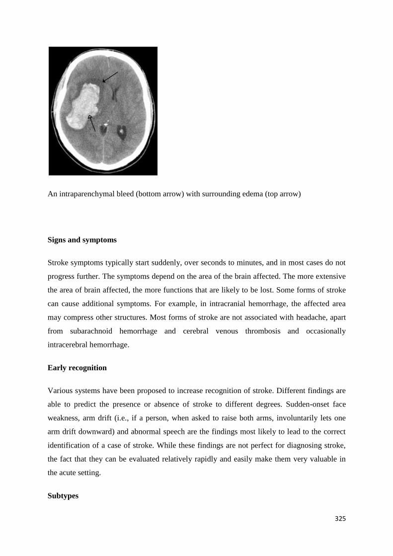
325
An intraparenchymal bleed (bottom arrow) with surrounding edema (top arrow)
Signs and symptoms
Stroke symptoms typically start suddenly, over seconds to minutes, and in most cases do not
progress further. The symptoms depend on the area of the brain affected. The more extensive
the area of brain affected, the more functions that are likely to be lost. Some forms of stroke
can cause additional symptoms. For example, in intracranial hemorrhage, the affected area
may compress other structures. Most forms of stroke are not associated with headache, apart
from subarachnoid hemorrhage and cerebral venous thrombosis and occasionally
intracerebral hemorrhage.
Early recognition
Various systems have been proposed to increase recognition of stroke. Different findings are
able to predict the presence or absence of stroke to different degrees. Sudden-onset face
weakness, arm drift (i.e., if a person, when asked to raise both arms, involuntarily lets one
arm drift downward) and abnormal speech are the findings most likely to lead to the correct
identification of a case of stroke. While these findings are not perfect for diagnosing stroke,
the fact that they can be evaluated relatively rapidly and easily make them very valuable in
the acute setting.
Subtypes

326
If the area of the brain affected contains one of the three prominent central nervous system
pathways—the spinothalamic tract, corticospinal tract, and dorsal column (medial lemniscus),
symptoms may include:
hemiplegia and muscle weakness of the face.
numbness
reduction in sensory or vibratory sensation
initial flaccidity (hypotonicity), replaced by spasticity (hypertonicity), hyperreflexia,
and obligatory synergies.
Hemiplegia means severe weakness of the limbs on one side of the body but the specific
features can vary tremendously from person to person. Problems may include:
Difficulty with gait
Difficulty with balance while standing or walking
Having difficulty with motor activities like holding, grasping or pinching
Increasing stiffness of muscles
Muscle spasticity.
The majority of children who develop hemiplegia also have abnormal mental
development
Behavior problems like anxiety, anger, irritability, lack of concentration or
comprehension
Emotions — depression
Shoulder pain — Often associated with a loss of external rotation of the glenohumeral
joint, commonly due to the increased tone of the Subscapularis muscle and Pectoralis
major muscle.
Shoulder Subluxation,
In most cases, the symptoms affect only one side of the body (unilateral). Depending on the
part of the brain affected, the defect in the brain is usually on the opposite side of the body.
However, since these pathways also travel in the spinal cord and any lesion there can also
produce these symptoms, the presence of any one of these symptoms does not necessarily
indicate a stroke.
327
In addition to the above CNS pathways, the brainstem gives rise to most of the twelve cranial
nerves. A stroke affecting the brain stem and brain therefore can produce symptoms relating
to deficits in these cranial nerves:
altered smell, taste, hearing, or vision (total or partial)
drooping of eyelid (ptosis) and weakness of ocular muscles
decreased reflexes: gag, swallow, pupil reactivity to light
decreased sensation and muscle weakness of the face
balance problems and nystagmus
altered breathing and heart rate
weakness in sternocleidomastoid muscle with inability to turn head to one side
weakness in tongue (inability to protrude and/or move from side to side)
If the cerebral cortex is involved, the CNS pathways can again be affected, but also can
produce the following symptoms:
aphasia (difficulty with verbal expression, auditory comprehension, reading and/or
writing Broca's or Wernicke's area typically involved)
dysarthria (motor speech disorder resulting from neurological injury)
apraxia (altered voluntary movements)
visual field defect
memory deficits (involvement of temporal lobe)
hemineglect (involvement of parietal lobe)
disorganized thinking, confusion, hypersexual gestures (with involvement of frontal
lobe)
lack of insight of his or her, usually stroke-related, disability
If the cerebellum is involved, the patient may have the following:
altered walking gait
altered movement coordination
vertigo and or disequilibrium
Associated symptoms
328
Loss of consciousness, headache, and vomiting usually occurs more often in hemorrhagic
stroke than in thrombosis because of the increased intracranial pressure from the leaking
blood compressing the brain.
If symptoms are maximal at onset, the cause is more likely to be a subarachnoid hemorrhage
or an embolic stroke.
Causes
Thrombotic stroke:
In thrombotic stroke a thrombus (blood clot) usually forms around atherosclerotic plaques.
Since blockage of the artery is gradual, onset of symptomatic thrombotic strokes is slower. A
thrombus itself (even if non-occluding) can lead to an embolic stroke ,if the thrombus breaks
off, at which point it is called an "embolus." Two types of thrombosis can cause stroke:
Large vessel disease involves the common and internal carotids, vertebral, and the
Circle of Willis. Diseases that may form thrombi in the large vessels include (in
descending incidence): atherosclerosis, vasoconstriction (tightening of the artery),
aortic, carotid or vertebral artery dissection, various inflammatory diseases of the
blood vessel wall (Takayasu arteritis, giant cell arteritis, vasculitis), noninflammatory
vasculopathy, Moyamoya disease and fibromuscular dysplasia.
Small vessel disease involves the smaller arteries inside the brain: branches of the
circle of Willis, middle cerebral artery, stem, and arteries arising from the distal
vertebral and basilar artery. Diseases that may form thrombi in the small vessels
include (in descending incidence): lipohyalinosis (build-up of fatty hyaline matter in
the blood vessel as a result of high blood pressure and aging) and fibrinoid
degeneration (stroke involving these vessels are known as lacunar infarcts) and
microatheroma (small atherosclerotic plaques).
Sickle-cell anemia, which can cause blood cells to clump up and block blood vessels, can
also lead to stroke. A stroke is the second leading killer of people under 20 who suffer from
sickle-cell anemia.
329
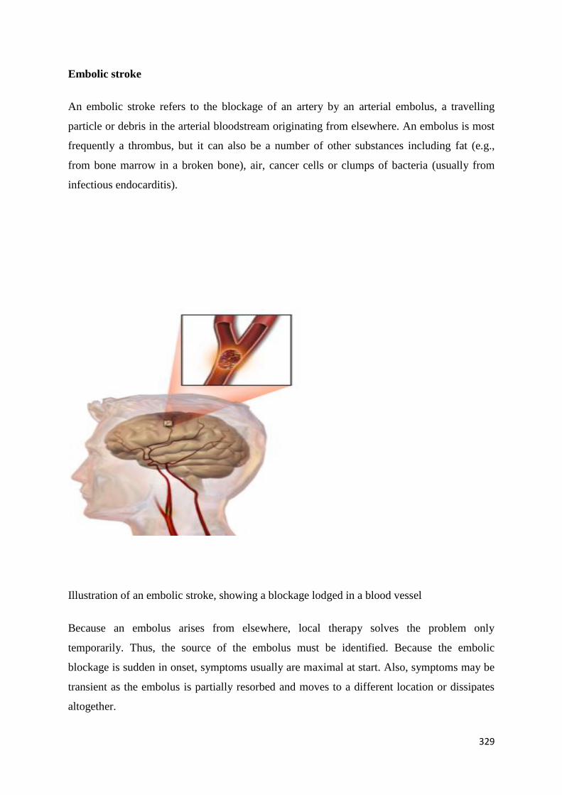
Embolic stroke
An embolic stroke refers to the blockage of an artery by an arterial embolus, a travelling
particle or debris in the arterial bloodstream originating from elsewhere. An embolus is most
frequently a thrombus, but it can also be a number of other substances including fat (e.g.,
from bone marrow in a broken bone), air, cancer cells or clumps of bacteria (usually from
infectious endocarditis).
Illustration of an embolic stroke, showing a blockage lodged in a blood vessel
Because an embolus arises from elsewhere, local therapy solves the problem only
temporarily. Thus, the source of the embolus must be identified. Because the embolic
blockage is sudden in onset, symptoms usually are maximal at start. Also, symptoms may be
transient as the embolus is partially resorbed and moves to a different location or dissipates
altogether.
330
Emboli most commonly arise from the heart (especially in atrial fibrillation) but may
originate from elsewhere in the arterial tree. In paradoxical embolism, a deep vein thrombosis
embolises through an atrial or ventricular septal defect in the heart into the brain.
Cardiac causes can be distinguished between high and low-risk:
High risk: atrial fibrillation and paroxysmal atrial fibrillation, rheumatic disease of the
mitral or aortic valve disease, artificial heart valves, known cardiac thrombus of the
atrium or ventricle, sick sinus syndrome, sustained atrial flutter, recent myocardial
infarction, chronic myocardial infarction together with ejection fraction <28 percent,
symptomatic congestive heart failure with ejection fraction <30 percent, dilated
cardiomyopathy, Libman-Sacks endocarditis, Marantic endocarditis, infective
endocarditis, papillary fibroelastoma, left atrial myxoma and coronary artery bypass
graft (CABG) surgery.
Low risk/potential: calcification of the annulus (ring) of the mitral valve, patent
foramen ovale (PFO), atrial septal aneurysm, atrial septal aneurysm with patent
foramen ovale, left ventricular aneurysm without thrombus, isolated left atrial
"smoke" on echocardiography (no mitral stenosis or atrial fibrillation), complex
atheroma in the ascending aorta or proximal arch.
Systemic hypoperfusion
Systemic hypoperfusion is the reduction of blood flow to all parts of the body. It is most
commonly due to heart failure from cardiac arrest or arrhythmias, or from reduced cardiac
output as a result of myocardial infarction, pulmonary embolism, pericardial effusion, or
bleeding. Hypoxemia (low blood oxygen content) may precipitate the hypoperfusion.
Because the reduction in blood flow is global, all parts of the brain may be affected,
especially "watershed" areas - border zone regions supplied by the major cerebral arteries. A
watershed stroke refers to the condition when blood supply to these areas is compromised.
Blood flow to these areas does not necessarily stop, but instead it may lessen to the point
where brain damage can occur.
Venous thrombosis
331
Cerebral venous sinus thrombosis leads to stroke due to locally increased venous pressure,
which exceeds the pressure generated by the arteries. Infarcts are more likely to undergo
hemorrhagic transformation (leaking of blood into the damaged area) than other types of
ischemic stroke.
Intracerebral hemorrhage
It generally occurs in small arteries or arterioles and is commonly due to hypertension,
intracranial vascular malformations (including cavernous angiomas or arteriovenous
malformations), cerebral amyloid angiopathy, or infarcts into which secondary haemorrhage
has occurred. Other potential causes are trauma, bleeding disorders, amyloid angiopathy,
illicit drug use (e.g., amphetamines or cocaine). The hematoma enlarges until pressure from
surrounding tissue limits its growth, or until it decompresses by emptying into the ventricular
system, CSF or the pial surface. A third of intracerebral bleed is into the brain's ventricles.
ICH has a mortality rate of 44 percent after 30 days, higher than ischemic stroke or
subarachnoid hemorrhage (which technically may also be classified as a type of stroke).
Silent stroke
A silent stroke is a stroke that does not have any outward symptoms, and the patients are
typically unaware they have suffered a stroke. Despite not causing identifiable symptoms, a
silent stroke still causes damage to the brain, and places the patient at increased risk for both
transient ischemic attack and major stroke in the future. Conversely, those who have suffered
a major stroke are also at risk of having silent strokes. In a broad study in 1998, more than 11
million people were estimated to have experienced a stroke in the United States.
Approximately 770,000 of these strokes were symptomatic and 11 million were first-ever
silent MRI infarcts or hemorrhages. Silent strokes typically cause lesions which are detected
via the use of neuroimaging such as MRI. Silent strokes are estimated to occur at five times
the rate of symptomatic strokes. The risk of silent stroke increases with age, but may also
affect younger adults and children, especially those with acute anemia.
Pathophysiology:
Ischemic:

332
Ischemic stroke occurs because of a loss of blood supply to part of the brain, initiating the
ischemic cascade. Brain tissue ceases to function if deprived of oxygen for more than 60 to
90 seconds, and after approximately three hours will suffer irreversible injury possibly
leading to death of the tissue, i.e., infarction. (This is why fibrinolytics such as alteplase are
given only until three hours since the onset of the stroke.) Atherosclerosis may disrupt the
blood supply by narrowing the lumen of blood vessels leading to a reduction of blood flow,
by causing the formation of blood clots within the vessel, or by releasing showers of small
emboli through the disintegration of atherosclerotic plaques. Embolic infarction occurs when
emboli formed elsewhere in the circulatory system, typically in the heart as a consequence of
atrial fibrillation, or in the carotid arteries, break off, enter the cerebral circulation, then lodge
in and occlude brain blood vessels. Since blood vessels in the brain are now occluded, the
brain becomes low in energy, and thus it resorts into using anaerobic metabolism within the
region of brain tissue affected by ischemia. Unfortunately, this kind of metabolism produces
less adenosine triphosphate (ATP) but releases a by-product called lactic acid. Lactic acid is
an irritant which could potentially destroy cells since it is an acid and disrupts the normal
acid-base balance in the brain. The ischemia area is referred to as the "ischemic penumbra".
Then, as oxygen or glucose becomes depleted in ischemic brain tissue, the production of high
energy phosphate compounds such as adenosine triphosphate (ATP) fails, leading to failure
of energy-dependent processes (such as ion pumping) necessary for tissue cell survival. This
sets off a series of interrelated events that result in cellular injury and death. A major cause of
neuronal injury is release of the excitatory neurotransmitter glutamate. The concentration of
glutamate outside the cells of the nervous system is normally kept low by so-called uptake
carriers, which are powered by the concentration gradients of ions (mainly Na+) across the
cell membrane. However, stroke cuts off the supply of oxygen and glucose which powers the
ion pumps maintaining these gradients. As a result the transmembrane ion gradients run
down, and glutamate transporters reverse their direction, releasing glutamate into the
extracellular space. Glutamate acts on receptors in nerve cells (especially NMDA receptors),
producing an influx of calcium which activates enzymes that digest the cells' proteins, lipids
and nuclear material. Calcium influx can also lead to the failure of mitochondria, which can
lead further toward energy depletion and may trigger cell death due to apoptosis.
Ischemia also induces production of oxygen free radicals and other reactive oxygen species.
These react with and damage a number of cellular and extracellular elements. Damage to the
333
blood vessel lining or endothelium is particularly important. In fact, many antioxidant
neuroprotectants such as uric acid and NXY-059 work at the level of the endothelium and not
in the brain per se. Free radicals also directly initiate elements of the apoptosis cascade by
means of redox signaling.
These processes are the same for any type of ischemic tissue and are referred to collectively
as the ischemic cascade. However, brain tissue is especially vulnerable to ischemia since it
has little respiratory reserve and is completely dependent on aerobic metabolism, unlike most
other organs.
In addition to injurious effects on brain cells, ischemia and infarction can result in loss of
structural integrity of brain tissue and blood vessels, partly through the release of matrix
metalloproteases, which are zinc- and calcium-dependent enzymes that break down collagen,
hyaluronic acid, and other elements of connective tissue. Other proteases also contribute to
this process. The loss of vascular structural integrity results in a breakdown of the protective
blood brain barrier that contributes to cerebral edema, which can cause secondary progression
of the brain injury.
Hemorrhagic
Hemorrhagic strokes arise from a number of possible brain lesions, mostly vascular in
nature.They result in tissue injury by causing compression of tissue from an expanding
hematoma or hematomas. This can distort and injure tissue. In addition, the pressure may
lead to a loss of blood supply to affected tissue with resulting infarction, and the blood
released by brain hemorrhage appears to have direct toxic effects on brain tissue and
vasculature. Inflammation contributes to the secondary brain injury after hemorrhage.
Diagnosis:
Stroke is diagnosed through several techniques: a neurological examination ,CT scans (most
often without contrast enhancements) or MRI scans, Doppler ultrasound, and arteriography.
The diagnosis of stroke itself is clinical, with assistance from the imaging techniques.
Imaging techniques also assist in determining the subtypes and cause of stroke. There is yet
334
no commonly used blood test for the stroke diagnosis itself, though blood tests may be of
help in finding out the likely cause of stroke.
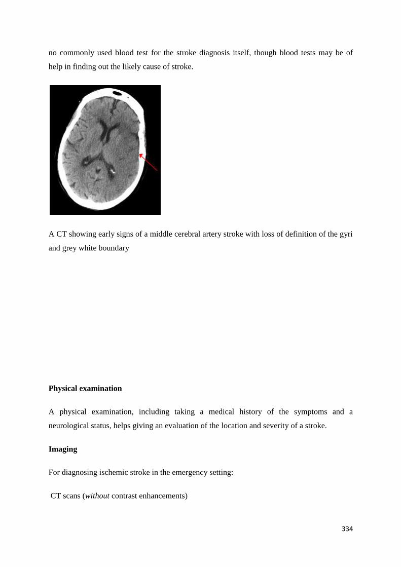
A CT showing early signs of a middle cerebral artery stroke with loss of definition of the gyri
and grey white boundary
Physical examination
A physical examination, including taking a medical history of the symptoms and a
neurological status, helps giving an evaluation of the location and severity of a stroke.
Imaging
For diagnosing ischemic stroke in the emergency setting:
CT scans (without contrast enhancements)
335
sensitivity= 16%
specificity= 96%
MRI scan
sensitivity= 83%
specificity= 98%
For diagnosing hemorrhagic stroke in the emergency setting:
CT scans (without contrast enhancements)
sensitivity= 89%
specificity= 100%
MRI scan
sensitivity= 81%
specificity= 100%
For detecting chronic hemorrhages, MRI scan is more sensitive.
For the assessment of stable stroke, nuclear medicine scans SPECT and PET/CT may be
helpful. SPECT documents cerebral blood flow and PET with FDG isotope the metabolic
activity of the neurons.
Underlying cause
336
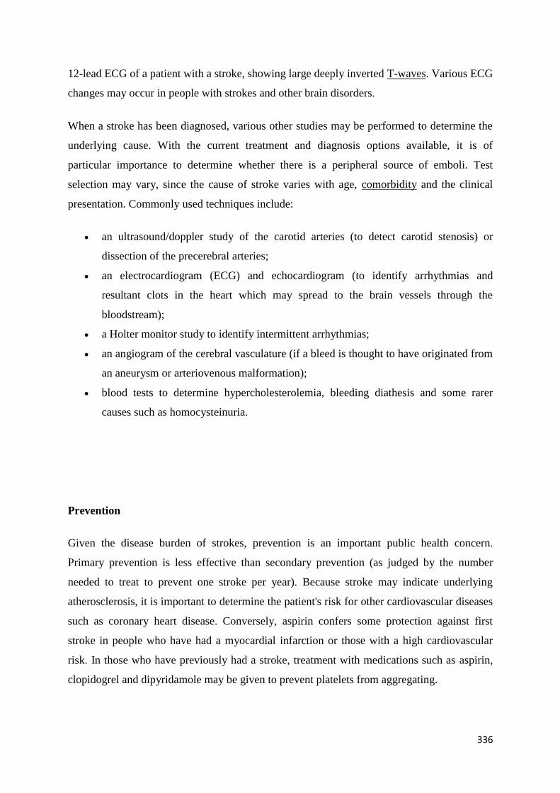
12-lead ECG of a patient with a stroke, showing large deeply inverted T-waves. Various ECG
changes may occur in people with strokes and other brain disorders.
When a stroke has been diagnosed, various other studies may be performed to determine the
underlying cause. With the current treatment and diagnosis options available, it is of
particular importance to determine whether there is a peripheral source of emboli. Test
selection may vary, since the cause of stroke varies with age, comorbidity and the clinical
presentation. Commonly used techniques include:
an ultrasound/doppler study of the carotid arteries (to detect carotid stenosis) or
dissection of the precerebral arteries;
an electrocardiogram (ECG) and echocardiogram (to identify arrhythmias and
resultant clots in the heart which may spread to the brain vessels through the
bloodstream);
a Holter monitor study to identify intermittent arrhythmias;
an angiogram of the cerebral vasculature (if a bleed is thought to have originated from
an aneurysm or arteriovenous malformation);
blood tests to determine hypercholesterolemia, bleeding diathesis and some rarer
causes such as homocysteinuria.
Prevention
Given the disease burden of strokes, prevention is an important public health concern.
Primary prevention is less effective than secondary prevention (as judged by the number
needed to treat to prevent one stroke per year). Because stroke may indicate underlying
atherosclerosis, it is important to determine the patient's risk for other cardiovascular diseases
such as coronary heart disease. Conversely, aspirin confers some protection against first
stroke in people who have had a myocardial infarction or those with a high cardiovascular
risk. In those who have previously had a stroke, treatment with medications such as aspirin,
clopidogrel and dipyridamole may be given to prevent platelets from aggregating.
337
Risk factors
The most important modifiable risk factors for stroke are high blood pressure and atrial
fibrillation .Other modifiable risk factors include high blood cholesterol levels, diabetes,
cigarette smoking (active and passive), heavy alcohol consumption and drug use, lack of
physical activity, obesity, processed red meat consumption and unhealthy diet. Alcohol use
could predispose to ischemic stroke, and intracerebral and subarachnoid hemorrhage via
multiple mechanisms (for example via hypertension, atrial fibrillation, rebound
thrombocytosis and platelet aggregation and clotting disturbances). The drugs most
commonly associated with stroke are cocaine, amphetamines causing hemorrhagic stroke, but
also over-the-counter cough and cold drugs containing sympathomimetics.
No high quality studies have shown the effectiveness of interventions aimed at weight
reduction, promotion of regular exercise, reducing alcohol consumption or smoking
cessation. Nonetheless, given the large body of circumstantial evidence, best medical
management for stroke includes advice on diet, exercise, smoking and alcohol use.
Medication or drug therapy is the most common method of stroke prevention; carotid
endarterectomy can be a useful surgical method of preventing stroke.
Blood pressure
Hypertension (high blood pressure) accounts for 35-50% of stroke risk. Blood pressure
reduction of 10 mmHg systolic or 5 mmHg diastolic reduces the risk of stroke by ~40%.
Lowering blood pressure has been conclusively shown to prevent both ischemic and
hemorrhagic strokes. It is equally important in secondary prevention. Even patients older than
80 years and those with isolated systolic hypertension benefit from antihypertensive therapy.
The available evidence does not show large differences in stroke prevention between
antihypertensive drugs —therefore, other factors such as protection against other forms of
cardiovascular disease should be considered .
Atrial fibrillation
Those with atrial fibrillation have a 5% a year risk of stroke, and this risk is higher in those
with valvular atrial fibrillation. Depending on the stroke risk, anticoagulation with
medications such as warfarin or aspirin is warranted for stroke prevention.
338
Blood lipids
High cholesterol levels have been inconsistently associated with (ischemic) stroke.[61][70]
Statins have been shown to reduce the risk of stroke by about 15%.[71]
Since earlier meta-
analyses of other lipid-lowering drugs did not show a decreased risk,[72]
statins might exert
their effect through mechanisms other than their lipid-lowering effects.[71]
Diabetes mellitus
Diabetes mellitus increases the risk of stroke by 2 to 3 times. While intensive control of blood
sugar has been shown to reduce microvascular complications such as nephropathy and
retinopathy it has not been shown to reduce macrovascular complications such as stroke.
Anticoagulation drugs
Oral anticoagulants such as warfarin have been the mainstay of stroke prevention for over 50
years. However, several studies have shown that aspirin and antiplatelet drugs are highly
effective in secondary prevention after a stroke or transient ischemic attack. Low doses of
aspirin (for example 75–150 mg) are as effective as high doses but have fewer side effects;
the lowest effective dose remains unknown. Thienopyridines (clopidogrel, ticlopidine) "might
be slightly more effective" than aspirin and have a decreased risk of gastrointestinal bleeding,
but they are more expensive. Their exact role remains controversial. Ticlopidine has more
skin rash, diarrhea, neutropenia and thrombotic thrombocytopenic purpura. Dipyridamole can
be added to aspirin therapy to provide a small additional benefit, even though headache is a
common side effect. Low-dose aspirin is also effective for stroke prevention after sustaining a
myocardial infarction. Except for in atrial fibrillation, oral anticoagulants are not advised for
stroke prevention —any benefit is offset by bleeding risk.
In primary prevention however, antiplatelet drugs did not reduce the risk of ischemic stroke
while increasing the risk of major bleeding. Further studies are needed to investigate a
possible protective effect of aspirin against ischemic stroke in women.
Surgery
Carotid endarterectomy or carotid angioplasty can be used to remove atherosclerotic
narrowing (stenosis) of the carotid artery. There is evidence supporting this procedure in

339
selected cases. Endarterectomy for a significant stenosis has been shown to be useful in the
prevention of further strokes in those who have already had one. Carotid artery stenting has
not been shown to be equally useful. Patients are selected for surgery based on age, gender,
degree of stenosis, time since symptoms and patients' preferences. Surgery is most efficient
when not delayed too long —the risk of recurrent stroke in a patient who has a 50% or greater
stenosis is up to 20% after 5 years, but endarterectomy reduces this risk to around 5%.
Screening for carotid artery narrowing has not been shown to be a useful screening test in the
general population. Studies of surgical intervention for carotid artery stenosis without
symptoms have shown only a small decrease in the risk of stroke. To be beneficial, the
complication rate of the surgery should be kept below 4%. Even then, for 100 surgeries, 5
patients will benefit by avoiding stroke, 3 will develop stroke despite surgery, 3 will develop
stroke or die due to the surgery itself, and 89 will remain stroke-free but would also have
done so without intervention.
Diet
Nutrition, specifically the Mediterranean-style diet, has the potential for decreasing the risk of
having a stroke by more than half. It does not appear that lowering levels of homocysteine
with folic acid affects the risk of stroke.
Women
A number of specific recommendation have been made for women including: taking aspirin
after the 11th week of pregnancy if there is a history of previous chronic high blood pressure,
blood pressure medications in pregnancy if the blood pressure is greater than 150 mmHg
systolic or greater than 100 mmHg diastolic. In those who have previously had preeclampsia
other risk factors should be treated more aggressively.
Secondary prevention
Anticoagulation can prevent recurrent ischemic strokes. Among patients with nonvalvular
atrial fibrillation, anticoagulation can reduce stroke by 60% while antiplatelet agents can
reduce stroke by 20%.However, a recent meta-analysis suggests harm from anti-coagulation
started early after an embolic stroke. The most widely used anticoagulant to prevent
thromboembolic stroke in patients with nonvalvular atrial fibrillation is the oral agent
340
warfarin while dabigatran is a new alternative which does not require prothrombin time
monitoring.
If studies show carotid stenosis, and the patient has residual function in the affected side,
carotid endarterectomy (surgical removal of the stenosis) may decrease the risk of recurrence
if performed rapidly after stroke.
Management of stroke:
Ischemic stroke
Definitive therapy is aimed at removing the blockage by breaking the clot down
(thrombolysis), or by removing it mechanically (thrombectomy). The philosophical premise
underlying the importance of rapid stroke intervention was crystallized as Time is Brain! in
the early 1990s. Years later, that same idea, that rapid cerebral blood flow restoration results
in fewer brain cells dying, has been proved and quantified.
Thrombolysis
Thrombolysis with recombinant tissue plasminogen activator (rtPA) in acute ischemic stroke,
when given before three hours of symptom onset increases the risk of death in the short term
but in the long term improves the rate of independence; the change in long term mortality is
not significant. When broken down by time to treatment it increases the chance of being alive
and living independently by 9% in those treated within three hours, however the benefit for
those treated between three and six hours is not significant. These benefits or lack of benefits
occurred regardless of the age of the person treated. There is no reliable way to determine
who will have an intracranial hemorrhage post treatment versus who will not.
Its use is endorsed by the American Heart Association and the American Academy of
Neurology as the recommended treatment for acute stroke within three hours of onset of
symptoms as long as there are not other contraindications (such as abnormal lab values, high
blood pressure, or recent surgery). When administered within the first three hours
341
thrombolysis improves functional outcome without affecting mortality. Intra-arterial
fibrinolysis, where a catheter is passed up an artery into the brain and the medication is
injected at the site of thrombosis, has been found to improve outcomes in people with acute
ischemic stroke.
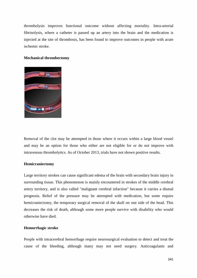
Mechanical thrombectomy
Removal of the clot may be attempted in those where it occurs within a large blood vessel
and may be an option for those who either are not eligible for or do not improve with
intravenous thrombolytics. As of October 2013, trials have not shown positive results.
Hemicraniectomy
Large territory strokes can cause significant edema of the brain with secondary brain injury in
surrounding tissue. This phenomenon is mainly encountered in strokes of the middle cerebral
artery territory, and is also called "malignant cerebral infaction" because it carries a dismal
prognosis. Relief of the pressure may be attempted with medication, but some require
hemicraniectomy, the temporary surgical removal of the skull on one side of the head. This
decreases the risk of death, although some more people survive with disability who would
otherwise have died.
Hemorrhagic stroke
People with intracerebral hemorrhage require neurosurgical evaluation to detect and treat the
cause of the bleeding, although many may not need surgery. Anticoagulants and
342
antithrombotics, key in treating ischemic stroke, can make bleeding worse. People are
monitored for changes in the level of consciousness, and their blood pressure, blood sugar,
and oxygenation are kept at optimum levels.[citation needed]
Stroke unit
Ideally, people who have had a stroke are admitted to a "stroke unit", a ward or dedicated
area in hospital staffed by nurses and therapists with experience in stroke treatment. It has
been shown that people admitted to a stroke unit have a higher chance of surviving than those
admitted elsewhere in hospital, even if they are being cared for by doctors without experience
in stroke. When an acute stroke is suspected by history and physical examination, the goal of
early assessment is to determine the cause. Treatment varies according to the underlying
cause of the stroke, thromboembolic (ischemic) or hemorrhagic. Good nursing care is
fundamental in maintaining skin care, feeding, hydration, positioning, and monitoring vital
signs such as temperature, pulse, and blood pressure.
Physiotherapy in the Acute stage:
Treatment of the adult with Hemiplegia, following stroke starts from the very acute stage,
when the patient in the ICU or Hospital bed. Treatment should be based on assessment by the
relevant health professionals, including physiotherapists, Physicians and occupational
therapists. Muscles with severe motor impairment including weakness need these therapists
to assist them with specific exercises.
The Physiotherapist as a treatment team member in the stroke unit aims the followings in
this stage:
Maintenance anti -spastic/ anti-synergistic posture (RIP).
Maintenance of Range of motion.
Maintenance of airways and facilitation of breathing.
Maintenance of muscle properties and strength.
Stimulation of upper limb and oro- facial function.
343
Facilitation of bed mobility such as lying to sitting.
Facilitation of early sitting.
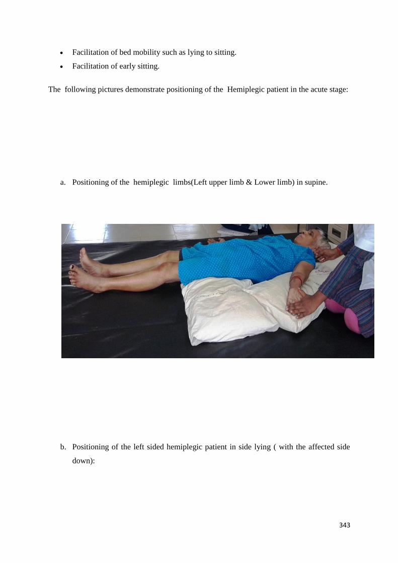
The following pictures demonstrate positioning of the Hemiplegic patient in the acute stage:
a. Positioning of the hemiplegic limbs(Left upper limb & Lower limb) in supine.
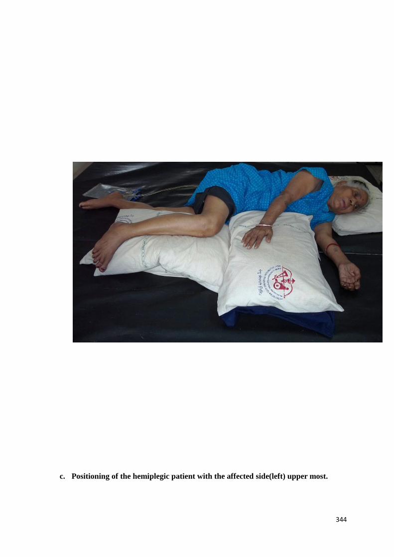
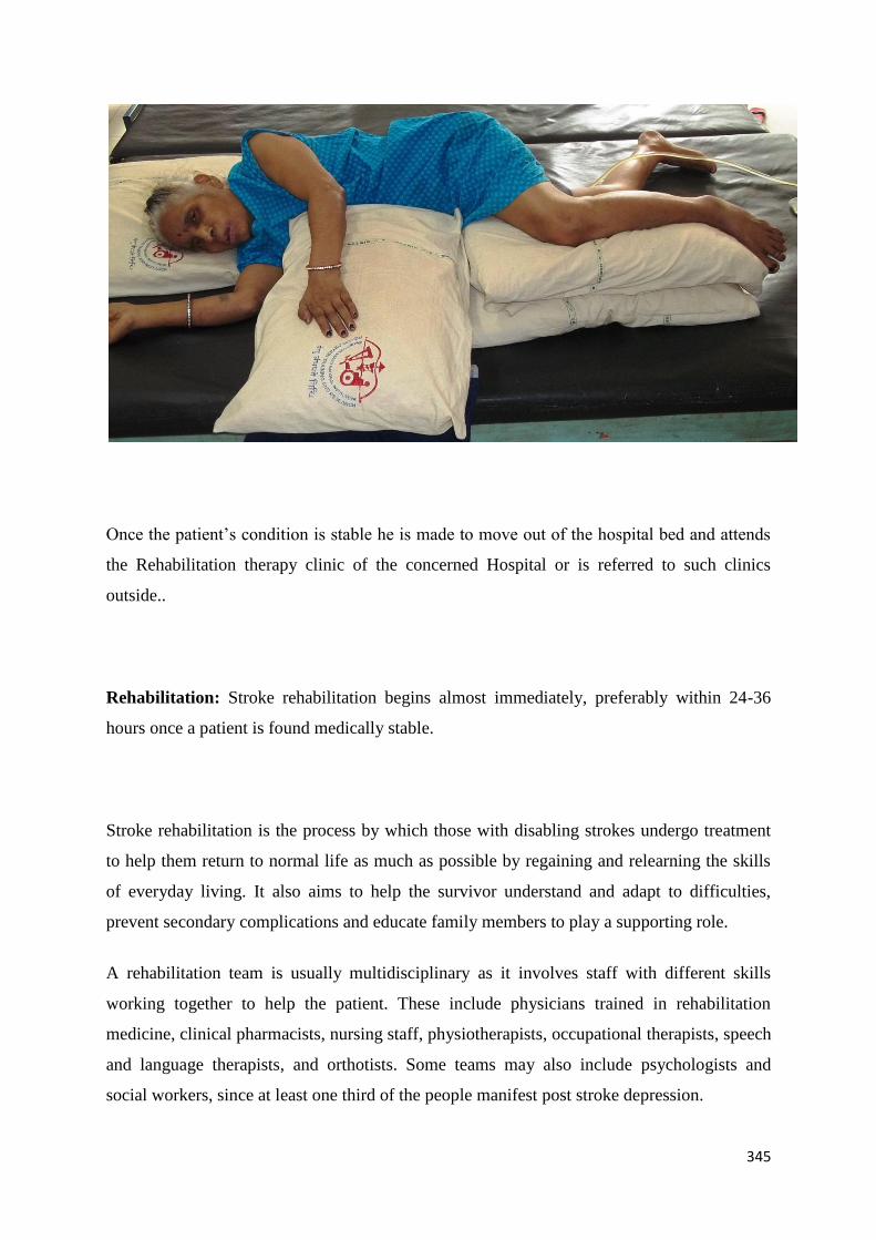
b. Positioning of the left sided hemiplegic patient in side lying ( with the affected side
down):
344
c. Positioning of the hemiplegic patient with the affected side(left) upper most.
345
Once the patient’s condition is stable he is made to move out of the hospital bed and attends
the Rehabilitation therapy clinic of the concerned Hospital or is referred to such clinics
outside..
Rehabilitation: Stroke rehabilitation begins almost immediately, preferably within 24-36
hours once a patient is found medically stable.
Stroke rehabilitation is the process by which those with disabling strokes undergo treatment
to help them return to normal life as much as possible by regaining and relearning the skills
of everyday living. It also aims to help the survivor understand and adapt to difficulties,
prevent secondary complications and educate family members to play a supporting role.
A rehabilitation team is usually multidisciplinary as it involves staff with different skills
working together to help the patient. These include physicians trained in rehabilitation
medicine, clinical pharmacists, nursing staff, physiotherapists, occupational therapists, speech
and language therapists, and orthotists. Some teams may also include psychologists and
social workers, since at least one third of the people manifest post stroke depression.

346
For most people with stroke, physical therapy (PT), occupational therapy (OT) and speech-
language pathology (SLP) are the cornerstones of the rehabilitation process. Often, assistive
technology such as wheelchairs, walkers and canes may be beneficial. Many mobility
problems can be improved by the use of ankle foot orthoses.
The Physiotherapist focuses on joint range of motion and strength by performing exercises
and re-learning functional tasks such as bed mobility, transferring, walking and other gross
motor functions. Physiotherapists can also work with patients to improve awareness and use
of the hemiplegic side. Rehabilitation involves working on the ability to produce strong
movements or the ability to perform tasks using normal patterns. Emphasis is often
concentrated on functional tasks and patient’s goals. One example physiotherapists employ to
promote motor learning involves constraint-induced movement therapy. Through continuous
practice the patient relearns to use and adapt the hemiplegic limb during functional activities
to create lasting permanent changes.
Physiotherapy in the Recovery/ Rehabilitation phase:
A. Assesment:
Before delivering Physical therapy, the Therapist assesses the patient’s tone, muscle power ,
range of motion, tightness /contracture and deformities and functional abilities. Therapist
gives more emphasis on patients functionl limitation and try to find the cause which is
responsible for the functional limitation. Therapist asks patients to perform all the bed
mobility,supine to sit ,sit to stand and finally walking. There are a variety of standardized
assessment scales available to physiotherapists and other health care professionals for use in
the ongoing evaluation of the status of a patient’s Hemiplegia. The use of standardized
assessment scales may help physiotherapists and other health care professionals during the
course of their treatment plant to:
Prioritize treatment interventions based on specific identifiable motor and sensory
deficits
347
Create appropriate short and long term goals for treatment based on the outcome of
the scales, their professional expertise and the desires of the patient
Evaluate the potential burden of care and monitor any changes based on either
improving or declining scores
Some of the most commonly used scales in the assessment of hemiplegia are:
The Fugl-Meyer Assessment of Physical Performance (FMA)
The FMA is often used as a measure of functional or physical impairment following a
cerebrovascular accident(CVA). It measures sensory and motor impairment of the upper and
lower extremities, balance in several positions, range of motion, and pain. This test is a
reliable and valid measure in measuring post-stroke impairments related to stroke recovery. A
lower score in each component of the test indicates higher impairment and a lower functional
level for that area. The maximum score for each component is 66 for the upper extremities,
34 for the lower extremities, and 14 for balance. Administration of the FMA should be done
after reviewing a training manual.
The Chedoke-McMaster Stroke Assessment (CMSA)
This test is a reliable measure of two separate components evaluating both motor impairment
and disability. The disability component assesses any changes in physical function including
gross motor function and walking ability. The disability inventory can have a maximum score
of 100 with 70 from the gross motor index and 30 from the walking index. Each task in this
inventory has a maximum score of seven except for the 2 minute walk test which is out of
two. The impairment component of the test evaluates the upper and lower extremities,
postural control and pain. The impairment inventory focuses on the seven stages of recovery
from stroke from flaccid paralysis to normal motor functioning. A training workshop is
recommended if the measure is being utilized for the purpose of data collection.
The Stroke Rehabilitation Assessment of Movement (STREAM)
The STREAM consists of 30 test items involving upper-limb movements, lower-limb
movements, and basic mobility items. It is a clinical measure of voluntary movements and
general mobility (rolling, bridging, sit-to-stand, standing, stepping, walking and stairs)
following a stroke. The voluntary movement part of the assessment is measured using a 3-
348
point ordinal scale (unable to perform, partial performance, and complete performance) and
the mobility part of the assessment uses a 4-point ordinal scale (unable, partial, complete with
aid, complete no aid). The maximum score one can receive on the STREAM is a 70 (20 for
each limb score and 30 for mobility score). The higher the score, the better movement and
mobility is available for the individual being scored.
B. Intervention:
Physical therapy (PT) can help improve muscle strength & coordination, mobility (such as
standing and walking), and other physical function using different sensorimotor techniques.
Physiotherapists can also help reduce shoulder pain by maintaining shoulder range of motion,
as well as using Functional electrical stimulation. Supportive devices, such as braces or
slings, can be used to help prevent or treat shoulder subluxation, in the hopes to minimize
disability and pain. It should be noted that although many individuals suffering from stroke
experience both shoulder pain and shoulder subluxation, the two are mutually exclusive. A
treatment method that can be implemented with the goal of helping to regain motor function
in the affected limb is constraint-induced movement therapy. This consists of constraining the
unaffected limb, forcing the affected limb to accomplish tasks of daily living.
Physiotherapy
– Conventional therapies.
– Neurophysiological therapies.
a. Conventional therapies(Therapeutic Exercises,Traditional Functional Retraining)
Range Of Motion (ROM) Exercises
Muscle Strengthening Exercises
Mobilization activities
Fitness training
349
Compensatory Techniques
b. Neurophysiological Approaches:
1. Muscle Re-education Approach.
2. Neurodevelopmental Approaches:
– Sensorimotor Approach (Rood, 1940S)
– Movement Therapy Approach (Brunnstrom, 1950S)
– NDT Approach (Bobath, 1960-70S)
– PNF Approach (Knot and Voss,1960-70S)
3. Motor Relearning Program for Stroke (1980S)
4. Contemporary Task Oriented Approach (1990S)
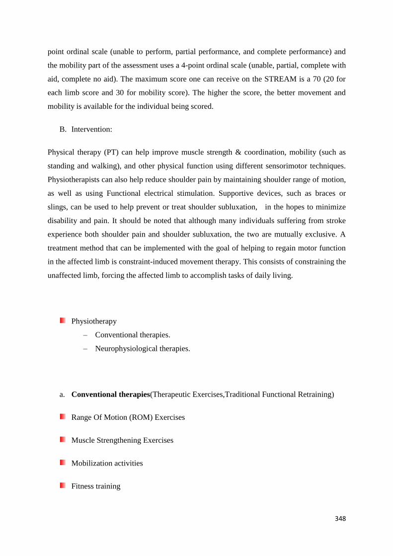
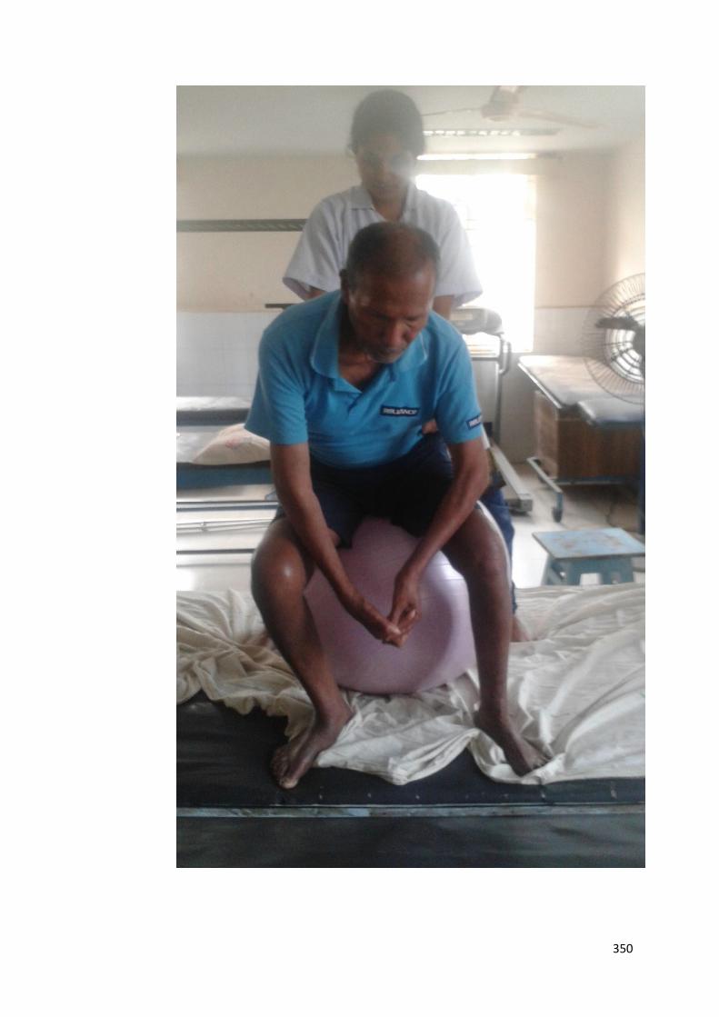
The following pictures demonstrate some of the techniques followed by the therapist to
improve function in Hemiplegic patients.
A. THERAPEUTIC ACTIVITIES TO IMPROVE TRUNK STABILITY.
350
351

352

353
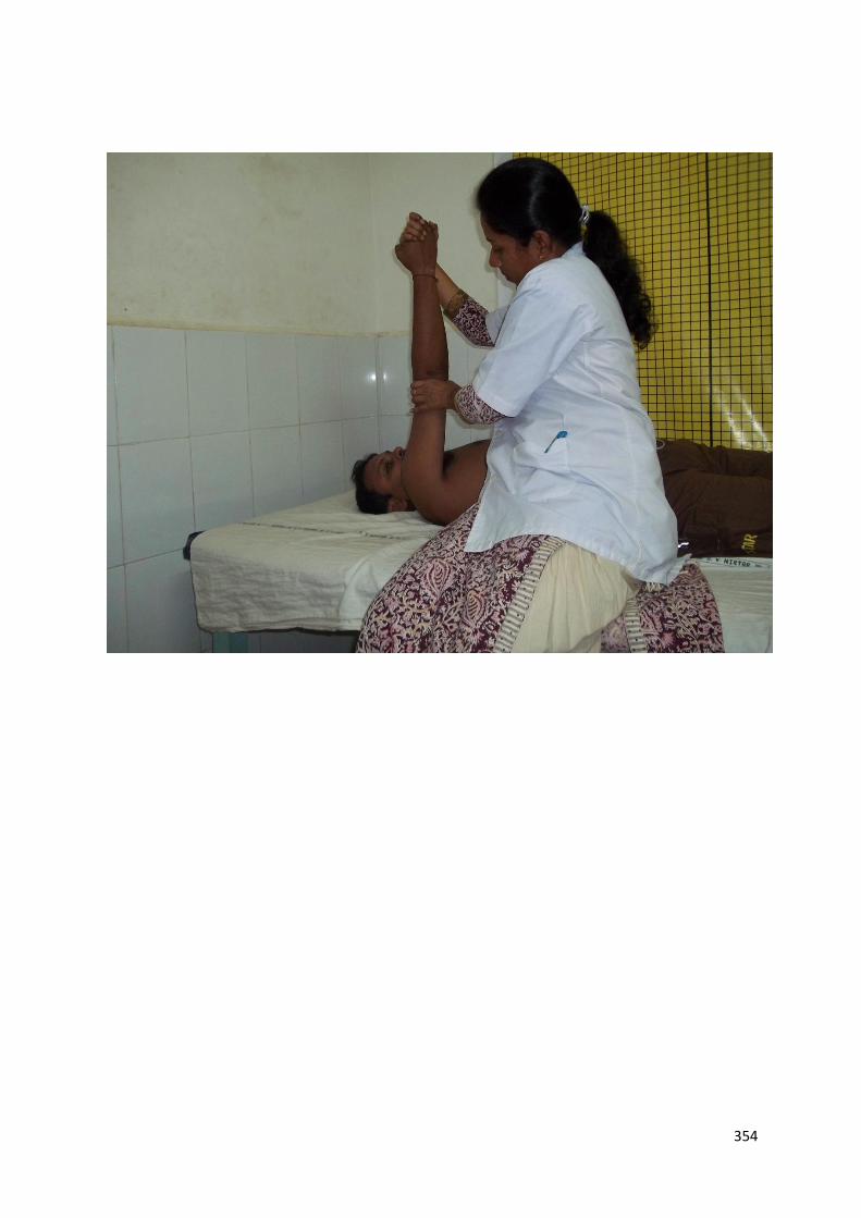
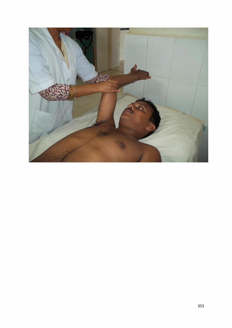
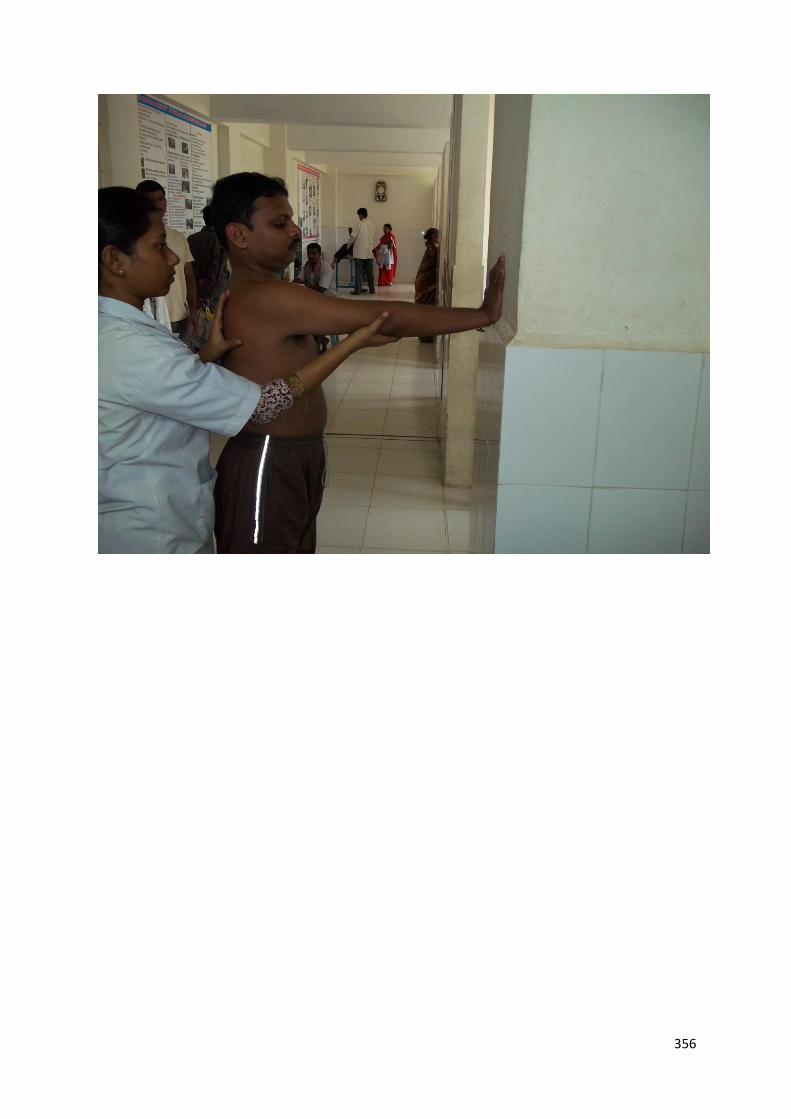
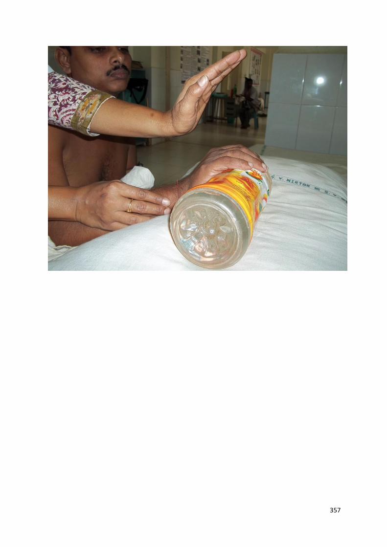
B. THERAPEUTIC ACTIVITIES TO IMPROVE UPPER LIMB FUNCTION.
354
355
356
357
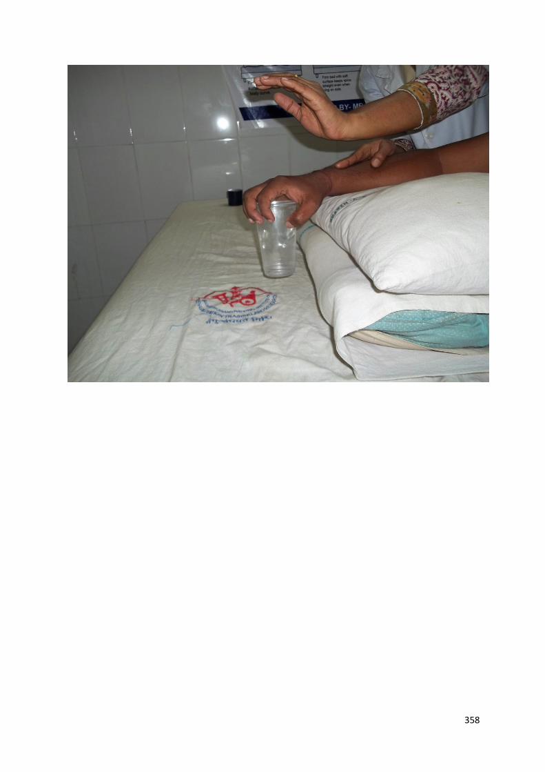
358
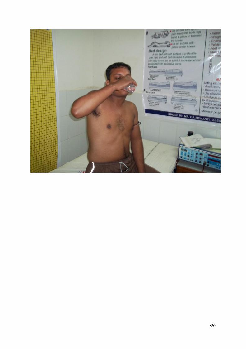
359
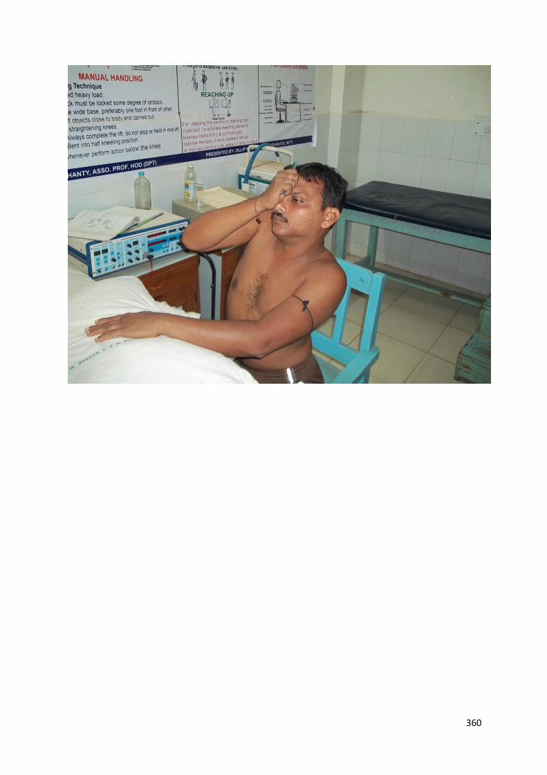
360
361
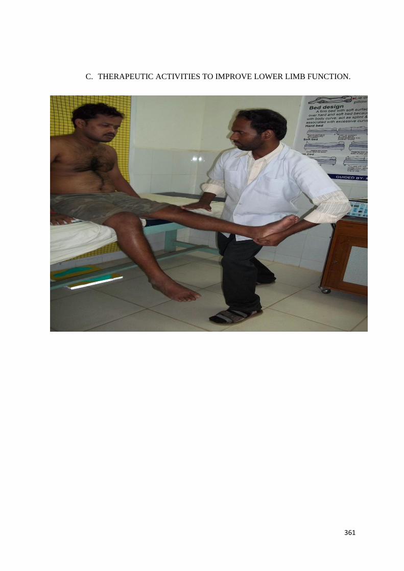
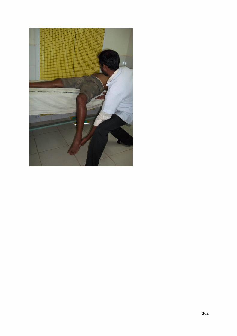
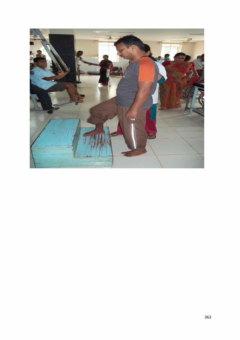
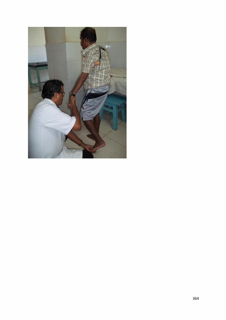
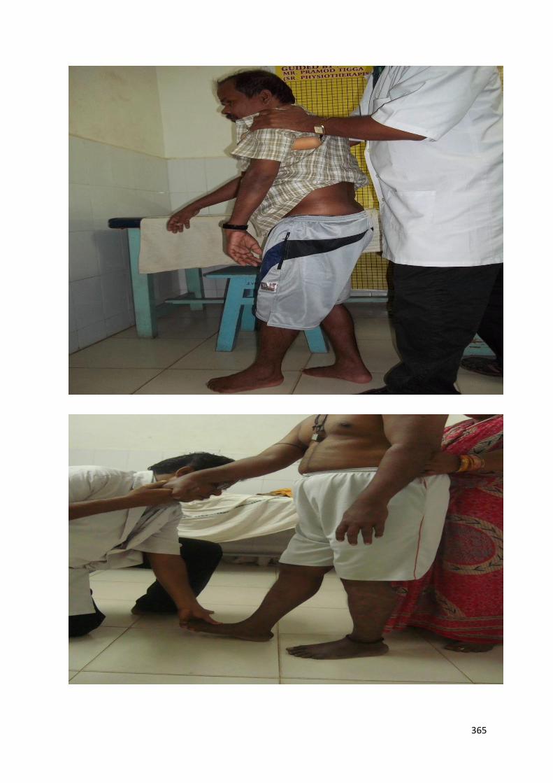
C. THERAPEUTIC ACTIVITIES TO IMPROVE LOWER LIMB FUNCTION.
362
363
364
365
366
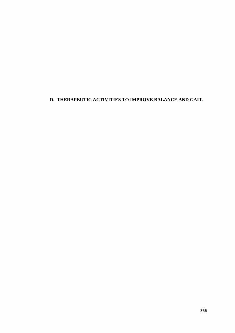
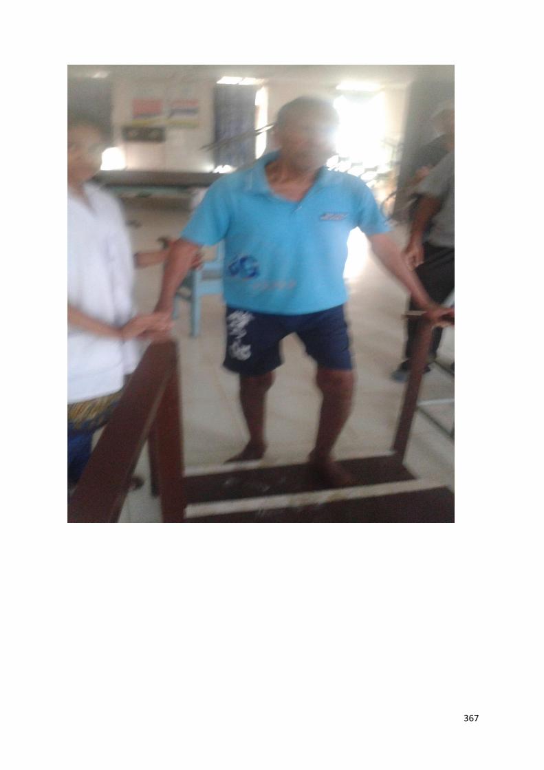
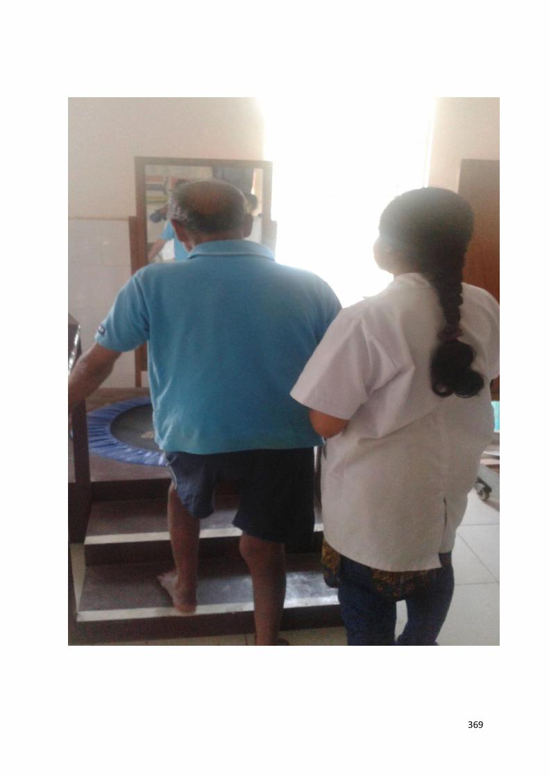
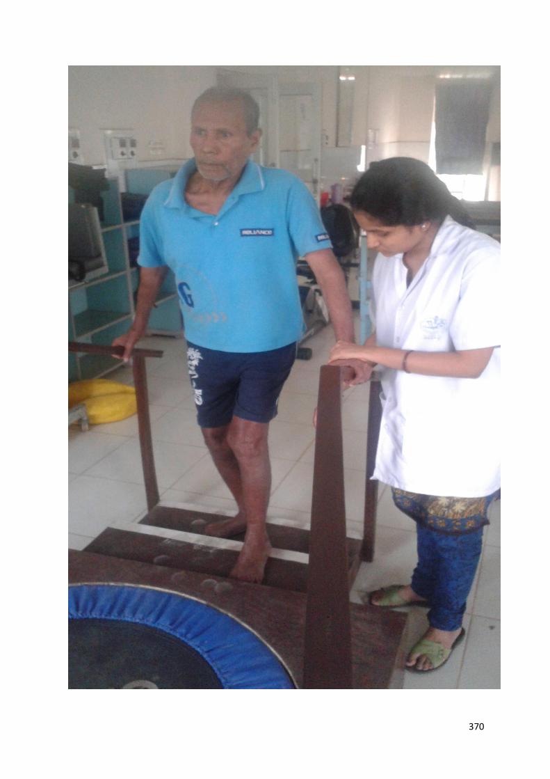
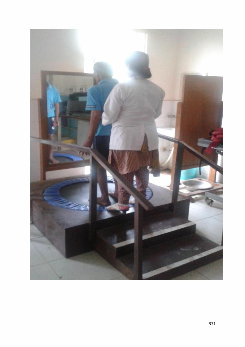
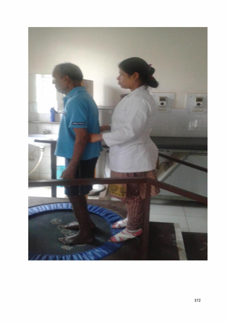
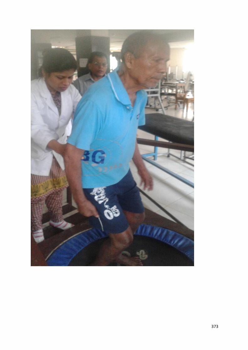
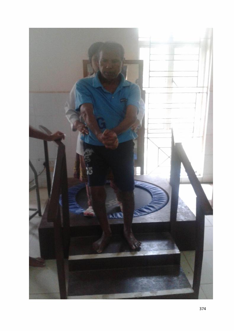
D. THERAPEUTIC ACTIVITIES TO IMPROVE BALANCE AND GAIT.
367

368
369
370
371
372
373
374
375
Physiotherapy for Shoulder pain:
Electrical stimulation
Shoulder strapping/ Shoulder Orthosis
Mobilization (esp. External rotator, abduction) to prevent frozen shoulder.
Modalities : ice, heat, massage
Strengthening exercise for the rotator cuff muscles and the muscles in the various
shoulder force couples.
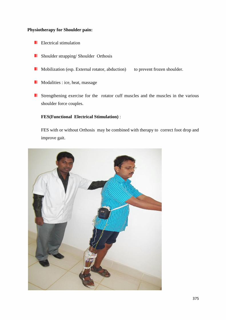
FES(Functional Electrical Stimulation) :
FES with or without Orthosis may be combined with therapy to correct foot drop and
improve gait.
376
Hemiplegic patient walking with FES and Orthosis.
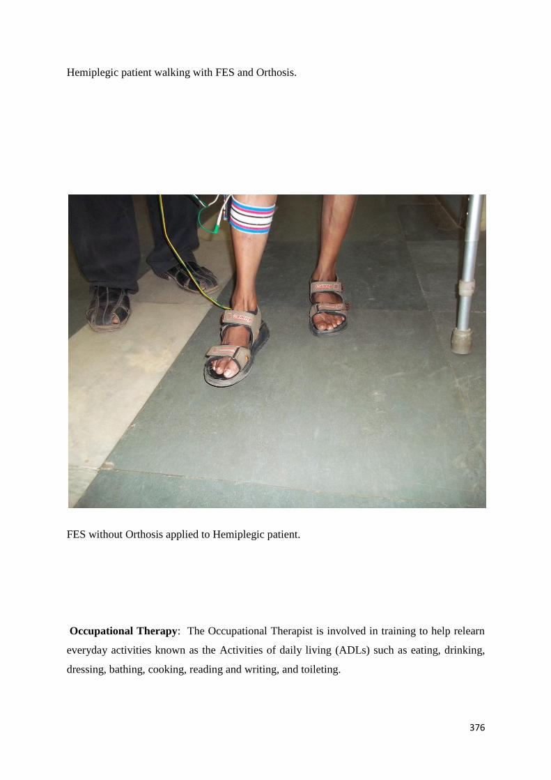
FES without Orthosis applied to Hemiplegic patient.
Occupational Therapy: The Occupational Therapist is involved in training to help relearn
everyday activities known as the Activities of daily living (ADLs) such as eating, drinking,
dressing, bathing, cooking, reading and writing, and toileting.
377
Speech and language therapy: This is appropriate for patients with the speech production
disorders: dysarthria and apraxia of speech, aphasia, cognitive-communication impairments
and/or dysphagia (problems with swallowing).
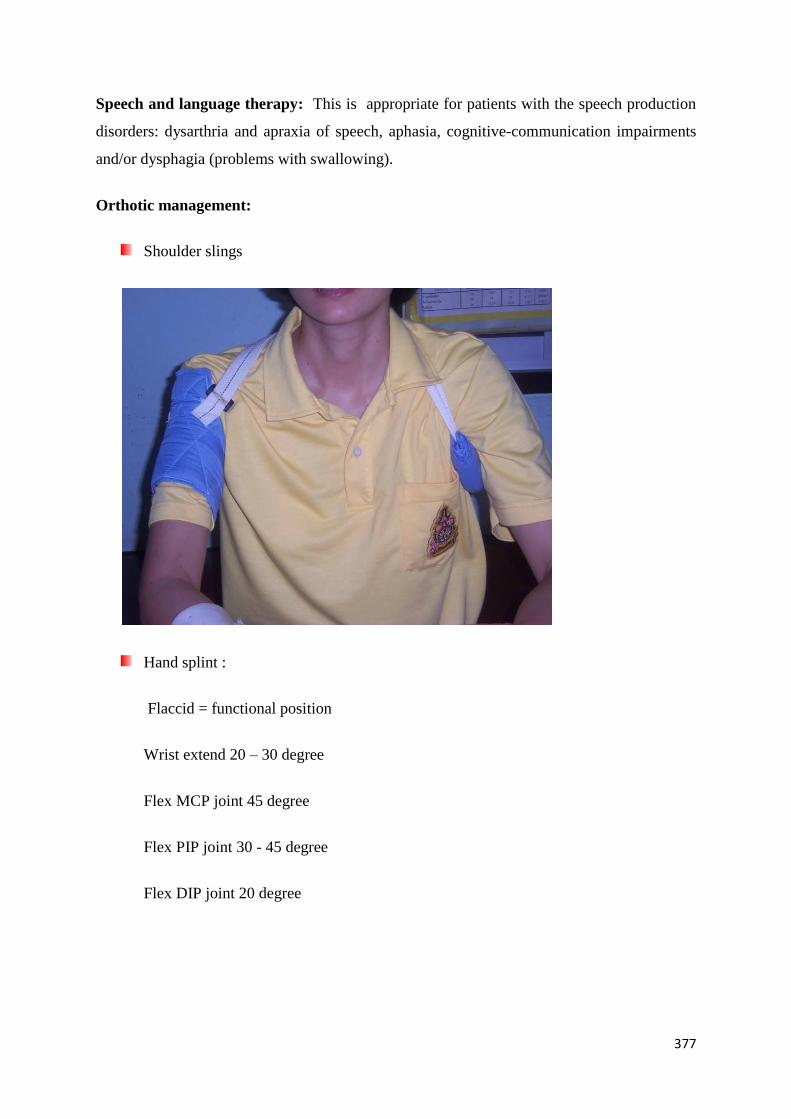
Orthotic management:
Shoulder slings
Hand splint :
Flaccid = functional position
Wrist extend 20 – 30 degree
Flex MCP joint 45 degree
Flex PIP joint 30 - 45 degree
Flex DIP joint 20 degree

378
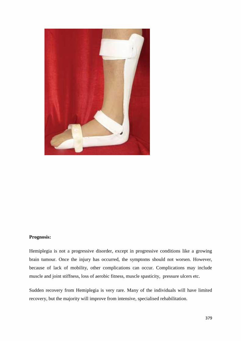
Foot slings:
Ankle foot orthosis :
379
Prognosis:
Hemiplegia is not a progressive disorder, except in progressive conditions like a growing
brain tumour. Once the injury has occurred, the symptoms should not worsen. However,
because of lack of mobility, other complications can occur. Complications may include
muscle and joint stiffness, loss of aerobic fitness, muscle spasticity, pressure ulcers etc.
Sudden recovery from Hemiplegia is very rare. Many of the individuals will have limited
recovery, but the majority will improve from intensive, specialised rehabilitation.
380
Rise beyond limitations with Occupational therapy
Rebuilding faith with OT
Undoing Paralysis with OT
Story : Mr. A, a 40 year old right handed , happy go lucky man, working as a sales man at a
departmental store, whose life was moving smoothly until a day when he suffered a (brain)
stroke, resulting in the paralysis of one side of his body (hemiplegia). That black day, his life
took a U- turn. The aftermath of stroke was felt once he was declared medically stable. As the
famous proverb saying ―only the bearer knows where the shoe pinches‖, Mr. A had to bear
the brunt of not being able to move the right side of his body. His limbs felt heavy and started
getting tight. Rolling in bed, moving his right upper and lower limbs were not possible for
him. He became dependent for the basic activities of life (ADL) like brushing, bathing,
toileting and even dressing. Sitting, while keeping an upright posture, was difficult for him
and so were standing and walking. Basically, he became dependent for almost everything.
This resulted in a situation where he stopped going to his job, meeting people and preferred
staying alone at home and not talking to others around.
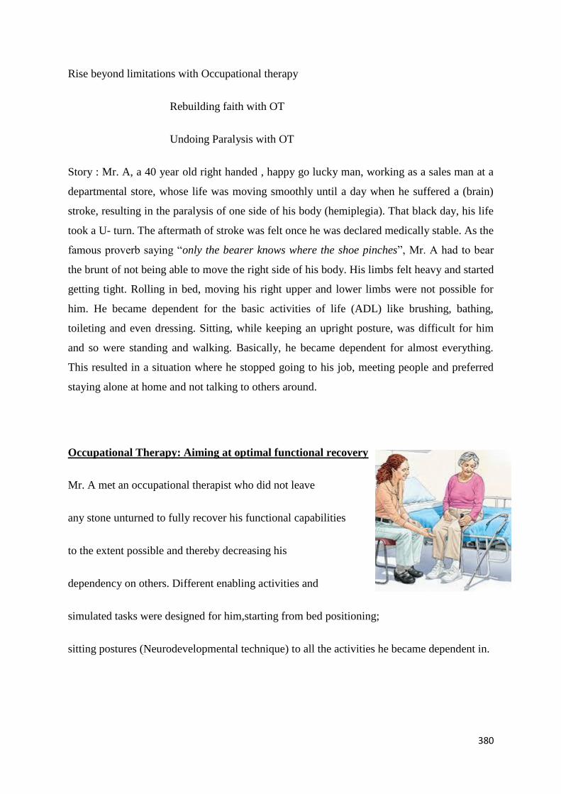
Occupational Therapy: Aiming at optimal functional recovery
Mr. A met an occupational therapist who did not leave
any stone unturned to fully recover his functional capabilities
to the extent possible and thereby decreasing his
dependency on others. Different enabling activities and
simulated tasks were designed for him,starting from bed positioning;
sitting postures (Neurodevelopmental technique) to all the activities he became dependent in.
381
Weight bearingactivities (exerting pressure over different joints helping in maintaining
normal length of muscles) were started at the earliest. Shoulder wheel, pulleys, static cycling
etc. were given only after it was found to be suitable for Mr. A’s physical status. All these
activities and tasks were based on myriad therapeutic approaches, to name a few, such
asNeurophysiological techniques (PROPRIOCEPTIVE NEUROMUSCULAR
FACILITATION, BRUNNSTROM APPROACH), task oriented approaches(MOTOR
LEARNING), PERSON-ENVIRONMENT-OCCUPATION model etc.
For example, in bathing, he was advised to use long handled sponge for rubbing and a towel
holder (which fixes the towel at one end and the person holds the other end in the functioning
hand) wiping the body.
Such simple modifications made wonders in his life.
He was taught one handed dressing techniques which helped him in donning and doffing
clothes, wearing socks and shoes. He was made to practice reaching and manipulating
activities with the help of objects used in daily living, like glass, boxes and jars etc.
Neurodevelopmental and Roods facilitatory and inhibitory Techniques were used for
reducing tightness in the muscles which helped in optimizing the tone and therefore helping
him gain his maximum functional status.
For restoring balance and gait training, various activities like floor ladder, step markers,
parallel bars, ergometer, correkta were given.These activities were found helpful as they were
very similar to real life situation, therefore, reducing the fear of falls in him.
Along with the ongoing treatment, splinting and orthotic devices were provided for
enhancing and maintaining functional status.
He was advised small changes at his working area like keeping the frequently used articles
within an arm reach. He was advised to keep non skid mats on the floor, at his home as well
as his office which further reduced any chances of imbalance.
Other simple architectural modifications suggested like encouraging using a western style
toilet instead of the conventional Indian style or simply using a cut out chair ( whichever was
found feasible for Mr.A) with grab bars installed around helping him to get up and down

382
safely. Another important precaution suggested was always to keep the area obstacle free and
well lighted.
With his active participation in Occupational Therapy Intervention along with proper
education and counselling, he was ready to be integrated back to the community and job as he
gradually regained his confidenceand he was asked to follow up after every three months.
Now, after a year of treatment, he has satisfactorily regained his previous role with minor
deficits.
Feed a man a fish, and you feed him for a day. Teach a man how to fish, and you feed him
for a lifetime.
A Chinese saying by Anne Isabella Thackeray
Ritchie(1837-1919) in her novel Mrs. Dymond