Embed Size (px)
Citation preview

Structure
Review
Structural Analysis of Supramolecular Assembliesby Cryo-Electron Tomography
Jan Harapin,1 Matthias Eibauer,1 and Ohad Medalia1,2,*1Department of Biochemistry, University of Zurich, Winterthurerstrasse 190, 8057 Zurich, Switzerland2Department of Life Sciences and the National Institute for Biotechnology in the Negev, Ben-Gurion University, Beer-Sheva, 84105 Israel*Correspondence: [email protected]://dx.doi.org/10.1016/j.str.2013.08.003
Structural analysis of macromolecular assemblies in their physiological environment is a challenging taskthat is instrumental in answering fundamental questions in cellular and molecular structural biology. Thecontinuous development of computational and analytical tools for cryo-electron tomography (cryo-ET)enables the study of these assemblies at a resolution of a few nanometers. Through the implementation ofthinning procedures, cryo-ET can nowbe applied to the reconstruction ofmacromolecular structures locatedinside thick regions of vitrified cells and tissues, thus becoming a central tool for structural determinations invarious biological disciplines. Here, we focus on the successful in situ applications of cryo-ET to reveal struc-tures of macromolecular complexes within eukaryotic cells.
Macromolecular assemblies are central players in many cellular
processes that influence the dynamic intracellular architecture of
cells. For instance, cytoskeletal processes occur within fractions
of a second, resulting in major remodeling of the cytoplasm and
the overall cellular morphology (Diez et al., 2005). Various exper-
imental tools for analyzing multicomponent macromolecular
structures inside cells have been developed in order to advance
the current understanding of functional interactions and dynamic
properties of different cellular processes. More specifically,
advances in imaging and image analysis techniques have
enabled the structural analysis of macromolecular assemblies
to reach resolutions on the previously unattainable nanometer
scale, thus providing novel insights into the local and global or-
ganization of functional modules and cellular networks (Fridman
et al., 2012; Luci�c et al., 2008).
Cryo-electron tomography (cryo-ET) has a pivotal role in
cellular biology (Al-Amoudi et al., 2007; Maimon et al., 2012;
Patla et al., 2010), microbiology (Dobro et al., 2013; Kurner
et al., 2005; Lieber et al., 2009; Swulius et al., 2011) and virology
(Bharat et al., 2012; Meyerson et al., 2011). It can depict a
particular cellular scene and provide a three-dimensional (3D)
structural map of an unperturbed, vitrified sample, i.e., in a
close-to-physiological state (Fridman et al., 2012; Luci�c et al.,
2005; Yahav et al., 2011). Preserving fine and delicate structural
details in a close-to-life state are made possible by the use of
rapid freezing, which thus circumvents the deleterious effects
of chemical fixatives and dehydration on cellular ultrastructures
(Adrian et al., 1984; Dubochet et al., 1988).
In this review, we will focus on the principles and implementa-
tion of cryo-ET in the field of cellular and molecular structural
biology and discuss the recent technical advances in recon-
structing 3D structures of macromolecular complexes within
intact cells and organelles. The combination of cryo-ET and
single-particle analysis approaches will be discussed in detail
with special emphasis on their potential for increasing the final
resolution of reconstructed images. Finally, we will discuss com-
plementary sample preparation procedures that enable the
application of cryo-ET to large cells and tissues. Although we
1522 Structure 21, September 3, 2013 ª2013 Elsevier Ltd All rights r
focus on the application of these techniques to eukaryotic
systems, cryo-ET has proven itself instrumental for reconstruct-
ing molecular structures in prokaryotes, yielding impressive
results (Abrusci et al., 2013; Briegel et al., 2012; Schlimpert
et al., 2012; Swulius et al., 2011).
Cryo-ET: Basic PrinciplesThe true power of cryo-ET lies in its ability to directly observe
macromolecular densities in situ due to phase contrast between
the biological material and the surrounding vitrified ice, bypass-
ing the use of fixatives as well as commonly used contrasting
agents such as heavy metal salts (Medalia et al., 2002). Thus,
the initial step in preparing biological samples for cryo-ET is vitri-
fication, which is typically performed by plunging into liquid-ni-
trogen-cooled liquid ethane or high-pressure freezing (HPF) to
ensure full hydration and ultrastructure preservation.
During the process of tomographic acquisition, vitrified sam-
ples of suitable thickness (<1 mm) are rotated around a defined
tilt axis in discrete increments inside the transmission electron
microscope (TEM), covering a maximal range of 140�, between
�70� and +70� (Frank, 1992). A series of two-dimensional
projections, i.e., a ‘‘tilt-series,’’ is collected under ‘‘low electron
dose’’ conditions (typically <100 e�/A2) to prevent radiation
damage to the sample (Dierksen et al., 1992, 1993). The tilt-
series is subsequently aligned to a common frame using fiducial
markers, i.e., colloidal gold of 10-15 nm in diameter (Amat et al.,
2008), or using cross-correlation-based strategies (Castano-
Dıez et al., 2010; Sorzano et al., 2009). The aligned tilt-series is
then used to reconstruct the 3D volume of the specimen, namely,
a tomogram (Frank, 1992). Although the most commonly used
algorithm for tomographic reconstruction is the weighted back
projection (Radermacher, 1988), alternative algorithms, such
as algebraic reconstruction technique (ART) (Gordon et al.,
1970) and simultaneous iterative reconstruction technique
(SIRT) (Gilbert, 1972), are also in use.
The quality of the final tomogram is directly dependent on the
angular increments and the number of recorded two-dimen-
sional images (Horowitz et al., 1997). However, the inherent
eserved
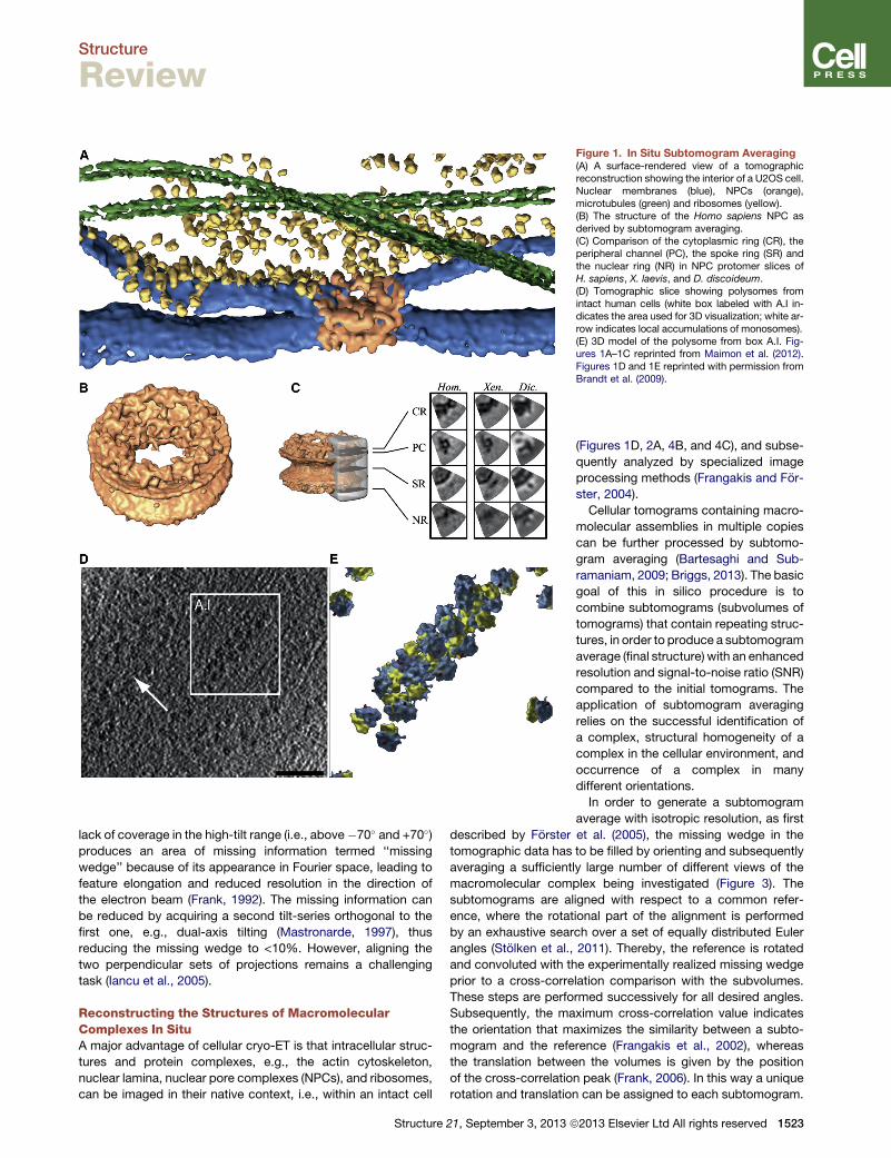
Figure 1. In Situ Subtomogram Averaging(A) A surface-rendered view of a tomographicreconstruction showing the interior of a U2OS cell.Nuclear membranes (blue), NPCs (orange),microtubules (green) and ribosomes (yellow).(B) The structure of the Homo sapiens NPC asderived by subtomogram averaging.(C) Comparison of the cytoplasmic ring (CR), theperipheral channel (PC), the spoke ring (SR) andthe nuclear ring (NR) in NPC protomer slices ofH. sapiens, X. laevis, and D. discoideum.(D) Tomographic slice showing polysomes fromintact human cells (white box labeled with A.I in-dicates the area used for 3D visualization; white ar-row indicates local accumulations of monosomes).(E) 3D model of the polysome from box A.I. Fig-ures 1A–1C reprinted from Maimon et al. (2012).Figures 1D and 1E reprinted with permission fromBrandt et al. (2009).
Structure
Review
lack of coverage in the high-tilt range (i.e., above�70� and +70�)produces an area of missing information termed ‘‘missing
wedge’’ because of its appearance in Fourier space, leading to
feature elongation and reduced resolution in the direction of
the electron beam (Frank, 1992). The missing information can
be reduced by acquiring a second tilt-series orthogonal to the
first one, e.g., dual-axis tilting (Mastronarde, 1997), thus
reducing the missing wedge to <10%. However, aligning the
two perpendicular sets of projections remains a challenging
task (Iancu et al., 2005).
Reconstructing the Structures of MacromolecularComplexes In SituA major advantage of cellular cryo-ET is that intracellular struc-
tures and protein complexes, e.g., the actin cytoskeleton,
nuclear lamina, nuclear pore complexes (NPCs), and ribosomes,
can be imaged in their native context, i.e., within an intact cell
Structure 21, September 3, 2013 ª
(Figures 1D, 2A, 4B, and 4C), and subse-
quently analyzed by specialized image
processing methods (Frangakis and For-
ster, 2004).
Cellular tomograms containing macro-
molecular assemblies in multiple copies
can be further processed by subtomo-
gram averaging (Bartesaghi and Sub-
ramaniam, 2009; Briggs, 2013). The basic
goal of this in silico procedure is to
combine subtomograms (subvolumes of
tomograms) that contain repeating struc-
tures, in order to produce a subtomogram
average (final structure) with an enhanced
resolution and signal-to-noise ratio (SNR)
compared to the initial tomograms. The
application of subtomogram averaging
relies on the successful identification of
a complex, structural homogeneity of a
complex in the cellular environment, and
occurrence of a complex in many
different orientations.
In order to generate a subtomogram
average with isotropic resolution, as first
described by Forster et al. (2005), the missing wedge in the
tomographic data has to be filled by orienting and subsequently
averaging a sufficiently large number of different views of the
macromolecular complex being investigated (Figure 3). The
subtomograms are aligned with respect to a common refer-
ence, where the rotational part of the alignment is performed
by an exhaustive search over a set of equally distributed Euler
angles (Stolken et al., 2011). Thereby, the reference is rotated
and convoluted with the experimentally realized missing wedge
prior to a cross-correlation comparison with the subvolumes.
These steps are performed successively for all desired angles.
Subsequently, the maximum cross-correlation value indicates
the orientation that maximizes the similarity between a subto-
mogram and the reference (Frangakis et al., 2002), whereas
the translation between the volumes is given by the position
of the cross-correlation peak (Frank, 2006). In this way a unique
rotation and translation can be assigned to each subtomogram.
2013 Elsevier Ltd All rights reserved 1523

Figure 2. Cryo-ET of Cytoskeletal Elements(A and B) A 10 nm tomographic slice through afocal adhesion site (A) and a correspondingrendered view of the cellular volume (B). Mem-branes (blue), actin (red) and adhesion-relatedparticles (green).(C and D) A 10 nm tomographic slice through aspread nuclear membrane of X. laevis ectopicallyexpressing ce-lamins (C) and a correspondingrendered view of the tomographic volume (D).Lamin filaments (yellow), NPCs (red). Figures 2Aand 2B reprinted from Patla et al. (2010); Figures2C and 2D reprinted from Grossman et al. (2012b).
Structure
Review
Next, the subtomograms are transformed according to the
found parameters, and the aligned stack of volumes is aver-
aged and serves as the reference for the next step. This iterative
procedure is performed until the structure no longer changes
with subsequent iterations. Finally, the iteratively refined trans-
formations are used to generate the final subtomogram average
(Walz et al., 1997). Obviously, the inverse of the calculated rota-
tions describe the original orientations of the macromolecular
complexes in the cellular environment. This information can
be useful for unveiling the spatial relationship between macro-
molecules in a physiological assembly such as polysomes
(Brandt et al., 2009, 2010) (Figures 1D and 1E). Because of a
high value of defocus used in cellular cryo-ET, typically in the
range of �6 to �15 mm, the maximal resolution of a subtomo-
gram average is around 3–5 nm. In order to extend the informa-
tion beyond the first destructive electron interference, a
contrast transfer function (CTF) correction can be applied to
the projection images, as it is routinely used in single-particle
analysis electron microscopy (van Heel et al., 2000). Here, the
resulting CTF-corrected tomograms serve as input data for sub-
tomogram averaging.
Two major challenges in applying the CTF correction to tomo-
graphic data sets should be resolved. First, the effective value of
defocus cannot be accurately determined in an image of thick
cells, because of the low electron-dose used to obtain each indi-
vidual projection. Second, the effective value of defocus varies
throughout the entire projection, because of the tilting of the
1524 Structure 21, September 3, 2013 ª2013 Elsevier Ltd All rights reserved
sample (Philippsen et al., 2007). In order
to tackle these challenges, an approach
called strip-based periodogram aver-
aging was developed by Fernandez
et al. (2006). The problem of defocus
determination can be solved by dividing
a projection into subimageswith constant
values of defocus. These are located par-
allel to the tilt axis when the tilt-series is
eucentric and no major jumps in the
beam direction occur. Depending on the
tilt angle, the target region for the extrac-
tion of the subimages has to be adjusted
with a precision threshold. For example,
one can use the entire 0� projection im-
age because it has a constant value of
defocus over the entire field of view. In
contrast, only a small region parallel to
the tilt axis of the projections acquired
at the highest tilt angles can be used for strip-based periodo-
gram averaging. Once all subimages with constant defocus
from a tilt-series have been extracted, they can be averaged.
The power spectrum of this periodogram average (Fernandez
et al., 1997) has an improved SNR and the Thon rings become
more clearly visible. The average defocus value of the tilt-series
can now be determined from the periodogram average, using
standard methods (Mindell and Grigorieff, 2003). Subsequently,
the average defocus value can be used to calculate the defocus
value of each pixel in the projections using geometrical parame-
ters of the tilt-series (tilt angle and tilt axis orientation). Finally, the
CTF gradient arising from the tilting of the sample can be cor-
rected locally (Winkler et al., 2003).
Other approacheswere recently developed that use advanced
schemes to determine the effective value of defocus in a tilt-
series and to perform proper CTF correction (Eibauer et al.,
2012; Xiong et al., 2009; Zanetti et al., 2009). The CTF correction
in combination with subtomogram averaging will allow the
reconstruction of images of macromolecular complexes in situ,
reaching resolutions close to 1 nm in the future.
In recent years additional algorithms were developed in order
to tackle the challenge of subtomogram averaging, mainly in
order to accelerate the angular part of the search. Examples
include approaches that employ spherical harmonics for angular
assignments (Bartesaghi et al., 2008; Chen et al., 2013; Xu et al.,
2012) and maximum likelihood-based methods (Scheres et al.,
2009; Stolken et al., 2011), as well as integrated open-source

Figure 3. Principles of SubtomogramAveraging(A–C) A cryo-ET analysis yields a 3D-re-constructed volume (A) (shown schematically). Astack of N subtomograms of a macromolecularassembly in different orientations is extracted fromthe cryo-tomograms. These subtomograms havea very low SNR (B, upper panel), and the proteincomplex is elongated in direction of the electronbeam because of the missing wedge, which hasthe same orientation for all subtomograms. Thesubtomograms are aligned with respect to acommon reference by an exhaustive angular
search. The missing wedges of the aligned particles indicate which regions in Fourier space are sampled in the related subtomogram (B, lower panel). Finally, thealigned subtomogram stack is averaged, which improves the SNR by a factor of
ffiffiffiffi
Np
(C, upper panel). Furthermore, the resolution is isotropic, because missingregions in Fourier space are filled (C, lower panel). Figure modified from Eibauer et al. (2012).
Structure
Review
software packages for subtomogram averaging that will surely
make the procedure more accessible to a wide audience
(Castano-Dıez et al., 2012; Hrabe et al., 2012).
The Structure of the NPC as Resolved by SubtomogramAveragingA successful application of subtomogram averaging, applied to
tomograms from intact human cells, is the structural analysis
of the NPC (Maimon et al., 2012). NPCs are composed of �30
different proteins termed nucleoporins (Nups), which are
arranged as multimers containing multiple copies (Alber et al.,
2007; Cronshaw et al., 2002; Ori et al., 2013; Terry et al.,
2007). The architecture of the NPC is largely conserved between
lower and higher eukaryotes (Figure 1C), comprising a pseudo-
8-fold symmetric central framework termed the spoke complex,
a central pore of about 50 nm in diameter, and filamentous struc-
tures on the cytoplasmic and nuclear sides of the complex (Elad
et al., 2009). On the nuclear face, the NPC is found in close
interaction with the nuclear lamina, a meshwork of filamentous
protein structures and other associated proteins (Burke and
Stewart, 2013).
This humongous macromolecular assembly of over 120 MDa
fuses the outer nuclear membrane and the inner nuclear mem-
brane to form aqueous translocation channels. The NPC allows
passive diffusion of small molecules and receptor-dependent
translocation of large proteins and ribonucleoproteins (Adams
and Wente, 2013; Grossman et al., 2012a). Macromolecular
cargo usually harbors a specific nuclear localization signal
(NLS) or nuclear export signal (NES) that are recognized by trans-
port receptors, mediating cargo passage through the NPC. Re-
ceptors referred to as karyopherins chaperone cargo during
transport across the NPC by means of hydrophobic interactions
with phenylalanine-glycine-rich nucleoporin repeat domains
(FG-repeats) (Stewart, 2006; Suntharalingam and Wente, 2003).
Over the last decade, cryo-ET has become a major tool for
reconstructing the structure of the NPC by using intact nuclei
and nuclear envelopes (NE) with minimal purification steps
(Beck et al., 2004; Stoffler et al., 2003). However, variability within
the complex and deviations from its 8-fold symmetrical structure
limited the resolution to �8.5 nm. Introducing symmetry-inde-
pendent averaging procedures allowed computational compen-
sation for the deviations of individual NPC protomers from their
putative positions in an 8-fold rotational symmetric structure in
both Dictyostelium discoideum and Xenopus laevis (Beck et al.,
2007; Frenkiel-Krispin et al., 2010).
Structure
Recently, we have deployed a similar approach to recon-
structing the human NPC, using intact cells (Maimon et al.,
2012). Tomograms were acquired at thin nuclear regions within
cells, �700 nm, followed by subtomogram averaging analysis
that used the symmetry-independent averaging approach
(Beck et al., 2007). The result was a detailed structure of the
human NPC at a resolution of 6.4 nm. NPCs from three different
species were reconstructed using the very same approach at
similar resolutions (Figure 1C). All three structures converged
to a pseudo-8-fold rotational symmetric architecture with similar
values for the outer diameter (�105 nm) and the central channel
(�50 nm). However, they exhibited substantial structural differ-
ences in the cytoplasmic ring and the peripheral channels, espe-
cially between the NPCs of higher and lower eukaryotes
(Figure 1C). The structure of the spoke ring shows overall struc-
tural similarity between the NPCs of X. laevis and D. discoideum.
Furthermore, the density of the NPCs in the nuclear envelope
differs widely across these species rising up to 50 NPCs/mm2
in X. laevis and is significantly less in human cells (Maimon
et al., 2012). A more detailed structure that allows for the fine
interpretation of the spatial organization and protein composition
of the NPCs will surely arise in the future. The combined
approach of cryo-ET and 3D averaging over a data set suffi-
ciently large, acquired on a thinner sample, should allow for a
final resolution of �2 nm and make possible the reliable fitting
of individual Nup crystal structures into the final tomographic
reconstruction.
Cellular ProcessesCryo-ET has been successfully applied to the study of cytoskel-
eton-based processes (Ben-Harush et al., 2010), allowing for a
detailed description of cellular events, such as adhesion (Patla
et al., 2010), virus infection (Ibiricu et al., 2013), endocytosis
(Swulius et al., 2011), and cytokinesis (Elad et al., 2011). The
exact architecture of the actin filaments and the macromolecular
assembly of adhesion sites could not be seen using conventional
electron microscopy sample preparation methods (Medalia and
Geiger, 2010). The architecture of focal adhesionswas described
in great detail, clearly showing that the actin cytoskeleton is not
directly connected to themembrane domain of the adhesion site,
namely, the integrin (Figures 2A and 2B). Another example is the
elucidation of the intricate organizational properties of the Cae-
norhabditis elegans lamin filaments assembled in X. laevis oo-
cytes that show the wild-type filaments to be almost half the pre-
viously reported diameter (Grossman et al., 2012b) (Figure 2D).
21, September 3, 2013 ª2013 Elsevier Ltd All rights reserved 1525

Figure 4. Focused Ion Beam Milling of Vitrified Eukaryotic Samples(A) Schematic illustration of FIB milling of cells and the production of thin la-mellas for subsequent cryo-ET analysis.(B) Cryo-EM image of a 250 nm thick vitreous lamella produced by FIB millingof a U2Os cell (left) and a higher magnification TEM image indicating crowdedcytoplasm.(C) The application of cryo-FIB to a multicellular specimen is exemplified byimaging the 300 nm thick lamella produced in an earlyC. elegans embryo (left),and a 4 nm slice through a tomographic reconstruction of such a sample. N,nucleus; Mt, mitochondria; V, vesicles; ER, endoplasmatic reticulum; whitearrowheads, Ga+ ion deposits at the edges of the lamella; black arrows indi-cate, NPCs; black arrow, direction of FIB milling.
Structure
Review
Cryo-ET has several fundamental limitations that warrant
the use of additional methods in order to produce high-quality
tomograms in which molecular complexes can be identified
and localized. These issues arise mainly from limited resolution,
low SNR, and a limited field of view (only covers 3–4 mm2 of
a cell) and can be overcome, for example, by a correlative
light and electron microscopy approach (Luci�c et al., 2008) (Frid-
man et al., 2012). In such a way, one can localize specific mol-
ecules with fluorescence-based microscopy and subsequently
acquire cryo-tomograms to reveal the cellular ultrastructure at
1526 Structure 21, September 3, 2013 ª2013 Elsevier Ltd All rights r
these sites (Jun et al., 2011; Patla et al., 2010; Swulius et al.,
2011).
Another example of such limitations is the specimen thick-
ness. Samples exceeding 1 mm in thickness can barely be stud-
ied in toto and require preprocessing, such as cryo-sectioning,
before tomographic analysis. Several laboratories have devoted
substantial efforts to establishing robust thinning procedures (Al-
Amoudi et al., 2007; Bokstad et al., 2012; Marko et al., 2006).
Cryo-electron microscopy of vitrified sections (CEMOVIS) was
successfully applied to reconstructing 3D images of both pro-
karyotic and eukaryotic cells, as well as some tissues (Al-Amoudi
et al., 2003, 2004, 2005, 2007; Hsieh et al., 2002; Pierson et al.,
2011). However, the production of thick cryo-sections (300–
500 nm) remains a challenging task, and the sections are riddled
with sectioning-associated artifacts, such as physical compres-
sions, knife marks, and other distortions. These perturbations
are readily observed in the tomographic reconstructions and
are keeping this approach from widespread use. In order to be
able to apply cryo-ET to thick specimens, such as whole cells
and tissues, one must be able to produce a vitrified area of suit-
able thickness (<500 nm). Thus, the need arose for a robust so-
lution to conventional sample thinning procedures.
Focused Ion Beam Milling for Cryo-ET of Cells andTissuesFocused ion beam (FIB) technology has already been routinely
applied in the field of material sciences in order to manipulate
sample thickness and gain insight into so far inaccessible inte-
riors of complex materials (MoberlyChan and Schalek, 2007).
In biology, this technique offers a broad range of applications
for sequential imaging of plastic embedded samples at room
temperature, allowing full-range structural reconstructions (Ben-
nett et al., 2009; Bushby et al., 2011; Heymann et al., 2006). It
provides highly resolved views on membranes, up to 5 nm (Kre-
shuk et al., 2011). However, a 3D structure of macromolecular
complexes cannot be reconstructed in great detail because of
fixation, dehydration, staining with heavy metal salts, and
embedding of the specimen into resins (Bushby et al., 2011).
The application of FIB technology to biological samples under
cryogenic conditions is an emerging technology for the produc-
tion of thin, vitrified specimens for cryo-ET analysis (Marko et al.,
2006, 2007; Rigort et al., 2012; Wang et al., 2012). Biological
material applied to EM grids can be vitrified by plunge-freezing
or high-pressure freezing (HPF) and directly transferred to the
cryo-FIB without further processing or use of chemical fixatives
and stains. The sample is then thinned by a Ga+ ion beam at a
velocity sufficiently high to overcome the surface binding
energies of the sample, consequentially ejecting atoms and
leaving a thinned surface (Giannuzzi, 2005). Thus, lamellas of
various thicknesses, suitable for cryo-tomography, can be
readily produced (Figure 4A). Finally, the EM grid is transferred
into the transmission electron microscope for tomographic
analysis. High-quality tomograms of biological samples (Rigort
et al., 2012), regardless of the original cellular thickness, can
be acquired, opening up a plethora of directions for studying
the structural and functional organization of both cells and
tissues, e.g., human cells and embryos of C. elegans (Figures
4B and 4C, respectively). The quality of these tomogramsmakes
possible the application of subtomogram averaging techniques
eserved

Structure
Review
(Rigort et al., 2012) to the reconstruction of macromolecular
complexes in tissues within reach.
Several considerations have to be taken into account
throughout the procedure. In order to avoid devitrification and
surface contamination of the thinned area, the sample needs
to be kept under high-vacuum and cryogenic conditions, as
well as be physically shielded with a shutter during the transfer
between the different devices used in this kind of a setup.
Furthermore, the duration of the milling and the intensity of
the ion beam should be kept at a minimum in order to ensure
minimal heat transfer to the sample (Marko et al., 2007). The
milling of biological samples is performed relatively fast
(compared with plastic embedded samples) and at ‘‘grazing’’
angles. Thus, the effects of SEM imaging, used primarily for
finding regions of interest, and the implantation of Ga+ ions
can be considered negligible (Rigort et al., 2010). The straight-
forward control over the milling process, the possibility of
choosing from a wide variety of acceleration voltages and
doses of the FIB column, and the design of a milling strategy,
depending on the topology of the vitrified biological sample,
will ensure the widespread use of this procedure in the near
future.
Future PerspectivesCryo-ET is a pivotal tool for studying cellular architecture and
macromolecules in their native environment. In conjunction
with subtomogram averaging, it can describe the structural
architecture of macromolecular complexes in situ. Extending
the application of this method and developing novel image
processing algorithms will surely provide an optimal interface
for determining cellular structures and following biological
processes at high resolution. Sample preparation techniques,
such as the cryo-FIB, will eventually make the study of develop-
mental processes from a structural perspective possible. Macro-
molecular complexes could then be reconstructed at specific
stages of development. This will give exciting insight into the
macromolecular remodeling within complex organisms.
The achievable resolution in cryo-tomography is hampered by
the large sample thickness and the low SNR, resulting in a
restricted resolution range (2–4 nm) compared to single-particle
analysis reconstructions (Luci�c et al., 2005). Thus, hardware
developments and improvements in electron microscopy
accessories will eventually make increasing the resolution and
the SNR of cellular tomograms possible. In particular, intro-
ducing direct-electron detectors will allow the acquisition of
higher quality data (McMullan et al., 2009). This will have
profound effects on the sample integrity because higher quality
data will decrease the amount of electrons required to obtain a
given resolution, thus ensuring less irradiation of the sample.
Data collection routines will be further optimized, leading to
the acquisition of larger data sets from which critical para-
meters, such as defocus, can be reliably determined. Within
such a body of data, striking the right balance of dose, focus,
and geometry for the problem being addressed will become
less demanding. More powerful and increasingly user-friendly
software for tomographic reconstruction and subtomogram
averaging is continuously being developed, making data anal-
ysis a routine task (Castano-Dıez et al., 2012; Chen et al.,
2013; Hrabe et al., 2012; Meyerson et al., 2011).
Structure
A major challenge, at the current resolution of cellular
tomograms, remains the identification of individual constituents
within macromolecular complexes. This can be overcome
by developing new labeling techniques that would enable the
identification of specific molecules by cryo-ET. Several
approaches have been described that use genetically encoded
tags, such as metallothionein (Mercogliano and DeRosier, 2006;
Nishino et al., 2007), ‘‘miniSOG’’ (Shu et al., 2011), and APEX
(Martell et al., 2012). However, these approaches are not yet
suitable for close-to-life cryo-ET of cells and tissues. Thus,
there is a fundamental need for the design of a GFP analog
for electron microscopy that would provide a general solution
for the in situ identification of complexes whose structure is
not yet determined or which intimately interact to form large
assemblies. The development of novel labeling approaches,
the improved resolution of cryo-electron microscopy, and the
ability to reconstruct 3D volumes of tissues and cells in a
hydrated state are likely to revolutionize cellular and molecular
structural biology and our understanding of basic processes
in biology.
ACKNOWLEDGMENTS
We thank Dr. Monika Zwerger and Dr. Tanuj K. Sapra for their critical reading ofour manuscript. This work was supported by a European Research CouncilStarting Grant (243047 INCEL) and a Swiss National Science Foundation Grant(SNSF 31003A_141083/1).
REFERENCES
Abrusci, P., Vergara-Irigaray, M., Johnson, S., Beeby, M.D., Hendrixson, D.R.,Roversi, P., Friede, M.E., Deane, J.E., Jensen, G.J., Tang, C.M., and Lea, S.M.(2013). Architecture of the major component of the type III secretion systemexport apparatus. Nat. Struct. Mol. Biol. 20, 99–104.
Adams, R.L., andWente, S.R. (2013). Uncovering nuclear pore complexity withinnovation. Cell 152, 1218–1221.
Adrian, M., Dubochet, J., Lepault, J., and McDowall, A.W. (1984). Cryo-elec-tron microscopy of viruses. Nature 308, 32–36.
Al-Amoudi, A., Dubochet, J., Gnaegi, H., Luthi, W., and Studer, D. (2003). Anoscillating cryo-knife reduces cutting-induced deformation of vitreous ultrathinsections. J. Microsc. 212, 26–33.
Al-Amoudi, A., Chang, J.J., Leforestier, A., McDowall, A., Salamin, L.M.,Norlen, L.P., Richter, K., Blanc, N.S., Studer, D., and Dubochet, J. (2004).Cryo-electron microscopy of vitreous sections. EMBO J. 23, 3583–3588.
Al-Amoudi, A., Studer, D., and Dubochet, J. (2005). Cutting artefacts andcutting process in vitreous sections for cryo-electron microscopy. J. Struct.Biol. 150, 109–121.
Al-Amoudi, A., Dıez, D.C., Betts, M.J., and Frangakis, A.S. (2007). The molec-ular architecture of cadherins in native epidermal desmosomes. Nature 450,832–837.
Alber, F., Dokudovskaya, S., Veenhoff, L.M., Zhang, W., Kipper, J., Devos, D.,Suprapto, A., Karni-Schmidt, O., Williams, R., Chait, B.T., et al. (2007). Themolecular architecture of the nuclear pore complex. Nature 450, 695–701.
Amat, F., Moussavi, F., Comolli, L.R., Elidan, G., Downing, K.H., and Horowitz,M. (2008). Markov random field based automatic image alignment for electrontomography. J. Struct. Biol. 161, 260–275.
Bartesaghi, A., and Subramaniam, S. (2009). Membrane protein structuredetermination using cryo-electron tomography and 3D image averaging.Curr. Opin. Struct. Biol. 19, 402–407.
Bartesaghi, A., Sprechmann, P., Liu, J., Randall, G., Sapiro, G., and Subrama-niam, S. (2008). Classification and 3D averaging with missing wedge correc-tion in biological electron tomography. J. Struct. Biol. 162, 436–450.
21, September 3, 2013 ª2013 Elsevier Ltd All rights reserved 1527
Structure
Review
Beck, M., Forster, F., Ecke, M., Plitzko, J.M., Melchior, F., Gerisch, G., Bau-meister, W., and Medalia, O. (2004). Nuclear pore complex structure anddynamics revealed by cryoelectron tomography. Science 306, 1387–1390.
Beck, M., Luci�c, V., Forster, F., Baumeister, W., and Medalia, O. (2007). Snap-shots of nuclear pore complexes in action captured by cryo-electron tomogra-phy. Nature 449, 611–615.
Ben-Harush, K., Maimon, T., Patla, I., Villa, E., and Medalia, O. (2010). Visual-izing cellular processes at the molecular level by cryo-electron tomography.J. Cell Sci. 123, 7–12.
Bennett, A.E., Narayan, K., Shi, D., Hartnell, L.M., Gousset, K., He, H., Lowe-kamp, B.C., Yoo, T.S., Bliss, D., Freed, E.O., and Subramaniam, S. (2009). Ion-abrasion scanning electron microscopy reveals surface-connected tubularconduits in HIV-infected macrophages. PLoS Pathog. 5, e1000591.
Bharat, T.A., Davey, N.E., Ulbrich, P., Riches, J.D., de Marco, A., Rumlova, M.,Sachse, C., Ruml, T., and Briggs, J.A. (2012). Structure of the immature retro-viral capsid at 8 A resolution by cryo-electron microscopy. Nature 487,385–389.
Bokstad, M., Sabanay, H., Dahan, I., Geiger, B., and Medalia, O. (2012).Reconstructing adhesion structures in tissues by cryo-electron tomographyof vitrified frozen sections. J. Struct. Biol. 178, 76–83.
Brandt, F., Etchells, S.A., Ortiz, J.O., Elcock, A.H., Hartl, F.U., and Baumeister,W. (2009). The native 3D organization of bacterial polysomes. Cell 136,261–271.
Brandt, F., Carlson, L.A., Hartl, F.U., Baumeister, W., and Grunewald, K.(2010). The three-dimensional organization of polyribosomes in intact humancells. Mol. Cell 39, 560–569.
Briegel, A., Li, X., Bilwes, A.M., Hughes, K.T., Jensen, G.J., and Crane, B.R.(2012). Bacterial chemoreceptor arrays are hexagonally packed trimers of re-ceptor dimers networked by rings of kinase and coupling proteins. Proc. Natl.Acad. Sci. USA 109, 3766–3771.
Briggs, J.A. (2013). Structural biology in situ—the potential of subtomogramaveraging. Curr. Opin. Struct. Biol. 23, 261–267.
Burke, B., and Stewart, C.L. (2013). The nuclear lamins: flexibility in function.Nat. Rev. Mol. Cell Biol. 14, 13–24.
Bushby, A.J., P’ng, K.M., Young, R.D., Pinali, C., Knupp, C., and Quantock,A.J. (2011). Imaging three-dimensional tissue architectures by focused ionbeam scanning electron microscopy. Nat. Protoc. 6, 845–858.
Castano-Dıez, D., Scheffer, M., Al-Amoudi, A., and Frangakis, A.S. (2010).Alignator: a GPU powered software package for robust fiducial-less alignmentof cryo tilt-series. J. Struct. Biol. 170, 117–126.
Castano-Dıez, D., Kudryashev, M., Arheit, M., and Stahlberg, H. (2012).Dynamo: a flexible, user-friendly development tool for subtomogram aver-aging of cryo-EM data in high-performance computing environments.J. Struct. Biol. 178, 139–151.
Chen, Y., Pfeffer, S., Hrabe, T., Schuller, J.M., and Forster, F. (2013). Fast andaccurate reference-free alignment of subtomograms. J. Struct. Biol. 182,235–245.
Cronshaw, J.M., Krutchinsky, A.N., Zhang, W., Chait, B.T., and Matunis, M.J.(2002). Proteomic analysis of the mammalian nuclear pore complex. J. CellBiol. 158, 915–927.
Dierksen, K., Typke, D., Hegerl, R., Koster, A.J., and Baumeister, W. (1992).Towards automatic electron tomography. Ultramicroscopy 40, 71–87.
Dierksen, K., Typke, D., Hegerl, R., and Baumeister, W. (1993). Towards auto-matic electron tomography II. Implementation of autofocus and low-doseprocedures. Ultramicroscopy 49, 109–120.
Diez, S., Gerisch, G., Anderson, K., Muller-Taubenberger, A., and Bretsch-neider, T. (2005). Subsecond reorganization of the actin network in cell motilityand chemotaxis. Proc. Natl. Acad. Sci. USA 102, 7601–7606.
Dobro, M.J., Samson, R.Y., Yu, Z., McCullough, J., Ding, H.J., Chong, P.L.,Bell, S.D., and Jensen, G.J. (2013). Electron cryotomography of ESCRTassemblies and dividing Sulfolobus cells suggests that spiraling filamentsare involved in membrane scission. Mol. Biol. Cell 24, 2319–2327.
1528 Structure 21, September 3, 2013 ª2013 Elsevier Ltd All rights r
Dubochet, J., Adrian, M., Chang, J.J., Homo, J.C., Lepault, J., McDowall,A.W., and Schultz, P. (1988). Cryo-electron microscopy of vitrified specimens.Q. Rev. Biophys. 21, 129–228.
Eibauer, M., Hoffmann, C., Plitzko, J.M., Baumeister, W., Nickell, S., andEngelhardt, H. (2012). Unraveling the structure of membrane proteins in situby transfer function corrected cryo-electron tomography. J. Struct. Biol.180, 488–496.
Elad, N., Maimon, T., Frenkiel-Krispin, D., Lim, R.Y., and Medalia, O. (2009).Structural analysis of the nuclear pore complex by integrated approaches.Curr. Opin. Struct. Biol. 19, 226–232.
Elad, N., Abramovitch, S., Sabanay, H., and Medalia, O. (2011). Microtubuleorganization in the final stages of cytokinesis as revealed by cryo-electrontomography. J. Cell Sci. 124, 207–215.
Fernandez, J.J., Sanjurjo, J.R., and Carazo, J.M. (1997). A spectral estimationapproach to contrast transfer function detection in electron microscopy. Ultra-microscopy 68, 267–295.
Fernandez, J.J., Li, S., and Crowther, R.A. (2006). CTF determination andcorrection in electron cryotomography. Ultramicroscopy 106, 587–596.
Forster, F., Medalia, O., Zauberman, N., Baumeister, W., and Fass, D. (2005).Retrovirus envelope protein complex structure in situ studied by cryo-electrontomography. Proc. Natl. Acad. Sci. USA 102, 4729–4734.
Frangakis, A.S., Bohm, J., Forster, F., Nickell, S., Nicastro, D., Typke, D.,Hegerl, R., and Baumeister, W. (2002). Identification of macromolecular com-plexes in cryoelectron tomograms of phantom cells. Proc. Natl. Acad. Sci.USA 99, 14153–14158.
Frangakis, A.S., and Forster, F. (2004). Computational exploration of structuralinformation from cryo-electron tomograms. Curr. Opin. Struct. Biol. 14,325–331.
Frank, J. (1992). Electron Tomography: Three-Dimensional Imaging with theTransmission Electron Microscope (New York: Plenum Press).
Frank, J. (2006). Three-Dimensional Electron Microscopy of MacromolecularAssemblies: Visualization of Biological Molecules in Their Native State,Second Edition (Oxford: Oxford University Press).
Frenkiel-Krispin, D., Maco, B., Aebi, U., and Medalia, O. (2010). Structuralanalysis of a metazoan nuclear pore complex reveals a fused concentric ringarchitecture. J. Mol. Biol. 395, 578–586.
Fridman, K., Mader, A., Zwerger, M., Elia, N., and Medalia, O. (2012).Advances in tomography: probing the molecular architecture of cells. Nat.Rev. Mol. Cell Biol. 13, 736–742.
Giannuzzi, L.A. (2005). Introduction to Focused Ion Beams: Instrumentation,Theory, Techniques and Practice, Softcover Edition (New York: Springer).
Gilbert, P. (1972). Iterative methods for the three-dimensional reconstructionof an object from projections. J. Theor. Biol. 36, 105–117.
Gordon, R.B., Bender, R., and Herman, G.T. (1970). Algebraic reconstructiontechniques (ART) for three-dimensional electron microscopy and x-rayphotography. J. Theor. Biol. 29, 471–481.
Grossman, E., Medalia, O., and Zwerger,M. (2012a). Functional architecture ofthe nuclear pore complex. Annu. Rev. Biophys. 41, 557–584.
Grossman, E., Dahan, I., Stick, R., Goldberg, M.W., Gruenbaum, Y., and Med-alia, O. (2012b). Filaments assembly of ectopically expressed Caenorhabditiselegans lamin within Xenopus oocytes. J. Struct. Biol. 177, 113–118.
Heymann, J.A., Hayles, M., Gestmann, I., Giannuzzi, L.A., Lich, B., and Subra-maniam, S. (2006). Site-specific 3D imaging of cells and tissues with a dualbeam microscope. J. Struct. Biol. 155, 63–73.
Horowitz, R.A., Koster, A.J., Walz, J., and Woodcock, C.L. (1997). Automatedelectron microscope tomography of frozen-hydrated chromatin: the irregularthree-dimensional zigzag architecture persists in compact, isolated fibers.J. Struct. Biol. 120, 353–362.
Hrabe, T., Chen, Y., Pfeffer, S., Cuellar, L.K., Mangold, A.V., and Forster, F.(2012). PyTom: a python-based toolbox for localization of macromoleculesin cryo-electron tomograms and subtomogram analysis. J. Struct. Biol. 178,177–188.
eserved

Structure
Review
Hsieh, C.E., Marko, M., Frank, J., and Mannella, C.A. (2002). Electron tomo-graphic analysis of frozen-hydrated tissue sections. J. Struct. Biol. 138, 63–73.
Iancu, C.V., Wright, E.R., Benjamin, J., Tivol, W.F., Dias, D.P., Murphy, G.E.,Morrison, R.C., Heymann, J.B., and Jensen, G.J. (2005). A ‘‘flip-flop’’ rotationstage for routine dual-axis electron cryotomography. J. Struct. Biol. 151,288–297.
Ibiricu, I., Maurer, U.E., and Grunewald, K. (2013). Characterization of herpessimplex virus type 1 L-particle assembly and egress in hippocampal neuronesby electron cryo-tomography. Cell. Microbiol. 15, 285–291.
Jun, S., Ke, D., Debiec, K., Zhao, G., Meng, X., Ambrose, Z., Gibson, G.A.,Watkins, S.C., and Zhang, P. (2011). Direct visualization of HIV-1 with correla-tive live-cell microscopy and cryo-electron tomography. Structure 19, 1573–1581.
Kreshuk, A., Straehle, C.N., Sommer, C., Koethe, U., Cantoni, M., Knott, G.,andHamprecht, F.A. (2011). Automated detection and segmentation of synap-tic contacts in nearly isotropic serial electron microscopy images. PLoS ONE6, e24899.
Kurner, J., Frangakis, A.S., and Baumeister, W. (2005). Cryo-electron tomog-raphy reveals the cytoskeletal structure of Spiroplasma melliferum. Science307, 436–438.
Lieber, A., Leis, A., Kushmaro, A., Minsky, A., and Medalia, O. (2009). Chro-matin organization and radio resistance in the bacteriumGemmata obscuriglo-bus. J. Bacteriol. 191, 1439–1445.
Luci�c, V., Forster, F., and Baumeister, W. (2005). Structural studies by electrontomography: from cells to molecules. Annu. Rev. Biochem. 74, 833–865.
Luci�c, V., Leis, A., and Baumeister, W. (2008). Cryo-electron tomography ofcells: connecting structure and function. Histochem. Cell Biol. 130, 185–196.
Maimon, T., Elad, N., Dahan, I., and Medalia, O. (2012). The human nuclearpore complex as revealed by cryo-electron tomography. Structure 20, 998–1006.
Marko, M., Hsieh, C., Moberlychan, W., Mannella, C.A., and Frank, J. (2006).Focused ion beam milling of vitreous water: prospects for an alternative tocryo-ultramicrotomy of frozen-hydrated biological samples. J. Microsc. 222,42–47.
Marko, M., Hsieh, C., Schalek, R., Frank, J., andMannella, C. (2007). Focused-ion-beam thinning of frozen-hydrated biological specimens for cryo-electronmicroscopy. Nat. Methods 4, 215–217.
Martell, J.D., Deerinck, T.J., Sancak, Y., Poulos, T.L., Mootha, V.K., Sosinsky,G.E., Ellisman, M.H., and Ting, A.Y. (2012). Engineered ascorbate peroxidaseas a genetically encoded reporter for electron microscopy. Nat. Biotechnol.30, 1143–1148.
Mastronarde, D.N. (1997). Dual-axis tomography: an approach with alignmentmethods that preserve resolution. J. Struct. Biol. 120, 343–352.
McMullan, G., Chen, S., Henderson, R., and Faruqi, A.R. (2009). Detectivequantum efficiency of electron area detectors in electron microscopy. Ultrami-croscopy 109, 1126–1143.
Medalia, O., and Geiger, B. (2010). Frontiers of microscopy-based researchinto cell-matrix adhesions. Curr. Opin. Cell Biol. 22, 659–668.
Medalia, O., Weber, I., Frangakis, A.S., Nicastro, D., Gerisch, G., and Bau-meister, W. (2002). Macromolecular architecture in eukaryotic cells visualizedby cryoelectron tomography. Science 298, 1209–1213.
Mercogliano, C.P., and DeRosier, D.J. (2006). Gold nanocluster formationusing metallothionein: mass spectrometry and electron microscopy. J. Mol.Biol. 355, 211–223.
Meyerson, J.R., White, T.A., Bliss, D., Moran, A., Bartesaghi, A., Borgnia, M.J.,de la Cruz, M.J., Schauder, D., Hartnell, L.M., Nandwani, R., et al. (2011).Determination of molecular structures of HIV envelope glycoproteins usingcryo-electron tomography and automated sub-tomogram averaging. J. Vis.Exp. Published online December 1, 2011. http://dx.doi.org/10.3791/2770.
Mindell, J.A., and Grigorieff, N. (2003). Accurate determination of local defocusand specimen tilt in electron microscopy. J. Struct. Biol. 142, 334–347.
Structure
MoberlyChan W.J., and Schalek, R. (2007). Ion beam induced surface modu-lations from nano to pico: optimizing deposition during erosion and erosionduring deposition. MRS Proceedings 1059/2007.
Nishino, Y., Yasunaga, T., and Miyazawa, A. (2007). A genetically encodedmetallothionein tag enabling efficient protein detection by electron micro-scopy. J. Electron Microsc. (Tokyo) 56, 93–101.
Ori, A., Banterle, N., Iskar, M., Andres-Pons, A., Escher, C., Khanh Bui, H.,Sparks, L., Solis-Mezarino, V., Rinner, O., Bork, P., et al. (2013). Cell type-spe-cific nuclear pores: a case in point for context-dependent stoichiometry ofmolecular machines. Mol. Syst. Biol. 9, 648.
Patla, I., Volberg, T., Elad, N., Hirschfeld-Warneken, V., Grashoff, C., Fassler,R., Spatz, J.P., Geiger, B., and Medalia, O. (2010). Dissecting the moleculararchitecture of integrin adhesion sites by cryo-electron tomography. Nat.Cell Biol. 12, 909–915.
Philippsen, A., Engel, H.A., and Engel, A. (2007). The contrast-imaging functionfor tilted specimens. Ultramicroscopy 107, 202–212.
Pierson, J., Ziese, U., Sani, M., and Peters, P.J. (2011). Exploring vitreous cryo-section-induced compression at the macromolecular level using electroncryo-tomography; 80S yeast ribosomes appear unaffected. J. Struct. Biol.173, 345–349.
Radermacher, M. (1988). Three-dimensional reconstruction of single particlesfrom random and nonrandom tilt series. J. Electron Microsc. Tech. 9, 359–394.
Rigort, A., Bauerlein, F.J., Leis, A., Gruska, M., Hoffmann, C., Laugks, T.,Bohm, U., Eibauer, M., Gnaegi, H., Baumeister, W., and Plitzko, J.M. (2010).Micromachining tools and correlative approaches for cellular cryo-electron to-mography. J. Struct. Biol. 172, 169–179.
Rigort, A., Bauerlein, F.J., Villa, E., Eibauer, M., Laugks, T., Baumeister, W.,and Plitzko, J.M. (2012). Focused ion beammicromachining of eukaryotic cellsfor cryoelectron tomography. Proc. Natl. Acad. Sci. USA 109, 4449–4454.
Scheres, S.H., Melero, R., Valle, M., and Carazo, J.M. (2009). Averaging ofelectron subtomograms and random conical tilt reconstructions through likeli-hood optimization. Structure 17, 1563–1572.
Schlimpert, S., Klein, E.A., Briegel, A., Hughes, V., Kahnt, J., Bolte, K., Maier,U.G., Brun, Y.V., Jensen, G.J., Gitai, Z., and Thanbichler, M. (2012). Generalprotein diffusion barriers create compartments within bacterial cells. Cell151, 1270–1282.
Shu, X., Lev-Ram, V., Deerinck, T.J., Qi, Y., Ramko, E.B., Davidson, M.W., Jin,Y., Ellisman, M.H., and Tsien, R.Y. (2011). A genetically encoded tag for corre-lated light and electron microscopy of intact cells, tissues, and organisms.PLoS Biol. 9, e1001041.
Sorzano, C.O., Messaoudi, C., Eibauer, M., Bilbao-Castro, J.R., Hegerl, R.,Nickell, S., Marco, S., and Carazo, J.M. (2009). Marker-free image registrationof electron tomography tilt-series. BMC Bioinformatics 10, 124.
Stewart, M. (2006). Structural basis for the nuclear protein import cycle. Bio-chem. Soc. Trans. 34, 701–704.
Stoffler, D., Feja, B., Fahrenkrog, B., Walz, J., Typke, D., and Aebi, U. (2003).Cryo-electron tomography provides novel insights into nuclear pore architec-ture: implications for nucleocytoplasmic transport. J. Mol. Biol. 328,119–130.
Stolken, M., Beck, F., Haller, T., Hegerl, R., Gutsche, I., Carazo, J.M., Bau-meister, W., Scheres, S.H., and Nickell, S. (2011). Maximum likelihood basedclassification of electron tomographic data. J. Struct. Biol. 173, 77–85.
Suntharalingam,M., andWente, S.R. (2003). Peering through the pore: nuclearpore complex structure, assembly, and function. Dev. Cell 4, 775–789.
Swulius, M.T., Chen, S., Jane Ding, H., Li, Z., Briegel, A., Pilhofer, M., Tocheva,E.I., Lybarger, S.R., Johnson, T.L., Sandkvist, M., and Jensen, G.J. (2011).Long helical filaments are not seen encircling cells in electron cryotomogramsof rod-shaped bacteria. Biochem. Biophys. Res. Commun. 407, 650–655.
Terry, L.J., Shows, E.B., and Wente, S.R. (2007). Crossing the nuclear enve-lope: hierarchical regulation of nucleocytoplasmic transport. Science 318,1412–1416.
van Heel, M., Gowen, B., Matadeen, R., Orlova, E.V., Finn, R., Pape, T., Cohen,D., Stark, H., Schmidt, R., Schatz, M., and Patwardhan, A. (2000).
21, September 3, 2013 ª2013 Elsevier Ltd All rights reserved 1529
Structure
Review
Single-particle electron cryo-microscopy: towards atomic resolution. Q. Rev.Biophys. 33, 307–369.
Walz, J., Typke, D., Nitsch, M., Koster, A.J., Hegerl, R., and Baumeister, W.(1997). Electron tomography of single ice-embedded macromolecules:three-dimensional alignment and classification. J. Struct. Biol. 120, 387–395.
Wang, K., Strunk, K., Zhao, G., Gray, J.L., and Zhang, P. (2012). 3D structuredetermination of native mammalian cells using cryo-FIB and cryo-electrontomography. J. Struct. Biol. 180, 318–326.
Winkler, H.H., Daugherty, R.M., and Audia, J.P. (2003). Cysteine-scanningmutagenesis and thiol modification of the Rickettsia prowazekii ATP/ADPtranslocase: evidence that TM VIII faces an aqueous channel. Biochemistry42, 12562–12569.
1530 Structure 21, September 3, 2013 ª2013 Elsevier Ltd All rights r
Xiong, Q., Morphew, M.K., Schwartz, C.L., Hoenger, A.H., and Mastronarde,D.N. (2009). CTF determination and correction for low dose tomographic tiltseries. J. Struct. Biol. 168, 378–387.
Xu, M., Beck, M., and Alber, F. (2012). High-throughput subtomogram align-ment and classification by Fourier space constrained fast volumetric match-ing. J. Struct. Biol. 178, 152–164.
Yahav, T., Maimon, T., Grossman, E., Dahan, I., and Medalia, O. (2011). Cryo-electron tomography: gaining insight into cellular processes by structuralapproaches. Curr. Opin. Struct. Biol. 21, 670–677.
Zanetti, G., Riches, J.D., Fuller, S.D., and Briggs, J.A. (2009). Contrast transferfunction correction applied to cryo-electron tomography and sub-tomogramaveraging. J. Struct. Biol. 168, 305–312.
eserved