Embed Size (px)
Citation preview
Structural Basis for Ligand-Receptor Recognition and
Dimerization
Moosa MohammadiDept. of PharmacologyMedical Science Building, 4th Floor, Rooms 425, [email protected]
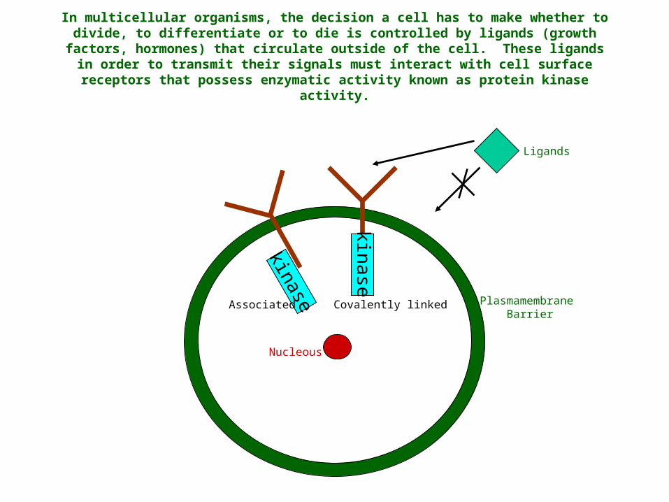
In multicellular organisms, the decision a cell has to make whether to divide, to differentiate or to die is controlled by ligands (growth factors, hormones) that circulate outside of the cell. These ligands in order to transmit their signals must interact with cell surface receptors that possess
enzymatic activity known as protein kinase activity.
Nucleous
Plasmamembrane Barrier
Ligands
Covalently linkedAssociatedkinase
kinase
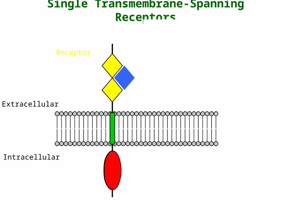
Single Transmembrane-Spanning Receptors
Extracellular
Intracellular
Ligand
Receptor
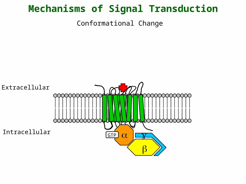
Mechanisms of Signal Transduction
Extracellular
Intracellular
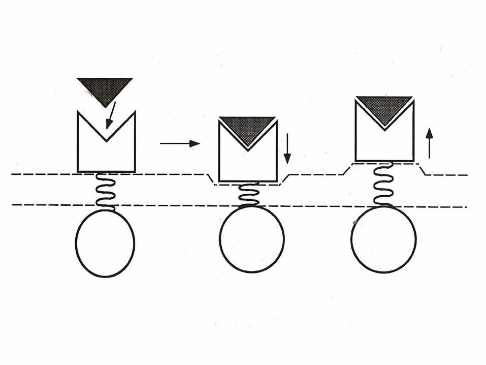
Conformational Change
GDPGTP

Receptor Dimerization
Extracellular
IntracellularY
YY
YY
Y
ATP
PP
P P
ATP
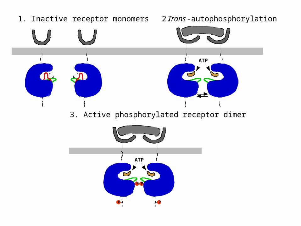
1. Inactive receptor monomers 2.Trans-autophosphorylation
3. Active phosphorylated receptor dimer
Early experiments suggesting that receptors undergo
dimerization
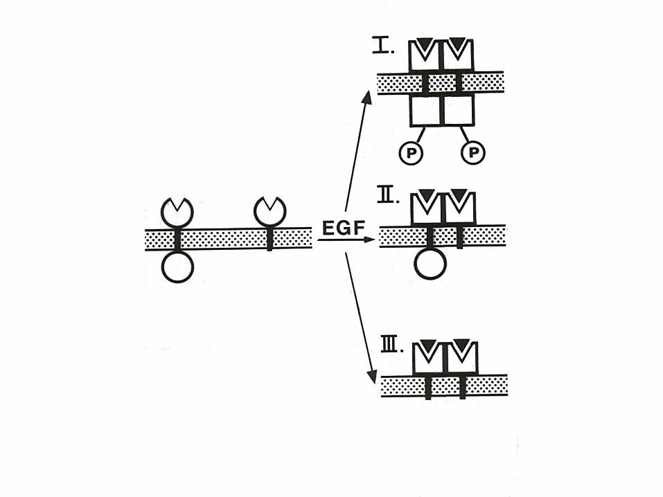
•Truncated receptors lacking the cytoplasmic domain inhibit signaling.
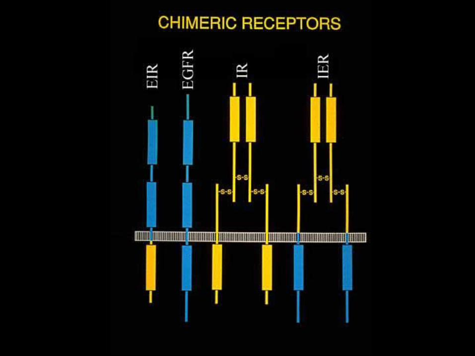
•Transmembrane helices are interchangeable betweendifferent receptors.
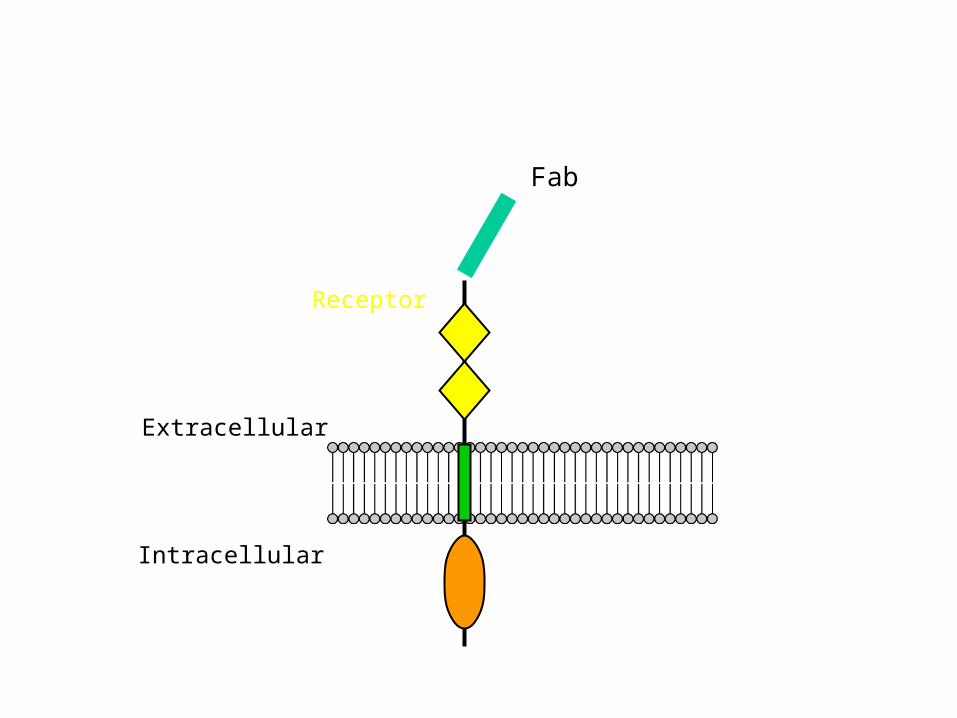
•Antibodies against the cytoplasmic domain activate the kinase domain.

Antibody-mediated activation
Extracellular
Intracellular
Receptor
Extracellular
Intracellular
Receptor
Fab

Receptor Ras
Raf
MAPKK
MAPKSos
Grb2
Growth factor
Shc
Jun Fos
NUCLEUS
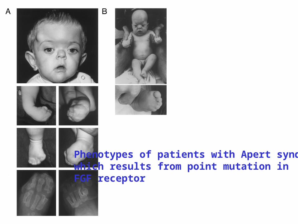
Phenotypes of patients with Apert syndromewhich results from point mutation in FGF receptor
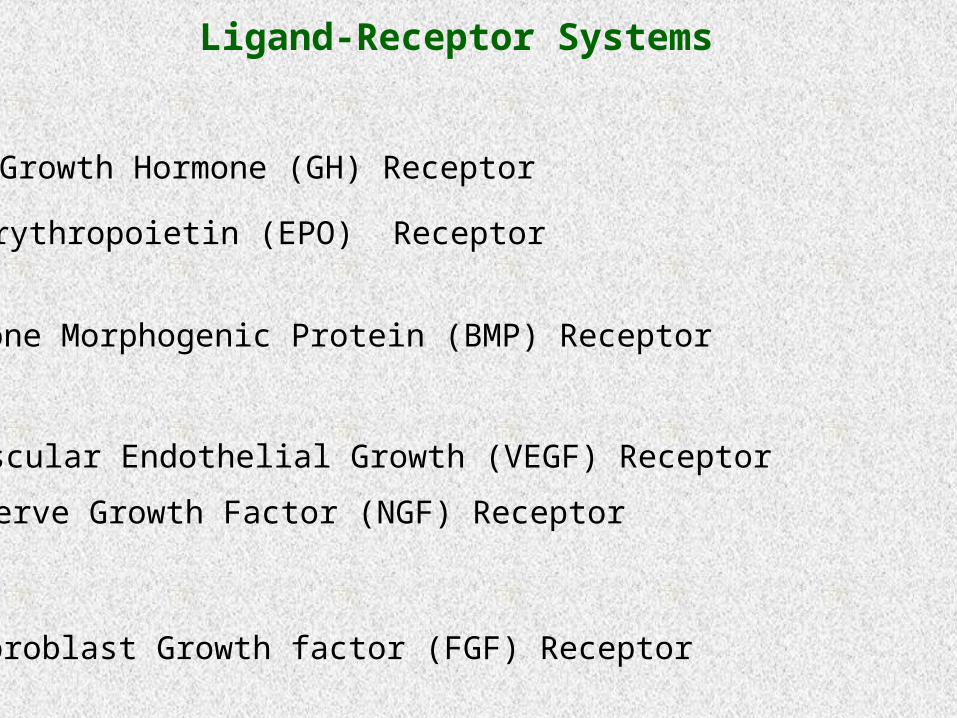
•Growth Hormone (GH) Receptor
•Erythropoietin (EPO) Receptor
•Bone Morphogenic Protein (BMP) Receptor
•Vascular Endothelial Growth (VEGF) Receptor
•Nerve Growth Factor (NGF) Receptor
•Fibroblast Growth factor (FGF) Receptor
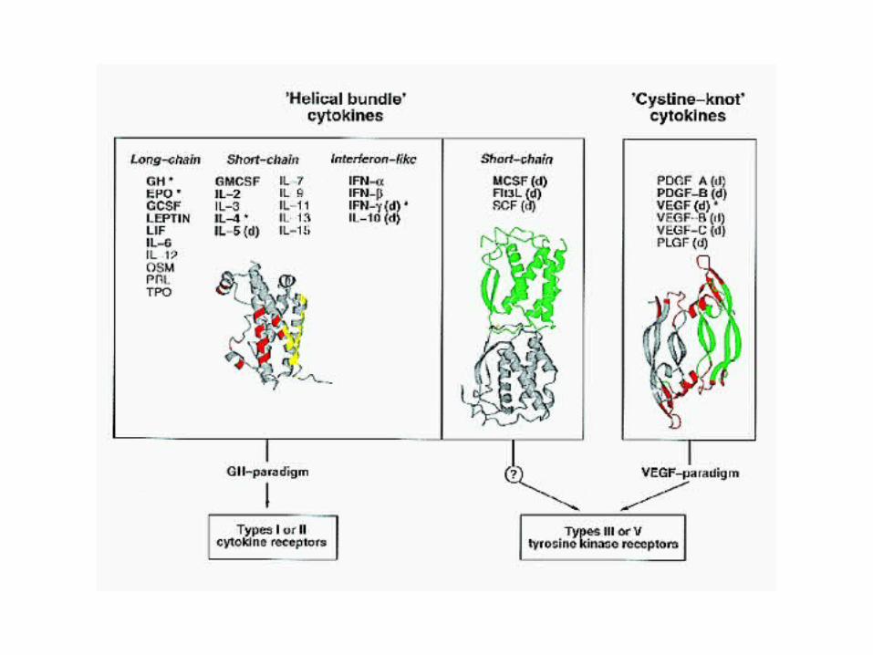
Ligand-Receptor Systems
• Large family of single-pass transmembrane receptors.
• Receptors bind polypeptide ligands: mediators of cell growth, differentiation and immune responses.
• Cytoplasmic domain does not contain intrinsic protein tyrosine kinase activity - associated with Jak tyrosine kinases.
Cytokine Receptors
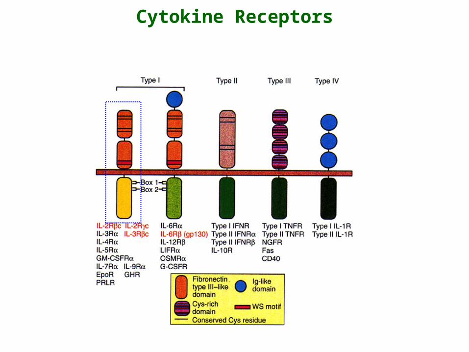
Cytokine Receptors
• GH stimulates the growth and metabolism of muscle, bone and cartilage cells.
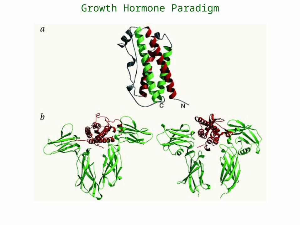
• GH is a member of the 4-helix bundle family.
• The active form of GH is a monomer. Stoichiometry of binding is 1:2 GH-GHR.
Activation Through Binding of a Monomeric Ligand – Growth Hormone
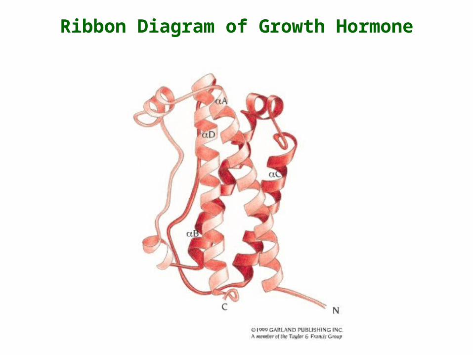
Ribbon Diagram of Growth Hormone
A
CD
B
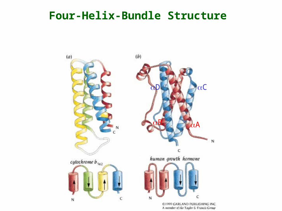
Four-Helix-Bundle Structure
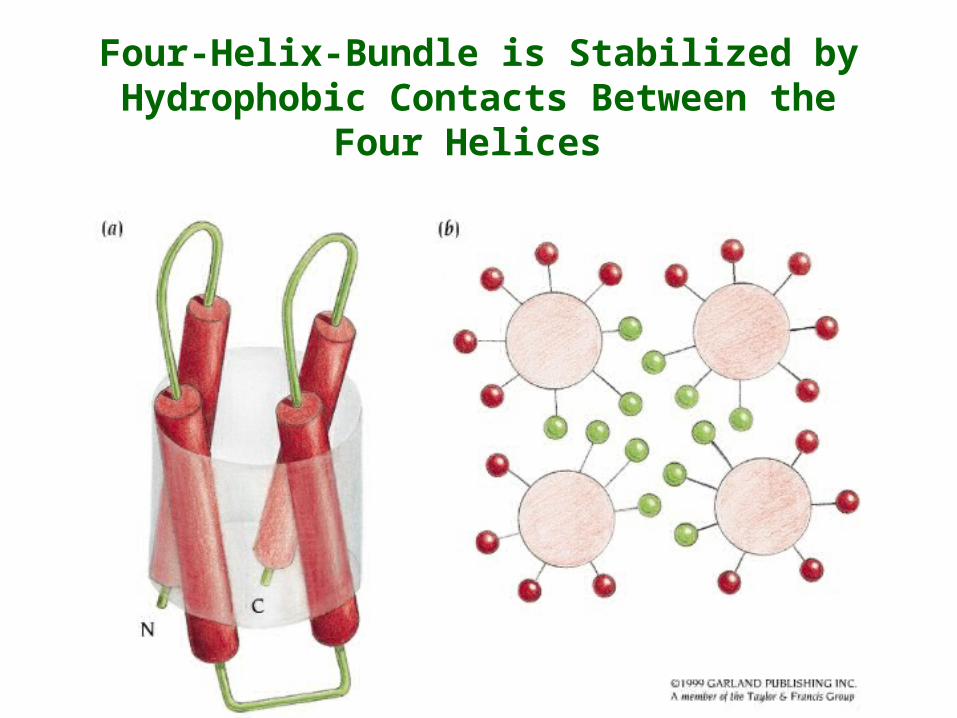
Four-Helix-Bundle is Stabilized by Hydrophobic Contacts Between the Four Helices
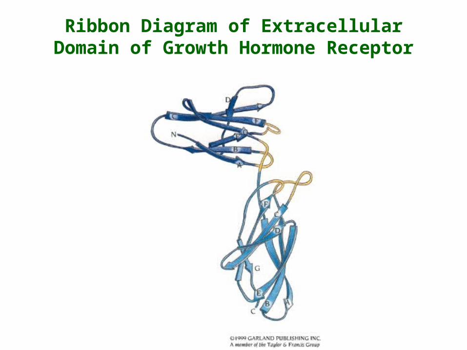
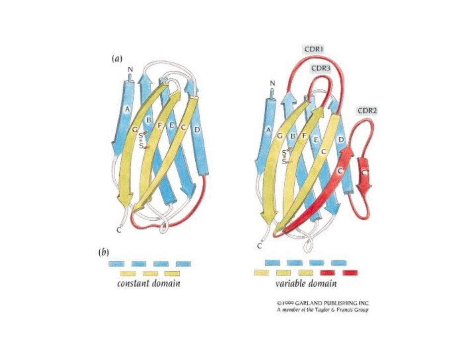
Ribbon Diagram of Extracellular Domain of Growth Hormone Receptor
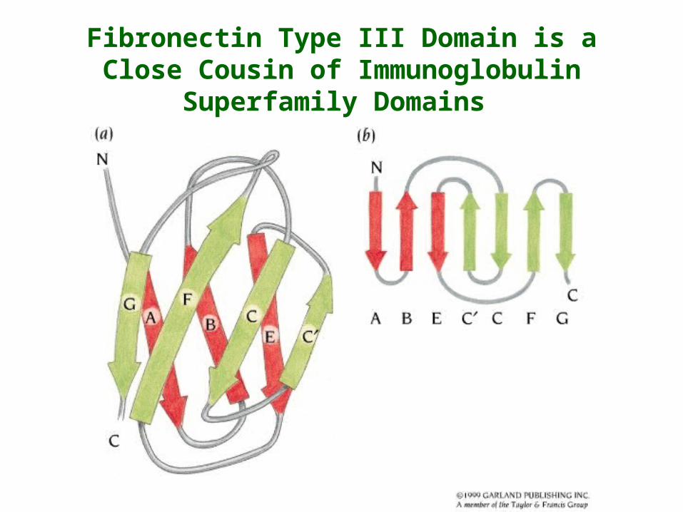
Fibronectin Type III Domain is a Close Cousin of Immunoglobulin Superfamily Domains
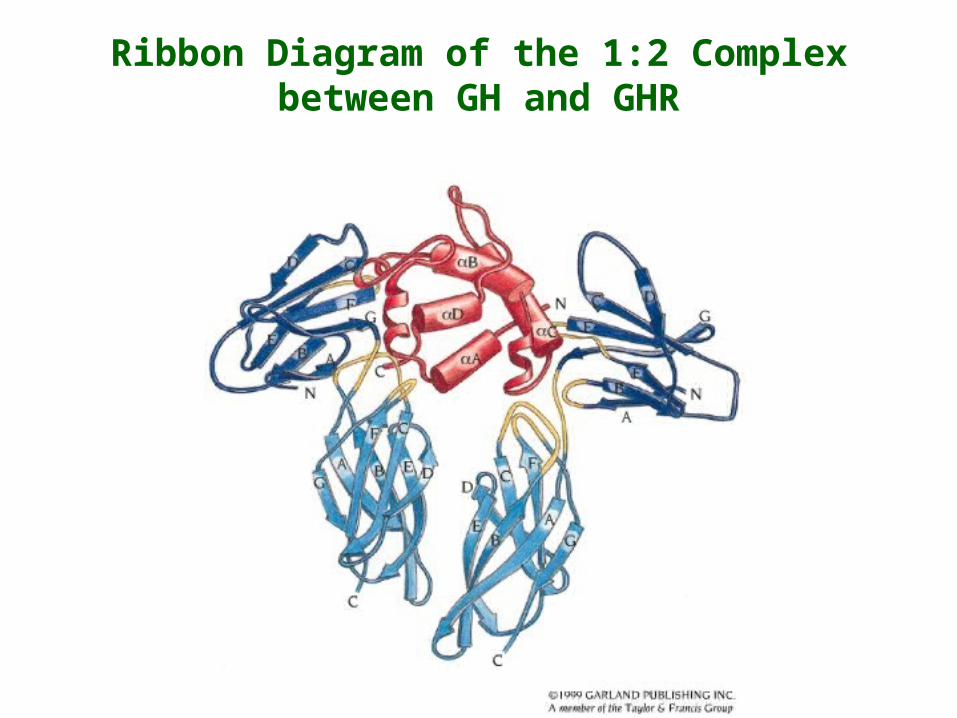
Ribbon Diagram of the 1:2 Complex between GH and GHR
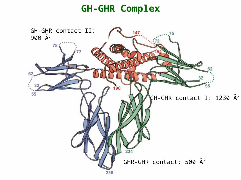
GH-GHR Complex
GH-GHR contact I: 1230 Å2
GHR-GHR contact: 500 Å2
GH-GHR contact II: 900 Å2

Details of GH-GHR Interactions
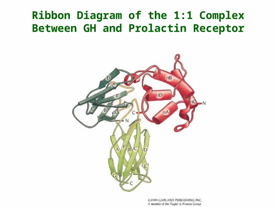
Ribbon Diagram of the 1:1 Complex Between GH and Prolactin Receptor
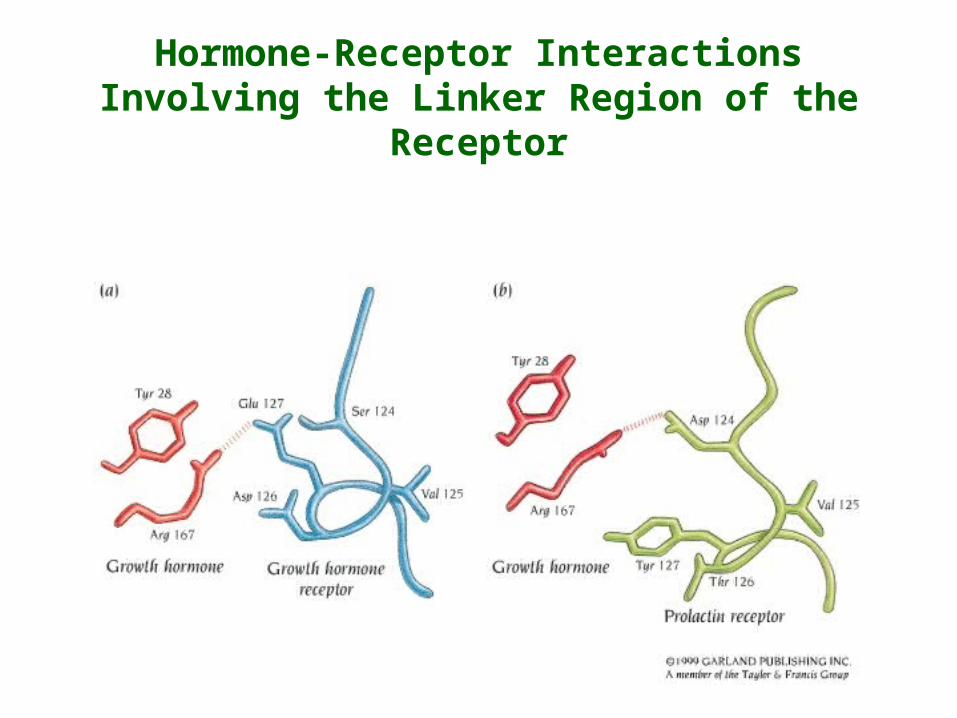
Hormone-Receptor Interactions Involving the Linker Region of the Receptor

Hormone-Receptor Interactions Involving the F-G Loop in the C-terminal Fibronectin Domain
Activation Through Binding of a Monomeric Ligand – Erythropoietin (EPO)
• EPO is a haematopoietic cytokine required for differentiation and proliferation of precursor cells into red blood cells.
• Like GH, EPO is monomeric and belongs to the 4-helix bundle family.
• EPO binds to its receptor (EPOR) with a stoichiometry of 1:2 EPO-EPOR.
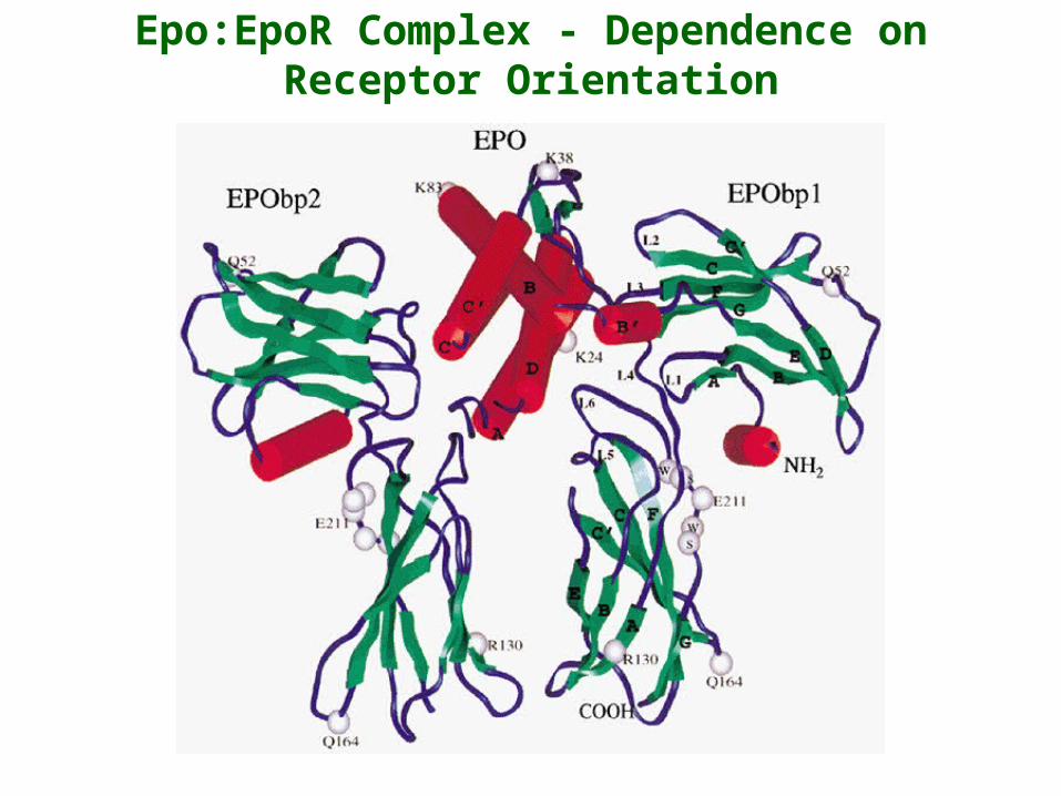
Epo:EpoR Complex - Dependence on Receptor Orientation
RasMol Presentation of the Dimeric EPO-EPOR Structure

Growth Hormone Paradigm
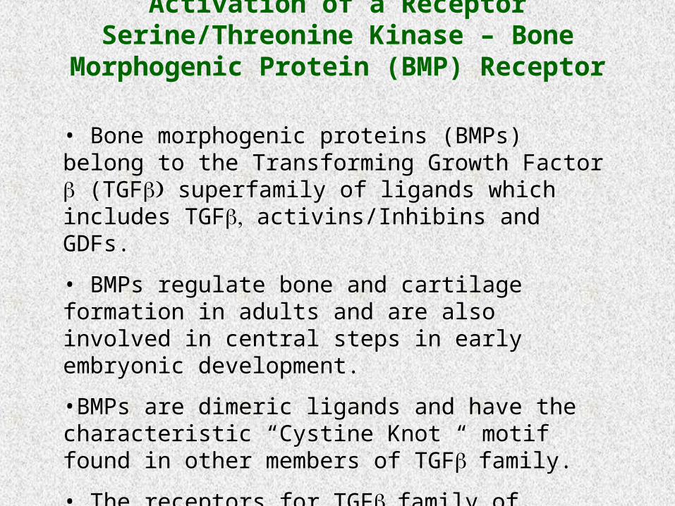
• Bone morphogenic proteins (BMPs) belong to the Transforming Growth Factor (TGF superfamily of ligands which includes TGF activins/Inhibins and GDFs.
• BMPs regulate bone and cartilage formation in adults and are also involved in central steps in early embryonic development.
•BMPs are dimeric ligands and have the characteristic “Cystine Knot “ motif found in other members of TGFfamily.
• The receptors for TGFfamily of ligands are transmembrane receptors with intrinsic serine/threonine kinase activity.
Activation of a Receptor Serine/Threonine Kinase – Bone Morphogenic Protein (BMP) Receptor
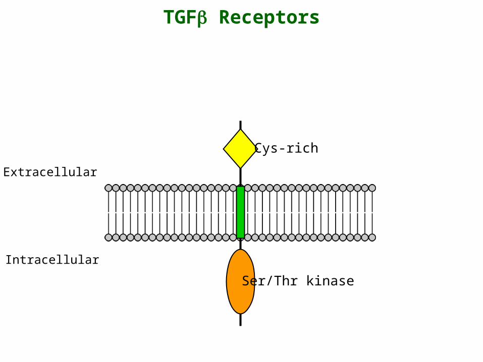
TGF Receptors
Extracellular
Intracellular
Ser/Thr kinase
Cys-rich
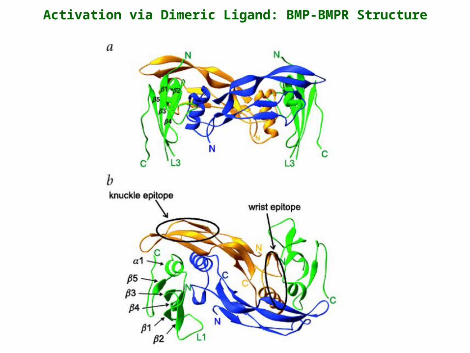
Activation via Dimeric Ligand: BMP-BMPR Structure

RasMol Presentation of the Dimeric BMP-BMPR Structure
• Large family of single-pass transmembrane receptors.
• Receptors are predominantly for growth factors but also for insulin.
• Cytoplasmic domain contains intrinsic protein tyrosine kinase activity.
Receptor Tyrosine Kinases
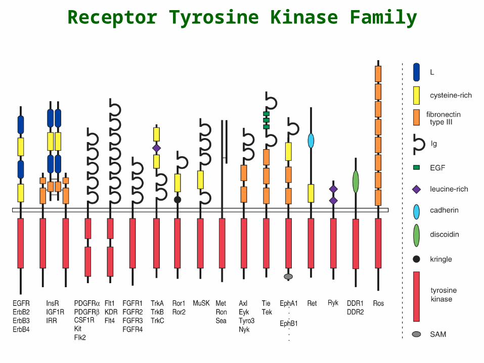
Receptor Tyrosine Kinase Family

• VEGF is a mitogen that is highly specific for endothelial cells.
• VEGF is a potent angiogenic factor involved in the development of the vascular system and also in tumor angiogenesis.
• VEGF is a covalent (disulfide-linked) dimer.
Activation Through Binding of a Dimeric Ligand – Vascular Endothelial Growth Factor
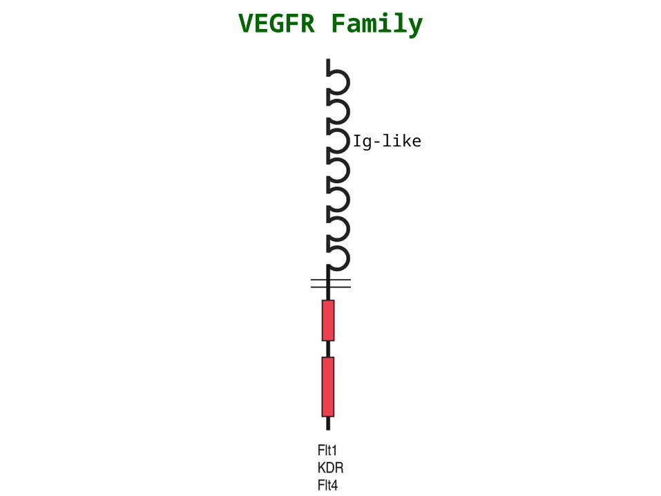
VEGFR Family
Ig-like
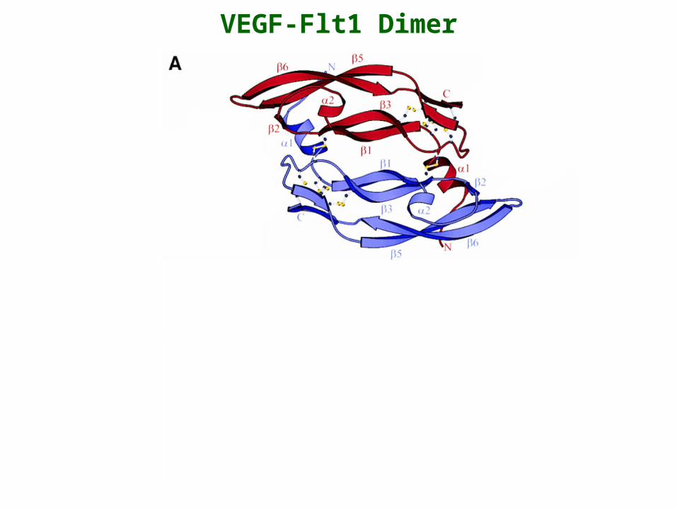
VEGF-Flt1 Dimer
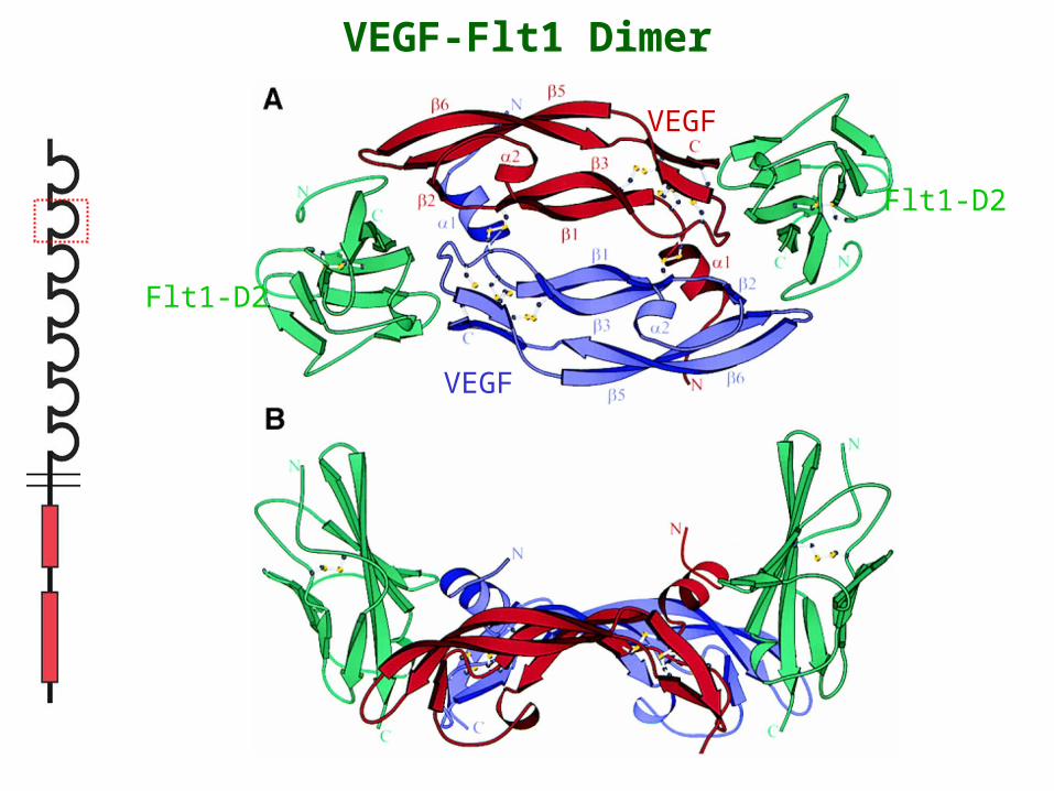
VEGF-Flt1 Dimer
Flt1-D2
Flt1-D2
VEGF
VEGF
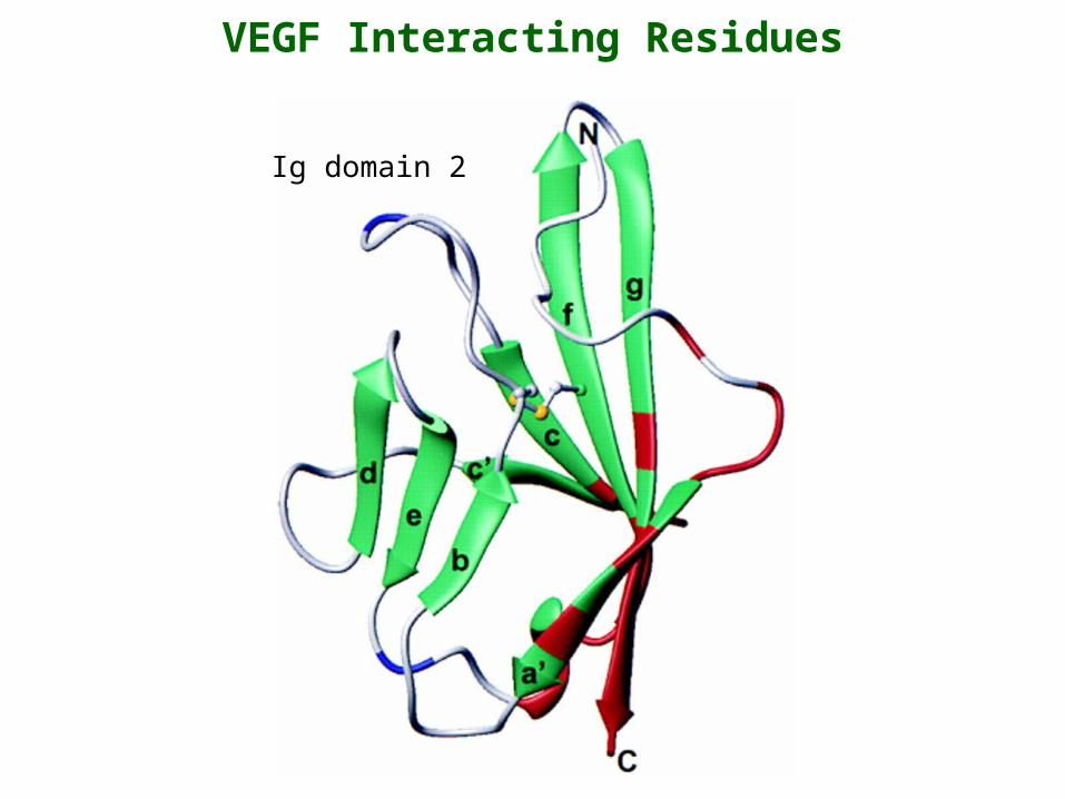
VEGF Interacting Residues
Ig domain 2
RasMol Presentation of the Dimeric VEGF-FLT1 Structure
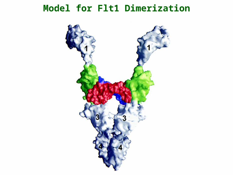
Model for Flt1 Dimerization
• NGF is a member of a family of neurotrophins which also includes brain-derived neurotrophic factor (BDNF), NT-3, NT-4/5 and NT-6.
• NGF mediates neuronal differentiation and survival.
• These neurotrophins are non-covalent dimers, members of the cystine knot family.
Activation Through Binding of a Dimeric Ligand – Neurotrophic Growth Factor

Trk (NGF Receptor) Family
Cys-richLeu-rich
Ig-like
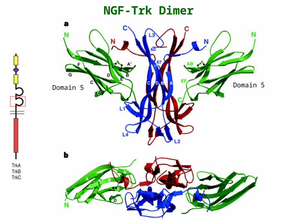
NGF-Trk Dimer
Domain 5 Domain 5

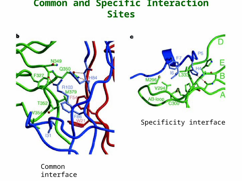
Common and Specific Interaction Sites
Common interface
Specificity interface
Common and Specific Interaction Sites
Specificity interface
Common interface
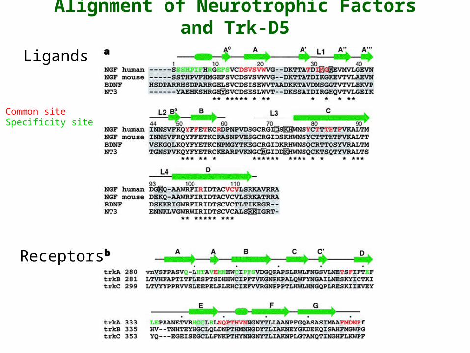
Alignment of Neurotrophic Factors and Trk-D5
Common siteSpecificity site
Ligands
Receptors
RasMol Presentation of the Dimeric NGF-TRK Structure
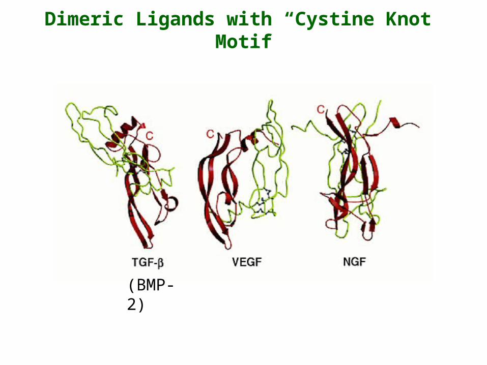
Dimeric Ligands with “Cystine Knot” Motif
(BMP-2)

Receptor Dimerization by Dimeric Ligands with “Cystine Knot” Motif
Growth Hormone Paradigm
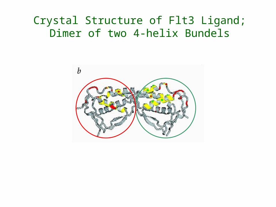
Crystal Structure of Flt3 Ligand;Dimer of two 4-helix Bundels
RasMol Presentation of FLT3 Ligand;Dimer of two 4-helix Bundles

Dimerization by Flt3L Versus VEGF
Flt3L-Flt3 (model) VEGF-Flt1 (partial model)


![Moosa Bin Naseer [PdfStuff.blogspot.com]](https://img.pdfslide.net/doc/110x75/55cf96ef550346d0338eba29/moosa-bin-naseer-pdfstuffblogspotcom.jpg)