Embed Size (px)
Citation preview
10. V. Southgate, A. Senju, G. Csibra, Psychol. Sci. 18, 587–592(2007).
11. D. C. Marticorena, A. M. Ruiz, C. Mukerji, A. Goddu,L. R. Santos, Dev. Sci. 14, 1406–1416 (2011).
12. A. Martin, L. R. Santos, Cognition 130, 300–308 (2014).13. F. Kano, J. Call, Psychol. Sci. 25, 1691–1698 (2014).14. F. Kano, S. Hirata, Curr. Biol. 25, 2513–2517 (2015).15. Materials and methods are available as supplementary
materials on Science Online.16. J. Perner, T. Ruffman, Science 308, 214–216 (2005).17. C. Heyes, Dev. Sci. 17, 647–659 (2014).18. R. Baillargeon, R. M. Scott, Z. He, Trends Cogn. Sci. 14, 110–118
(2010).
19. A. Senju, V. Southgate, C. Snape, M. Leonard, G. Csibra,Psychol. Sci. 22, 878–880 (2011).
20. K. Karg, M. Schmelz, J. Call, M. Tomasello, Anim. Behav. 105,211–221 (2015).
ACKNOWLEDGMENTS
We thank the staff at the Wolfgang Kohler Primate ResearchCenter and Kumamoto Sanctuary for assistance. Financial supportwas provided by the NSF Graduate Research Fellowship Program(grant DGE-1106401 to C.K.), the Ministry of Education, Culture,Sports, Science and Technology of Japan (grant K-CONNEX toF.K.), the Japan Society for the Promotion of Science (grantsKAKENHI 26885040 and 16K21108 to F.K. and grants KAKENHI
26245069 and 24000001 to S.H.), and the European ResearchCouncil (Synergy grant 609819 to J.C.). Data are available in themain text and supplementary materials.
SUPPLEMENTARY MATERIALS
www.sciencemag.org/content/354/6308/110/suppl/DC1Materials and MethodsSupplementary TextFigs. S1 to S5Tables S1 to S5Movies S1 and S2
27 April 2016; accepted 26 August 201610.1126/science.aaf8110
STRUCTURAL BIOLOGY
The methanogenic CO2 reducing-and-fixingenzyme is bifunctional and contains46 [4Fe-4S] clustersTristan Wagner,1 Ulrich Ermler,2 Seigo Shima1,3*
Biological methane formation starts with a challenging adenosine triphosphate (ATP)–independent carbon dioxide (CO2) fixation process.We explored this enzymatic processby solving the x-ray crystal structure of formyl-methanofuran dehydrogenase, determined hereas Fwd(ABCDFG)2 and Fwd(ABCDFG)4 complexes, from Methanothermobacter wolfeii. Thelatter 800-kilodalton apparatus consists of four peripheral catalytic sections and an electron-supplying core with 46 electronically coupled [4Fe-4S] clusters. Catalysis is separatelyperformed by subunits FwdBD (FwdB and FwdD), which are related to tungsten-containingformate dehydrogenase, and subunit FwdA, a binuclear metal center carrying amidohydrolase.CO2 is first reduced to formate in FwdBD, which then diffuses through a 43-angstrom-longtunnel to FwdA, where it condenses with methanofuran to formyl-methanofuran.Thearrangement of [4Fe-4S] clusters functions as an electron relay but potentially also couplesthe four tungstopterin active sites over 206 angstroms.
Methanogenic archaea produce ~1 billiontons of methane per year and thus playan important ecological role in the globalcarbon cycle (1). Biological methane isproduced mainly from acetate and CO2-
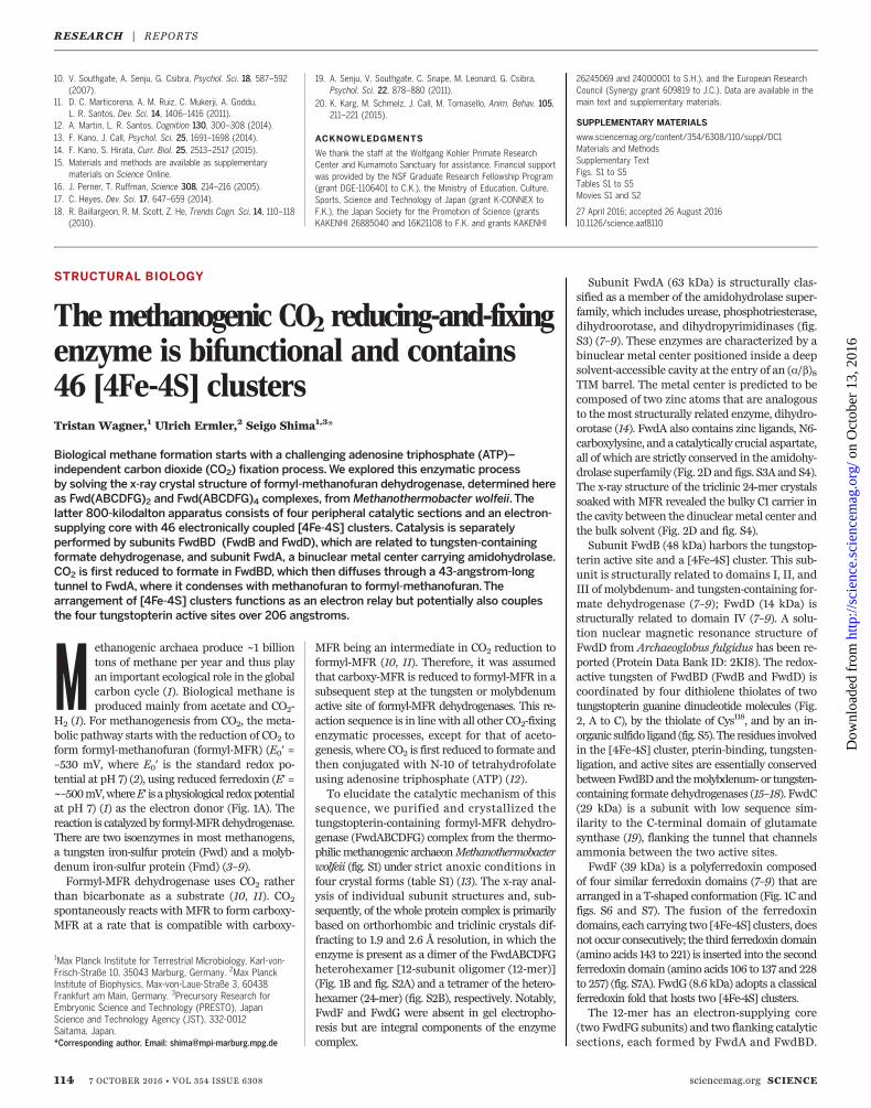
H2 (1). For methanogenesis from CO2, the meta-bolic pathway starts with the reduction of CO2 toform formyl-methanofuran (formyl-MFR) (E0′ =–530 mV, where E0′ is the standard redox po-tential at pH 7) (2), using reduced ferredoxin (E′ =~–500mV,whereE′ is aphysiological redoxpotentialat pH 7) (1) as the electron donor (Fig. 1A). Thereaction is catalyzedby formyl-MFRdehydrogenase.There are two isoenzymes in most methanogens,a tungsten iron-sulfur protein (Fwd) and a molyb-denum iron-sulfur protein (Fmd) (3–9).Formyl-MFR dehydrogenase uses CO2 rather
than bicarbonate as a substrate (10, 11). CO2
spontaneously reacts with MFR to form carboxy-MFR at a rate that is compatible with carboxy-
MFR being an intermediate in CO2 reduction toformyl-MFR (10, 11). Therefore, it was assumedthat carboxy-MFR is reduced to formyl-MFR in asubsequent step at the tungsten or molybdenumactive site of formyl-MFR dehydrogenases. This re-action sequence is in line with all other CO2-fixingenzymatic processes, except for that of aceto-genesis, where CO2 is first reduced to formate andthen conjugated with N-10 of tetrahydrofolateusing adenosine triphosphate (ATP) (12).To elucidate the catalytic mechanism of this
sequence, we purified and crystallized thetungstopterin-containing formyl-MFR dehydro-genase (FwdABCDFG) complex from the thermo-philic methanogenic archaeonMethanothermobacterwolfeii (fig. S1) under strict anoxic conditions infour crystal forms (table S1) (13). The x-ray anal-ysis of individual subunit structures and, sub-sequently, of the whole protein complex is primarilybased on orthorhombic and triclinic crystals dif-fracting to 1.9 and 2.6 Å resolution, in which theenzyme is present as a dimer of the FwdABCDFGheterohexamer [12-subunit oligomer (12-mer)](Fig. 1B and fig. S2A) and a tetramer of the hetero-hexamer (24-mer) (fig. S2B), respectively. Notably,FwdF and FwdG were absent in gel electropho-resis but are integral components of the enzymecomplex.
Subunit FwdA (63 kDa) is structurally clas-sified as a member of the amidohydrolase super-family, which includes urease, phosphotriesterase,dihydroorotase, and dihydropyrimidinases (fig.S3) (7–9). These enzymes are characterized by abinuclear metal center positioned inside a deepsolvent-accessible cavity at the entry of an (a/b)8TIM barrel. The metal center is predicted to becomposed of two zinc atoms that are analogousto themost structurally related enzyme, dihydro-orotase (14). FwdA also contains zinc ligands, N6-carboxylysine, and a catalytically crucial aspartate,all of which are strictly conserved in the amidohy-drolase superfamily (Fig. 2D and figs. S3A and S4).The x-ray structure of the triclinic 24-mer crystalssoaked with MFR revealed the bulky C1 carrier inthe cavity between the dinuclearmetal center andthe bulk solvent (Fig. 2D and fig. S4).Subunit FwdB (48 kDa) harbors the tungstop-
terin active site and a [4Fe-4S] cluster. This sub-unit is structurally related to domains I, II, andIII of molybdenum- and tungsten-containing for-mate dehydrogenase (7–9); FwdD (14 kDa) isstructurally related to domain IV (7–9). A solu-tion nuclear magnetic resonance structure ofFwdD from Archaeoglobus fulgidus has been re-ported (Protein Data Bank ID: 2KI8). The redox-active tungsten of FwdBD (FwdB and FwdD) iscoordinated by four dithiolene thiolates of twotungstopterin guanine dinucleotide molecules (Fig.2, A to C), by the thiolate of Cys118, and by an in-organic sulfido ligand (fig. S5). The residues involvedin the [4Fe-4S] cluster, pterin-binding, tungsten-ligation, and active sites are essentially conservedbetweenFwdBDand themolybdenum-or tungsten-containing formate dehydrogenases (15–18). FwdC(29 kDa) is a subunit with low sequence sim-ilarity to the C-terminal domain of glutamatesynthase (19), flanking the tunnel that channelsammonia between the two active sites.FwdF (39 kDa) is a polyferredoxin composed
of four similar ferredoxin domains (7–9) that arearranged in a T-shaped conformation (Fig. 1C andfigs. S6 and S7). The fusion of the ferredoxindomains, each carrying two [4Fe-4S] clusters, doesnot occur consecutively; the third ferredoxin domain(amino acids 143 to 221) is inserted into the secondferredoxin domain (amino acids 106 to 137 and 228to 257) (fig. S7A). FwdG (8.6 kDa) adopts a classicalferredoxin fold that hosts two [4Fe-4S] clusters.The 12-mer has an electron-supplying core
(two FwdFG subunits) and two flanking catalyticsections, each formed by FwdA and FwdBD.
114 7 OCTOBER 2016 • VOL 354 ISSUE 6308 sciencemag.org SCIENCE
1Max Planck Institute for Terrestrial Microbiology, Karl-von-Frisch-Straße 10, 35043 Marburg, Germany. 2Max PlanckInstitute of Biophysics, Max-von-Laue-Straße 3, 60438Frankfurt am Main, Germany. 3Precursory Research forEmbryonic Science and Technology (PRESTO), JapanScience and Technology Agency (JST), 332-0012Saitama, Japan.*Corresponding author. Email: [email protected]
RESEARCH | REPORTS
on
Oct
ober
13,
201
6ht
tp://
scie
nce.
scie
ncem
ag.o
rg/
Dow
nloa
ded
from
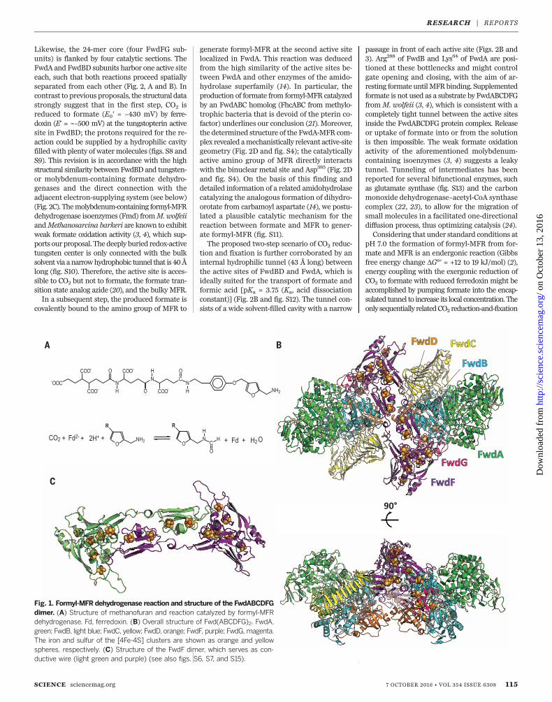
Likewise, the 24-mer core (four FwdFG sub-units) is flanked by four catalytic sections. TheFwdA and FwdBD subunits harbor one active siteeach, such that both reactions proceed spatiallyseparated from each other (Fig. 2, A and B). Incontrast to previous proposals, the structural datastrongly suggest that in the first step, CO2 isreduced to formate (E0′ = –430 mV) by ferre-doxin (E′ = ~–500 mV) at the tungstopterin activesite in FwdBD; the protons required for the re-action could be supplied by a hydrophilic cavityfilled with plenty of watermolecules (figs. S8 andS9). This revision is in accordance with the highstructural similarity between FwdBD and tungsten-or molybdenum-containing formate dehydro-genases and the direct connection with theadjacent electron-supplying system (see below)(Fig. 2C). Themolybdenum-containing formyl-MFRdehydrogenase isoenzymes (Fmd) fromM.wolfeiiandMethanosarcina barkeri are known to exhibitweak formate oxidation activity (3, 4), which sup-ports our proposal. The deeply buried redox-activetungsten center is only connected with the bulksolvent via anarrowhydrophobic tunnel that is 40Ålong (fig. S10). Therefore, the active site is acces-sible to CO2 but not to formate, the formate tran-sition state analog azide (20), and the bulky MFR.In a subsequent step, the produced formate is
covalently bound to the amino group of MFR to
generate formyl-MFR at the second active sitelocalized in FwdA. This reaction was deducedfrom the high similarity of the active sites be-tween FwdA and other enzymes of the amido-hydrolase superfamily (14). In particular, theproduction of formate from formyl-MFR catalyzedby an FwdABC homolog (FhcABC from methylo-trophic bacteria that is devoid of the pterin co-factor) underlines our conclusion (21). Moreover,the determined structure of the FwdA-MFR com-plex revealed amechanistically relevant active-sitegeometry (Fig. 2D and fig. S4); the catalyticallyactive amino group of MFR directly interactswith the binuclear metal site and Asp385 (Fig. 2Dand fig. S4). On the basis of this finding anddetailed information of a related amidohydrolasecatalyzing the analogous formation of dihydro-orotate from carbamoyl aspartate (14), we postu-lated a plausible catalytic mechanism for thereaction between formate and MFR to gener-ate formyl-MFR (fig. S11).The proposed two-step scenario of CO2 reduc-
tion and fixation is further corroborated by aninternal hydrophilic tunnel (43 Å long) betweenthe active sites of FwdBD and FwdA, which isideally suited for the transport of formate andformic acid [pKa = 3.75 (Ka, acid dissociationconstant)] (Fig. 2B and fig. S12). The tunnel con-sists of a wide solvent-filled cavity with a narrow
passage in front of each active site (Figs. 2B and3). Arg288 of FwdB and Lys64 of FwdA are posi-tioned at these bottlenecks and might controlgate opening and closing, with the aim of ar-resting formate untilMFRbinding. Supplementedformate is not used as a substrate by FwdABCDFGfromM. wolfeii (3, 4), which is consistent with acompletely tight tunnel between the active sitesinside the FwdABCDFG protein complex. Releaseor uptake of formate into or from the solutionis then impossible. The weak formate oxidationactivity of the aforementioned molybdenum-containing isoenzymes (3, 4) suggests a leakytunnel. Tunneling of intermediates has beenreported for several bifunctional enzymes, suchas glutamate synthase (fig. S13) and the carbonmonoxide dehydrogenase–acetyl-CoA synthasecomplex (22, 23), to allow for the migration ofsmall molecules in a facilitated one-directionaldiffusion process, thus optimizing catalysis (24).Considering that under standard conditions at
pH 7.0 the formation of formyl-MFR from for-mate and MFR is an endergonic reaction (Gibbsfree energy change DG°′ = +12 to 19 kJ/mol) (2),energy coupling with the exergonic reduction ofCO2 to formate with reduced ferredoxin might beaccomplished by pumping formate into the encap-sulated tunnel to increase its local concentration. Theonly sequentially relatedCO2 reduction-and-fixation
SCIENCE sciencemag.org 7 OCTOBER 2016 • VOL 354 ISSUE 6308 115
Fig. 1. Formyl-MFRdehydrogenase reaction and structure of the FwdABCDFGdimer. (A) Structure of methanofuran and reaction catalyzed by formyl-MFRdehydrogenase. Fd, ferredoxin. (B) Overall structure of Fwd(ABCDFG)2. FwdA,green; FwdB, light blue; FwdC, yellow; FwdD, orange; FwdF, purple; FwdG, magenta.The iron and sulfur of the [4Fe-4S] clusters are shown as orange and yellowspheres, respectively. (C) Structure of the FwdF dimer, which serves as con-ductive wire (light green and purple) (see also figs. S6, S7, and S15).
RESEARCH | REPORTS
on
Oct
ober
13,
201
6ht
tp://
scie
nce.
scie
ncem
ag.o
rg/
Dow
nloa
ded
from
process found in acetogenesis is catalyzed by twoseparate enzymes, formate dehydrogenase andformyl-tetrahydrofolate synthetase, the latter ofwhich requires ATP for formate activation.In the 24-mer, two 12-mers are associated in
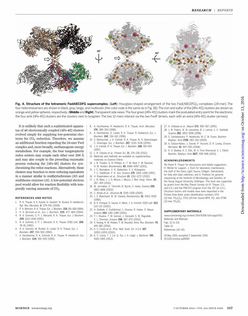
an hourglass-like arrangement (Fig. 4), therebyfurnished with an additional [4Fe-4S] clusterper each 12-mer that covalently links the twoFwdF subunits (fig. S6). The two extra [4Fe-4S]clusters in the 24-mer are 7.7 Å apart from eachother and 10.3 Å away from the next [4Fe-4S]
cluster (figs. S2B and S14), which allows inter-dodecameric electron shuttling (Fig. 4 and fig.S15). The cysteine ligands of the two additional[4Fe-4S] clusters originating from both FwdFsubunits are fully conserved in methanogenswithout cytochromes, except for those belongingto Methanomicrobiales. The residues of the sur-rounding loop, which are involved in interdo-decameric interactions, are also conserved to agreat extent (fig. S14). In addition, the 24-merwas observed in another crystal form [P3221 (fig.
S2B and table S1], which grew in a different crys-tallization solution (see supplementary materials).Finally, formyl-MFR dehydrogenase appears tomake a huge complex with heterodisulfide re-ductase and other catabolic enzymes (25, 26),which may stabilize the 24-mer FwdABCDFGstructure. Therefore, the 24-meric supercomplexis assumed to be a physiologically active stateand not a crystallographic artifact.The 46 [4Fe-4S] clusters in the 24-mer are
arranged in a stringlike distribution (Fig. 4).The edge-to-edge distances of 8.6 to 12.4 Å arecharacteristic of electrically connected redox cen-ters (fig. S15) (27, 28). Because polar and chargedresidues are known to affect the redox potentialof iron-sulfur clusters (29), their similar surround-ings concerning structure and electrostatics suggestthat electrons are conducted almost isopoten-tially, without obvious thermodynamic barriers.The formed electron wire extends over distancesof 188 Å between the redox-active tungsten cen-ters of the 12-mer. The 24-mer uses the inter-dodecamer bridge between the two wires of the12-mers to extend over 206 Å between activesites. The outer cluster of the two [4Fe-4S] clus-ters in the branched peripheral arm of the T-shaped FwdF subunits might serve as the entrypoint for electrons (Fig. 4) from where the elec-trons flow to the tungsten center via a chain ofoptimally spaced [4Fe-4S] clusters. Associationbetween the formyl-MFR dehydrogenase complexand the electron-bifurcating [NiFe]-hydrogenase–heterodisulfide reductase complex (MvhABD-HdrABC) (25) implicates a long, direct electrontransfer route from the site of electron bifurcation(HdrA) to the site of CO2 reduction (FwdBD), per-haps via the FwdF entry point.
116 7 OCTOBER 2016 • VOL 354 ISSUE 6308 sciencemag.org SCIENCE
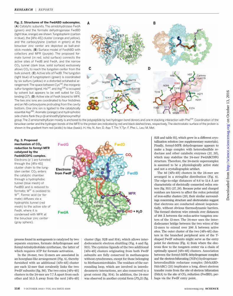
Fig. 3. Proposedmechanism of CO2
reduction to formyl-MFRcatalyzed by theFwdABCDFG complex.Electrons (e–) are funneledthrough the [4Fe-4S]cluster chain to the tung-sten center. CO2 entersthe catalytic chamberthrough a hydrophobictunnel (blue mesh) ofFwdBD and is reduced toformate; WIV is oxidized toWVI. Formic acid (or for-mate) diffuses via ahydrophilic tunnel (redmesh) to the active site ofFwdA, where it iscondensed with MFR atthe binuclear zinc center(gray sphere).
Fig. 2. Structures of the FwdABD subcomplex.(A) Catalytic subunits. The amidohydrolase FwdA(green) and the formate dehydrogenase FwdBD(light blue, orange) are shown.Tungstopterin (carbonin blue), the [4Fe-4S] cluster (orange and yellow),and the carboxylysine (carbon in green) at thebinuclear zinc center are depicted as ball-and-stick models. (B) Surface model of FwdABD withcofactors and MFR (purple). The proposed for-mate tunnel (in red, solid surface) connects theactive sites of FwdB and FwdA, and the narrowCO2 tunnel (dark blue, solid surface) exclusivelyallows CO2 to reach the tungsten center from thebulk solvent. (C) Active site of FwdB.The tungsten(light blue) of tungstopterin (green) is coordinatedby six sulfurs (yellow) in a distorted octahedral ar-rangement.The space between Cys118, the inorganicsulfur tungsten ligand, His119, and Arg228 is occupiedby solvent but appears to be well suited for CO2
binding (17). (D) Active site of FwdA bound to MFR.The two zinc ions are coordinated to four histidinesand an N6-carboxylysine protruding from the cavitybottom. One zinc ion is ligated to the catalyticallyessential Asp385. Aromatic (orange) and hydrophobicside chains flank the p-(b-aminoethyl)phenoxymethylgroup.The 2-aminomethylfuran moiety is anchored to the polypeptide by two hydrogen bond donors and one π-stacking interaction with Phe274. Coordination of thebinuclear center and the hydrogen bonds of theMFR to the protein are indicated by red and black dashed lines, respectively.The electrostatic surface of the protein isshown in the gradient from red (acidic) to blue (basic). H, His; N, Asn; D, Asp; T,Thr; Y,Tyr; F, Phe; L, Leu; M, Met.
RESEARCH | REPORTS
on
Oct
ober
13,
201
6ht
tp://
scie
nce.
scie
ncem
ag.o
rg/
Dow
nloa
ded
from
It is unlikely that such a sophisticated appara-tus of 46 electronically coupled [4Fe-4S] clustersevolved simply for supplying low-potential elec-trons for CO2 reduction. Therefore, we assumean additional function regarding the 24-mer Fwdcomplex and,more broadly,methanogenic energymetabolism. For example, the four tungstopterinredox centers may couple each other over 206 Åand may also couple to the preceding enzymaticprocess reducing the [4Fe-4S] clusters for syn-chronizing the redox reactions. Alternatively, theseclusters may function to store reducing equivalentsin a manner similar to multicytochromes (30) andmultiheme enzymes (31). A low-potential electronpool would allow for reaction flexibility with tem-porally varying amounts of CO2.
REFERENCES AND NOTES
1. R. K. Thauer, A. K. Kaster, H. Seedorf, W. Buckel, R. Hedderich,Nat. Rev. Microbiol. 6, 579–591 (2008).
2. P. A. Bertram, R. K. Thauer, Eur. J. Biochem. 226, 811–818 (1994).3. P. A. Bertram et al., Eur. J. Biochem. 220, 477–484 (1994).4. R. A. Schmitz, S. P. J. Albracht, R. K. Thauer, Eur. J. Biochem.
209, 1013–1018 (1992).5. R. A. Schmitz, S. P. J. Albracht, R. K. Thauer, FEBS Lett. 309,
78–81 (1992).6. R. A. Schmitz, M. Richter, D. Linder, R. K. Thauer, Eur. J.
Biochem. 207, 559–565 (1992).7. A. Hochheimer, R. A. Schmitz, R. K. Thauer, R. Hedderich, Eur.
J. Biochem. 234, 910–920 (1995).
8. A. Hochheimer, R. Hedderich, R. K. Thauer, Arch. Microbiol.170, 389–393 (1998).
9. A. Hochheimer, D. Linder, R. K. Thauer, R. Hedderich, Eur. J.Biochem. 242, 156–162 (1996).
10. S. Bartoschek, J. A. Vorholt, R. K. Thauer, B. H. Geierstanger,C. Griesinger, Eur. J. Biochem. 267, 3130–3138 (2000).
11. J. A. Vorholt, R. K. Thauer, Eur. J. Biochem. 248, 919–924(1997).
12. L. R. Celeste et al., Protein Sci. 21, 219–228 (2012).13. Materials and methods are available as supplementary
materials on Science Online.14. J. B. Thoden, G. N. Phillips Jr., T. M. Neal, F. M. Raushel,
H. M. Holden, Biochemistry 40, 6989–6997 (2001).15. J. C. Boyington, V. N. Gladyshev, S. V. Khangulov,
T. C. Stadtman, P. D. Sun, Science 275, 1305–1308 (1997).16. H. Raaijmakers et al., Structure 10, 1261–1272 (2002).17. L. B. Maia, J. J. G. Moura, I. Moura, J. Biol. Inorg. Chem. 20,
287–309 (2015).18. M. Jormakka, S. Törnroth, B. Byrne, S. Iwata, Science 295,
1863–1868 (2002).19. C. Binda et al., Structure 8, 1299–1308 (2000).20. J. S. Blanchard, W. W. Cleland, Biochemistry 19, 3543–3550
(1980).21. B. K. Pomper, O. Saurel, A. Milon, J. A. Vorholt, FEBS Lett. 523,
133–137 (2002).22. H. Dobbek, V. Svetlitchnyi, L. Gremer, R. Huber, O. Meyer,
Science 293, 1281–1285 (2001).23. T. I. Doukov, T. M. Iverson, J. Seravalli, S. W. Ragsdale,
C. L. Drennan, Science 298, 567–572 (2002).24. X. Huang, H. M. Holden, F. M. Raushel, Annu. Rev. Biochem. 70,
149–180 (2001).25. K. C. Costa et al., Proc. Natl. Acad. Sci. U.S.A. 107,
11050–11055 (2010).26. K. C. Costa, T. J. Lie, Q. Xia, J. A. Leigh, J. Bacteriol. 195,
5160–5165 (2013).
27. A. Volbeda et al., Nature 373, 580–587 (1995).28. J. W. Peters, W. N. Lanzilotta, B. J. Lemon, L. C. Seefeldt,
Science 282, 1853–1858 (1998).29. S. Santabarbara, P. Heathcote, M. C. W. Evans, Biochim.
Biophys. Acta 1708, 283–310 (2005).30. A. Esteve-Núñez, J. Sosnik, P. Visconti, D. R. Lovley, Environ.
Microbiol. 10, 497–505 (2008).31. K. D. Bewley, K. E. Ellis, M. A. Firer-Sherwood, S. J. Elliott,
Biochim. Biophys. Acta 1827, 938–948 (2013).
ACKNOWLEDGMENTS
We thank R. Thauer for discussions and helpful suggestions,H. Michel for support, J. Koch for laboratory maintenance,the staff of the Swiss Light Source (Villigen, Switzerland)for help with data collection, and A. Poehlein for genomesequencing at the Institute of Microbiology and Genetics ofthe Georg August University Göttingen. This work was supportedby grants from the Max Planck Society (to R. Thauer, S.S.,and U.E.) and the PRESTO program from the JST (to S.S.).Structure factors and models have been deposited in theProtein Data Bank under identification numbers 5T5I(12-mer, P212121), 5T61 (24-mer bound MFR, P1), and 5T5M(12-mer, P3221).
SUPPLEMENTARY MATERIALS
www.sciencemag.org/content/354/6308/114/suppl/DC1Materials and MethodsFigs. S1 to S15Table S1References (32–43)
18 May 2016; accepted 2 September 201610.1126/science.aaf9284
SCIENCE sciencemag.org 7 OCTOBER 2016 • VOL 354 ISSUE 6308 117
Fig. 4. Structure of the tetrameric FwdABCDFG supercomplex. (Left) Hourglass-shaped arrangement of the two Fwd(ABCDFG)2 complexes (24-mer). Thefour heterohexamers are shown in black, gray, beige, andmulticolor (the color code is the same as in Fig. 1B).The iron and sulfur of the [4Fe-4S] clusters are shown asorange and yellow spheres, respectively. (Middle andRight) Transparent side views.The four green [4Fe-4S] clustersmark thepostulated entry point for the electrons;the four pink [4Fe-4S] clusters are the clusters next to tungsten.The two 12-mers interact via the two FwdFdimers, each with an extra [4Fe-4S] cluster (arrows).
RESEARCH | REPORTS
on
Oct
ober
13,
201
6ht
tp://
scie
nce.
scie
ncem
ag.o
rg/
Dow
nloa
ded
from
(6308), 114-117. [doi: 10.1126/science.aaf9284]354Science Tristan Wagner, Ulrich Ermler and Seigo Shima (October 6, 2016) bifunctional and contains 46 [4Fe-4S] clusters
reducing-and-fixing enzyme is2The methanogenic CO
Editor's Summary
, this issue p. 114Sciencetungsten redox centers.clusters. Although the exact function of this chain is unclear, it may electronically couple the four
reduction. The complex also contains a chain of 46 iron-sulfur2transferring the formate made after COcomplex. Two active sites in the complex are separated by a 43-Å tunnel, which is responsible for
solved the x-ray crystal structure of a tungsten-containing formyl-MFR dehydrogenaseet al.Wagner and methanofuran (MFR) are reduced to formyl-MFR by an as yet unresolved mechanism.2COThe process by which archaea make methane involves a series of reactions and enzymes. First,
The long and winding road to methane
This copy is for your personal, non-commercial use only.
Article Tools
http://science.sciencemag.org/content/354/6308/114article tools: Visit the online version of this article to access the personalization and
Permissionshttp://www.sciencemag.org/about/permissions.dtlObtain information about reproducing this article:
is a registered trademark of AAAS. ScienceAdvancement of Science; all rights reserved. The title Avenue NW, Washington, DC 20005. Copyright 2016 by the American Association for thein December, by the American Association for the Advancement of Science, 1200 New York
(print ISSN 0036-8075; online ISSN 1095-9203) is published weekly, except the last weekScience
on
Oct
ober
13,
201
6ht
tp://
scie
nce.
scie
ncem
ag.o
rg/
Dow
nloa
ded
from
www.sciencemag.org/content/354/6308/114/suppl/DC1
Supplementary Materials for
The methanogenic CO2 reducing-and-fixing enzyme is bifunctional and
contains 46 [4Fe-4S] clusters
Tristan Wagner, Ulrich Ermler, Seigo Shima*
*Corresponding author. Email: [email protected]
Published 7 October 2016, Science 354, 114 (2016)
DOI: 10.1126/science.aaf9284
This PDF file includes:
Materials and Methods
Figs. S1 to S15
Table S1
References
2
Materials and Methods
Methanothermobacter wolfeii culture
We structurally analyzed the formyl-methanofuran (formyl-MFR) dehydrogenase
complex from Methanothermobacter wolfeii because this enzyme has already been well
characterized biochemically (4-6). M. wolfeii (DSM 2970) was obtained from the
Deutsche Sammlung von Mikroorganismen und Zellkulturen (DSMZ, Braunschweig,
Germany) and was routinely grown at 65 °C in two 2 liter fermenters containing 1.5 liters
of medium each. The growth medium corresponds to that used by Schmitz et al. (4), with
a mixture of 0.33 mg/l Na2WO4•2H2O and 0.24 mg/l Na2MoO4•2H2O . The cultures were
gassed with 80% H2/20% CO2/0.01% H2S at a rate of 0.8 liter per min and stirred at
1,000 rpm. When the culture reached an optical density of ca. 5 at 600 nm, the cells were
harvested by centrifugation under anoxic conditions at 8 °C, yielding approximately 30–
35 g cells (wet mass). The cell pellets were stored at −80 °C before enzyme purification.
Purification of the Fwd complex
The cell pellets were thawed at room temperature. Lysis buffer (50 mM
MOPS/NaOH, pH 7.0, 10 mM MgCl2 and 2 mM dithiothreitol) was added at a ratio of 2
ml per g pellet. The cells were disrupted in a French press at 7 MPa under 100% N2 gas.
The lysate was centrifuged twice at 10,000 ×g for 60 min, and then the supernatant was
ultracentrifuged at 110,000 ×g for 90 min at 4 °C. The subsequent steps were performed
under anoxic conditions (95% N2/5% H2) at 18 °C, without any freezing steps. First, the
soluble fraction was passed through a DEAE Sepharose fast-flow column equilibrated
with 50 mM Tricine-NaOH pH 8.0 containing 2 mM dithiothreitol (DTT) (buffer A). The
protein was eluted at a flow rate of 4 ml/min with a step-wise increasing concentration of
NaCl: two column volumes of 270 mM NaCl, four column volumes of 310 mM NaCl,
and four column volumes of 420 mM NaCl. The 420 mM NaCl fractions containing Fwd
were pooled and diluted with an equal volume of buffer A. The sample was loaded onto a
Q-Sepharose fast-flow column (GE Healthcare, Freiburg) pre-equilibrated in buffer A;
the column was washed with 400 mM NaCl, and the proteins were eluted at a flow rate of
3.5 ml/min with a gradient of 400–500 mM NaCl in six column volumes. Fwd typically
eluted at 450–475 mM NaCl. Fractions containing Fwd were diluted with an equal
volume of 25 mM sodium phosphate buffer, pH 7.6, containing 2 mM DTT (buffer B)
and loaded onto a hydroxyapatite ceramic type I column (macroprep; Bio-Rad; München,
Germany) equilibrated with buffer B. The column was washed with 50 mM sodium
phosphate buffer, pH 7.6, and the proteins were eluted at a flow rate of 2 ml/min with a
gradient of 50–150 mM sodium phosphate buffer, pH 7.6, in four column volumes. Fwd
eluted at 90–140 mM sodium phosphate buffer, pH 7.6. The Fwd fractions were pooled,
diluted with two volumes of 25 mM Tris/HCl, pH 7.6, containing 2.0 M (NH4)2SO4 and 2
mM DTT, and applied to a Source 15 Phe column that had been pre-equilibrated in the
same buffer. The column was washed with 0.5 M (NH4)2SO4, and the protein was eluted
at a flow rate of 1 ml/min with a gradient of 0.5–0.18 M (NH4)2SO4 in six column
volumes. Fwd eluted in two fractions: in an “early” fraction at 0.46–0.32 M (NH4)2SO4
along with methyl-viologen-reducing hydrogenase (Mvh) (32) and in a “late” fraction at
0.31–0.26 M (NH4)2SO4 alone. Both the “early” and “late” fractions were separately
concentrated by passing them through a 50 kDa cut-off filter (Merck Millipore,
3
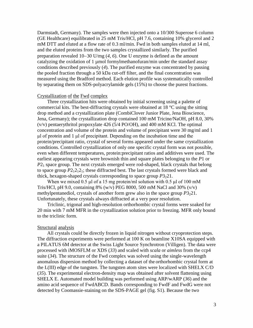
Darmstadt, Germany). The samples were then injected onto a 10/300 Superose 6 column
(GE Healthcare) equilibrated in 25 mM Tris/HCl, pH 7.6, containing 10% glycerol and 2
mM DTT and eluted at a flow rate of 0.3 ml/min. Fwd in both samples eluted at 14 ml,
and the eluted proteins from the two samples crystallized similarly. The purified
preparation revealed 1030 U/mg (4, 6). One U enzyme is defined as the amount
catalyzing the oxidation of 1 µmol formylmethanofuran/min under the standard assay
conditions described previously (4). The purified enzyme was concentrated by passing
the pooled fraction through a 50 kDa cut-off filter, and the final concentration was
measured using the Bradford method. Each elution profile was systematically controlled
by separating them on SDS-polyacrylamide gels (15%) to choose the purest fractions.
Crystallization of the Fwd complex
Three crystallization hits were obtained by initial screening using a palette of
commercial kits. The best-diffracting crystals were obtained at 18 °C using the sitting
drop method and a crystallization plate (CombiClover Junior Plate, Jena Bioscience,
Jena, Germany); the crystallization drop contained 100 mM Tricine/NaOH, pH 8.0, 30%
(v/v) pentaerythritol propoxylate 426 (5/4 PO/OH), and 400 mM KCl. The optimal
concentration and volume of the protein and volume of precipitant were 30 mg/ml and 1
μl of protein and 1 μl of precipitant. Depending on the incubation time and the
protein/precipitant ratio, crystal of several forms appeared under the same crystallization
conditions. Controlled crystallization of only one specific crystal form was not possible,
even when different temperatures, protein:precipitant ratios and additives were used. The
earliest appearing crystals were brownish thin and square plates belonging to the P1 or
P21 space group. The next crystals emerged were rod-shaped, black crystals that belong
to space group P212121; these diffracted best. The last crystals formed were black and
thick, hexagon-shaped crystals corresponding to space group P3221.
When we mixed 0.5 μl of a 15 mg protein/ml solution with 0.5 μl of 100 mM
Tris/HCl, pH 9.0, containing 8% (w/v) PEG 8000, 500 mM NaCl and 30% (v/v)
methylpentanediol, crystals of another form grew also in the space group P3221.
Unfortunately, these crystals always diffracted at a very poor resolution.
Triclinic, trigonal and high-resolution orthorhombic crystal forms were soaked for
20 min with 7 mM MFR in the crystallization solution prior to freezing. MFR only bound
to the triclinic form.
Structural analysis
All crystals could be directly frozen in liquid nitrogen without cryoprotection steps.
The diffraction experiments were performed at 100 K on beamline X10SA equipped with
a PILATUS 6M detector at the Swiss Light Source Synchrotron (Villigen). The data were
processed with iMOSFLM or XDS (33) and scaled with scala or aimless from the ccp4
suite (34). The structure of the Fwd complex was solved using the single-wavelength
anomalous dispersion method by collecting a dataset of the orthorhombic crystal form at
the L(III) edge of the tungsten. The tungsten atom sites were localized with SHELX C/D
(35). The experimental electron-density map was obtained after solvent flattening using
SHELX E. Automated model building was performed using ARP/wARP (36) and the
amino acid sequence of FwdABCD. Bands corresponding to FwdF and FwdG were not
detected by Coomassie-staining on the SDS-PAGE gel (fig. S1). Because the two
4
subunits were not expected to be part of the Fwd complex structure, FwdF and FwdG
were first traced as a glycine backbone. The electron-density map was sufficiently
accurate to recognize the sequence in the chain and to identify FwdF and FwdG.
ARP/wARP was used to complete the model using the FwdF and FwdG sequences.
The structures of the other crystal forms were determined by molecular replacement
using Molrep (ccp4 suite) (37) or Phaser (Phenix package) (38) and the coordinates of the
orthorhombic crystal form as the search model. All models were manually constructed
with COOT (39) and refined by REFMAC (40), except for the poorly diffracting trigonal
crystal form, for which the resolution was too low for further refinement. The final
refinements of orthorhombic crystal data was perfomed using BUSTER (Bricogne G.,
Blanc E., Brandl M., Flensburg C., Keller P., Paciorek W., Roversi P, Sharff A., Smart
O.S., Vonrhein C., Womack T.O. (2016). BUSTER version 2.10.1. Cambridge, United
Kingdom: Global Phasing Ltd.). The two other crystal forms, triclinic and trigonal were
refined by Phenix (38). Restraints for non-crystallographic symmetry (NCS) were applied
to all models (except for the better-diffracting trigonal crystal form, which contained only
one heterohexamer in the asymmetric unit), and translation-liberation-screw-rotation
(TLS) was only applied to the orthorhombic and trigonal crystal forms. The final models
were validated through the MolProbity server (http://molprobity.biochem.duke.edu). Data
collection, the refinement statistics of the deposited models, and the structure factors are
listed in Table S1.
The figures were generated and rendered with PyMOL (Version 1.5, Schrödinger,
LLC). All superposition studies were calculated using the ccp4 suite. Both tunnel cavities
possibly for CO2 and formate transfer in the protein were detected using the program
CAVER 3.0 (41) by applying the threshold of 1.2 Å radius.
5
+- Heated
15 kDa
25 kDa
35 kDa
40 kDa
55 kDa
70 kDa
10 kDa
100 kDa130 kDa170 kDa
FwdAFwdB
FwdC
FwdD
MvhD
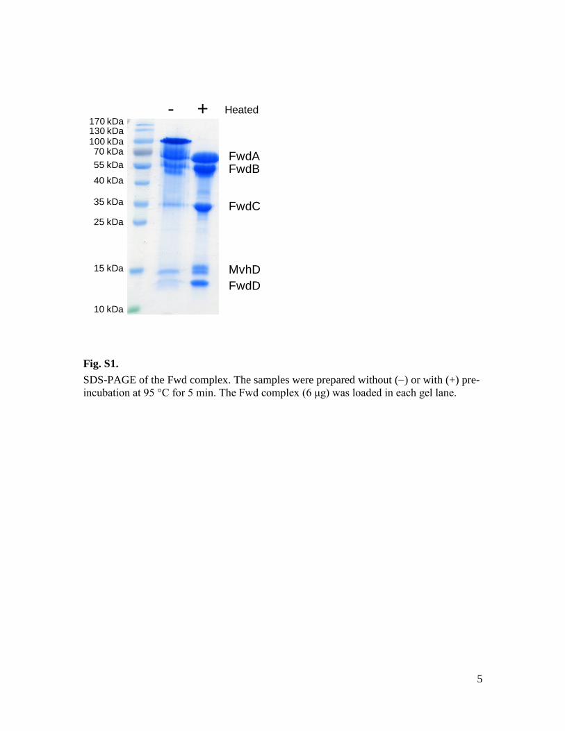
Fig. S1.
SDS-PAGE of the Fwd complex. The samples were prepared without () or with (+) pre-
incubation at 95 °C for 5 min. The Fwd complex (6 μg) was loaded in each gel lane.
6
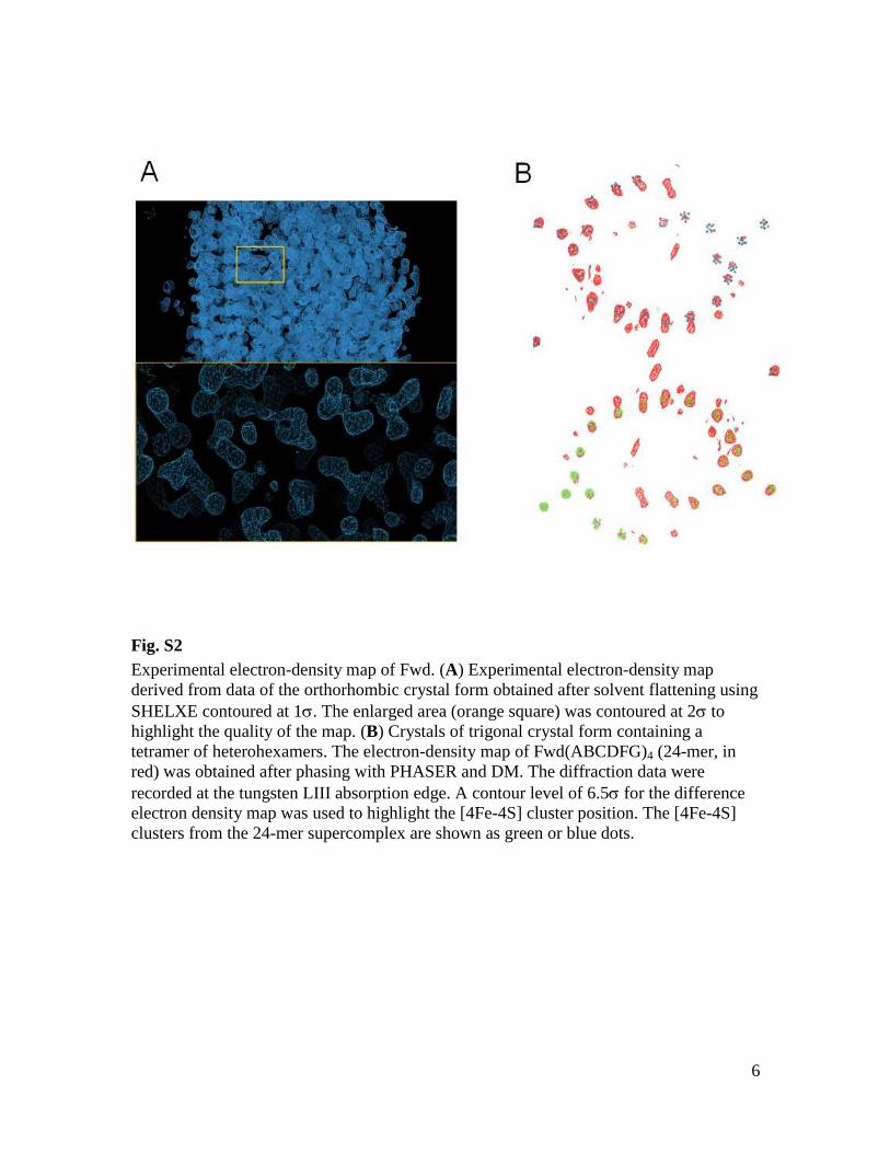
Fig. S2
Experimental electron-density map of Fwd. (A) Experimental electron-density map
derived from data of the orthorhombic crystal form obtained after solvent flattening using
SHELXE contoured at 1. The enlarged area (orange square) was contoured at 2 to
highlight the quality of the map. (B) Crystals of trigonal crystal form containing a
tetramer of heterohexamers. The electron-density map of Fwd(ABCDFG)4 (24-mer, in
red) was obtained after phasing with PHASER and DM. The diffraction data were
recorded at the tungsten LIII absorption edge. A contour level of 6.5 for the difference
electron density map was used to highlight the [4Fe-4S] cluster position. The [4Fe-4S]
clusters from the 24-mer supercomplex are shown as green or blue dots.
7
8
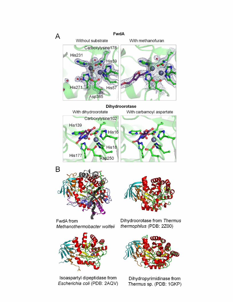
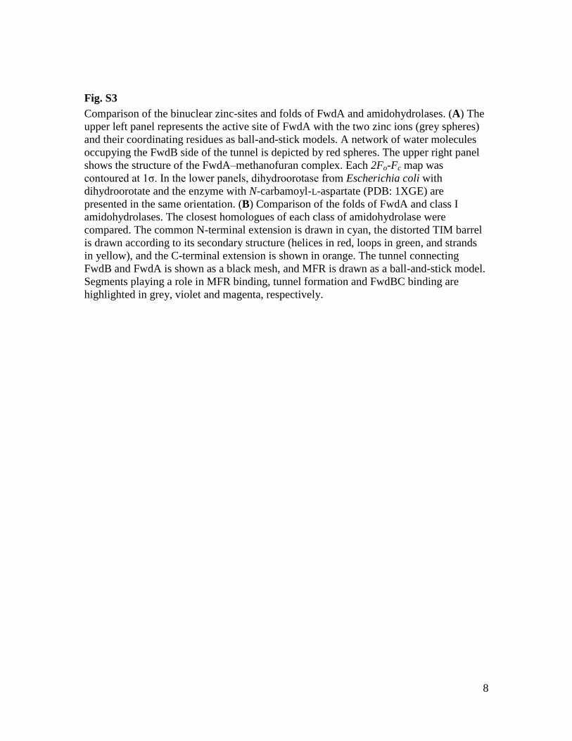
Fig. S3
Comparison of the binuclear zinc-sites and folds of FwdA and amidohydrolases. (A) The
upper left panel represents the active site of FwdA with the two zinc ions (grey spheres)
and their coordinating residues as ball-and-stick models. A network of water molecules
occupying the FwdB side of the tunnel is depicted by red spheres. The upper right panel
shows the structure of the FwdA–methanofuran complex. Each 2Fo-Fc map was
contoured at 1σ. In the lower panels, dihydroorotase from Escherichia coli with
dihydroorotate and the enzyme with N-carbamoyl-L-aspartate (PDB: 1XGE) are
presented in the same orientation. (B) Comparison of the folds of FwdA and class I
amidohydrolases. The closest homologues of each class of amidohydrolase were
compared. The common N-terminal extension is drawn in cyan, the distorted TIM barrel
is drawn according to its secondary structure (helices in red, loops in green, and strands
in yellow), and the C-terminal extension is shown in orange. The tunnel connecting
FwdB and FwdA is shown as a black mesh, and MFR is drawn as a ball-and-stick model.
Segments playing a role in MFR binding, tunnel formation and FwdBC binding are
highlighted in grey, violet and magenta, respectively.
9
D358MFR
H59
H57
Carboxy-
Lys178
H271
H231
D358
MFR
H59
H57
H271
H231 L235
M323
F274
L330
W282
N388
T316
Y353 F351
Q363
W419
R283
R422
V359
P358
H417
A
B
Carboxy-
Lys178
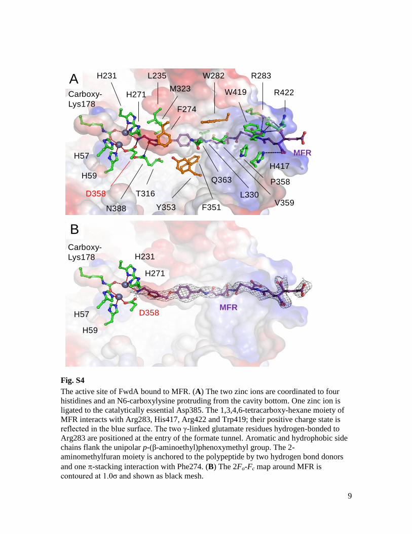
Fig. S4
The active site of FwdA bound to MFR. (A) The two zinc ions are coordinated to four
histidines and an N6-carboxylysine protruding from the cavity bottom. One zinc ion is
ligated to the catalytically essential Asp385. The 1,3,4,6-tetracarboxy-hexane moiety of
MFR interacts with Arg283, His417, Arg422 and Trp419; their positive charge state is
reflected in the blue surface. The two γ-linked glutamate residues hydrogen-bonded to
Arg283 are positioned at the entry of the formate tunnel. Aromatic and hydrophobic side
chains flank the unipolar p-(β-aminoethyl)phenoxymethyl group. The 2-
aminomethylfuran moiety is anchored to the polypeptide by two hydrogen bond donors
and one -stacking interaction with Phe274. (B) The 2Fo-Fc map around MFR is
contoured at 1.0σ and shown as black mesh.
10
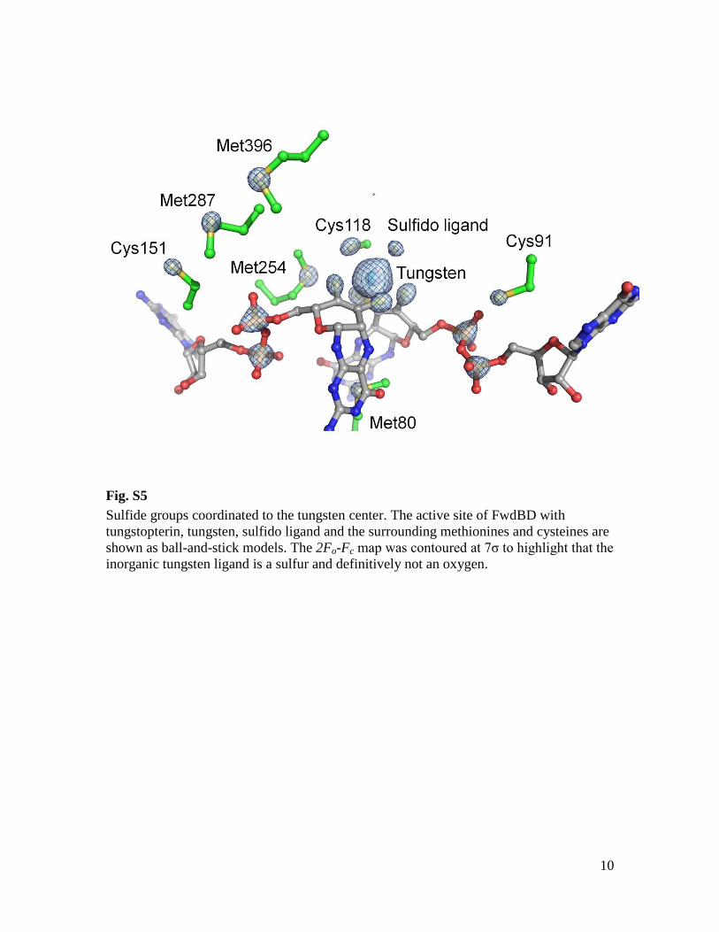
Fig. S5
Sulfide groups coordinated to the tungsten center. The active site of FwdBD with
tungstopterin, tungsten, sulfido ligand and the surrounding methionines and cysteines are
shown as ball-and-stick models. The 2Fo-Fc map was contoured at 7σ to highlight that the
inorganic tungsten ligand is a sulfur and definitively not an oxygen.
11
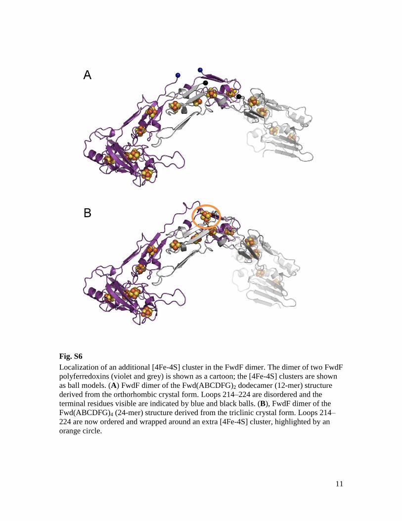
Fig. S6
Localization of an additional [4Fe-4S] cluster in the FwdF dimer. The dimer of two FwdF
polyferredoxins (violet and grey) is shown as a cartoon; the [4Fe-4S] clusters are shown
as ball models. (A) FwdF dimer of the Fwd(ABCDFG)2 dodecamer (12-mer) structure
derived from the orthorhombic crystal form. Loops 214–224 are disordered and the
terminal residues visible are indicated by blue and black balls. (B), FwdF dimer of the
Fwd(ABCDFG)4 (24-mer) structure derived from the triclinic crystal form. Loops 214–
224 are now ordered and wrapped around an extra [4Fe-4S] cluster, highlighted by an
orange circle.
12
a
b
120
A
B
228
135
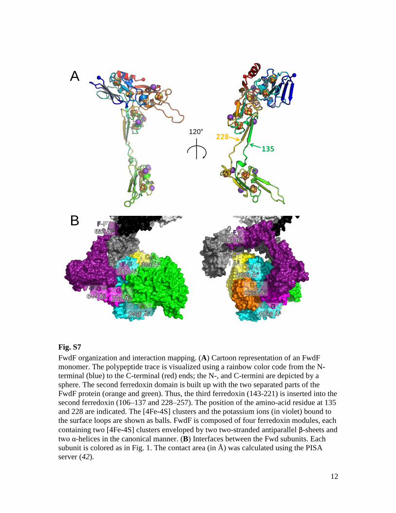
Fig. S7
FwdF organization and interaction mapping. (A) Cartoon representation of an FwdF
monomer. The polypeptide trace is visualized using a rainbow color code from the N-
terminal (blue) to the C-terminal (red) ends; the N-, and C-termini are depicted by a
sphere. The second ferredoxin domain is built up with the two separated parts of the
FwdF protein (orange and green). Thus, the third ferredoxin (143-221) is inserted into the
second ferredoxin (106–137 and 228–257). The position of the amino-acid residue at 135
and 228 are indicated. The [4Fe-4S] clusters and the potassium ions (in violet) bound to
the surface loops are shown as balls. FwdF is composed of four ferredoxin modules, each
containing two [4Fe-4S] clusters enveloped by two two-stranded antiparallel β-sheets and
two α-helices in the canonical manner. (B) Interfaces between the Fwd subunits. Each
subunit is colored as in Fig. 1. The contact area (in Å) was calculated using the PISA
server (42).
13
C91
H290
Tungstopterin
C118
CO2
entry point
Solvated
channel
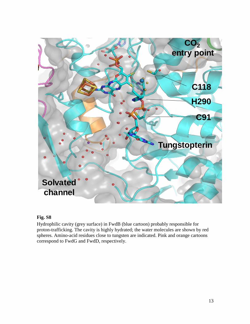
Fig. S8
Hydrophilic cavity (grey surface) in FwdB (blue cartoon) probably responsible for
proton-trafficking. The cavity is highly hydrated; the water molecules are shown by red
spheres. Amino-acid residues close to tungsten are indicated. Pink and orange cartoons
correspond to FwdG and FwdD, respectively.
14
TungstopterinH
CO
O-FwdA
FwdB
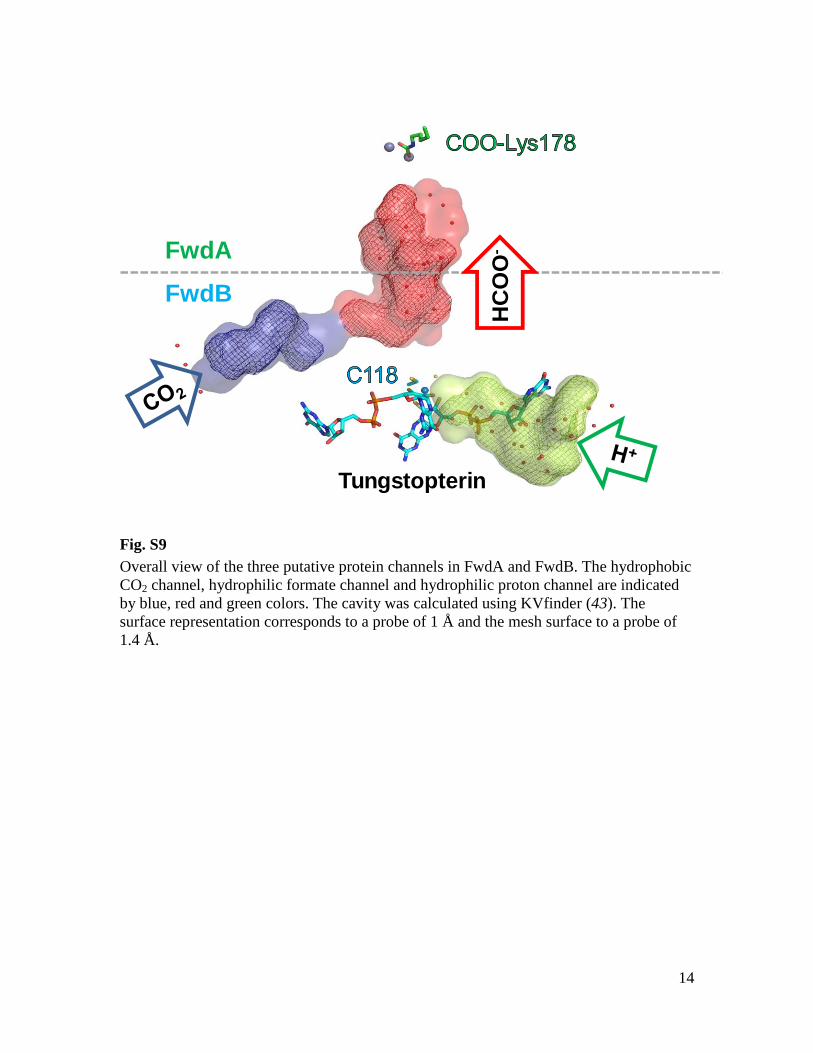
Fig. S9
Overall view of the three putative protein channels in FwdA and FwdB. The hydrophobic
CO2 channel, hydrophilic formate channel and hydrophilic proton channel are indicated
by blue, red and green colors. The cavity was calculated using KVfinder (43). The
surface representation corresponds to a probe of 1 Å and the mesh surface to a probe of
1.4 Å.
15
Tungstopterin
C118
R288
H119
V123
L126Y131L284I270
P286L250
V273
M272
L242
M213
V241
A238
I197
L199I182
V147
L210
W149I256
F252I266
S122
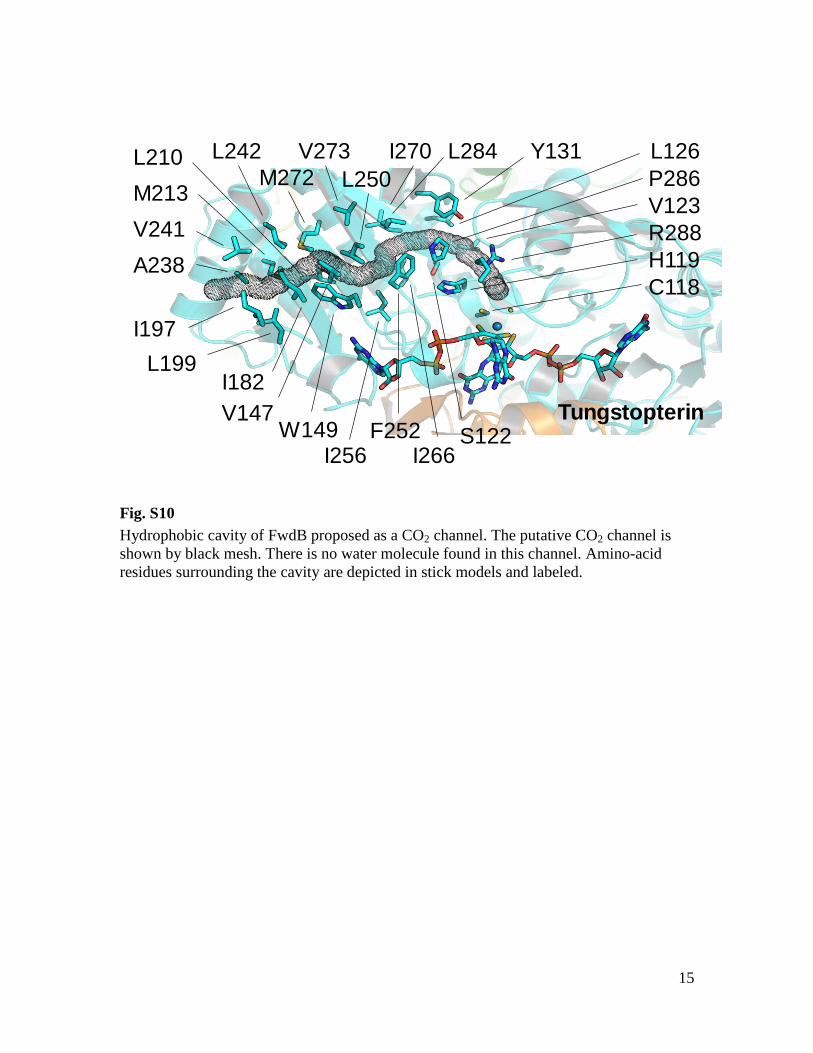
Fig. S10
Hydrophobic cavity of FwdB proposed as a CO2 channel. The putative CO2 channel is
shown by black mesh. There is no water molecule found in this channel. Amino-acid
residues surrounding the cavity are depicted in stick models and labeled.
16
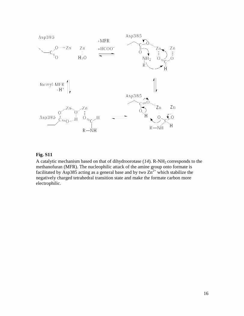
Fig. S11
A catalytic mechanism based on that of dihydroorotase (14). R-NH2 corresponds to the
methanofuran (MFR). The nucleophilic attack of the amine group onto formate is
facilitated by Asp385 acting as a general base and by two Zn2+
which stabilize the
negatively charged tetrahedral transition state and make the formate carbon more
electrophilic.
17
Tungstopterin
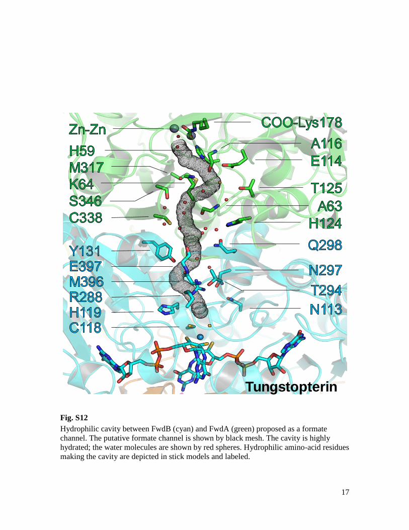
Fig. S12
Hydrophilic cavity between FwdB (cyan) and FwdA (green) proposed as a formate
channel. The putative formate channel is shown by black mesh. The cavity is highly
hydrated; the water molecules are shown by red spheres. Hydrophilic amino-acid residues
making the cavity are depicted in stick models and labeled.
18
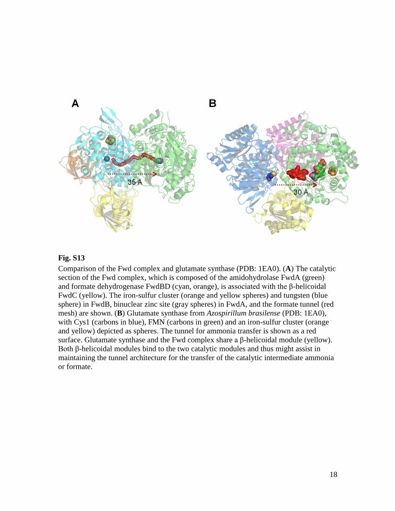
Fig. S13
Comparison of the Fwd complex and glutamate synthase (PDB: 1EA0). (A) The catalytic
section of the Fwd complex, which is composed of the amidohydrolase FwdA (green)
and formate dehydrogenase FwdBD (cyan, orange), is associated with the β-helicoidal
FwdC (yellow). The iron-sulfur cluster (orange and yellow spheres) and tungsten (blue
sphere) in FwdB, binuclear zinc site (gray spheres) in FwdA, and the formate tunnel (red
mesh) are shown. (B) Glutamate synthase from Azospirillum brasilense (PDB: 1EA0),
with Cys1 (carbons in blue), FMN (carbons in green) and an iron-sulfur cluster (orange
and yellow) depicted as spheres. The tunnel for ammonia transfer is shown as a red
surface. Glutamate synthase and the Fwd complex share a β-helicoidal module (yellow).
Both β-helicoidal modules bind to the two catalytic modules and thus might assist in
maintaining the tunnel architecture for the transfer of the catalytic intermediate ammonia
or formate.
19
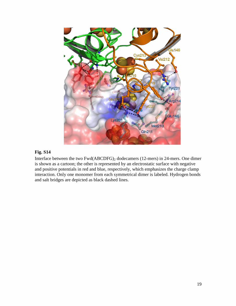
Fig. S14
Interface between the two Fwd(ABCDFG)2 dodecamers (12-mers) in 24-mers. One dimer
is shown as a cartoon; the other is represented by an electrostatic surface with negative
and positive potentials in red and blue, respectively, which emphasizes the charge clamp
interaction. Only one monomer from each symmetrical dimer is labeled. Hydrogen bonds
and salt bridges are depicted as black dashed lines.
20
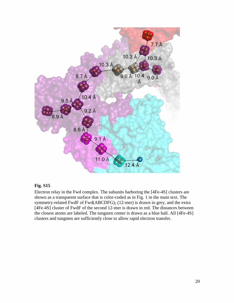
Fig. S15
Electron relay in the Fwd complex. The subunits harboring the [4Fe-4S] clusters are
shown as a transparent surface that is color-coded as in Fig. 1 in the main text. The
symmetry-related FwdF of Fwd(ABCDFG)2 (12-mer) is drawn in grey, and the extra
[4Fe-4S] cluster of FwdF of the second 12-mer is drawn in red. The distances between
the closest atoms are labeled. The tungsten center is drawn as a blue ball. All [4Fe-4S]
clusters and tungsten are sufficiently close to allow rapid electron transfer.
21
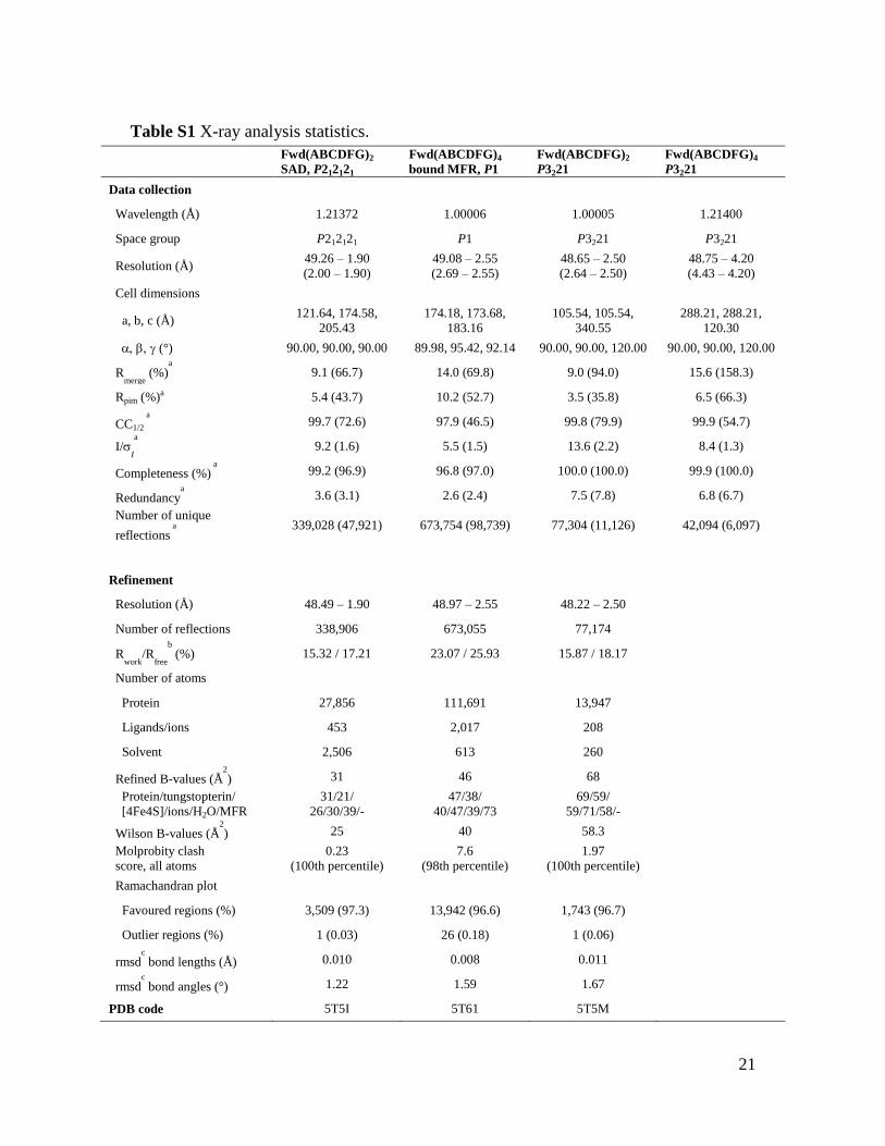
Table S1 X-ray analysis statistics.
Fwd(ABCDFG)2
SAD, P212121
Fwd(ABCDFG)4
bound MFR, P1
Fwd(ABCDFG)2
P3221
Fwd(ABCDFG)4
P3221
Data collection
Wavelength (Å) 1.21372 1.00006 1.00005 1.21400
Space group P212121 P1 P3221 P3221
Resolution (Å) 49.26 – 1.90
(2.00 – 1.90)
49.08 – 2.55
(2.69 – 2.55)
48.65 – 2.50
(2.64 – 2.50)
48.75 – 4.20
(4.43 – 4.20)
Cell dimensions
a, b, c (Å) 121.64, 174.58,
205.43
174.18, 173.68,
183.16
105.54, 105.54,
340.55
288.21, 288.21,
120.30
(°) 90.00, 90.00, 90.00 89.98, 95.42, 92.14 90.00, 90.00, 120.00 90.00, 90.00, 120.00
Rmerge
(%)a
9.1 (66.7) 14.0 (69.8) 9.0 (94.0) 15.6 (158.3)
Rpim (%)a 5.4 (43.7) 10.2 (52.7) 3.5 (35.8) 6.5 (66.3)
CC1/2
a
99.7 (72.6) 97.9 (46.5) 99.8 (79.9) 99.9 (54.7)
I/I
a
9.2 (1.6) 5.5 (1.5) 13.6 (2.2) 8.4 (1.3)
Completeness (%) a
99.2 (96.9) 96.8 (97.0) 100.0 (100.0) 99.9 (100.0)
Redundancya
3.6 (3.1) 2.6 (2.4) 7.5 (7.8) 6.8 (6.7)
Number of unique
reflections a
339,028 (47,921) 673,754 (98,739) 77,304 (11,126) 42,094 (6,097)
Refinement
Resolution (Å) 48.49 – 1.90 48.97 – 2.55 48.22 – 2.50
Number of reflections 338,906 673,055 77,174
Rwork
/Rfree
b
(%) 15.32 / 17.21 23.07 / 25.93 15.87 / 18.17
Number of atoms
Protein 27,856 111,691 13,947
Ligands/ions 453 2,017 208
Solvent 2,506 613 260
Refined B-values (Å2
) 31 46 68
Protein/tungstopterin/
[4Fe4S]/ions/H2O/MFR
31/21/
26/30/39/-
47/38/
40/47/39/73
69/59/
59/71/58/-
Wilson B-values (Å2
) 25 40 58.3
Molprobity clash
score, all atoms
0.23
(100th percentile)
7.6
(98th percentile)
1.97
(100th percentile)
Ramachandran plot
Favoured regions (%) 3,509 (97.3) 13,942 (96.6) 1,743 (96.7)
Outlier regions (%) 1 (0.03) 26 (0.18) 1 (0.06)
rmsdc
bond lengths (Å) 0.010 0.008 0.011
rmsdc
bond angles (°) 1.22 1.59 1.67
PDB code 5T5I 5T61 5T5M
22
a Values relative to the highest resolution shell are within parentheses.
b Rfree was
calculated as the Rwork for 5% of the reflections that were not included in the refinement. c
rmsd, root mean square deviation.
References and Notes
1. R. K. Thauer, A. K. Kaster, H. Seedorf, W. Buckel, R. Hedderich, Methanogenic archaea:
Ecologically relevant differences in energy conservation. Nat. Rev. Microbiol. 6, 579–
591 (2008). Medline doi:10.1038/nrmicro1931
2. P. A. Bertram, R. K. Thauer, Thermodynamics of the formylmethanofuran dehydrogenase
reaction in Methanobacterium thermoautotrophicum. Eur. J. Biochem. 226, 811–818
(1994). Medline doi:10.1111/j.1432-1033.1994.t01-1-00811.x
3. P. A. Bertram, M. Karrasch, R. A. Schmitz, R. Böcher, S. P. Albracht, R. K. Thauer,
Formylmethanofuran dehydrogenases from methanogenic Archaea. Substrate specificity,
EPR properties and reversible inactivation by cyanide of the molybdenum or tungsten
iron-sulfur proteins. Eur. J. Biochem. 220, 477–484 (1994). Medline doi:10.1111/j.1432-
1033.1994.tb18646.x
4. R. A. Schmitz, S. P. J. Albracht, R. K. Thauer, A molybdenum and a tungsten isoenzyme of
formylmethanofuran dehydrogenase in the thermophilic archaeon Methanobacterium
wolfei. Eur. J. Biochem. 209, 1013–1018 (1992). Medline doi:10.1111/j.1432-
1033.1992.tb17376.x
5. R. A. Schmitz, S. P. J. Albracht, R. K. Thauer, Properties of the tungsten-substituted
molybdenum formylmethanofuran dehydrogenase from Methanobacterium wolfei. FEBS
Lett. 309, 78–81 (1992). Medline doi:10.1016/0014-5793(92)80743-Z
6. R. A. Schmitz, M. Richter, D. Linder, R. K. Thauer, A tungsten-containing active
formylmethanofuran dehydrogenase in the thermophilic archaeon Methanobacterium
wolfei. Eur. J. Biochem. 207, 559–565 (1992). Medline doi:10.1111/j.1432-
1033.1992.tb17082.x
7. A. Hochheimer, R. A. Schmitz, R. K. Thauer, R. Hedderich, The tungsten
formylmethanofuran dehydrogenase from Methanobacterium thermoautotrophicum
contains sequence motifs characteristic for enzymes containing molybdopterin
dinucleotide. Eur. J. Biochem. 234, 910–920 (1995). Medline doi:10.1111/j.1432-
1033.1995.910_a.x
8. A. Hochheimer, R. Hedderich, R. K. Thauer, The formylmethanofuran dehydrogenase
isoenzymes in Methanobacterium wolfei and Methanobacterium thermoautotrophicum:
Induction of the molybdenum isoenzyme by molybdate and constitutive synthesis of the
tungsten isoenzyme. Arch. Microbiol. 170, 389–393 (1998). Medline
doi:10.1007/s002030050658
9. A. Hochheimer, D. Linder, R. K. Thauer, R. Hedderich, The molybdenum
formylmethanofuran dehydrogenase operon and the tungsten formylmethanofuran
dehydrogenase operon from Methanobacterium thermoautotrophicum. Structures and
transcriptional regulation. Eur. J. Biochem. 242, 156–162 (1996). Medline
doi:10.1111/j.1432-1033.1996.0156r.x
10. S. Bartoschek, J. A. Vorholt, R. K. Thauer, B. H. Geierstanger, C. Griesinger, N-
carboxymethanofuran (carbamate) formation from methanofuran and CO2 in
methanogenic archaea. Thermodynamics and kinetics of the spontaneous reaction. Eur. J.
Biochem. 267, 3130–3138 (2000). Medline doi:10.1046/j.1432-1327.2000.01331.x
11. J. A. Vorholt, R. K. Thauer, The active species of ‘CO2’ utilized by formylmethanofuran
dehydrogenase from methanogenic Archaea. Eur. J. Biochem. 248, 919–924 (1997).
Medline doi:10.1111/j.1432-1033.1997.00919.x
12. L. R. Celeste, G. Chai, M. Bielak, W. Minor, L. L. Lovelace, L. Lebioda, Mechanism of N10
-
formyltetrahydrofolate synthetase derived from complexes with intermediates and
inhibitors. Protein Sci. 21, 219–228 (2012). Medline doi:10.1002/pro.2005
13. Materials and methods are available as supplementary materials on Science Online.
14. J. B. Thoden, G. N. Phillips Jr., T. M. Neal, F. M. Raushel, H. M. Holden, Molecular
structure of dihydroorotase: A paradigm for catalysis through the use of a binuclear metal
center. Biochemistry 40, 6989–6997 (2001). Medline doi:10.1021/bi010682i
15. J. C. Boyington, V. N. Gladyshev, S. V. Khangulov, T. C. Stadtman, P. D. Sun, Crystal
structure of formate dehydrogenase H: Catalysis involving Mo, molybdopterin,
selenocysteine, and an Fe4S4 cluster. Science 275, 1305–1308 (1997). Medline
doi:10.1126/science.275.5304.1305
16. H. Raaijmakers, S. Macieira, J. M. Dias, S. Teixeira, S. Bursakov, R. Huber, J. J. G. Moura,
I. Moura, M. J. Romão, Gene sequence and the 1.8 Å crystal structure of the tungsten-
containing formate dehydrogenase from Desulfovibrio gigas. Structure 10, 1261–1272
(2002). Medline doi:10.1016/S0969-2126(02)00826-2
17. L. B. Maia, J. J. G. Moura, I. Moura, Molybdenum and tungsten-dependent formate
dehydrogenases. J. Biol. Inorg. Chem. 20, 287–309 (2015). Medline doi:10.1007/s00775-
014-1218-2
18. M. Jormakka, S. Törnroth, B. Byrne, S. Iwata, Molecular basis of proton motive force
generation: Structure of formate dehydrogenase-N. Science 295, 1863–1868 (2002).
Medline doi:10.1126/science.1068186
19. C. Binda, R. T. Bossi, S. Wakatsuki, S. Arzt, A. Coda, B. Curti, M. A. Vanoni, A. Mattevi,
Cross-talk and ammonia channeling between active centers in the unexpected domain
arrangement of glutamate synthase. Structure 8, 1299–1308 (2000). Medline
doi:10.1016/S0969-2126(00)00540-2
20. J. S. Blanchard, W. W. Cleland, Kinetic and chemical mechanisms of yeast formate
dehydrogenase. Biochemistry 19, 3543–3550 (1980). Medline doi:10.1021/bi00556a020
21. B. K. Pomper, O. Saurel, A. Milon, J. A. Vorholt, Generation of formate by the
formyltransferase/hydrolase complex (Fhc) from Methylobacterium extorquens AM1.
FEBS Lett. 523, 133–137 (2002). Medline doi:10.1016/S0014-5793(02)02962-9
22. H. Dobbek, V. Svetlitchnyi, L. Gremer, R. Huber, O. Meyer, Crystal structure of a carbon
monoxide dehydrogenase reveals a [Ni-4Fe-5S] cluster. Science 293, 1281–1285 (2001).
Medline doi:10.1126/science.1061500
23. T. I. Doukov, T. M. Iverson, J. Seravalli, S. W. Ragsdale, C. L. Drennan, A Ni-Fe-Cu center
in a bifunctional carbon monoxide dehydrogenase/acetyl-CoA synthase. Science 298,
567–572 (2002). Medline doi:10.1126/science.1075843
24. X. Huang, H. M. Holden, F. M. Raushel, Channeling of substrates and intermediates in
enzyme-catalyzed reactions. Annu. Rev. Biochem. 70, 149–180 (2001). Medline
doi:10.1146/annurev.biochem.70.1.149
25. K. C. Costa, P. M. Wong, T. Wang, T. J. Lie, J. A. Dodsworth, I. Swanson, J. A. Burn, M.
Hackett, J. A. Leigh, Protein complexing in a methanogen suggests electron bifurcation
and electron delivery from formate to heterodisulfide reductase. Proc. Natl. Acad. Sci.
U.S.A. 107, 11050–11055 (2010). Medline doi:10.1073/pnas.1003653107
26. K. C. Costa, T. J. Lie, Q. Xia, J. A. Leigh, VhuD facilitates electron flow from H2 or formate
to heterodisulfide reductase in Methanococcus maripaludis. J. Bacteriol. 195, 5160–5165
(2013). Medline doi:10.1128/JB.00895-13
27. A. Volbeda, M. H. Charon, C. Piras, E. C. Hatchikian, M. Frey, J. C. Fontecilla-Camps,
Crystal structure of the nickel-iron hydrogenase from Desulfovibrio gigas. Nature 373,
580–587 (1995). Medline doi:10.1038/373580a0
28. J. W. Peters, W. N. Lanzilotta, B. J. Lemon, L. C. Seefeldt, X-ray crystal structure of the Fe-
only hydrogenase (CpI) from Clostridium pasteurianum to 1.8 angstrom resolution.
Science 282, 1853–1858 (1998). Medline doi:10.1126/science.282.5395.1853
29. S. Santabarbara, P. Heathcote, M. C. W. Evans, Modelling of the electron transfer reactions
in Photosystem I by electron tunnelling theory: The phylloquinones bound to the PsaA
and the PsaB reaction centre subunits of PS I are almost isoenergetic to the iron-sulfur
cluster FX. Biochim. Biophys. Acta 1708, 283–310 (2005). Medline
doi:10.1016/j.bbabio.2005.05.001
30. A. Esteve-Núñez, J. Sosnik, P. Visconti, D. R. Lovley, Fluorescent properties of c-type
cytochromes reveal their potential role as an extracytoplasmic electron sink in Geobacter
sulfurreducens. Environ. Microbiol. 10, 497–505 (2008). Medline doi:10.1111/j.1462-
2920.2007.01470.x
31. K. D. Bewley, K. E. Ellis, M. A. Firer-Sherwood, S. J. Elliott, Multi-heme proteins: Nature’s
electronic multi-purpose tool. Biochim. Biophys. Acta 1827, 938–948 (2013). Medline
doi:10.1016/j.bbabio.2013.03.010
32. J. N. Reeve, G. S. Beckler, D. S. Cram, P. T. Hamilton, J. W. Brown, J. A. Krzycki, A. F.
Kolodziej, L. Alex, W. H. Orme-Johnson, C. T. Walsh, A hydrogenase-linked gene in
Methanobacterium thermoautotrophicum strain ΔH encodes a polyferredoxin. Proc. Natl.
Acad. Sci. U.S.A. 86, 3031–3035 (1989). Medline doi:10.1073/pnas.86.9.3031
33. W. Kabsch, Xds. Acta Crystallogr. D 66, 125–132 (2010). Medline
doi:10.1107/S0907444909047337
34. M. D. Winn, C. C. Ballard, K. D. Cowtan, E. J. Dodson, P. Emsley, P. R. Evans, R. M.
Keegan, E. B. Krissinel, A. G. W. Leslie, A. McCoy, S. J. McNicholas, G. N.
Murshudov, N. S. Pannu, E. A. Potterton, H. R. Powell, R. J. Read, A. Vagin, K. S.
Wilson, Overview of the CCP4 suite and current developments. Acta Crystallogr. D 67,
235–242 (2011). Medline doi:10.1107/S0907444910045749
35. G. M. Sheldrick, A short history of SHELX. Acta Crystallogr. A 64, 112–122 (2008).
Medline doi:10.1107/S0108767307043930
36. G. Langer, S. X. Cohen, V. S. Lamzin, A. Perrakis, Automated macromolecular model
building for X-ray crystallography using ARP/wARP version 7. Nat. Protoc. 3, 1171–
1179 (2008). Medline doi:10.1038/nprot.2008.91
37. A. Vagin, A. Teplyakov, MOLREP: An automated program for molecular replacement. J.
Appl. Cryst. 30, 1022–1025 (1997). doi:10.1107/S0021889897006766
38. P. V. Afonine, R. W. Grosse-Kunstleve, V. B. Chen, J. J. Headd, N. W. Moriarty, J. S.
Richardson, D. C. Richardson, A. Urzhumtsev, P. H. Zwart, P. D. Adams,
phenix.model_vs_data: A high-level tool for the calculation of crystallographic model
and data statistics. J. Appl. Crystallogr. 43, 669–676 (2010). Medline
doi:10.1107/S0021889810015608
39. P. Emsley, K. Cowtan, Coot: Model-building tools for molecular graphics. Acta Crystallogr.
D 60, 2126–2132 (2004). Medline doi:10.1107/S0907444904019158
40. G. N. Murshudov, A. A. Vagin, E. J. Dodson, Refinement of macromolecular structures by
the maximum-likelihood method. Acta Crystallogr. D 53, 240–255 (1997). Medline
doi:10.1107/S0907444996012255
41. E. Chovancova, A. Pavelka, P. Benes, O. Strnad, J. Brezovsky, B. Kozlikova, A. Gora, V.
Sustr, M. Klvana, P. Medek, L. Biedermannova, J. Sochor, J. Damborsky, CAVER 3.0:
A tool for the analysis of transport pathways in dynamic protein structures. PLOS
Comput. Biol. 8, e1002708 (2012). Medline doi:10.1371/journal.pcbi.1002708
42. E. Krissinel, K. Henrick, Inference of macromolecular assemblies from crystalline state. J.
Mol. Biol. 372, 774–797 (2007). Medline doi:10.1016/j.jmb.2007.05.022
43. S. H. P. Oliveira, F. A. N. Feraz, R. V. Honorato, J. Xavier-Neto, T. J. P. Sobreira, P. S. de
Oliveira, KVFinder: Steered identification of protein cavities as a PyMOL plugin. BMC
Bioinformatics 15, 197 (2014). Medline doi:10.1186/1471-2105-15-197