Embed Size (px)
Citation preview

Biophysical Journal Volume 97 September 2009 1709–1718 1709
Structural Changes to Monomeric CuZn Superoxide Dismutase Causedby the Familial Amyotrophic Lateral Sclerosis-Associated Mutation A4V
Tom Schmidlin,† Brian K. Kennedy,† and Valerie Daggett†‡*†Department of Biochemistry and ‡Department of Bioengineering, University of Washington, Seattle, Washington
ABSTRACT Amyotrophic lateral sclerosis (ALS) is a progressive motor neuron degenerative disease, and the inherited form,familial ALS (fALS), has been linked to over 100 different point mutations scattered throughout the Cu-Zn superoxide dismutaseprotein (SOD1). The disease is likely due to a toxic gain of function caused by the misfolding, oligomerization, and eventualaggregation of mutant SOD1, but it is not yet understood how the structurally diverse mutations result in a common diseasephenotype. The behavior of the apo-monomer fALS-associated mutant protein A4V was explored using molecular-dynamicssimulations to elucidate characteristic structural changes to the protein that may allow the mutant form to improperly associatewith other monomer subunits. Simulations showed that the mutant protein is less stable than the WT protein overall, with shifts inresidue-residue contacts that lead to destabilization of the dimer and metal-binding sites, and stabilization of nonnative contactsthat leads to a misfolded state. These findings provide a unifying explanation for disparate experimental observations, allowa better understanding of alterations of residue contacts that accompany loss of SOD1 structural integrity, and suggest siteswhere compensatory changes may stabilize the mutant structure.
INTRODUCTION
A subset of familial amyotrophic lateral sclerosis (fALS)
cases has been tied to mutations in the Cu-Zn superoxide dis-
mutase protein (SOD1). Though the mechanism of toxicity
has been under intense scrutiny for 15 years, it remains
largely undetermined. One current model involves the mis-
folding of the mutant protein (1), which results in a new
structural species that directly confers toxicity and/or seques-
ters cellular machinery that is important for protein homeo-
stasis, such as chaperones or proteasome components.
SOD1 is normally responsible for the disproportionation
of superoxide to molecular oxygen and hydrogen peroxide,
in which one superoxide molecule is oxidized and then
another is reduced by copper in the active site of the enzyme.
Superoxide is a naturally occurring byproduct of respiration.
Although SOD1 is abundant and ubiquitous in the cyto-
plasm, it has also been found in the mitochondrial intermem-
brane space. The concentrations are especially high in certain
subcellular locations, such as motor neuron axons (2).
Only minimal changes have been observed in the crystal
structure of fully metallated disease-associated mutant forms
of SOD1 in comparison to wild-type (WT) SOD1. Many
SOD1 mutants display lower melting temperatures than their
WT counterparts, regardless of the metallation state (3), and
they unfold more easily with urea or guanidine-HCL (4).
However, some of the metal-binding mutants have higher
melting temperatures than WT (5). Both sporadic and
familial forms of ALS show evidence of cytoplasmic inclu-
sion bodies—deposits that are characteristic of many neuro-
degenerative disorders, including Huntington’s disease,
Submitted December 12, 2008, and accepted for publication June 15, 2009.
*Correspondence: [email protected]
Editor: Kathleen B. Hall.
� 2009 by the Biophysical Society
0006-3495/09/09/1709/10 $2.00
Alzheimer’s disease, and Parkinson’s disease (6)—and
aggregates have been found in mouse models of the disease
(7–9). The familial ALS aggregates contain SOD1, neurofi-
lament proteins, ubiquitin, and a host of other cellular
components, but it is not clear whether zinc or copper is
present (10). The current thinking is that the aggregation
is a cellular protective mechanism and the most toxic form
is either a misfolded monomer or a soluble oligomeric
species or protofibril (6). Similar models have been proposed
for other neurodegenerative diseases (11).
The unfolding and aggregation pathway of the protein
involves the dissociation of the dimer and loss of metal
binding, followed by the subsequent oligomeric assembly
of the protein (12–14). It is also possible that the aggregates
are favored when the amount of misfolded protein reaches
a point where the ubiquitin proteolytic machinery becomes
unable to handle the load (15,16). Although the aforemen-
tioned experimental approaches have been enlightening,
there are other opportunities to investigate the effects of
mutation on the protein. An atomic-level look at the protein
dynamics through molecular-dynamics (MD) simulations
can be informative in determining the effect the mutations
have on the structure of the protein, allowing investigation
of how changing one residue to another can create a ripple
effect through the protein that eventually affects dimeriza-
tion, metal binding, and/or overall protein stability.
SOD1 conforms to the Greek key b-barrel folding
topology, and each monomer subunit of the homodimer
binds one copper and one zinc ion. Although the crystal
structures of several ALS-causing mutants have been solved,
the structure of WT SOD1 in solution differs from the crystal
structure (17). These average structures are very informative;
however, proteins are dynamic and important conforma-
tional states may have low sampling rates.
doi: 10.1016/j.bpj.2009.06.043

1710 Schmidlin et al.
Previous MD studies have included short (<1 ns) simula-
tions of WT bovine and human SOD1 dimers (18–20), as
well as comparisons of simulations of the WT monomer
and dimer (21,22). In addition, 5 ns simulations of WT and
mutant (A4V, G37R, and H46R) dimers and monomers
(23) have provided the first direct comparison of mutant
and WT MD simulations. These results indicate that altered
long-range communication within the protein structure could
be an underlying cause of aggregation of mutant SOD1 in
fALS. Also, a recent study examined several 100 ns simula-
tions of WT apo and holo monomers and dimers, and the
effects of disulfide bonds on SOD1 stability (24).
Here we report findings from multiple long (60 ns) simu-
lations of WT and A4V apo (demetallated) monomers to
explore the dominant patterns of structural changes upon
mutation in the monomer after loss of metallation. A4V is
very close to the dimer interface and represents one of the
most common and most lethal mutations, with rapid disease
progression and death occurring on average at 1.4 years vs.
3–5 years for other mutants (25). These simulations reveal
significant changes in the mutant protein structure, which
is discussed in terms of potential effects on dimer destabili-
zation, loss of metallation, and the aberrant oligomerization
of the misfolded monomer.
MATERIALS AND METHODS
Models
The starting structure for WT SOD1 monomer was obtained from a 1.8 A
crystal structure of human SOD1 (1HL5 Chain A) (26). The A4V SOD1
starting structure was obtained from a 1.9 A crystal structure of the A4V
mutant of human SOD1 (1UXM Chain A) (17). Before energy minimization
was performed, the Cu2þ and Zn2þ ions were removed and H63 was left un-
protonated. The ionization states of the amino acids corresponded to neutral
pH (Asp�, Glu�, His0, Lysþ, and Argþ). The C57-C146 disulfide bond was
intact to match in vitro studies.
The holo 1HL5 structure was chosen over the apo 1HL4 structure for
a variety of reasons. In brief, of the four SOD1 proteins available in the
1HL4 tetramer, chains C and D are missing many residues (26 and 28 out
of 153, respectively). Chains A and B are missing many heavy atoms
(18 and 12 atoms, respectively) and they also have bound zinc. The model-
building required to render these structures suitable for simulation would
be quite extensive, and the resultant starting structure would be inferior to
the 1HL5 structure used.
MD simulations
Simulations of apo WT and A4V SOD1 were performed with the in lucem
Molecular Mechanics (ilmm) (27) simulation software using protocols
described elsewhere (27–32). In brief, the simulations included all hydro-
gens and explicit flexible three-center waters (29). The protein was solvated
in a box extending at least 10 A from any protein atom, with the solvent
density set to the experimental value at 310 K of 0.993 g/mL (33). Periodic
boundary conditions were employed to minimize edge effects. The microca-
nonical constant number of molecules, volume, and energy (NVE) ensemble
was employed. A 10 A force-shifted cutoff range was used for all nonbonded
atom interactions, and the interaction pair list was updated every three steps.
The potential energy functions were used to propagate MD trajectories with
a 2 fs time step, and structures were saved every 1 ps for analysis. Three
Biophysical Journal 97(6) 1709–1718
60 ns simulations were run for each WT and A4V apoprotein at 310 K,
yielding ~180,000 structures each for analysis. The long simulation time
allows the different simulations to reach a ‘‘converged’’ state for each mono-
mer in solution with enough sampling of the state to be statistically sound.
Analyses
Except where noted, all analyses were performed over all structures. The
Define Secondary Structure of Proteins (DSSP) algorithm (34) was used
to assign secondary structure based on hydrogen-bond energies. The
DSSP data used to generate the graphs were sampled at 1 ps intervals;
however, conformational time-based populations from DSSP were calcu-
lated at 100 ps intervals.
The CONGENEAL structural dissimilarity scores (35) for each simula-
tion were calculated at each time point based on the minimized structure
of the protein. CONGENEAL is based on the weighted distance maps of
two structures, such that the weight is higher for atoms that are closer
together. Each element of the matrix is calculated using the equation
wi,j ¼ di,j�p, where w is the weight, d is the Ca distance between the two
residues, and p ¼ 2.
Differences between the total contact time of residue pairs in the WT and
A4V simulations were considered to be statistically significant if the differ-
ence in the averages of the two different types of simulations was greater
than the combined standard deviation (SD) of those values. Atoms were
considered to be in contact if the heavy atom distance was <4.6 A, except
for C-C, where the cutoff was 5.4 A. Protein images were produced using
UCSF Chimera (36).
RESULTS AND DISCUSSION
Simulations of the fALS-associated SOD1 mutant A4V
showed the effects of the mutation on the kinetic stability
of the protein when compared with WT under similar condi-
tions (neutral pH, 310 K, apo, monomer). For each WT and
mutant protein, three simulations were run for 60.5–63.0 ns
and then analyzed. The differences between the two
sequences may be illustrated in a number of ways to provide
clues for the previously reported effects of this mutation on
dimer stability (14) and metal binding (37,38).
Overall protein instability
The simulations reveal that during the course of simulation,
the A4V mutant protein undergoes larger structural changes
than WT. An examination of the Ca root mean-square devi-
ation (RMSD) (see Fig. S1, A and B, in the Supporting
Material), total solvent accessible surface area (SASA)
(Fig. S1, C and D), radius of gyration (Fig. S1, E and F),
and CONGENEAL structural dissimilarity score (Fig. S1,
G and H) for the simulations shows a general increase in
all of these measures across the simulations for the mutant
versus WT. The smaller changes in the SASA and radius
of gyration data indicate that the mutant protein sample
conformations are somewhat different but still fairly
compact. A breakdown of the various components that
comprise the total SASA of the protein reveals that each of
the components of the total SASA is generally higher in
the mutant than in the WT (data not shown). Overall, larger
changes in the structure are observed for A4V than for WT.
fALS Mutation A4V Alters SOD1 Structure 1711
Perturbations of residues normally at the dimerinterface
Dimerization of the individual monomer subunits increases
the stability of the SOD1 enzyme (39), and thus mutations
that destabilize the dimer may decrease the overall protein
stability. Dimer destabilization may be viewed in two
ways: instability in the dimer itself, or a decreased affinity
for dimerization by the individual monomer subunits. Pertur-
bations of the native dimer interface residues may either
prevent the initial dimerization or decrease the lifetime of
the dimer. Although the effects of the mutation on the dimer
would be better studied using dimer simulations, our intent
was to study the A4V-associated structural changes in the
monomer that occur after the known loss of dimerization,
which may have implications for the ability of the dimer to
reform.
One way to measure perturbations of the protein in MD
simulations is to examine the changes in contacts between
residues over time. A minimum increase or decrease in total
contact time of 50% over the entire simulation between any
residue pair in the protein was used as a cutoff, allowing for
the creation of a map showing the major changes in intramo-
nomer residue-residue contacts as the effects propagated
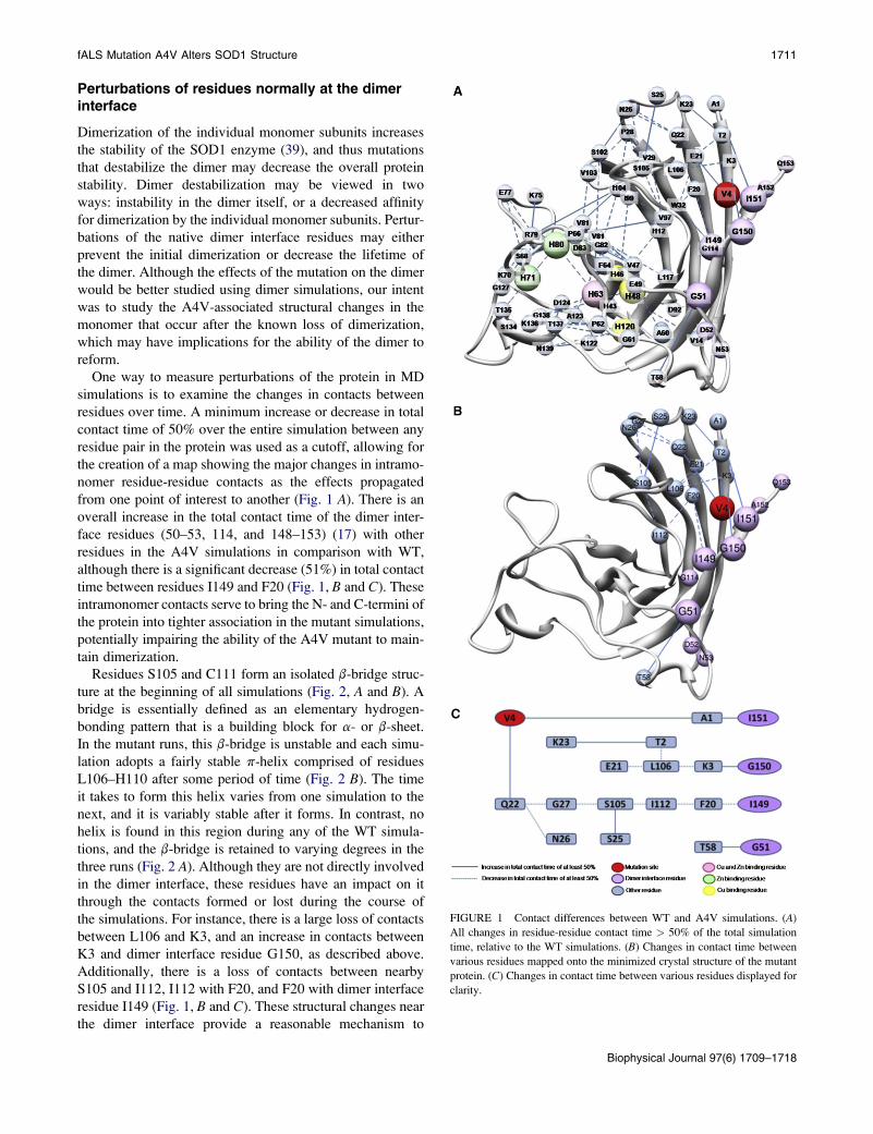
from one point of interest to another (Fig. 1 A). There is an
overall increase in the total contact time of the dimer inter-
face residues (50–53, 114, and 148–153) (17) with other
residues in the A4V simulations in comparison with WT,
although there is a significant decrease (51%) in total contact
time between residues I149 and F20 (Fig. 1, B and C). These
intramonomer contacts serve to bring the N- and C-termini of
the protein into tighter association in the mutant simulations,
potentially impairing the ability of the A4V mutant to main-
tain dimerization.
Residues S105 and C111 form an isolated b-bridge struc-
ture at the beginning of all simulations (Fig. 2, A and B). A
bridge is essentially defined as an elementary hydrogen-
bonding pattern that is a building block for a- or b-sheet.
In the mutant runs, this b-bridge is unstable and each simu-
lation adopts a fairly stable p-helix comprised of residues
L106–H110 after some period of time (Fig. 2 B). The time
it takes to form this helix varies from one simulation to the
next, and it is variably stable after it forms. In contrast, no
helix is found in this region during any of the WT simula-
tions, and the b-bridge is retained to varying degrees in the
three runs (Fig. 2 A). Although they are not directly involved
in the dimer interface, these residues have an impact on it
through the contacts formed or lost during the course of
the simulations. For instance, there is a large loss of contacts
between L106 and K3, and an increase in contacts between
K3 and dimer interface residue G150, as described above.
Additionally, there is a loss of contacts between nearby
S105 and I112, I112 with F20, and F20 with dimer interface
residue I149 (Fig. 1, B and C). These structural changes near
the dimer interface provide a reasonable mechanism to
FIGURE 1 Contact differences between WT and A4V simulations. (A)
All changes in residue-residue contact time > 50% of the total simulation
time, relative to the WT simulations. (B) Changes in contact time between
various residues mapped onto the minimized crystal structure of the mutant
protein. (C) Changes in contact time between various residues displayed for
clarity.
Biophysical Journal 97(6) 1709–1718

1712 Schmidlin et al.
FIGURE 2 Protein secondary structure. Hydrogen-bond
analysis and secondary structure assignment of representa-
tive (A) WT and (B) A4V simulations using DSSP. (C) The
starting structure of the WT protein with the b-strands
labeled.
explain the substantial destabilization of the A4V mutant
SOD1 dimer (17), which is the first step of the proposed olig-
omerization pathway.
Perturbations of the metal-binding sites
Copper-binding site
The residues involved in coordinating the metal (Fig. S2) are
distal to the A4V mutation, yet they show greater movement
during simulation in the mutant than in WT. The Ca root
mean-square fluctuation (RMSF) (Fig. 3, A and B) and Ca
RMSD (not shown) plots show higher values for these resi-
dues, although the distances between the Cu2þ ion-binding
atoms are not significantly different between the WT and
mutant proteins (Fig. 3 C). This is not surprising given the
known role of the Cu2þ ion in the enzymatic reaction, and
the normal level of catalytic activity of this mutant (37,40).
Motion of the electrostatic loop in WT apo dimer simula-
tions was previously reported (22) and we also observe this
motion in our monomer simulations. However, the helix in
the electrostatic loop near the copper-binding site is more
stable and has less apparent motion in the WT form
(Fig. S3 A) than in the mutant (Fig. S3B). The rotation of
this helix may be explained by a large decrease in the con-
tacts between the residues of the helix and the body of the
protein, specifically residue H63, which coordinates both
Zn2þ and Cu2þ binding. Table S1 shows that H63 has
greatly reduced total contact times with S134 (57%
FIGURE 3 Changes to metal-binding residues. (A)
Ca-RMSF by residue for three each WT (light gray, shown
in red online) and A4V (dark gray, blue online) simulation.
(B) Differences in Ca-RMSF are mapped onto the protein
structure. Regions with a higher RMSF in the mutant than
in the WT are shown in dark gray (blue online), and regions
with higher RMSF in the WT than in the mutant are shown
in light gray (red online). White indicates no difference.
The four residues displayed are the Zn2þ-binding residues.
H71, H80, and D83 each show higher RMSF and RMSD
(not shown) in the A4V mutant simulations than in the
WT. (C) Average pairwise distances between the Cu2þ-
binding atoms for three simulations of WT (diamonds)and A4V (squares) proteins. In each case the error bars
represent the SD of the three simulations, and in each
case the error bars overlap. The WT crystal structure
distances are shown as triangles. Mutant crystal structure
distances differ from WT by <0.2 A in all cases, including
those shown in panel D (not shown). (D) Average pairwise
distances between the Zn2þ-binding atoms for three simu-
lations of WT (diamonds) and A4V (squares) proteins. In
each case the error bars represent the SD of the three simu-
lations. There are significant differences between the
distances of the pairs: 63ND1-71ND1, 63ND1-83OD1,
71ND1-80ND1, and 80ND1-83OD1. Triangles are as in
panel C.
Biophysical Journal 97(6) 1709–1718

fALS Mutation A4V Alters SOD1 Structure 1713
reduction), T135 (43%), T137 (27%), and G138 (58%), and
H120 has less contact with G138 (57%) and N139 (75%)
during the mutant simulations compared to WT. Residues
in this region (residues 122–140) that differ by>50% in total
contact time between the WT and A4V simulations are
shown in Fig. 4. Toward the end of the third WT simulation,
the helix moves into a similar position as seen in the mutant,
which may mean that this motion is not an important effect
of the mutation. However, the apo form of the protein is
less stable (3,5,39,41), and this may simply be a consequence
of the lower stability. The mutant protein spends far more
time with the helix in the new position than does WT. There
is also greater movement of the C-terminal residues in WT
than in the mutant, as discussed above.
The native a-helix in the electrostatic loop, comprised of
residues E132–T137 (Fig. 5 A), is somewhat more stable in
the WT simulations than in the mutant runs. Toward the end
of one WT run at ~56 ns, the N-terminus of the helix
extends to include neighboring residues through K128. In
another WT run, the helix converts to p-helix intermittently
from ~7 to 23.5 ns, spending ~57% of this time in a p-helical
structure before returning more stably to the a-helix struc-
ture. The mutant simulations are somewhat different. In
one case, the a-helix in this region is very stable. In another,
a fairly stable p-helix forms that lasts from ~27.5 ns until the
FIGURE 4 Contact differences of the helix near the Cu2þ-binding site.
All changes are relative to WT simulations. (A) Changes in contact time
between various residues mapped onto the minimized crystal structure of
the mutant protein. (B) Changes in contact time between various residues
displayed for clarity.
end of the run (60 ns). In the third simulation, the helix
falls apart after only 3.5 ns. It returns stably for over 20 ns
beginning around 27 ns into the run, but after that it comes
apart again and does not refold before the end of the simu-
lation.
Although the A4V mutant has roughly WT levels of dis-
mutase activity per copper, it also has only 50% as much
copper bound (37). The destabilization of the electrostatic
loop observed by MD may affect the ability of the copper
chaperone to adequately load the copper ion into the binding
site. Alternatively, the deformations of the loop may affect
the protein’s ability to hold onto the metal, accounting for
the drop in observed copper binding.
FIGURE 5 Helix near the Zn2þ-binding residues. Locations of the native
(E132-T137) and nonnative (G73-E77) helix are shown in WT (A) in the
starting structure and (B) at 11.6 ns. Three of the zinc-binding residues are
shown as spheres.
Biophysical Journal 97(6) 1709–1718

1714 Schmidlin et al.
Zinc-binding site
The change in average distance between some of the zinc-
binding atoms is significant in the mutant simulations
(Fig. 3 D). Although there is no difference in the distance
between atoms 63ND1 and 80ND1 or between 71ND1 and
83OD1, there is a difference in the distances between
63ND1 and both 71ND1 and 83OD1, as well as between
80ND1 and both 71ND1 and 83OD1. These results indicate
that residues H63 and H80 move together in the mutant, as
do H71 and D83. This motion and the increased atom-
atom distance may prevent the mutant from properly binding
Zn2þ, consistent with the experimentally observed 30-fold
decrease in zinc affinity in the A4V mutant (38). Zinc
binding is known to stabilize the protein (3,5,41), and loss
of metal binding precedes oligomerization. Recent evidence
suggests that loss of zinc binding occurs simultaneously with
dimer dissociation on the millisecond timescale of the exper-
imental work (42).
A nonnative helix is formed around residues 73–77 in WT
(Fig. 5 B), a stretch that is unstructured in the crystal struc-
ture. Although these residues are not themselves involved
with zinc binding, they are close to the zinc-binding residues
H71 and H80. The different WT runs show a helical structure
forming in this region for varying amounts of time. The
nature of the helix also varies, alternating between a-helix,
p-helix, and 3/10 helix; however, the formation of some
helical structure at this location is consistent across all three
simulations, whereas no significant amount of helix is found
in this location during any of the mutant simulations. Struc-
tural stability in this region may be increased by the helix in
the WT simulations, accounting for the smaller changes in
the zinc-coordinating atom distances compared to the mutant
simulations. Movements of the zinc-binding loop were noted
in a previous work (22), but no mention was made of a helix
in this area.
Contact network analysis revealed a potential propagation
pathway for structural changes across the protein, which may
also lead to the observed differences in the pairwise distances
of the Zn2þ-binding residues (Fig. 6 A). A maximum of
seven contacts are needed for the four Zn2þ residues in the
map (H63, H71, H80, and D83) at a 45% cutoff. The three
Cu2þ-binding residues (H48, H63, and H120) required
a maximum of nine contacts, whereas the fourth residue
(H46) could not be included at that cutoff.
In Fig. 6 B this contact network is mapped onto the struc-
ture to illustrate how the A4V mutation causes a ripple effect
through the protein. The contacts shift between residues, and
the changes propagate throughout the protein. For example,
G27 loses contact with S105 and moves to S102. The loss of
contact between G27 and S105 causes S105 to lose contact
with residues I112 and P28, both of which then lose contact
with residue I104. Loss of contact between residues I112 and
I104 causes residue I112 to come into contact with residue
P66, a contact that almost never occurs in the WT simula-
Biophysical Journal 97(6) 1709–1718
tions (2% 5 3.46%). Residue I104 then loses contact with
G82 and has a significant increase in contact with residues
H80 and R79, and R79 loses contact with H71. Note that
H80 and H71 are zinc-binding residues. A list of the contacts
gained and lost during the simulations, as reflected in Fig. 6,
is provided in Table S1.
Through this type of analysis, we are able to identify key
residues between the mutation site and the site of interest,
such as residue I104, which serves as a branch point on
the map leading to all three of the Zn2þ-binding residues.
This residue could be mutated to halt the propagation of
the changes and, it is hoped, eliminate the phenotypes caused
by the A4V mutation. However, even though I104 is highly
conserved (43), there is a known fALS mutation at this
residue: I104F. If I104 serves as a key residue in maintaining
FIGURE 6 Contact differences between WT and A4V simulations. All
changes shown represent a minimum of a 45% absolute change in contact
time relative to WT simulations. (A) Map of the changes in contact time dis-
played for clarity. (B) The starting structure of the A4V mutant shown with
selected changes in contact between residues. The changes displayed repre-
sent the shortest pathways of change between the mutation site and the
metal-binding residues. Metal-binding residue not shown (Cu2þ: H46) did
not meet the threshold of 45% change in absolute contact time between
WT and A4V simulations.

fALS Mutation A4V Alters SOD1 Structure 1715
SOD1 stability, mutation at this site may only further exac-
erbate the instability of the monomer.
Potential effects on oligomeric assembly
Exposure of cysteines
Previous research indicates that C6 and C111 play a role in
SOD1 aggregation, although reports vary. It is not clear
whether both Cys residues are important for oligomerization
(44,45), C111 is important but C6 is not (46), or C6
and C111 play some role but are not the only important
factors (47).
Our results indicate that C111 has a ~20% higher average
solvent exposure in A4V compared to WT (69.1 A2 for
A4V vs. 57.1 A2 for WT), and the close association of b8
with b1 in the mutant simulations effectively prevents a signif-
icant change in solvent accessibility for C6 (1.1 A2 for A4V
vs. 1.5 A2 for WT). The increase in solvent accessibility for
C111 was predicted via Carr-Purcell-Meiboom-Gill 15N
nuclear spin relaxation dispersion measurements by K. Tei-
lum, M. H. Smith, E. Schulz, G. Solomentsev, M. Oliveberg,
and M. Akke (unpublished). This solvent exposure of the free
cysteine C111 may allow for nonnative monomer-monomer
interactions.
b-Sheet instability
The starting structures of both the WT (Fig. 2 C) and mutant
proteins contain b-strand comprising residues G85–A89
(strand b5) and V94–D101 (strand b6). b5 is an edge strand
of the sheet formed with strands b4, b7, and b8, whereas
strand b6 is an edge strand of the sheet formed with
strands b1, b2, and b3. Strands b5 and b6 are more stable
in WT than in mutant simulations (Fig. 2, A and B). Previous
MD simulations also found destabilization of b5 and b6 (23).
In the first two WT runs, the b-sheets are largely retained
at b5 and b6, with some loss of strand from the N-terminal
end of strand b6. In the third simulation, strand b6 loses
most hydrogen bonds with b3 by 26 ns, although some
b-bridge is retained through the end of the simulation. In
the A4V simulations, it is b5 that is less stable. In the first
mutant run, the hydrogen bonds formed with b4 have largely
disappeared after only 2.2 ns, although some b-bridges
appear infrequently. Strand b6 is much more stable, retaining
some b-sheet contacts for ~97% of the simulation and at a
minimum some b-bridges for >99% of the 60 ns simulation.
The second simulation reveals a loss of b-sheet beginning
with b5 (residues G85–A89), which loses most of the
b-structure after only a few nanoseconds. Some degree of
b-structure is retained in this region for nearly 40 ns,
however, alternating between fragments of b-sheet and
b-bridges during that time. After 40 ns, what little structure
is found in this area is a in nature, mostly a-bridges formed
between residues N86 on b5 and H43 on b4. Strand b6 also
begins to lose structure early in the simulation; however, the
ends of the strand remain in contact with b3 for much of the
first 40 ns of simulation. By 52.5 ns, there is a complete loss
of b-structure in b6, but by 44.6 ns residue D96 begins to
form frequent a-bridge contact with S34 on neighboring
b3. This contact is present ~94% of the time through the
end of the simulation. The third mutant simulation also
rapidly loses b-sheet contacts involving b5, as well as
a slower but substantial loss of b-sheet contacts from b6.
Strand b5 loses most of its b-structure by 4 ns, whereas b6
loses b-structure at ~30 ns. In each of the mutant simulations,
the loss of b-sheet contacts in b5 and b6 are accompanied by
some loss of contacts in neighboring strands b4 and b3,
respectively; however, each simulation retains b-sheet in
these regions through the end of the runs. The observed
destabilization of b5 and b6 is consistent with previous
MD studies (23).
The typical forms of edge b-strands protect proteins from
edge-edge aggregation (48). The perturbation and loss of the
native edge strands in the mutant simulations provide
another potential mechanism by which the mutant monomers
can improperly interact with other monomers. The b-barrel,
specifically b3 and b4 (49) and b5 and b6, was previously
identified experimentally as a potential hotspot for local un-
folding leading to aggregation in fALS cases (50), and our
results are in good agreement with recent findings of b-strand
destabilization of apo SOD1 monomers (24).
a-Strand and a-bridges
A hydrogen-bond analysis of the first A4V run shows non-
helical local a-structure beginning after 31 ns and continuing
at some level through the end of the simulation. This local
a-structure, when alternating between aL and aR, can give
rise to strands similar to b-strands, but in this case the main
chain carbonyl oxygens are aligned on one side and the amide
hydrogens along the other, rather than alternating. We refer
to this structure as a-sheet. From 31.1 ns to 51.3 ns, a-strand
or bridges are present in the area of residues F50–G51 and
S59–A60, near the dimer interface, for ~44% of the time. At
this point there is a ~6 ns period during which there is
no sampling of this structure; sampling then resumes at
~57.6 ns and continues for roughly 64% of the time through
the end of the run. A representative structure (32.8 ns) was
chosen for further analysis of the a-sheet hydrogen-bond
energies, particularly between residues 49–52 and 58–61
(Fig. 7). Note that residues G51 and D52 are dimer interface
residues.
In the second mutant simulation, intermittent a-bridges
form between residues G51 and A60, H43 and N86, and
S34 and D96, beginning at different time points but
continuing through the end of the simulation. The a-bridge
between residues G51 and A60 forms first, initially appear-
ing at ~6.6 ns into the simulation and persisting for ~44%
of the time. The a-bridge between residues H43 and N86
forms next at ~40 ns of the run and is present ~40% of the
time through the end of the run. Finally, the a-bridge
between residues S34 and D96 does not form until ~45 ns
Biophysical Journal 97(6) 1709–1718
1716 Schmidlin et al.
FIGURE 7 a-Sheet in the mutant simulations. A repre-
sentative structure of the A4V simulations shows one
location of a-sheet formation. In the right panel, the back-
ground structure and atoms have been removed for clarity.
into the run, but it is present ~94% of the time thereafter.
None of these a-bridges require either of the others to be
present for formation. In the third A4V run, a-sheet again
forms between residues F50–G51 and S59–A60, much like
in the first mutant run. There is an a-bridge between residues
G51 and A60 beginning around 4 ns of the simulation and
continuing nearly 73% of the time through 8.4 ns of the
simulation. At this point, it breaks and no significant amount
of a-bridge or a-sheet appears until after nearly 27 ns of
simulation time. After this time, there is an a-bridge or
a-sheet present in this region >83% of the time through
the end of the run.
In contrast to A4V, the a-bridge is barely populated
(<5%) in the second WT simulation, and none is found in
either of the other two WT runs. When we examine the
data by (4, j) angles, we can see that there is no a-strand
comprised of four or more consecutive residues in any of
the three neutral pH WT simulations that were run (data
not shown); however, it is present in all three of the A4V
simulations. In the first A4V simulation, this strand is
predominantly found from residues T88 to D92, forming
in the loop between b5 and b6. The other two simulations
form a-strand in different locations, specifically residues
L42–F45 in the second simulation (in strand b4) and at
a lower level in the third simulation residues E40–H43
(beginning in the loop between b3 and b4 and continuing
into b4). Some a-strand is also formed at residues V103–
L106 in the second simulation, which is part of the b-barrel
crossover loop between strands b6 and b7. This change may
be permitted due to the significant changes in the contacts of
residues V103–S105 in all of the mutant simulations, as dis-
cussed above.
It is not clear what role, if any, the presence of a-bridge or
a-sheet may play in protein aggregation, although it has been
observed in simulations of amyloidogenic proteins (32,
51–53), leading to the suggestion that aggregation can be
mediated by a-sheet (54). This structure is frequently seen
in the A4V mutant SOD1 simulations and conspicuously
absent from the WT simulations. As such, a-sheet could
provide a catalyst for aberrant oligomerization.
Biophysical Journal 97(6) 1709–1718
CONCLUSIONS
MD simulations of the fALS-associated SOD1 mutant A4V
in comparison with WT simulations indicate significant
structural differences. We found lower overall stability in
the mutant by a variety of measures, as well as perturbations
of important dimer interface residues that explain the
reduced dimerization observed experimentally. The zinc-
binding site was disrupted, which could account for the
reported 30-fold decrease in zinc-binding affinity in the
mutant. This may be caused by a failure of the mutant to
form a stabilizing a-helix in the zinc-binding loop. The helix
in the electrostatic loop also undergoes greater conforma-
tional changes in the mutant simulations. It may be that the
movement of this helix and the lack of helix in the zinc-
binding loop contribute to the loss of b-strand observed in
the mutant simulations. We also observed increased solvent
exposure of a free cysteine residue and the presence of
a-sheet in the A4V simulations, but not in the WT simula-
tions. Taken together, these results provide possible explana-
tions for dimer destabilization, loss of metallation, and
oligomerization of the mutant protein.
SUPPORTING MATERIAL
One table and three figures are available at http://www.biophysj.org/
biophysj/supplemental/S0006-3495(09)1219-3.
This study was supported by a Genetic Approaches to Aging Training Grant
from the Nathan Shock Center of Excellence in the Basic Biology of Aging
(AG 00057 to T.S.) and a grant from the National Institutes of Health
(GM 81407 to V.D.).
REFERENCES
1. Rakhit, R., and A. Chakrabartty. 2006. Structure, folding, and misfold-ing of Cu,Zn superoxide dismutase in amyotrophic lateral sclerosis.Biochim. Biophys. Acta. 1762:1025–1037.
2. Pardo, C. A., Z. Xu, D. R. Borchelt, D. L. Price, S. S. Sisodia, et al.1995. Superoxide dismutase is an abundant component in cell bodies,dendrites, and axons of motor neurons and in a subset of other neurons.Proc. Natl. Acad. Sci. USA. 92:954–958.

fALS Mutation A4V Alters SOD1 Structure 1717
3. Rodriguez, J. A., J. S. Valentine, D. K. Eggers, J. A. Roe, A. Tiwari,et al. 2002. Familial amyotrophic lateral sclerosis-associated mutationsdecrease the thermal stability of distinctly metallated species of humancopper/zinc superoxide dismutase. J. Biol. Chem. 277:15932–15937.
4. Lindberg, M. J., L. Tibell, and M. Oliveberg. 2002. Common denomi-nator of Cu/Zn superoxide dismutase mutants associated with amyotro-phic lateral sclerosis: decreased stability of the apo state. Proc. Natl.Acad. Sci. USA. 99:16607–16612.
5. Rodriguez, J. A., B. F. Shaw, A. Durazo, S. H. Sohn, P. A. Doucette,et al. 2005. Destabilization of apoprotein is insufficient to explainCu,Zn-superoxide dismutase-linked ALS pathogenesis. Proc. Natl.Acad. Sci. USA. 102:10516–10521.
6. Ross, C. A., and M. A. Poirier. 2005. Opinion: what is the role of proteinaggregation in neurodegeneration? Nat. Rev. Mol. Cell Biol. 6:891–898.
7. Bruijn, L. I., M. K. Houseweart, S. Kato, K. L. Anderson, S. D. Ander-son, et al. 1998. Aggregation and motor neuron toxicity of an ALS-linked SOD1 mutant independent from wild-type SOD1. Science.281:1851–1854.
8. Durham, H. D., J. Roy, L. Dong, and D. A. Figlewicz. 1997. Aggrega-tion of mutant Cu/Zn superoxide dismutase proteins in a culture modelof ALS. J. Neuropathol. Exp. Neurol. 56:523–530.
9. Johnston, J. A., M. J. Dalton, M. E. Gurney, and R. R. Kopito. 2000.Formation of high molecular weight complexes of mutant Cu, Zn-super-oxide dismutase in a mouse model for familial amyotrophic lateral scle-rosis. Proc. Natl. Acad. Sci. USA. 97:12571–12576.
10. Valentine, J. S., P. A. Doucette, and S. Zittin Potter. 2005. Copper-zincsuperoxide dismutase and amyotrophic lateral sclerosis. Annu. Rev.Biochem. 74:563–593.
11. Walsh, D. M., I. Klyubin, J. V. Fadeeva, M. J. Rowan, and D. J. Selkoe.2002. Amyloid-b oligomers: their production, toxicity and therapeuticinhibition. Biochem. Soc. Trans. 30:552–557.
12. Khare, S. D., M. Caplow, and N. V. Dokholyan. 2004. The rate andequilibrium constants for a multistep reaction sequence for the aggrega-tion of superoxide dismutase in amyotrophic lateral sclerosis. Proc.Natl. Acad. Sci. USA. 101:15094–15099.
13. Rakhit, R., J. P. Crow, J. R. Lepock, L. H. Kondejewski, N. R. Cash-man, et al. 2004. Monomeric Cu,Zn-superoxide dismutase is a commonmisfolding intermediate in the oxidation models of sporadic and familialamyotrophic lateral sclerosis. J. Biol. Chem. 279:15499–15504.
14. Ray, S. S., R. J. Nowak, K. Strokovich, R. H. Brown, Jr., T. Walz, et al.2004. An intersubunit disulfide bond prevents in vitro aggregation ofa superoxide dismutase-1 mutant linked to familial amytrophic lateralsclerosis. Biochemistry. 43:4899–4905.
15. Johnston, J. A., and K. Madura. 2004. Rings, chains and ladders: ubiq-uitin goes to work in the neuron. Prog. Neurobiol. 73:227–257.
16. Sherman, M. Y., and A. L. Goldberg. 2001. Cellular defenses againstunfolded proteins: a cell biologist thinks about neurodegenerativediseases. Neuron. 29:15–32.
17. Hough, M. A., J. G. Grossmann, S. V. Antonyuk, R. W. Strange, P. A.Doucette, et al. 2004. Dimer destabilization in superoxide dismutasemay result in disease-causing properties: structures of motor neurondisease mutants. Proc. Natl. Acad. Sci. USA. 101:5976–5981.
18. Chillemi, G., M. Falconi, A. Amadei, G. Zimatore, A. Desideri, et al.1997. The essential dynamics of Cu, Zn superoxide dismutase: sugges-tion of intersubunit communication. Biophys. J. 73:1007–1018.
19. Falconi, M., R. Gallimbeni, and E. Paci. 1996. Dimer asymmetry insuperoxide dismutase studied by molecular dynamics simulation.J. Comput. Aided Mol. Des. 10:490–498.
20. Khare, S. D., K. C. Wilcox, P. Gong, and N. V. Dokholyan. 2005.Sequence and structural determinants of Cu, Zn superoxide dismutaseaggregation. Proteins. 61:617–632.
21. Khare, S. D., F. Ding, and N. V. Dokholyan. 2003. Folding of Cu, Znsuperoxide dismutase and familial amyotrophic lateral sclerosis. J. Mol.Biol. 334:515–525.
22. Strange, R. W., C. W. Yong, W. Smith, and S. S. Hasnain. 2007. Molec-ular dynamics using atomic-resolution structure reveal structural fluctu-
ations that may lead to polymerization of human Cu-Zn superoxide dis-
mutase. Proc. Natl. Acad. Sci. USA. 104:10040–10044.
23. Khare, S. D., and N. V. Dokholyan. 2006. Common dynamical signa-tures of familial amyotrophic lateral sclerosis-associated structurally
diverse Cu, Zn superoxide dismutase mutants. Proc. Natl. Acad. Sci.USA. 103:3147–3152.
24. Ding, F., and N. V. Dokholyan. 2008. Dynamical roles of metal ions
and the disulfide bond in Cu, Zn superoxide dismutase folding andaggregation. Proc. Natl. Acad. Sci. USA. 105:19696–19701.
25. Broom, W. J., D. V. Johnson, K. E. Auwarter, A. J. Iafrate, C. Russ,
et al. 2008. SOD1A4V-mediated ALS: absence of a closely linked
modifier gene and origination in Asia. Neurosci. Lett. 430:241–245.
26. Strange, R. W., S. Antonyuk, M. A. Hough, P. A. Doucette, J. A.
Rodriguez, et al. 2003. The structure of holo and metal-deficient wild-
type human Cu, Zn superoxide dismutase and its relevance to familialamyotrophic lateral sclerosis. J. Mol. Biol. 328:877–891.
27. Beck, D.A.C., Alonso, D.O.V., Daggett, V. 2000–2008. in lucemMolecular Mechanics (ilmm). University of Washington, Seattle, WA.
28. Levitt, M., M. Hirshberg, R. Sharon, and V. Daggett. 1995. Potentialenergy function and parameters for simulations of the molecular
dynamics of proteins and nucleic acids in solution. Comput. Phys.Commun. 91:215–231.
29. Levitt, M., M. Hirshberg, R. Sharon, K. E. Laidig, and V. Daggett.
1997. Calibration and testing of a water model for simulation of the
molecular dynamics of proteins and nucleic acids in solution. J. Phys.Chem. B. 101:5051–5061.
30. Beck, D. A., R. S. Armen, and V. Daggett. 2005. Cutoff size need not
strongly influence molecular dynamics results for solvated polypep-tides. Biochemistry. 44:609–616.
31. Beck, D. A., and V. Daggett. 2004. Methods for molecular dynamics
simulations of protein folding/unfolding in solution. Methods.34:112–120.
32. Armen, R. S., B. M. Bernard, R. Day, D. O. Alonso, and V. Daggett.
2005. Characterization of a possible amyloidogenic precursor in gluta-mine-repeat neurodegenerative diseases. Proc. Natl. Acad. Sci. USA.102:13433–13438.
33. Kell, G. S. 1967. Precise representation of volume properties of water at
one atmosphere. J. Chem. Eng. Data. 12:66–69.
34. Kabsch, W., and C. Sander. 1983. Dictionary of protein secondary
structure: pattern recognition of hydrogen-bonded and geometrical
features. Biopolymers. 22:2577–2637.
35. Yee, D. P., and K. A. Dill. 1993. Families and the structural relatedness
among globular proteins. Protein Sci. 2:884–899.
36. Pettersen, E. F., T. D. Goddard, C. C. Huang, G. S. Couch, D. M.
Greenblatt, et al. 2004. UCSF Chimera–a visualization system forexploratory research and analysis. J. Comput. Chem. 25:1605–1612.
37. Hayward, L. J., J. A. Rodriguez, J. W. Kim, A. Tiwari, J. J. Goto, et al.
2002. Decreased metallation and activity in subsets of mutant super-oxide dismutases associated with familial amyotrophic lateral sclerosis.
J. Biol. Chem. 277:15923–15931.
38. Crow, J. P., J. B. Sampson, Y. Zhuang, J. A. Thompson, and J. S. Beck-man. 1997. Decreased zinc affinity of amyotrophic lateral sclerosis-
associated superoxide dismutase mutants leads to enhanced catalysis
of tyrosine nitration by peroxynitrite. J. Neurochem. 69:1936–1944.
39. Doucette, P. A., L. J. Whitson, X. Cao, V. Schirf, B. Demeler, et al.
2004. Dissociation of human copper-zinc superoxide dismutase dimers
using chaotrope and reductant. Insights into the molecular basis fordimer stability. J. Biol. Chem. 279:54558–54566.
40. Borchelt, D. R., M. K. Lee, H. S. Slunt, M. Guarnieri, Z. S. Xu, et al.
1994. Superoxide dismutase 1 with mutations linked to familial amyo-trophic lateral sclerosis possesses significant activity. Proc. Natl. Acad.Sci. USA. 91:8292–8296.
41. Potter, S. Z., H. Zhu, B. F. Shaw, J. A. Rodriguez, P. A. Doucette, et al.2007. Binding of a single zinc ion to one subunit of copper-zinc super-
oxide dismutase apoprotein substantially influences the structure and
Biophysical Journal 97(6) 1709–1718
1718 Schmidlin et al.
stability of the entire homodimeric protein. J. Am. Chem. Soc.129:4575–4583.
42. Mulligan, V. K., A. Kerman, S. Ho, and A. Chakrabartty. 2008. Dena-turational stress induces formation of zinc-deficient monomers of cu,znsuperoxide dismutase: implications for pathogenesis in amyotrophiclateral sclerosis. J. Mol. Biol. 383:424–436.
43. Ikeda, M., K. Abe, M. Aoki, M. Sahara, M. Watanabe, et al. 1995. Vari-able clinical symptoms in familial amyotrophic lateral sclerosis witha novel point mutation in the Cu/Zn superoxide dismutase gene.Neurology. 45:2038–2042.
44. Niwa, J., S. Yamada, S. Ishigaki, J. Sone, M. Takahashi, et al. 2007.Disulfide bond mediates aggregation, toxicity, and ubiquitylation offamilial amyotrophic lateral sclerosis-linked mutant SOD1. J. Biol.Chem. 282:28087–28095.
45. Banci, L., I. Bertini, A. Durazo, S. Girotto, E. B. Gralla, et al. 2007.Metal-free superoxide dismutase forms soluble oligomers under physi-ological conditions: a possible general mechanism for familial ALS.Proc. Natl. Acad. Sci. USA. 104:11263–11267.
46. Cozzolino, M., I. Amori, M. G. Pesaresi, A. Ferri, M. Nencini, et al.2008. Cysteine 111 affects aggregation and cytotoxicity of mutantCu,Zn-superoxide dismutase associated with familial amyotrophiclateral sclerosis. J. Biol. Chem. 283:866–874.
47. Karch, C. M., and D. R. Borchelt. 2008. A limited role for disulfidecross-linking in the aggregation of mutant SOD1 linked to familialamyotrophic lateral sclerosis. J. Biol. Chem. 283:13528–13537.
Biophysical Journal 97(6) 1709–1718
48. Richardson, J. S., and D. C. Richardson. 2002. Natural b-sheet proteins
use negative design to avoid edge-to-edge aggregation. Proc. Natl.
Acad. Sci. USA. 99:2754–2759.
49. Shaw, B. F., A. Durazo, A. M. Nersissian, J. P. Whitelegge, K. F. Faull,
et al. 2006. Local unfolding in a destabilized, pathogenic variant of
superoxide dismutase 1 observed with H/D exchange and mass spec-
trometry. J. Biol. Chem. 281:18167–18176.
50. Nordlund, A., and M. Oliveberg. 2006. Folding of Cu/Zn superoxide
dismutase suggests structural hotspots for gain of neurotoxic function
in ALS: parallels to precursors in amyloid disease. Proc. Natl. Acad.
Sci. USA. 103:10218–10223.
51. Armen, R. S., and V. Daggett. 2005. Characterization of two distinct b2-
microglobulin unfolding intermediates that may lead to amyloid fibrils
of different morphology. Biochemistry. 44:16098–16107.
52. Armen, R. S., M. L. DeMarco, D. O. Alonso, and V. Daggett. 2004.
Pauling and Corey’s a-pleated sheet structure may define the prefibrillar
amyloidogenic intermediate in amyloid disease. Proc. Natl. Acad. Sci.
USA. 101:11622–11627.
53. Armen, R. S., D. O. Alonso, and V. Daggett. 2004. Anatomy of an amy-
loidogenic intermediate: conversion of b-sheet to a-sheet structure in
transthyretin at acidic pH. Structure. 12:1847–1863.
54. Daggett, V. 2006. a-Sheet: the toxic conformer in amyloid diseases?
Acc. Chem. Res. 39:594–602.