Embed Size (px)
Citation preview

Structural comparison of Ntn-hydrolases
CARITA OINONEN and JUHA ROUVINENDepartment of Chemistry, University of Joensuu, P.O. Box 111, FIN-80101 Joensuu, Finland
~Received April 5, 2000; Final Revision October 3, 2000;Accepted October 3, 2000!
Abstract
The Ntn-hydrolases~N-terminal nucleophile! are a superfamily of diverse enzymes that has recently been characterized.All of the proteins in this family are activated autocatalytically; they contain an N-terminally located catalytic nucleo-phile, and they cleave an amide bond. In the present study, the structures of four enzymes of this superfamily arecompared in more detail. Although the amino acid sequence homology is almost completely absent, the enzymes sharea similarabba-core structure. The centralb-sheets in the core were found to have different packing angles, rangingfrom 5 to 358. In the Ntn-hydrolases under study, eight totally conserved secondary structure units were found~regionC!. Five of them were observed to contain the greatest number of conserved and functionally important residues andare therefore crucial for the structure and function of Ntn-hydrolases. Two additional regions, consisting of secondarystructure units~regions A and B!, were found to be in structurally similar locations, but in different orders in thepolypeptide chain. The catalytic machinery is located in the structures in a similar manner, and thus the catalyticmechanisms of all of the enzymes are probably similar. However, the substrate binding and the oxyanion hole differedpartially.
Keywords: abba-fold; aspartylglucosaminidase; glutamine amidotransferase; glycosylasparaginase; Ntn-fold;Ntn-hydrolase; penicillin acylase; proteasome
In 1995, Brannigan et al.~1995! recognized a new protein struc-tural superfamily called the Ntn-hydrolases~Ntn 5 N-terminalnucleophile!. The typical fold of this superfamily consists of afour-layered catalytically activeabba-core structure. This core isformed by two antiparallelb-sheets packed against each other, andtheseb-sheets are covered by a layer of antiparallela-helices onone side~Artymiuk, 1995; Brannigan et al., 1995!. All the knownNtn-hydrolases go through post-translational processes, which leadto an autocatalytically activated enzyme~Zwickl et al., 1994; Guanet al., 1996; Seemüller et al., 1996; Tikkanen et al., 1996; Xu et al.,1999!. Furthermore, they all catalyze the amide bond hydrolysis,but they differ in their substrate specificities. The enzymes sharesimilar catalytic residues and therefore probably catalyze substratehydrolysis in a similar way~Brannigan et al., 1995; Dugglebyet al., 1995!.
Apart from the great similarities of the Ntn-hydrolases, the se-quence homology between them is absent. This makes the struc-tural comparison very difficult, but also very interesting. Severalthree-dimensional~3D! structures of Ntn-hydrolases have beensolved thus far. These structures represent very different types ofenzymes:~1! Penicillin G acylases~PA! from Escherichia coli~Duggleby et al., 1995! and fromProvidencia rettgeri~McDon-ough et al., 1999!; Penicillin V acylase fromBacillus sphaericus
~Suresh et al., 1999!. ~2! Proteasomes~Pr! from Saccharomycescerevisiae~Groll et al., 1997!, from Thermoplasma acidophilum~Löwe et al., 1995! and fromE. coli ~Bochtler et al., 1997!. ~3!Glutamine 5-phosphoribosyl-1-pyrophosphate amidotransferases~Grpp! from Bacillus subtilis~Smith et al., 1994! and fromE. coli~Muchmore et al., 1998!, glucosamine 6-phosphate synthase fromE. coli ~Gat! ~Isupov et al., 1996!. ~4! Aspartylglucosaminidase~AGA! from human~Oinonen et al., 1995! and fromFlavobacte-rium meningosepticum~Guo et al., 1998; Xuan et al., 1998!. ~5!l-Aminopeptidase-d-Ala-esterase0amidase fromOchrobactrum an-thropi ~Bompard-Gilles et al., 2000!. With new structural data inhand, in this paper we are able to present a more detailed structuralcomparison and study of the Ntn-hydrolase superfamily.
Results and discussion
Selection of protein structures for study
To begin with, all the available Ntn-hydrolase structures were com-pared by inspecting the topology of proteins. Many of the proteinsclearly resemble each other, such as the Penicillin acylases G andV. The structure of thel-aminopeptidase-d-Ala-esterase0amidasefrom O. anthropi has much in common functionally with otherNtn-hydrolases, but the topology and structure of this protein arevery distinct compared to other Ntn-hydrolases, thus making struc-tural comparison difficult. As a consequence, we decided not toinclude this protein in our study.
Reprint requests to: Carita Oinonen, Department of Chemistry, Uni-versity of Joensuu, P.O. Box 111, FIN-80101 Joensuu, Finland; e-mail:[email protected].
Protein Science~2000!, 9:2329–2337. Cambridge University Press. Printed in the USA.Copyright © 2000 The Protein Society
2329
For our more detailed study, we selected four proteins to rep-resent different enzymes. The structures chosen were aspartylglu-cosaminidase from human~AGA, Protein Data Bank~PDB! code1apy!, proteasome fromS. cerevisiae~Pr, 1ryp, chain PRE2!, Peni-cillin acylase fromE. coli ~1pnk!, and glutamine phosphoribosyl-pyrophosphate amidotransferase fromE. coli ~Grpp, 1ecf!.
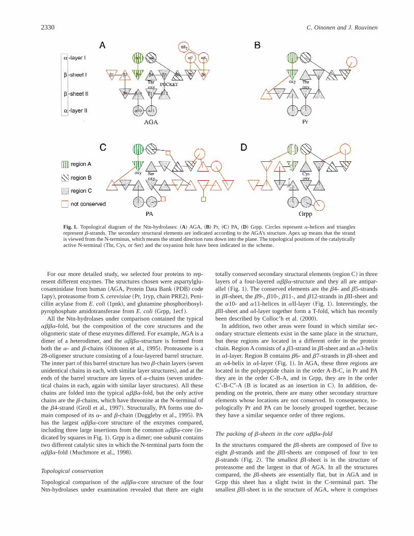
All the Ntn-hydrolases under comparison contained the typicalabba-fold, but the composition of the core structures and theoligomeric state of these enzymes differed. For example, AGA is adimer of a heterodimer, and theabba-structure is formed fromboth thea- andb-chains~Oinonen et al., 1995!. Proteasome is a28-oligomer structure consisting of a four-layered barrel structure.The inner part of this barrel structure has twob-chain layers~sevenunidentical chains in each, with similar layer structures!, and at theends of the barrel structure are layers ofa-chains~seven uniden-tical chains in each, again with similar layer structures!. All thesechains are folded into the typicalabba-fold, but the only activechains are theb-chains, which have threonine at the N-terminal ofthe b4-strand~Groll et al., 1997!. Structurally, PA forms one do-main composed of itsa- andb-chain~Duggleby et al., 1995!. PAhas the largestabba-core structure of the enzymes compared,including three large insertions from the commonabba-core~in-dicated by squares in Fig. 1!. Grpp is a dimer; one subunit containstwo different catalytic sites in which the N-terminal parts form theabba-fold ~Muchmore et al., 1998!.
Topological conservation
Topological comparison of theabba-core structure of the fourNtn-hydrolases under examination revealed that there are eight
totally conserved secondary structural elements~region C! in threelayers of a four-layeredabba-structure and they all are antipar-allel ~Fig. 1!. The conserved elements are theb4- andb5-strandsin bI-sheet, theb9-, b10-, b11-, andb12-strands inbII-sheet andthe a10- anda11-helices inaII-layer ~Fig. 1!. Interestingly, thebII-sheet andaI-layer together form a T-fold, which has recentlybeen described by Colloc’h et al.~2000!.
In addition, two other areas were found in which similar sec-ondary structure elements exist in the same place in the structure,but these regions are located in a different order in the proteinchain. Region A consists of ab3-strand inbI-sheet and ana3-helixin aI-layer. Region B containsb6- andb7-strands inbI-sheet andan a4-helix in aI-layer ~Fig. 1!. In AGA, these three regions arelocated in the polypeptide chain in the order A-B-C, in Pr and PAthey are in the order C-B-A, and in Grpp, they are in the orderC9-B-C0-A ~B is located as an insertion in C!. In addition, de-pending on the protein, there are many other secondary structureelements whose locations are not conserved. In consequence, to-pologically Pr and PA can be loosely grouped together, becausethey have a similar sequence order of three regions.
The packing ofb-sheets in the coreabba-fold
In the structures compared thebI-sheets are composed of five toeight b-strands and thebII-sheets are composed of four to tenb-strands~Fig. 2!. The smallestbI-sheet is in the structure ofproteasome and the largest in that of AGA. In all the structurescompared, thebI-sheets are essentially flat, but in AGA and inGrpp this sheet has a slight twist in the C-terminal part. ThesmallestbII-sheet is in the structure of AGA, where it comprises
Fig. 1. Topological diagram of the Ntn-hydrolases:~A! AGA, ~B! Pr, ~C! PA, ~D! Grpp. Circles representa-helices and trianglesrepresentb-strands. The secondary structural elements are indicated according to the AGA’s structure. Apex up means that the strandis viewed from the N-terminus, which means the strand direction runs down into the plane. The topological positions of the catalyticallyactive N-terminal~Thr, Cys, or Ser! and the oxyanion hole have been indicated in the scheme.
2330 C. Oinonen and J. Rouvinen

only the four conservedb-strands~strandsb9–b12; Figs. 1, 2!. Inboth AGA and Pr, thebII-sheet is predominantly flat; but in Grppit is slightly twisted, and in the case of PA, which has the largestbII-sheet, it is highly twisted at the N-terminal part.
In the study of Brannigan et al.~1995!, in the Ntn-hydrolases theb-sheets were found to be packed in such a way that there is a 308rotation between theb-sheets. However, in AGA, theb-sheetspack against each other face-to-face in an almost pure parallel
Fig. 2. The packing ofb-sheets in the coreabba-fold among the Ntn-hydrolases in two orientations:~A! Pr, ~B! PA, ~C! Grpp, and~D! AGA. The catalytically active N-terminal has been indicated in the illustrations and all the diagrams are on the same scale.
Structural comparison of Ntn-hydrolases 2331

fashion~b-b-packing angle around 58!. In PA the angle betweenthe b-sheets is 308, while in the others~Pr and Grpp! it is 358~Figs. 2, 3!.
We analyzed this different packing of theb-sheets by calculat-ing their amino acid composition on the basis of the topologicallyconservedb-strands and their interactions with each other. In ad-dition, we measured the distances between theb-sheets to explainthe variations in their packing~Table 1!. The number of calculatedcontacts between theb-sheets in AGA is only half that of the otherproteins. The contact residues are smaller in AGA, and thus thedistance between theb-sheets is 1–1.6 Å shorter~Table 1!. Theslightly different packing angle of theb-sheets in PA~308! and theother two structures~358! might be explained by the different ratios
of the small and large hydrophobic residues in the interface be-tween theb-sheets. In PA, the ratio of the small to large residuesis 40:30~in AGA 50:20!, while in the case of the other two~Pr andGrpp! the ratio is equal~50:50! ~Table 1!.
Essential components of Ntn-hydrolases
The 3D structural superimposition of the topologically conservedsecondary elements is a good fit, as we can see by looking at thebII-sheet~Fig. 3!. In thebI-sheet, the exception is AGA, which isthe result of the different packing of theb-sheets, although itsnucleophile and the important surrounding residues are well super-imposed. In the third topologically conserved layer, theaII-layer,the most prominest exception is PA, possibly because of the largeinsertion between the layersbII and aII ~indicated with square inFig. 1!.
On the basis of the structural superimposition, we did a se-quence comparison of the topologically conservedb-strands anda-helices ~Fig. 4!, which revealed that there were no identicalresidues at corresponding locations. Instead, at three locations inthree of the compared structures~PA was always the exception!,there was found to be an identical residue. The 235Gly was situ-ated in theb11-strand and forms a part of the oxyanion hole. The193Gly is situated in a turn just before theb5-strand and its role,as also the role of 262Ala in the middle of thea11-helix, may bestructural.
As a result of the sequence comparison, a total of 18 similarresidues were found. These were distributed in such a way that inthe b4-, b5-, andb11-strands, they were 3, 6, and 3 similar resi-dues, respectively~Fig. 4!. The rest of the similar residues werefound to be equally distributed among the rest of the topologically
Fig. 3. The superimposition of the topologically conserved secondary elements of the Ntn-hydrolases~region C!. Pr is in blue, PA ingreen, Grpp in purple and AGA in yellow.A: The left-hand-side diagram represents the side view in which the yellow-colored AGA’scloser packing is clearly prominent.B: The same is seen, but rotated by 908. The nucleophilic residues have been indicated as aball-and-stick model. The figure was generated with the program MOLSCRIPT~Kraulis, 1991!.
Table 1. The amino acid composition and number of contactsbetween the topologically conservedb-sheetsa
AGA Pr PA Grpp
Hydrophilic ~%! ~N,Q,T,S,H,C! 24 19 28 26Small hydrophobic~%! ~A,G,P,V! 52 29 39 35Large hydrophobic~%! ~I,W,F,Y,L,M! 19 33 28 39Charged~%! ~D,E,R,K! 5 19 6 —Contacts 63 135 133 122b-b-distance~Å! 6.2 7.8 7.8 7.2
aThe residues have been divided into four groups~hydrophilic, largehydrophobic, small hydrophobic and charged!. The distances between theb-sheets are the averages calculated from three different measurementpoints.
2332 C. Oinonen and J. Rouvinen

conserved secondary structural elements. The important residuesfor catalysis and substrate binding were located mostly in theb5-strand~residues 210–204!, b12-strand~221–223!, and b11-strand~233–235!, and also in thea11-helix ~Fig. 4!. In conse-quence, a total of five secondary structure elements~b4-, b5-,b12-, b12-strand, anda11-helix in region C! seem to be essentialfor the structure and function of the Ntn-hydrolases~Fig. 4!.
The active site and the catalytic machinery
The reaction mechanism of the Ntn-hydrolases was first describedby Duggleby et al.~1995!. The mechanism resembles that of ser-ine proteases, though instead of the catalytic triad, there is onlyone amino acid catalytic center, the autocatalytically uncoveredN-terminal Thr, Ser, or Cys~Fig. 5!. This N-terminal residue func-tions as a nucleophile and as a catalytic base. The reactivity of thisnucleophile is affected by the amino acid residues interacting in thevicinity. During the reaction, a covalent intermediate is formed viaa transition state, which is stabilized by residues from the so-calledoxyanion hole~Peräkylä & Rouvinen, 1996; Peräkylä & Kollman,1997!.
The nucleophile always originates from the N-terminus of theb4-strand, and is well superimposed~colored grey in Fig. 6!. Nocommon features have been found in the course of the presentstudy that would indicate which nucleophile~Ser, Thr, Cys! isused. The residues that interact with the nucleophile are all locatedin theb5-strand~colored light blue in Fig. 6!. In three of the fourproteins, these residues are located in the same position~23GlnNin PA, 19ArgO in Pr, and 74TyrN in Grpp; Table 2; Fig. 4!. InAGA, this residue~201ThrOg1! is situated only one residue before~Table 2; Fig. 4!. The reason for this may be that in the AGA theside chain of 201Thr is responsible for stabilization, while in theothers the main chain N or O interacts with the nucleophile. Inaddition, AGA and Pr each include a second interacting residue,but they are topologically situated in different places, namely 215Ser~b5 r b12 loop! and 33Arg~b12-strand! in AGA and Pr, respec-tively ~Table 2; Fig. 6!.
The amino acid residues, which are responsible for Na-stabilization~colored light blue in Fig. 6!, are located mainly in thea4r b6 loop~region B!. AGA’s 49Ser is located in the same placeas Pr’s 170Tyr and Grpp’s 26Arg. Another common location is theb3r a3 loop~region A!, where there is 241Asn in PA and 131Serin Pr, again located in the same place~Figs. 4, 6!.
The oxyanion hole is normally formed by two residues. The dis-tances between the nucleophile and the residues that form the oxy-anion hole are almost the same, approximately 4.5–5.5 Å. Only inthe proteasome is the distance greater, around 6.1 Å. One of the twooxyanion hole formers is located in a structurally and topologicallyequivalent place, near the end of theb11-strand and in three of thefour Ntn-structures under comparison this oxyanion hole former isthe main chain N of glycine. PA is the only exception to this rulesince it contains the main chain N of alanine in the same location~the residues of the oxyanion hole are colored pink in Fig. 6!.
The position of the second oxyanion hole former is different. InAGA and Grpp this residue is the glycine preceding residue in theb11-strand~Table 2; Figs. 4, 6!. In PA it is formed by 241Asnwhich is located in theb3 r a3-loop. In proteasome the possiblecandidate for the second oxyanion hole former is 131SerOg, orig-inating from the same theb3-a3-loop. It is important to note thatPr and PA, which are topologically close to each other, sharesimilar formations of the oxyanion hole.
Fig. 4. Sequence comparison of the topologically conserved secondarystructural elements.b4, b5, etc. refer to the secondary elements of AGA~Fig. 1!. Secondary structure elements are boxed and the residues that aresituated in similar positions in the structure are also shown. Similar resi-dues have been indicated with asterisks~* !. The nucleophile, its stabiliza-tion, substrate binding, and oxyanion hole formers have been indicatedwith the letters N, S, B, and O. These residues have also been indicated inbold on the appropriate protein sequence. The coloring of the amino acidresidues follows the classification used by Branden and Tooze~1991! e.g.,hydrophobic-green, polar-blue, charged-red.
Structural comparison of Ntn-hydrolases 2333

All of the structures compared catalyze the hydrolysis of theamide bond, but they differ in substrate specificity. Accordingly,the shape and the size of the substrate binding pocket differ, as dothe locations of interacting residues~Fig. 4!. However, in additionto their similar types of oxyanion hole formation, Pr and PA havesubstrate binding residues in the same places in theb5-strand~20Ala in Pr and 24Phe in PA!, in theb11-strand~45Met in Pr and67Ser in PA! and in almost the same places in theb12-strand~33Arg and 35Ile in Pr and 57Phe in PA!. On the other hand, bothAGA and Grpp have similar substrate binding residues, Arg andAsn, although these residues are located in different places in thestructures and the sequences~Table 2; Figs. 4, 6!.
Conclusions
Although the amino acid sequence homology is almost completelyabsent, we have been able to compare the sequences and 3D struc-tures of four Ntn-hydrolases. Ntn-hydrolases shares a similar cen-tral abba-core structure. Eight totally conserved secondary structureunits can be found~region C! in this core. Most of the conservedand functionally important residues are located in 5 of the 8 to-pologically conserved elements. Hence, we assumed that theb4,b5, b11, b12, anda11 elements are crucial for the structure andfunction of Ntn-hydrolases. Among the proteins under study, twoadditional regions of secondary structure units~regions A and B!were found that were located in structurally similar places but in adifferent order in the polypeptide chain.l-Aminopeptidase-d-Ala-esterase0amidase is a clear and distant exception from this fold.
The catalytic machinery is located in the same secondary struc-ture elements, but the residues for substrate binding and also theoxyanion hole formation differ to some extent. However, the cat-
alytic mechanism cleaving the amide bond is probably similar inall Ntn-hydrolases. The major unanswered question concerning theNtn-hydrolases is connected with the autocatalytic mechanism lead-ing to the formation of a free, catalytically active N-terminal. It hasbeen very difficult to identify those residues among the Ntn-hydrolases that would participate in autocatalysis. Hence, in thepresent study, we can only suggest that the components whichpossibly participate in the autocatalysis are located in the con-served area of the Ntn-fold, namely in the 5 most conserved ele-ments of the C-region.
Materials and methods
The structural superimposition and the sequence comparison
As a result of the different packing angle of theb-sheets, theavailable superimposition programs~for example STAMP; Russell& Barton, 1992! did not work properly, and so the superimposi-tions were carried out by means of manual fitting, using the pro-grams O~Jones et al., 1991! and Xtalview ~McRee, 1992!. Theclassification of residues in the sequence comparison was per-formed on the basis of Branden and Tooze~1991!, except that theglycines were regarded as hydrophobic amino acid residues.
The packing of theb-sheets in the coreabba-fold
The different packing angle of theb-sheets was measured betweentheb4- andb10-strands or between theb4- andb11-strands~Fig. 2!.To calculate the proportions of the different types of residues andthe number of contacts they made with the opposingb-sheet, weused the amino acids from the conservedb-strands~b4, b5, b9,
Fig. 5. Catalytic mechanism of Ntn-hydrolases. Y represents oxygen or sulfur, and X represents nitrogen or oxygen. The reactionbegins when the nucleophilic oxygen0sulfur of Thr0Ser0Cys donates its proton to its owna-amino group and attacks the carbonylcarbon of the substrate. The negatively charged tetrahedral intermediate is stabilized by hydrogen bonding~oxyanion hole formers!.The acylation step is complete when thea-amino group of the Thr0Ser0Cys donates the proton to the nitrogen of the scissile peptidebond. The covalent bond between the part of the substrate and the enzyme is formed and the part of the substrate is released. Thedeacylation step begins when the hydroxyl group of water attacks the carbonyl carbon of the acyl-enzyme product, and the basica-amino group of the nucleophile accepts the proton from the water molecule. The negatively charged intermediate is stabilized, as inthe acylation step. The reaction is complete when thea-amino group donates the proton to the nucleophile.
2334 C. Oinonen and J. Rouvinen

b10, b11, b12!. All of the residues that formed contacts at dis-tances of,4.0 Å from the oppositeb-sheet were included in theclassification and counted. The residues were classified as hydro-philic ~N, Q, T, S, H, C!, small hydrophobic~A, G, P, V!, largehydrophobic~I, W, F, Y, L, M!, and charged~D, E, K, R!. Thereported distance between theb-sheets was based on the averagedistance between three measurement points, and it represented thevalue of the nearest Ca-atoms from the oppositeb-sheets.
Acknowledgment
This study was supported by the Academy of Finland.
References
Artymiuk PJ. 1995. A sting in the~N-terminal! tail. Nat Struct Biol 2:1035–1037.Bochtler M, Ditzel L, Groll M, Huber R. 1997. Crystal structure of heat shock
locus V ~HsIV! from Escherichia coli. Proc Natl Acad Sci USA 94:6070–6074.
Bompard-Gilles C, Villeret V, Davies GJ, Fanuel L, Joris B, Frère J-M, VanBeeumen J. 2000. A new variant of the Ntn hydrolase fold revealed by thecrystal structure ofl-aminopeptidased-Ala-esterase fromOchrobactrumanthropi. Struct Fold Des 8:153–162.
Branden C, Tooze J. 1991.Introduction of protein structure. New York: GarlandPublishing.
Brannigan JA, Dodson G, Duggleby HJ, Moody PCE, Smith JL, Tomchick DR,Murzin AG. 1995. A protein catalytic framework with an N-terminal nu-cleophile is capable of self-activation.Nature 378:416–419.
Fig. 6. Diagrams of the active sites of the Ntn-hydrolases:~A! AGA, ~B! PA, ~C! Pr, and~D! Grpp. The grey-colored nucleophilesThr0Ser0Cys are shown on the left. Those residues that interact with the nucleophile or its Na-group are indicated in light blue. Thosecolored green represent the substrate binding residues. For the sake of clarity, not all of the reported substrate binding residues areshown. The oxyanion hole formers have been colored pink. The figure was generated with the program MOLSCRIPT~Kraulis, 1991!.
Structural comparison of Ntn-hydrolases 2335

Colloc’h N, Poupon A, Mornon J-P. 2000. Sequence and structural features ofthe T-fold, an original tunnelling building unit.Proteins 39:142–154.
Duggleby HJ, Tolley SP, Hill CP, Dodson EJ, Dodson G, Moody PCE. 1995.Penicillin acylase has a single-amino-acid catalytic centre.Nature 373:264–268.
Groll M, Ditzel L, Löwe J, Stock D, Bochtler M, Bartunik HD, Huber R. 1997.Structure of 20S proteasome from yeast at 2.4 Å resolution.Nature 386:463–471.
Guan C, Cui T, Rao V, Liao W, Benner J, Lin C-L, Comb D. 1996. Activationof glycosylasparaginase: Formation of active N-terminal threonine by in-tramolecular autoproteolysis.J Biol Chem 271:1732–1737.
Guo H-C, Xu Q, Buckley D, Guan C. 1998. Crystal structures ofFlavobacte-rium glycosylasparaginase. An N-terminal nucleophile hydrolase activatedby intramolecular proteolysis.J Biol Chem 273:20205–20212.
Isupov MN, Obmolova G, Butterworth S, Badet-Denisot M-A, Badet B, Po-likarpov I, Littlechild JA, Teplyakov A. 1996. Substrate binding is requiredfor assembly of the active conformation of the catalytic site in Ntn amido-transferases: Evidence from the 1.8 Å crystal structure of the glutaminasedomain of glucosamine 6-phosphate synthase.Structure 4:801–810.
Jones TA, Zou J-Y, Cowan SW, Kjeldgaard M. 1991. Improved methods forbuilding protein models in electron density maps and the location of errorsin these models.Acta Crystallogr A47:110–119.
Kraulis PJ. 1991. MOLSCRIPT: A program to produce both detailed and sche-matic plots of protein structures.J Appl Crystallogr 24:946–950.
Löwe J, Stock D, Jap B, Zwickl P, Baumeister W, Huber R. 1995. Crystalstructure of the 20S proteasome from the archaeonT. acidophilumat 3.4 Åresolution.Science 268:533–539.
McDonough MA, Klei HE, Kelly JA. 1999. Crystal structure of penicillin Gacylase from the Bro1 mutant strain ofProvidencia rettgeri. Protein Sci8:1971–1981.
McRee DE. 1992. A visual protein crystallographic software system for X110Xview. J Mol Graph 10:44–46.
Muchmore CR, Krahn JM, Kim JH, Zalkin H, Smith JL. 1998. Crystal structureof glutamine phosphoribosylpyrophosphate amidotransferase fromEsche-richia coli. Protein Sci 7:39–51.
Oinonen C, Tikkanen R, Rouvinen J, Peltonen L. 1995. Three-dimensionalstructure of human lysosomal aspartylglucosaminidase.Nat Struct Biol2:1102–1108.
Table 2. The catalytically important residues among the Ntn-hydrolasesa
NucleophileClose to
nucleophile Close to Na Oxyanion holeSubstratebinding
AGA 183ThrOg 201ThrOg 2.8 Å 49SerOg 2.9 Å 235GlyN 5.4 Å 211Argb4 b5 a4 r b6 loop b11 b5 r b12 loop
215SerOg 3.9 Å 49SerO 3.0 Å 234ThrOg 4.4 Å 214Aspb5r b12 loop a4 r b6 loop b11 b5 r b12 loop
PA 1SerOg 23GlnN 2.9 Å 241AsnOd1 3.0 Å 69AlaN 4.5 Å 142Metb4 b5 b3 r a3 loop b11 a-dimer
23GlnOE1 3.2 Å 241AsnNd2 4.5 Å 146Pheb5 b3 r a3 loop a-dimer
24Pheb5 r b14 loop
57Pheb12
67Serb11
154Trpa11
177Ileb10
Pr 1ThrOg 19ArgO 3.6 Å 170TyrO 2.7 Å 47GlyN 6.1 Å 20Alab4 b5 a4 r b6 loop b11 b5
33Arg-pl 3.0 Å 131SerOg 2.7 Å 131SerOg 5.2 Å 21Thrb12 b3 r a3 loop b3 r a3 loop b5
171SerOg 3.0 Å 31Vala4 r b6 loop b5 r b12 loop
35Ileb12
45Metb11
49Alaa11
53Glna11
Grpp 1CysSg 74TyrN 3.6 Å 26ArgO 3.0 Å 102GlyN 5.6 Å 73Argb4 b5 a4 r b6 loop b11 b5
101AsnNd2 4.7 Å 127Aspb11 a11 r a11 loop
aThe location of particular residues in the proteins’ secondary structures has also been indicated.
2336 C. Oinonen and J. Rouvinen

Peräkylä M, Kollman PA. 1997. A simulation of the catalytic mechanism ofaspartylglucosaminidase using ab initio quantum mechanics and moleculardynamics.J Am Chem Soc 119:1189–1196.
Peräkylä M, Rouvinen J. 1996. Ab initio quantum mechanical model calcula-tions on the catalytic mechanism of aspartylglucosaminidase~AGA!: Aserine protease-like mechanism with an N-terminal threonine and substrate-assisted catalysis.Chem Eur J 2:1548–1551.
Russell RB, Barton GJ. 1992. Multiple protein sequence alignment from tertiarystructure comparison: Assignment of global and residue confidence levels.Proteins 14:309–323.
Seemüller E, Lupas A, Baumeister W. 1996. Autocatalytic processing of the 20Sproteasome.Nature 382:468–470.
Smith JL, Zaluzec EJ, Wery J-P, Niu L, Switzer RL, Zalkin H, Satow Y. 1994.Structure of the allosteric regulatory enzyme of purine biosynthesis.Science264:1427–1433.
Suresh CG, Pundle AV, SivaRaman H, Rao KN, Brannigan JA, McVey CE,Verma CS, Dauter Z, Dodson EJ, Dodson GG. 1999. Penicillin V acylasecrystal structure reveals new Ntn-hydrolase family members.Nat Struct Biol6:414–416.
Tikkanen R, Riikonen A, Oinonen C, Rouvinen J, Peltonen L. 1996. Functionalanalyses of active site residues of human lysosomal aspartylglucosamini-dase: Implications for catalytic mechanism and autocatalytic activation.EMBO J 15:2954–2960.
Xu Q, Buckley D, Guan C, Guo H-C. 1999. Structural insights into the mech-anism of intramolecular proteolysis.Cell 98:651–661.
Xuan J, Tarentino AL, Grimwood BG, Plummer TH Jr, Cui T, Guan C, VanRoey P. 1998. Crystal structure of glycosylasparaginase fromFlavobacte-rium meningosepticum. Protein Sci 7:774–781.
Zwickl P, Kleinz J, Baumeister W. 1994. Critical elements in proteasome as-sembly.Nat Struct Biol 1:765–770.
Structural comparison of Ntn-hydrolases 2337