Embed Size (px)
Citation preview
Structural DisordersStructural Disorders
Fetal Demise / Intrauterine Fetal DeathFetal Demise / Intrauterine Fetal Death
Structural DisordersStructural Disorders
Fetal Demise / Intrauterine Fetal DeathFetal Demise / Intrauterine Fetal Death
DEFINITION:
Death of a fetus after the age of viability
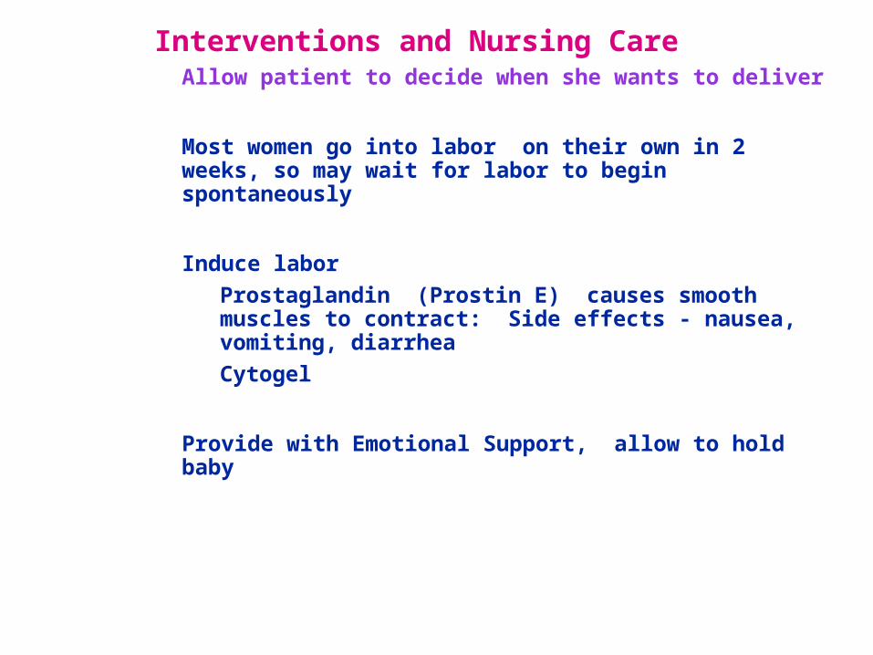
Interventions and Nursing Care• Allow patient to decide when she wants to deliver
• Most women go into labor on their own in 2 weeks, so may wait for labor to begin spontaneously
• Induce labor
• Prostaglandin (Prostin E) causes smooth muscles to contract: Side effects - nausea, vomiting, diarrhea
• Cytogel
• Provide with Emotional Support, allow to hold baby
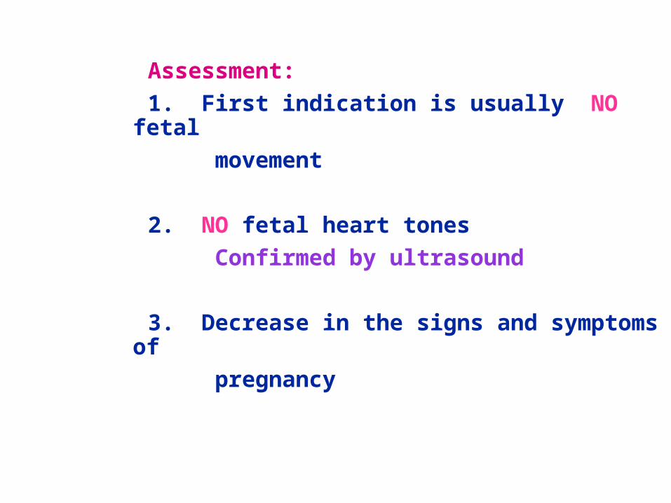
Assessment:
1. First indication is usually NO fetal
movement
2. NO fetal heart tones
Confirmed by ultrasound
3. Decrease in the signs and symptoms of
pregnancy
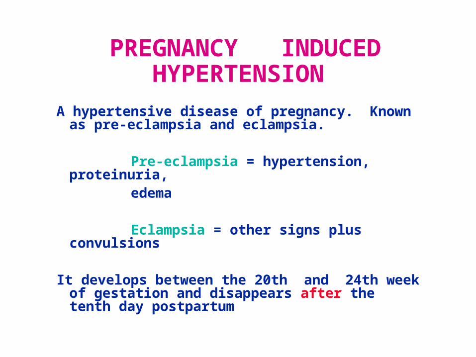
PREGNANCY INDUCED HYPERTENSION
A hypertensive disease of pregnancy. Known as pre-eclampsia and eclampsia.
Pre-eclampsia = hypertension, proteinuria,
edema
Eclampsia = other signs plus convulsions
It develops between the 20th and 24th week of gestation and disappears after the tenth day postpartum
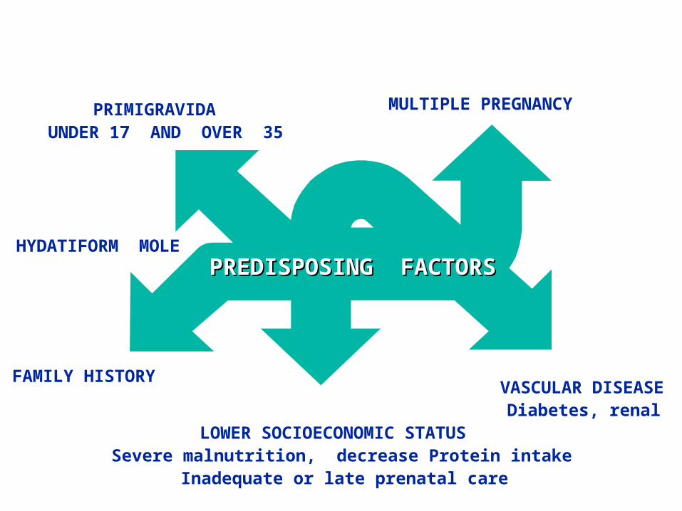
PREDISPOSING FACTORSPREDISPOSING FACTORS
PRIMIGRAVIDA MULTIPLE PREGNANCY
VASCULAR DISEASE
UNDER 17 AND OVER 35
LOWER SOCIOECONOMIC STATUSSevere malnutrition, decrease Protein intake
Inadequate or late prenatal care
FAMILY HISTORY
HYDATIFORM MOLE
Diabetes, renal
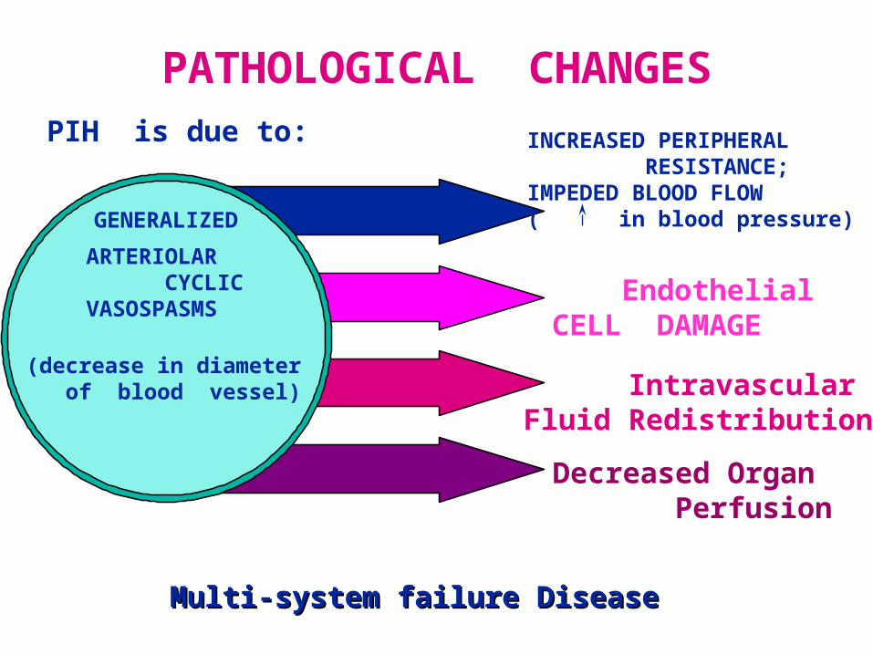
PATHOLOGICAL CHANGESPIH is due to:
GENERALIZED ARTERIOLAR CYCLICVASOSPASMS
INCREASED PERIPHERAL RESISTANCE; IMPEDED BLOOD FLOW( in blood pressure)
Endothelial CELL DAMAGE
Intravascular Fluid Redistribution
(decrease in diameter of blood vessel)
Decreased Organ Perfusion
Multi-system failure DiseaseMulti-system failure Disease
Clinical Manifestations
Clinical Manifestation
HYPERTENSIONHYPERTENSION
Earliest and The Most Earliest and The Most Dependable IndicatorDependable Indicator of PIHof PIH
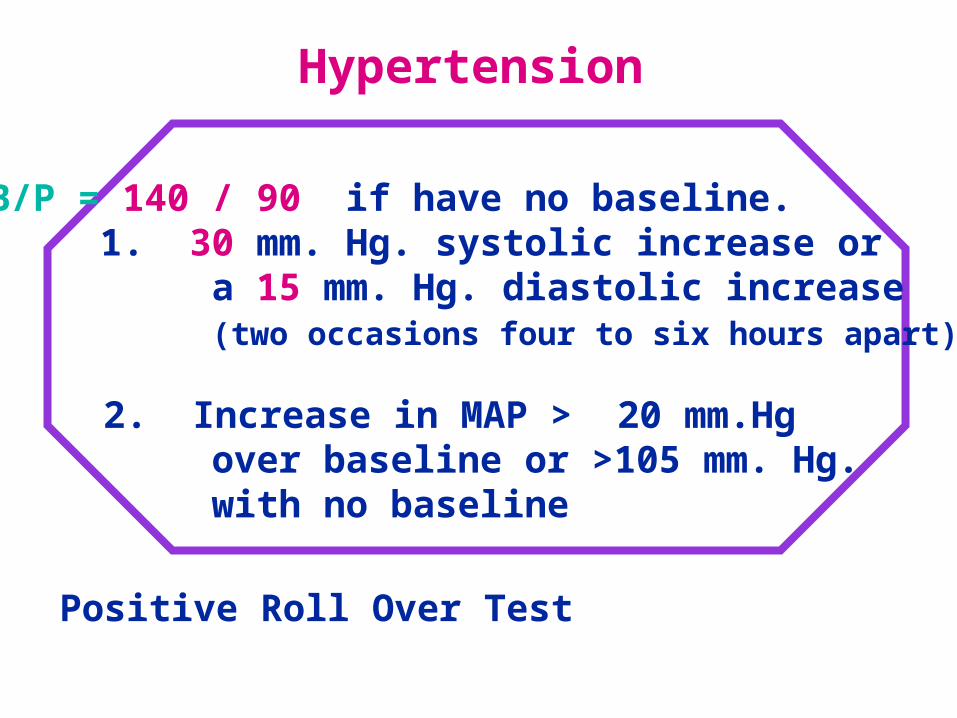
Hypertension
B/P = 140 / 90 if have no baseline. 1. 30 mm. Hg. systolic increase or a 15 mm. Hg. diastolic increase (two occasions four to six hours apart)
2. Increase in MAP > 20 mm.Hg over baseline or >105 mm. Hg. with no baseline
Positive Roll Over Test
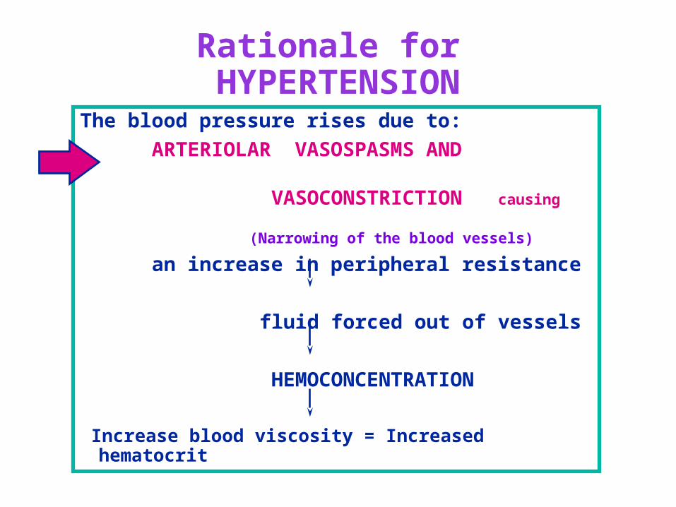
Rationale for HYPERTENSION
The blood pressure rises due to:
ARTERIOLAR VASOSPASMS AND VASOCONSTRICTION causing
(Narrowing of the blood vessels)
an increase in peripheral resistance
fluid forced out of vessels
HEMOCONCENTRATION
Increase blood viscosity = Increased hematocrit
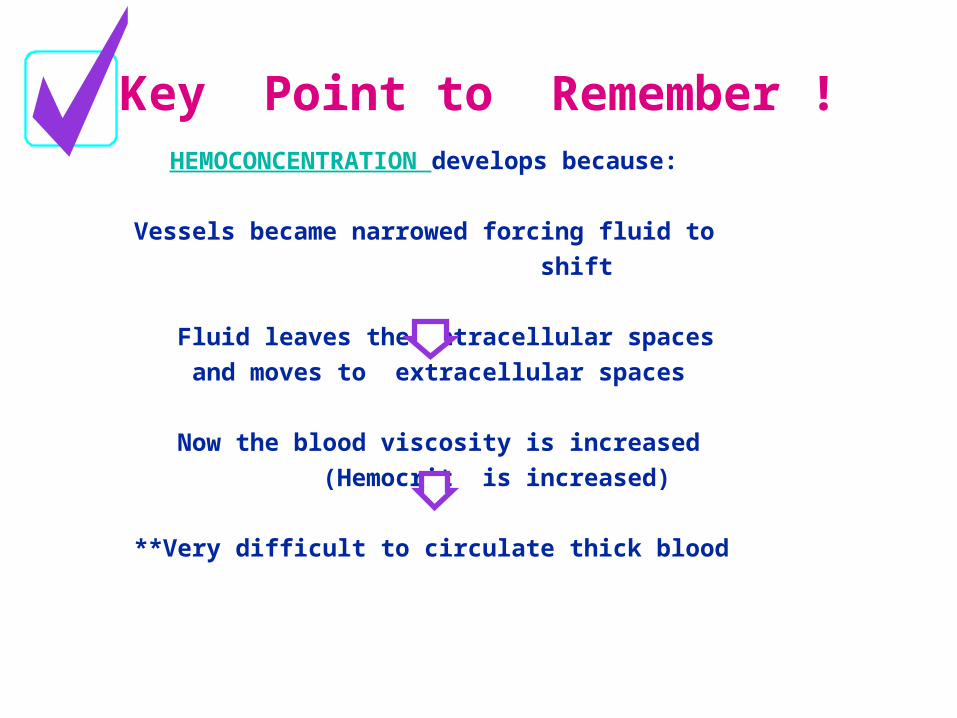
Key Point to Remember ! HEMOCONCENTRATION develops because:
Vessels became narrowed forcing fluid to
shift
Fluid leaves the intracellular spaces
and moves to extracellular spaces
Now the blood viscosity is increased
(Hemocrit is increased)
**Very difficult to circulate thick blood
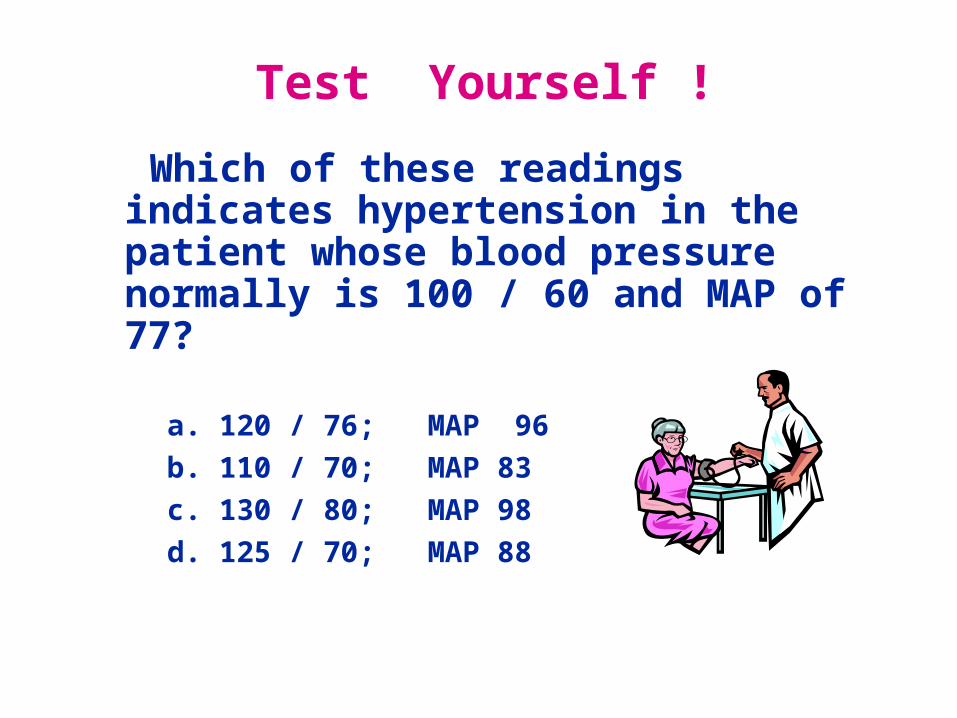
Test Yourself !
Which of these readings indicates hypertension in the patient whose blood pressure normally is 100 / 60 and MAP of 77?
a. 120 / 76; MAP 96
b. 110 / 70; MAP 83
c. 130 / 80; MAP 98
d. 125 / 70; MAP 88
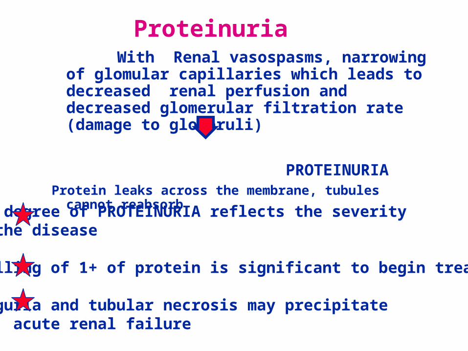
Proteinuria With Renal vasospasms, narrowing of
glomular capillaries which leads to decreased renal perfusion and decreased glomerular filtration rate (damage to glomeruli)
PROTEINURIA Protein leaks across the membrane, tubules cannot reabsorb
The degree of PROTEINURIA reflects the severity of the disease
Spilling of 1+ of protein is significant to begin treatment
Oliguria and tubular necrosis may precipitate acute renal failure
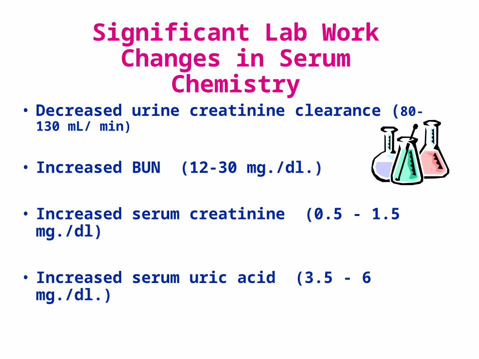
Significant Lab WorkChanges in Serum Chemistry
• Decreased urine creatinine clearance (80-130 mL/ min)
• Increased BUN (12-30 mg./dl.)
• Increased serum creatinine (0.5 - 1.5 mg./dl)
• Increased serum uric acid (3.5 - 6 mg./dl.)
Weight Gain and Edema
• Clinical Manifestation:
–Edema may appear rapidly
–Begins in lower extremities and moves upward
–Pitting edema and facial edema are late signs
–Weight gain is directly related to accumulation of fluid
WEIGHT GAIN AND EDEMA
Rationale:• Decreased blood flow to the kidneys causes a
loss of plasma proteins and albumin• This leads to a decreased colloid osmotic
pressure. • A in COP allows fluid to shift from from
intravascular to extravascular. • Now there is an accumulation of fluid in the
tissues.• Increased angiotensin and aldostersone
triggers retention of sodium and water.
The difference between dependent edema and generalized edema is important.
The patient with PIH has generalized edema because fluid is in all tissues.
The Nurse Must Know
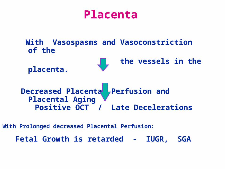
Placenta
With Vasospasms and Vasoconstriction of the
the vessels in the placenta.
Decreased Placental Perfusion and Placental Aging
Fetal Growth is retarded - IUGR, SGA
Positive OCT / Late Decelerations
With Prolonged decreased Placental Perfusion:

Condition
is
Worsening
• Oliguria – 100ml./4 hrs or less than 30 cc. / hour
• Edema moves upward and becomes generalized (face, periorbital, sacral)
• Excessive weight gain – greater than 2 pounds per week
Central Nervous System Changes
• Cerebral edema -- forcing of fluids to extracellular
–Headaches -- severe, continuous
–Hyperreflexia
–Level of Consciousness changes – changes in affect
–Convulsions / seizures
Visual Changes
Retinal Edema and spasms leads to:
• Blurred vision
• Double vision
• Retinal detachment
• Scotoma (areas of absent or depressed vision)
•Nausea and Vomiting
• Epigastric pain –often sign of impending coma
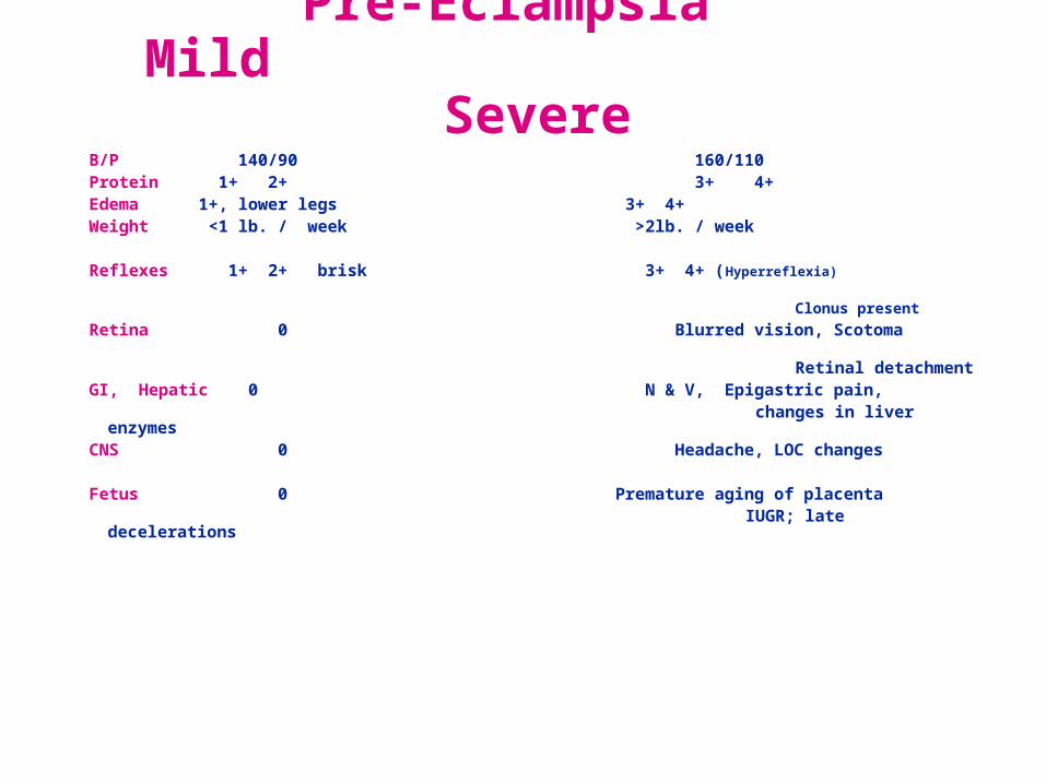
Pre-Eclampsia Mild Severe
B/P 140/90 160/110Protein 1+ 2+ 3+ 4+Edema 1+, lower legs 3+ 4+Weight <1 lb. / week >2lb. / week Reflexes 1+ 2+ brisk 3+ 4+ (Hyperreflexia) Clonus present
Retina 0 Blurred vision, Scotoma
Retinal detachmentGI, Hepatic 0 N & V, Epigastric pain, changes in liver enzymesCNS 0 Headache, LOC changes
Fetus 0 Premature aging of placenta IUGR; late decelerations
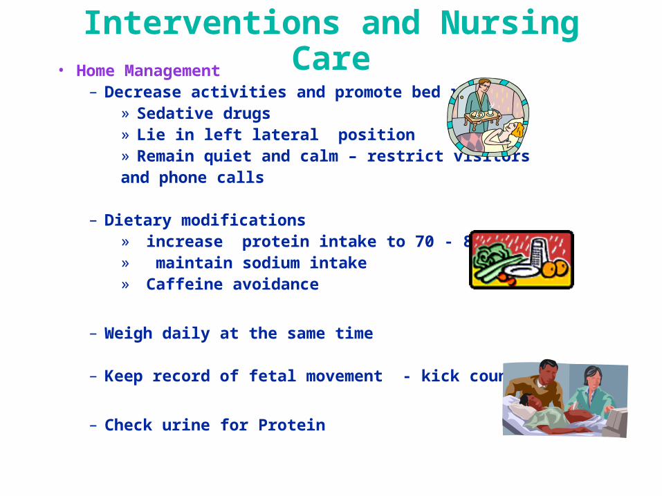
Interventions and Nursing Care• Home Management
– Decrease activities and promote bed rest » Sedative drugs» Lie in left lateral position» Remain quiet and calm – restrict visitors and phone calls
– Dietary modifications » increase protein intake to 70 - 80 g/day» maintain sodium intake» Caffeine avoidance
– Weigh daily at the same time
– Keep record of fetal movement - kick counts
– Check urine for Protein
Hospitalization
• If symptoms do not get better then the patient needs to be hospitalized in order to further evaluate her condition.
• Common lab studies:
–CBC, platelets; type and cross match
–Renal blood studies -- BUN, creatitine, uric acid
– Liver studies -- AST, LDH, Bilirubin
–DIC profile -- platelets, fibrinogen, FSP, D-Dimer
Hospital ManagementNursing Care Goal
1. Decrease CNS Irritability
2. Control Blood Pressure
3. Promote Diuresis
4. Monitor Fetal Well-Being
5. Deliver the Infant
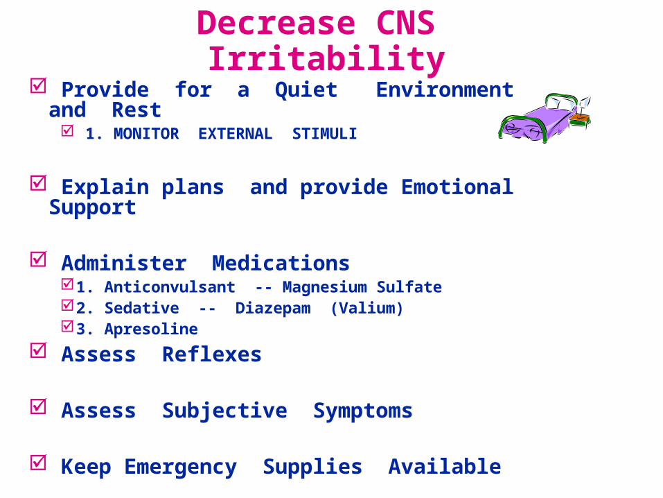
Decrease CNS Irritability Provide for a Quiet Environment and Rest
1. MONITOR EXTERNAL STIMULI
Explain plans and provide Emotional Support
Administer Medications1. Anticonvulsant -- Magnesium Sulfate2. Sedative -- Diazepam (Valium)3. Apresoline
Assess Reflexes
Assess Subjective Symptoms
Keep Emergency Supplies Available
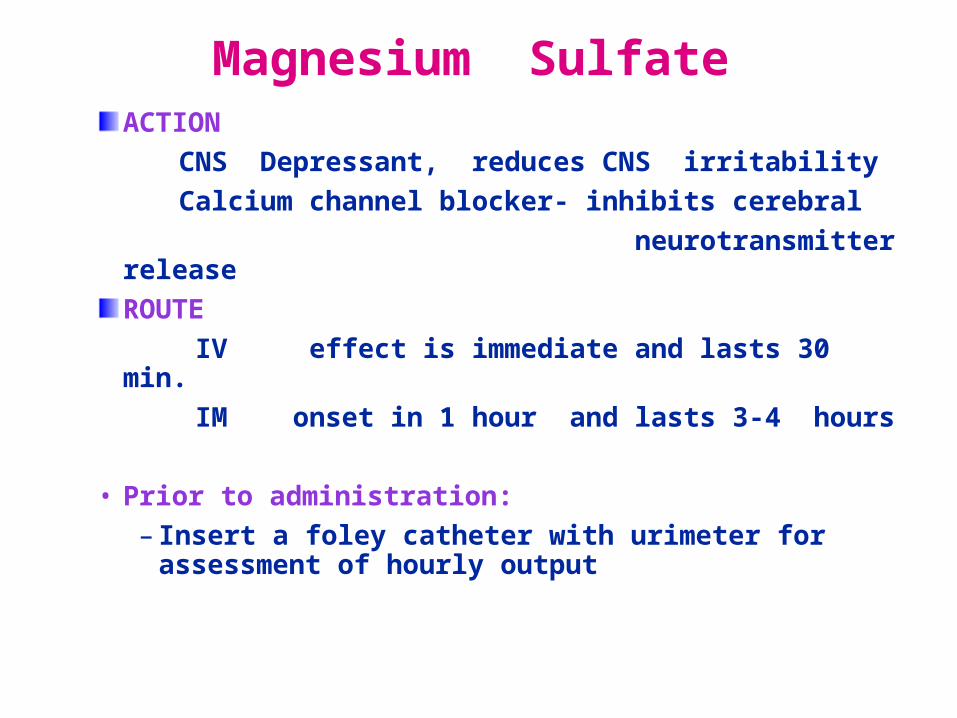
Magnesium SulfateACTION
CNS Depressant, reduces CNS irritability
Calcium channel blocker- inhibits cerebral
neurotransmitter release
ROUTE
IV effect is immediate and lasts 30 min.
IM onset in 1 hour and lasts 3-4 hours
• Prior to administration:
– Insert a foley catheter with urimeter for assessment of hourly output

Magnesium Sulfate NURSING IMPLICATIONS 1. Monitor respirations > 14-16; < 12 is critical
2. Assess reflexes for hyporeflexia -- D/C for
hyporeflexia
3. Measure Urinary Output >100cc in 4 hrs.
4. Measure Magnesium levels – normal is 1.5-2.5 mg/dl
Therapeutic is 4-8mg/dl. Toxicity - >9mg/dl; Absence of reflexes is >10 mg/dl; Respiratory arrest is 12-15 mg/dl; cardiac arrest is
> 15 mg/dl.
• Have Calcium Gluconate available as antagonist
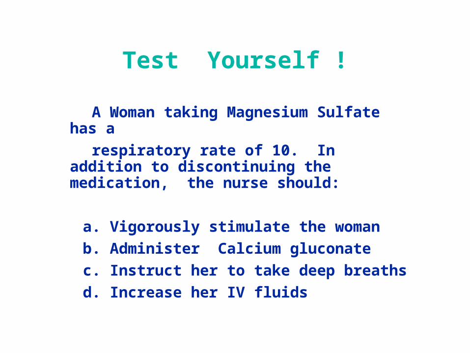
Test Yourself !
A Woman taking Magnesium Sulfate has a
respiratory rate of 10. In addition to discontinuing the medication, the nurse should:
a. Vigorously stimulate the woman
b. Administer Calcium gluconate
c. Instruct her to take deep breaths
d. Increase her IV fluids
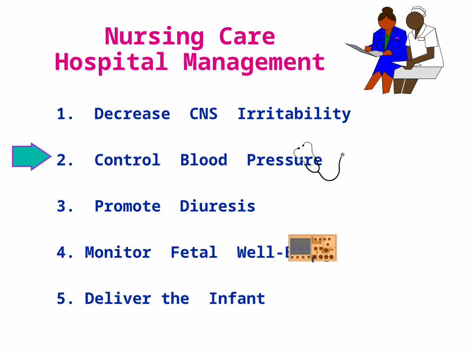
Nursing CareHospital Management
1. Decrease CNS Irritability
2. Control Blood Pressure
3. Promote Diuresis
4. Monitor Fetal Well-Being
5. Deliver the Infant
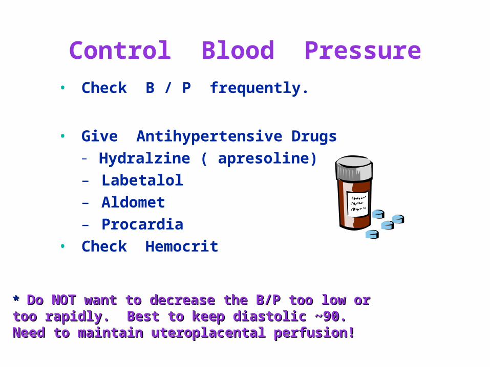
Control Blood Pressure
• Check B / P frequently.
• Give Antihypertensive Drugs– Hydralzine ( apresoline)
– Labetalol
– Aldomet
– Procardia
• Check Hemocrit
* * Do NOT want to decrease the B/P too low orDo NOT want to decrease the B/P too low ortoo rapidly. Best to keep diastolic ~90.too rapidly. Best to keep diastolic ~90.Need to maintain uteroplacental perfusion!Need to maintain uteroplacental perfusion!
Nursing CareHospital Management
1. Decrease CNS Irritability
2. Control Blood Pressure
3. Promote Diuresis
4. Monitor Fetal Well-Being
5. Deliver the Infant
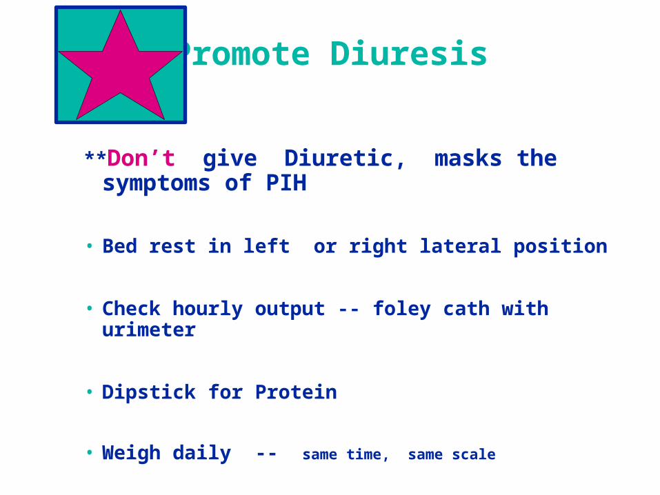
Promote Diuresis
**Don’t give Diuretic, masks the symptoms of PIH
• Bed rest in left or right lateral position
• Check hourly output -- foley cath with urimeter
• Dipstick for Protein
• Weigh daily -- same time, same scale
Nursing CareHospital Management
1. Decrease CNS Irritability
2. Control Blood Pressure
3. Promote Diuresis
4. Monitor Fetal Well-Being
5. Deliver the Infant
Monitor Fetal Well-Being
FETAL MONITORING-- assessing for late decelerations.
NST -- Non-stress test
OCT --oxytocin challenge test
If all else fails ---- Deliver the baby
Key Point to Remember !
SEVERE COMPLICATIONS OF PIH:
PLACENTAL SEPARATION - ABRUPTIO PLACENTA; DIC
PULMONARY EDEMA
RENAL FAILURE
CARDIOVASCULAR ACCIDENT
IUGR; FETAL DEATH
HELLP SYNDROME
HELLP Syndrome
• A multisystem condition that is a form of severe preeclampsia - eclampsia
• H = hemolysis of RBC
• EL = elevated liver enzymes
• LP = low platelets <100,000mm (thrombocytopenia)
Etiology of HELLPHemolysis occurs from destruction of RBC’s
Release of bilirubin
Elevated liver enzymes occur from blood flow that is obstructed in the liver due to fibrin deposits
Vascular vasoconstriction endothelial damage platelet aggregation at the sites of damage low platelets.
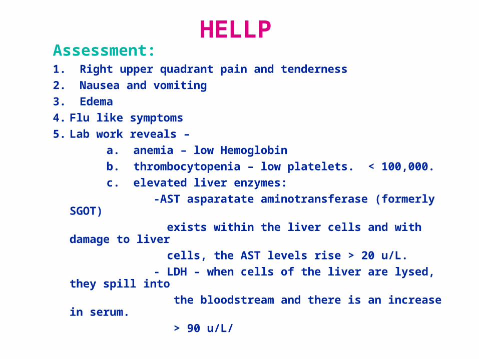
HELLPAssessment:1. Right upper quadrant pain and tenderness
2. Nausea and vomiting
3. Edema
4. Flu like symptoms
5. Lab work reveals –
a. anemia – low Hemoglobin
b. thrombocytopenia – low platelets. < 100,000.
c. elevated liver enzymes:
-AST asparatate aminotransferase (formerly SGOT)
exists within the liver cells and with damage to liver
cells, the AST levels rise > 20 u/L.
- LDH – when cells of the liver are lysed, they spill into
the bloodstream and there is an increase in serum.
> 90 u/L/
HELLP
• Intervention:• 1. Bedrest – any trauma or increase in intra-
abdominal pressure could lead to rupture
of the liver capsule hematoma.
• 2. Volume expanders
• 3. Antithrombic medications
Heart Disease in
Pregnancy
Cardiac Response in All Pregnancies
Increase in Cardiac Output 30% - 50%
Expanded Plasma Volume
Increase in Blood (Intravascular) Volume
Every Pregnancy affects the cardiovascular system
A woman with a healthy heart can tolerate the stress of pregnancy, but a woman with a compromised heart is challenged Hemodynamically and will have complications

Effects of Heart Disease on Pregnancy
Growth Retarded Fetus
Spontaneous Abortion
Premature Labor and Delivery
Effects of Pregnancy onHeart Disease
The Stress of Pregnancy on an already weakened heart may lead to cardiac decompensation (failure).
The effect may be varied depending upon the classification of the disease
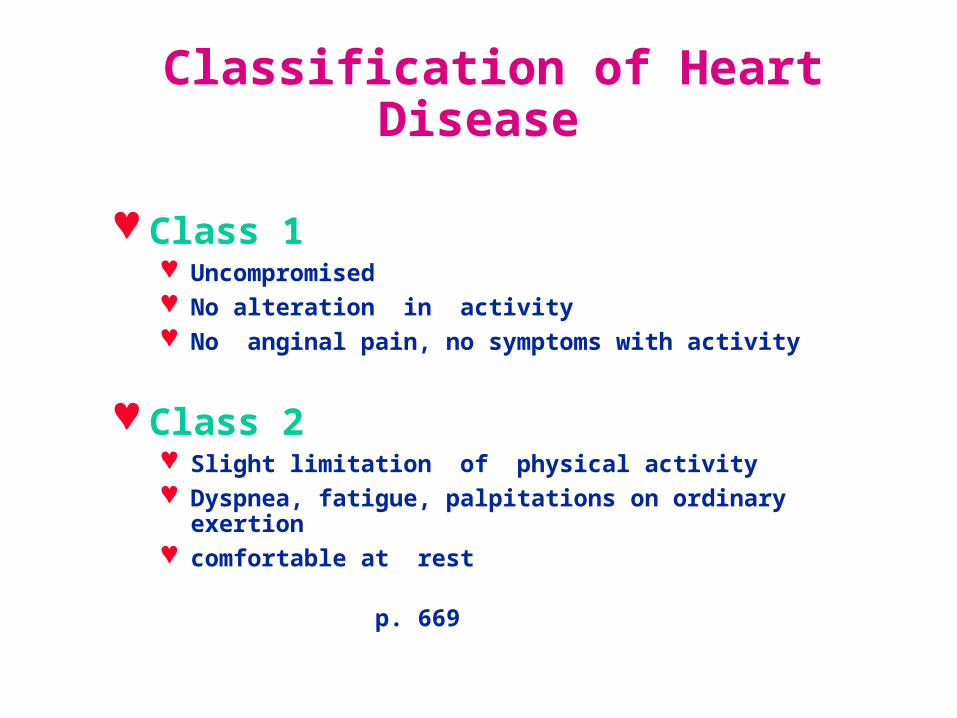
Classification of Heart Disease
Class 1 Uncompromised No alteration in activity No anginal pain, no symptoms with activity
Class 2 Slight limitation of physical activity Dyspnea, fatigue, palpitations on ordinary exertion comfortable at rest
p. 669
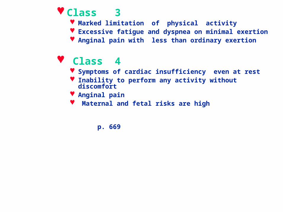
Class 3 Marked limitation of physical activity Excessive fatigue and dyspnea on minimal exertion Anginal pain with less than ordinary exertion
Class 4 Symptoms of cardiac insufficiency even at rest Inability to perform any activity without discomfort Anginal pain Maternal and fetal risks are high p. 669
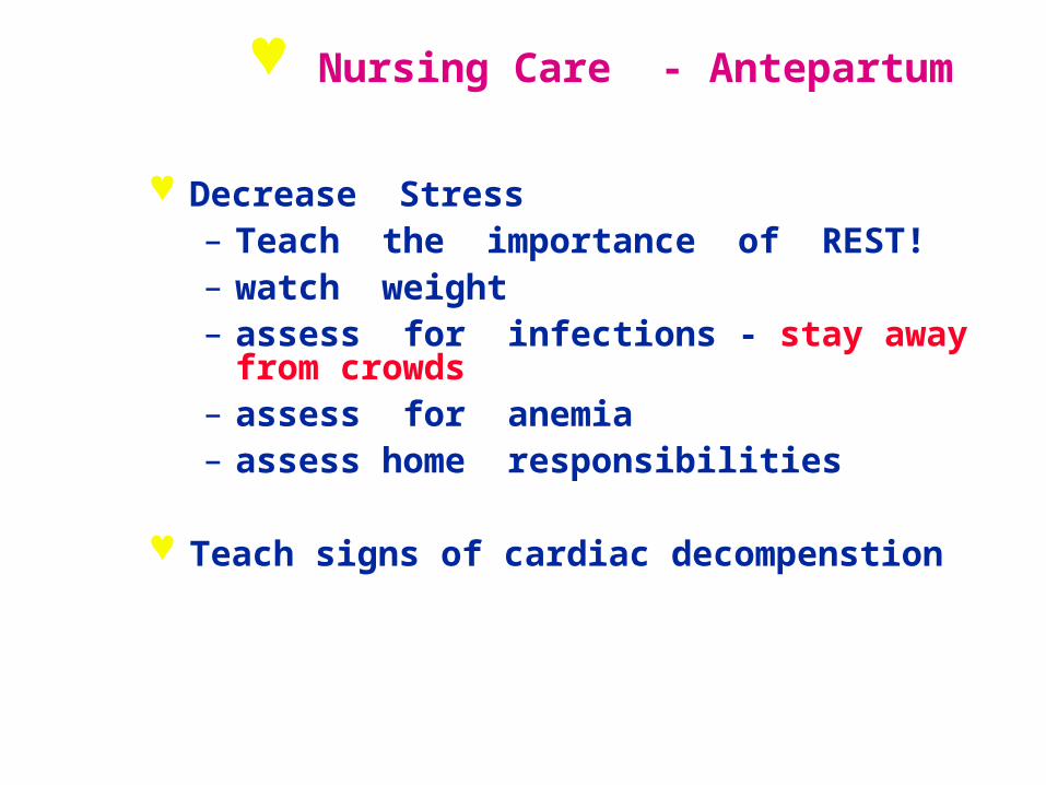
Nursing Care - Antepartum
Decrease Stress– Teach the importance of REST! – watch weight– assess for infections - stay away from
crowds– assess for anemia– assess home responsibilities
Teach signs of cardiac decompenstion
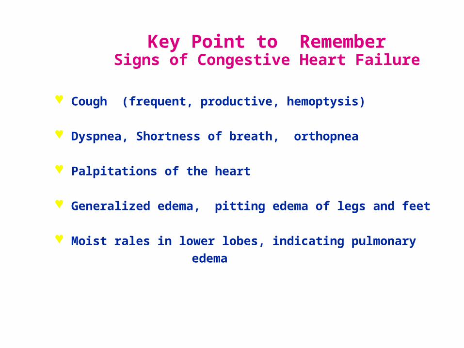
Key Point to RememberSigns of Congestive Heart Failure
Cough (frequent, productive, hemoptysis)
Dyspnea, Shortness of breath, orthopnea
Palpitations of the heart
Generalized edema, pitting edema of legs and feet
Moist rales in lower lobes, indicating pulmonary
edema
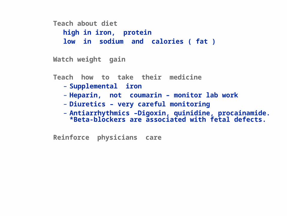
Teach about diethigh in iron, proteinlow in sodium and calories ( fat )
Watch weight gain
Teach how to take their medicine– Supplemental iron– Heparin, not coumarin – monitor lab work– Diuretics – very careful monitoring– Antiarrhythmics –Digoxin, quinidine, procainamide.
*Beta-blockers are associated with fetal defects.
Reinforce physicians care

Key point to remember !
Never eat foods high in Vitamin K while on
an anticoagulant!
( raw green leafy vegetables)
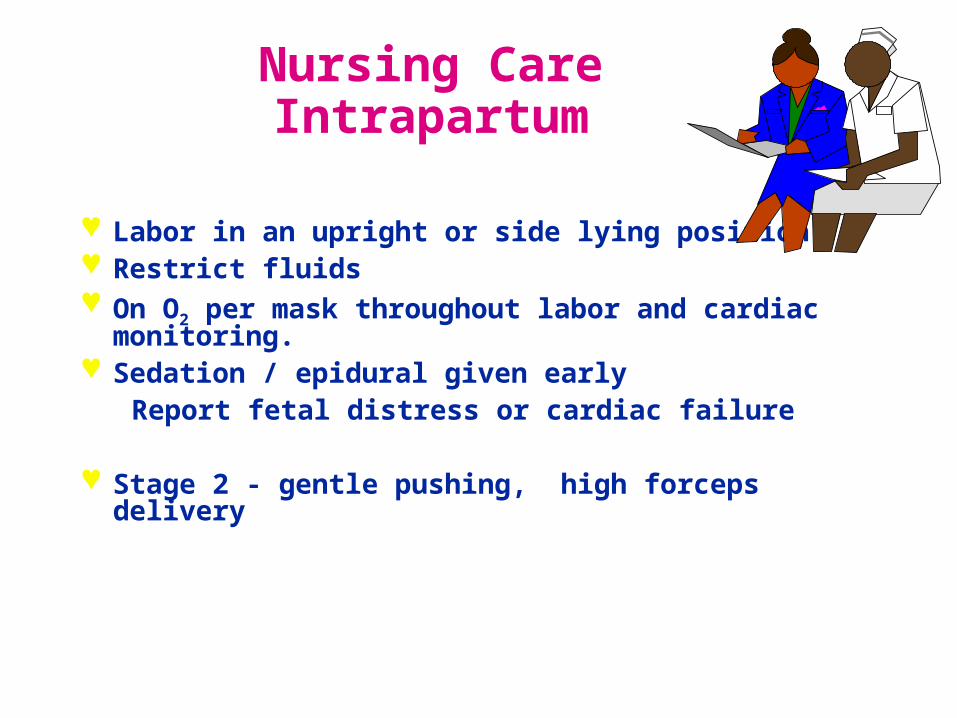
Nursing Care Intrapartum
Labor in an upright or side lying position Restrict fluids On O2 per mask throughout labor and cardiac
monitoring. Sedation / epidural given early Report fetal distress or cardiac failure
Stage 2 - gentle pushing, high forceps delivery
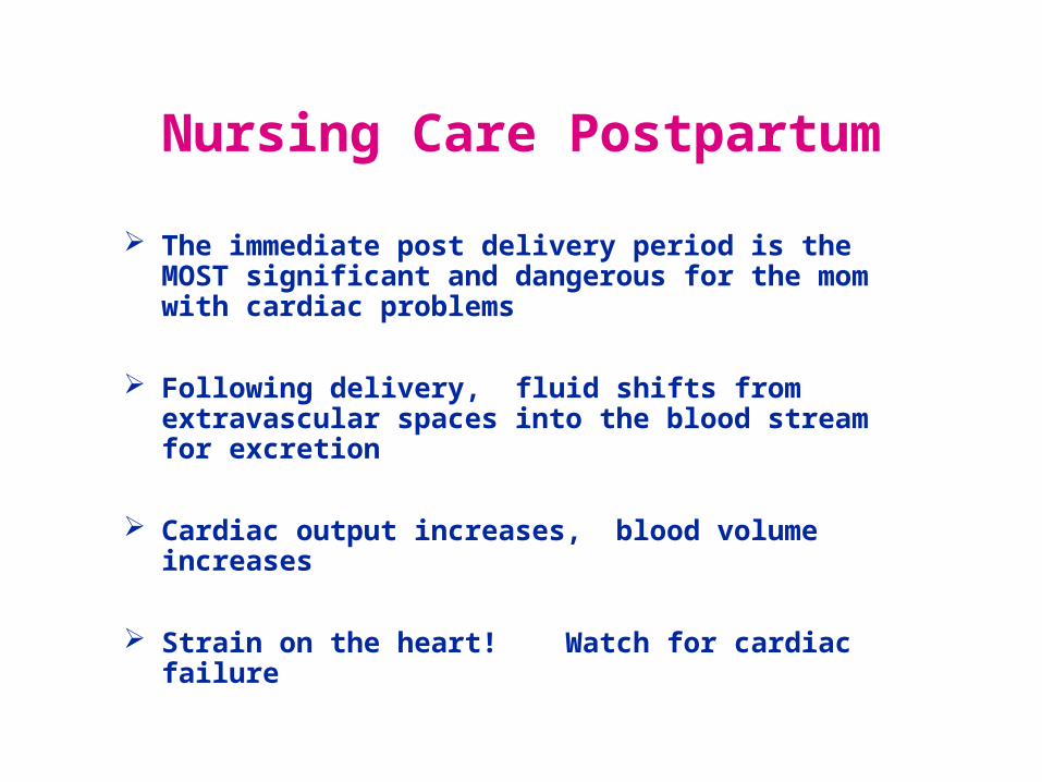
Nursing Care Postpartum
The immediate post delivery period is the MOST significant and dangerous for the mom with cardiac problems
Following delivery, fluid shifts from extravascular spaces into the blood stream for excretion
Cardiac output increases, blood volume increases
Strain on the heart! Watch for cardiac failure
Test Yourself !
• Mrs. B. has mitral valve prolapse. During the second trimester of pregnancy, she reports fatigue and palpitations during routine housework. As a cardiac patient, what would her functional classification be at this time?
a. Class I
b. Class II
c. Class III
d. Class IV

The End
return