Embed Size (px)
Citation preview

1
Structural elucidation of a novel phosphoglycolipid isolated from six species of Halomonas genus Assunta Giordano*, Filomena M. Vella, Ida Romano and Agata Gambacorta Istituto di Chimica Biomolecolare, CNR, via Campi Flegrei, 34 80078 Pozzuoli (NA), Italy Running title: phosphoglycolipid from Halomonas species. *Corresponding author. Assunta Giordano, Istituto di Chimica Biomolecolare, CNR, via Campi Flegrei 34, 80078 Pozzuoli (NA), Italy; Phone: 0039 081 8675309; Fax: 0039 081 8041770; email: [email protected]
by guest, on June 5, 2018w
ww
.jlr.orgD
ownloaded from
2
Abstract
The structure of a new phosphoglycolipid from the halophilic Gram-negative bacteria
Halomonas elongata ATCC 33173T, Halomonas eurihalina ATCC 49336T, Halomonas almeriensis
CECT 7050T, strain Sharm (AM238662), Halomonas halophila DSM 4770T, and Halomonas salina
ATCC 49509T, was elucidated by NMR and mass spectroscopy studies. In all the species examined,
the polar lipid composition consisted of 1,2 diacylglycero-3-phosphorylethanolamine, 1,2
diacylglycero-3-phosphoryl-glycerol, bisphosphatidylglycerol and the new phosphoglycolipid
PGL1. The structure of PGL1 was established to be 2-(α-D-glucopyranosyloxy)-3-hydroxy-
propyl)-phosphatidyl diacylglycerol. C16:0;C18:1 and C16:0;C19cyclopropane are the most
abundant acyl chains linked to the phosphatidylglycerol moiety of each isolated PGL1. All the
species presenting the lipid PGL1 belong to Halomonas rRNA group 1, suggesting that the new
phosphoglycolipid could be a chemotaxonomic marker of this phylogenetic group.
Structural elucidation of a new phosphoglycolipid from six strains belonging to the Halomonas
genus.
Supplementary key words Halomonas elongata ATCC 33173T. Halomonas eurihalina ATCC 49336T. Halomonas almeriensis CECT 7050T. Strain Sharm (AM238662). Halomonas halophila DSM 4770T. Halomonas salina ATCC 49509T. nuclear magnetic resonance spectroscopy. tandem mass spectrometry. Abbreviations: FAME, fatty acid methyl ester; DQF-COSY, double quantum filtered-correlation spectroscopy; HSQC, heteronuclear single quantum coherence; HMBC, heteronuclear multiple bond coherence; ESI, electrospray ionization.
by guest, on June 5, 2018w
ww
.jlr.orgD
ownloaded from

3
Introduction Halophile organisms are able to withstand fluctuations of extracellular salinity by using two
strategies that can operate simultaneously. The first one involves the cytoplasmic accumulation of
small organic compounds, called osmolites or compatible solutes, which enable the cell to reduce
the water loss and to maintain the cell pressure by reducing the osmotic potential between the cell
and the cytoplasm (1, 2). The second one consists in modifying the membrane structure, generally
affecting phospholipids and their fatty acid composition. In halophilic Gram-negative bacteria the
rise in salt concentration usually corresponds to a decrease of the zwitterionic phospholipid
phosphatidylethanolamine (PEA), and an increase of the anionic lipids, phosphatidylglycerol (PG)
and/or bisphosphatidyl glycerol (DPG), as well as the incorporation of cyclopropanic rings and
double bonds in fatty acid chains (3, 4). Regulation of the phospholipids composition of the cell
membrane is also utilized by the microorganisms to adjust the proton permeability of the membrane
to the growth temperature of the cell (5, 6).
Phosphoglycolipids have a relatively limited distribution among Gram-negative bacteria in
comparison with Gram-positive bacteria. Among euryhaline microorganisms, Halomonas species
are widely distributed throughout saline environments and comprise a remarkably high percentage
of the total microbial community in hydrothermal-vent habitats. The polar lipid composition shows
a general pattern, since PEA, PG and DPG are the major lipid components. The presence of
unknown lipids was firstly described in in 1990 by Franzmann and Tindall (7), which reported the
occurrence of unidentified periodate-Schiff positive phospholipids in some species of the
Halomonas genus. In 1998 Yagi and Maruyama described in a patent (8) the presence of new
glucosyl phosphatidyl glycerol derivatives in Halomonas marina. In this study the structure of a
new phosphoglycolipid present in six strains belonging to the genus Halomonas was elucidated by
NMR and mass spectroscopy studies.
Materials and Methods
Isolation and culture conditions
During the summer 2005, samples of water with mat were collected from the salt lake inside
Ras Muhammad Park in Egypt, pH was 8.5 and temperature 30°C.
Isolation of Sharm strain was carried out on a saline medium (medium 1) containing the
following components (g l-1): KCl, 2.0; MgSO4.7H2O, 20; NaCl, 150; sodium citrate, 3.0; yeast
by guest, on June 5, 2018w
ww
.jlr.orgD
ownloaded from
4
extract, 5.0; casamino acids 5.0; (µg l-1) MnCl2.4 H20, 0.36; FeSO4, 50. The strain was isolated by
dilution-plating technique on solid medium 1. Solid medium for purification and maintenance of the
strain was prepared by the addition of 1.8 % agar to the medium 1. Routinely the strain was grown
on liquid and/or solid medium 1. In liquid medium, growth was followed by measuring the
absorbance at 540 nm. Halomonas elongata (ATCC 33173T) (9), Halomonas eurihalina (ATCC
49336T) (10), Halomonas salina (11), Halomonas halophila (DSM 4770T) (12) were obtained from
the Deutsche Sammlung von Mikroorganismen und Zellkulturen, Brunschweig, Germany (DSMZ).
Halomonas almeriensis (CECT 7050 T) was kindly provided by the authors (13). All the strains
were grown in the media at their optimal growth conditions, as suggested by authors.
Lipid extraction and characterization
Complex lipid analyses were performed using freshly harvested cells of the strains (5 g),
lyophilised and extracted. Cells in the stationary phase were collected by centrifugation and
lyophilized. Dry cells were extracted with chloroform:methanol:water mixture (65:25:4, v/v) and
evaporated under reduced pressure. The lipid extract was analysed by two-dimensional thin layer
chromatography (TLC) on silica gel plates cut to 10 cm x 10 cm. The TLCs were developed in
chloroform:methanol:water (65:25:4, first dimension) and chloroform:methanol:acetic acid:water
(80:12:15:4, second dimension). The lipid extract was then fractionated by flash chromatography on
silica gel with methanol: chloroform (1:9, v/v), methanol: chloroform (2:8), methanol: chloroform
(3:7), methanol: chloroform: water (25:75:4). The fraction eluted with chloroform: methanol: water
25:75:4 was purified by preparative TLC eluting with chloroform: methanol: water 25:75:4 (Rf of
phosphoglycolipid PGL1: 0.25). Polar lipids were quantified by weight of isolated fractions. 13C NMR spectra were acquired on a Bruker DPX-300 operating at 300 MHz, using a dual
probe. 1H, 1H-1H DQF-COSY, HMBC and HSQC experiments were recorded on a 600 MHz
Bruker instrument. Chemical shifts were given in ppm (δ) scale; the MeOH signal was used as
internal standard (δ 3.34 1H; δ 49.0 13C). Mass spectra of the unknown phosphoglycolipid were
recorded on a QTof-/micro/mass spectrometer (Waters) equipped with an ESI source in negative
ion mode.
Fatty acid methyl esters (FAMEs) were obtained from complex lipids by acid methanolysis
incubating phosphoglycolipids with 0.5 M methanol-HCl at 80 °C for 1 h. The solvent was removed
under vacuum, and the residue was mixed with chloroform and water. FAMEs partitioned into the
organic phase. GC-MS analysis of FAMEs was performed with an HP5890 series II plus-5989B
by guest, on June 5, 2018w
ww
.jlr.orgD
ownloaded from
5
equipped with an HP-V column with a flux of 45 ml/min. The analysis program was 60 °C for 1
min, 60–140 °C at 25 °C/min, 140–200 °C at 5 °C/min, and 200–300 °C at 10 °C/min.
PGL1 (2 mg) was incubated in 1 M methanol-HCl (1 mL) at 60° C for 12 h. The reaction
mixture was dried and partitioned between chloroform and H2O/MeOH 8:2. The aqueous layer
containing the sugar was concentrated, filtered on a Sep-Pak cartridge and dried. Benzoyl chloride
(0.5 ml) and dry pyridine (0.5 ml) were added to the methanolysis products. After 12 h at room
temperature the mixture was dried and purified on silica gel (hexane/diethyl ether gradient) to
afford 0.7 mg and 0.4 mg of pure α- and β-tetra-benzoate. The absolute stereochemistry of the
sugar was determined by comparison of the circular dichroism (CD) spectra of the benzoate
derivatives with samples prepared from commercial α- and β-methyl-D-glucopyranose
respectively. CD (n-hexane): [θ]248 1640 for α derivative and [θ]235 613 for the β derivative.
Results
The strain Sharm was isolated from samples of water with mat collected from saline lake
inside Ras Muhammad Park in the promontory of Sinai Peninsula in Egypt. The Gram-negative
microorganism grew aerobically in enrichment medium containing up to 30 % of NaCl with an
optimum at 5-15% NaCl. The 16S rRNA gene sequence (EMBL database accession number
AM238662) was determined (data not shown) and compared with all sequences currently available
for members of the genus Halomonas and related taxa. The results were presented as a phylogenetic
dendrogram (Fig. 1) showing that strain Sharm is a member of the genus Halomonas. The strain
clustered together with the species H. elongata and H. eurihalina, and other species of the rRNA
group 1, according to the phylogenetic classification of Halomonas (14).
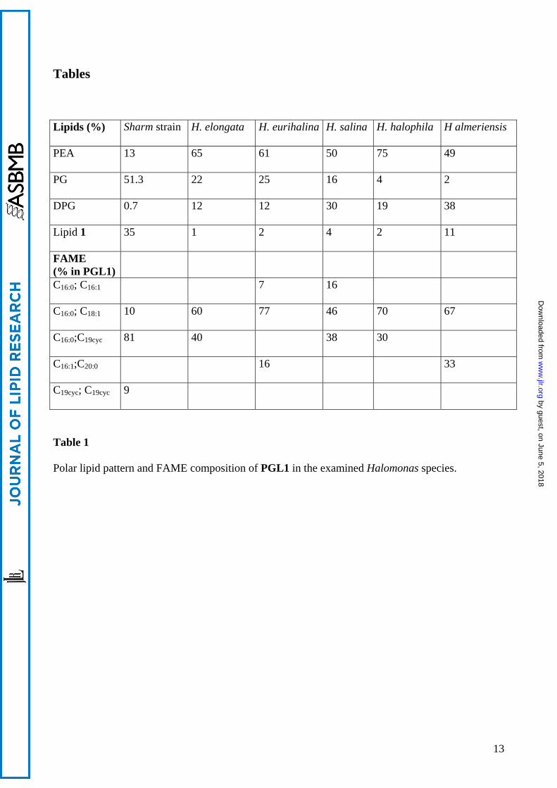
The total lipid composition of strain Sharm and that of the five related species H. elongata,
H. eurihalina, H. salina, H. almeriensis and H. halophila, are reported in Table 1. The isolate
Sharm possessed three phospholipids, 1,2 diacylglycero-3-phosphorylethanolamine (PEA, 13%),
1,2 diacylglycero-3-phosphoryl-glycerol (PG; 51.4%) and bisphosphatidyl glycerol (DPG, 0.7%),
and the new phosphoglycolipid PGL1 (33.0%), identified by NMR studies. Polar lipid analysis of
the other Halomonas strains showed that these species contain the same lipid pattern but with
different distribution (Table 1).
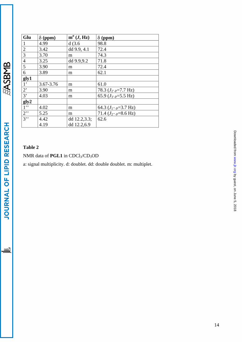
The structure of phosphoglycolipid PGL1 and NMR assignments are reported in Fig. 2 and
Table 2. The presence of spin systems corresponding to one sugar unit (H1-H6), two glycerols
(H1’-H3’and H1”-H3”), and fatty acids were suggested from 1H, 13C and 1H-1H DQF-COSY
spectra. The HSQC spectrum gave the assignment of carbons directly bonded to protons. The 1H-
by guest, on June 5, 2018w
ww
.jlr.orgD
ownloaded from
6
and 13C-NMR spectra contained methyls (δH 0.88), methylene chains (δH 1.25-2.42), double bonds
(δH 5.33, δC 130.0) in methylene chains, and carbonyl carbons (δC 173.2, 173.4), suggesting the
presence of fatty acid moieties. Moreover, the presence of 1H signals at about –0.4 ppm indicated
the presence of cyclopropanic ring.
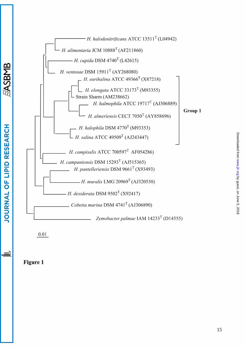
The HSQC spectrum (Figure 3) also revealed one anomeric methine (δH 4.99, δC 98.8) thus
suggesting the presence of one sugar unit with α-configuration (J1-2=3.6). Starting from the
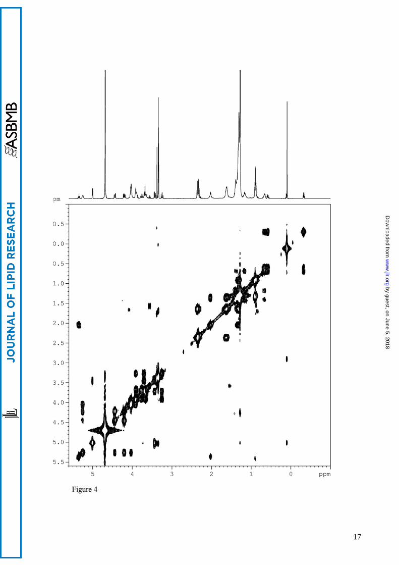
anomeric proton H1, interpretation of COSY correlations (Figure 4) allowed to assign the δH of the
protons of the sugar ring (Figure 4). The high values of the coupling constants J2-3 (9.90 Hz), J3-4
and J4-5 (9.0 and 9.2 Hz) indicated the axial orientation of H2, H3, H4, and H5, thus the sugar was
assigned as glucopyranose ring in 4C1 conformation.
Four oxy-methylenes and two oxy-methine were assigned as two glycerol units on the basis
of spin system from 1H-1H COSY and their directly bonded carbons (Figure 4). Coupling costants
of the carbon signals indicated the presence of a phosphate unit linking the two glycerols. In
particular, the small values of JC3’-P an JC1”-P are diagnostic for the presence of a phosphate group
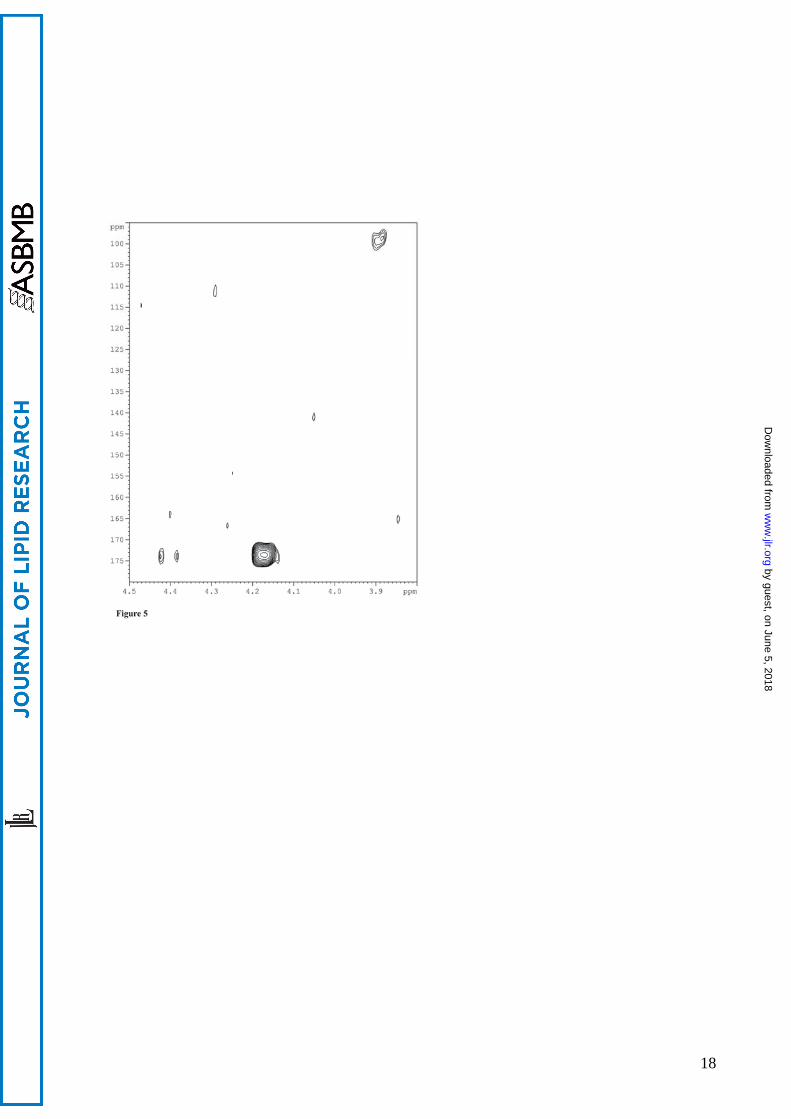
directly linked to these carbons. HMBC cross peak H2’/C1 (3.90/ 98.8) showed that the glucose
molecule is attached to the C2 of the first glycerol moiety (Figure 5). Furthermore, long range
correlations of the H2” and H3” with the carbonyl carbons demonstrated that the two acyl groups
were located on C-2” and C-3” oxydrils (5.25 and 4.42-4.19/ 169.2).
The stereochemistry of the glucose unit was determined by comparison of the CD spectra of
the purified benzoylated α- and β-methyl glucopyranoses. The CD curves of the benzoyl
derivatives were identical to those of the corresponding compounds obtained from benzoylation of
commercial α- and β-methyl-D-glucopyranoses, thus indicating the D absolute stereochemistry of
the sugar unit.
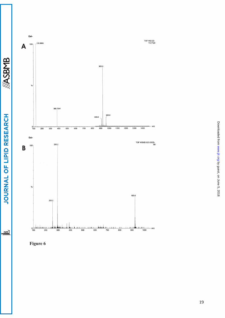
The fatty acid moieties were determined by tandem MS/MS spectra and GCMS analysis
(Table 1). The mass spectra contained several molecular ion peaks indicating differences in the type
and distribution of the acyl chains on the phosphatidylglycerol backbone. For Sharm strain the most
abundant were determined to be C16:0 and C19:cyclopropane, corresponding to the highest peak of
923 m/z in MS spectrum (Figure 6, A). In MS/MS spectra using negative ion electrospray
ionization (Figure 6, B), fragmentation of the molecular ion m/z 923 (M-H) led to fragment ions
295 (C19:cyc-H) and 255 (C16-H). Mass spectrometry analysis of phosphoglycolipids isolated
from the other Halomonas species allowed to establish the identity of acyl chains linked to the
glyceryl moiety. It was shown that all lipids carry two acyl chains, the most abundant being
C16:0;C18:1. The fatty acid composition was also confirmed by GCMS analysis of methyl esters
obtained by acid methanolysis of the phosphoglycolipids. The structure of the phosphoglycolipid
by guest, on June 5, 2018w
ww
.jlr.orgD
ownloaded from
7
present in Sharm strain, H. elongata, H. eurihalina, H. salina, H. almeriensis and H. halophila, was
established to be 2-(α-D-glucopyranosyloxy)-3-hydroxy-propyl)-phosphatidyl diacylglycerol.
Discussion
Polar lipids and their fatty acids can be used as a biochemical marker since many of them
have unique structures. Lipid structures, related to that described for Sharm strain, were found in
microorganisms classified in genera not-related to Halomonas genus, such as Mycoplasma,
Acholeplasma, Streptococcus (15-19). The presence of phosphoglycolipids in several strains
belonging to the Halomonas genus was detected by Franzmann and Tindall (7), which analysed the
general composition of polar lipids of many members of the family Halomonadaceae. In 1998 Yagi
and Maruyama described in a patent (8) new glucosyl phosphatidyl glycerol derivatives in
Halomonas marina.
In connection with our ongoing project on the search for new halophilic and alkalohalophilic
phenotypes in different continents (20- 23), we isolated a new halophilic strain from a salt lake
inside Ras Muhammad Park, Egypt. Phylogenetic analysis of Sharm strain indicated that the strain
clustered together with the species H. elongata and H. eurihalina, thus suggesting that it could
belong to the rRNA group 1, according to Arahal et al. (14) classification of Halomonas.
Chemotaxonomic characterization of this strain showed the presence of a large amount of an
unidentified compound in the polar lipid extract, positive to sugar and phosphate reaction on TLC.
Interestingly, lipid analysis of the species most related to Sharm strain shows that this new
lipid is present in other five members of Halomonas rRNA group 1, confirming a relationship
between these species. H. elongata, H. eurihalina, H. salina, H. almeriensis and H. halophila,
grown at their optimal growth conditions, possess the same polar lipids of the egyptian isolate,
although in different proportions. PEA is the major lipid in all the species with the exception of the
egyptian isolate. Phosphoglycolipid PGL1 comprises the 35% of the total lipid composition of
Sharm strain, while is present in lower amounts (between 2 and 11%) in the other Halomonas
species.
NMR and mass spectroscopic analyses allowed to establish the structure of PGL1. The
chemical shifts and the coupling constants of 1H NMR and 13C spectra indicate the presence of one
residue of glucopyranose in α configuration. The anomeric carbon is directly linked to a diacylated
phosphatidylglycerol molecule. An interesting feature of PGL1 is its unusual linkage of the sugar to
the secondary oxydryl group of phosphatidylglycerol, a structure resembling that of glycosylated
teichoic acids of Gram-positive bacteria. Halomonas species are usually rich of C18:1 and C16:0
by guest, on June 5, 2018w
ww
.jlr.orgD
ownloaded from
8
acyl chains, while an increment of cyclopropanic rings is a common response to higher salinity. The
FAME composition of each purified phosphoglycolipid respect this general rule and is quite
superimposable to that of the total lipid fraction. In all the species the C16:0;C18:1 and
C16:0;C19cyclopropane are the most abundant acyl chains linked to the phosphatidylglycerol
moiety.
To survive in harsh environments, microorganisms modify their membrane structures
modulating the chemical composition of lipids. One of the possible biological roles of
phosphoglycolipids in halophilic bacteria is to ensure the osmotic stability of the cellular
membrane. Bulky head groups would enhance steric protection through hydrogen bonding via
glycosyl head groups (24). This study elucidate the structure of a new phosphoglycolipid, which is
present in all the species of Halomonas rRNA group 1. It would be interesting to extend detailed
lipid characterization of all Halomonas species to ensure if the new phosphoglycolipid is a
chemotaxonomic marker of group 1 or if it is a common feature of Halomonas genus.
by guest, on June 5, 2018w
ww
.jlr.orgD
ownloaded from
9
Acknowledgements The paper was supported by “Centro Regionale di Competenza in applicazioni tecnologiche-industriali di biomolecole e biosistemi”. The authors thank Eduardo Pagnotta for technical assistance, Vincenzo Mirra, Salvatore Zambardino and Dominique Melck for NMR-ICB service, Maurizio Zampa for MS analyses and Emilio P. Castelluccio for computer system maintenance
by guest, on June 5, 2018w
ww
.jlr.orgD
ownloaded from
10
References
1. Galinski, E. A. 1995. Osmoadaptation in bacteria. In Advances in Microbial Physiology. R. K. Poole editors. Academic Press, London. 273–328.
2. Ventosa, A., J. J. Nieto, and A. Oren. 1998. Biology of moderately halophilic aerobic bacteria. Microbiol. Mol. Biol. Rev. 62: 504–544.
3. Kates, M. 1986. Influence of salt concentration on membrane lipids of halophilic bacteria. FEMS Microbiol. Rev. 39: 95–101.
4. Russell, N. J. 1993. Lipids of halophilic and halotolerant microorganisms. In The Biology of Halophilic Bacteria. R. H. Vreeland and L. I. Hochstein editors. CRC Press, Boca Raton, FL. 163–210.
5. Van de Vossenberg J. L. C. M. , T. Ubbink-Kok, M. G. L. Elferink, A. J. M. Driessen, and W. N. Konings. 1995. Ion permeability of the cytoplasmic membrane limits the maximum growth temperature of bacteria and archaea. Mol. Microbiol. 18: 925-932.
6. Kaye, J. Z., and J. A. Baross. 2004. Synchronous effects of temperature, hydrostatic pressure, and salinity on growth, phospholipid profiles, and protein patterns of four Halomonas species isolated from deep-sea hydrothermal-vent and sea surface environments. Appl. Envirom. Microbiol. 70: 6220–6229.
7. Franzmann, P. D., and B. J. Tindall. 1990. A chemotaxonomic study of members of the family Halomonadaceae. Syst. Appl. Microbiol. 13: 142-147.
8. Yagi, H. and A. Maruyama. New glucosyl phosphatidyl glycerol derivatives from Deleya marina. Jpn. Kokai Tokkyo Koho JP 2001226393 (C12P19/44), 2001-08-21, Appl. JP 2000-232915.
9. Vreeland, R.H., E.L. Litchfield, E. Martin, and E. Elliot. 1980. Halomonas elongata, a new genus and species of extremely salt-tolerant bacteria. Int J. Syst. Bacteriol. 30: 488-495.
10. Mellado, E., E.R. Moore, J.J. Nieto, and A. Ventosa. 1995. Phylogenetic inferences and taxonomic consequences of 16S ribosomal DNA sequence comparison of Chromohalobacter marismortui, Volcaniella eurihalina, and Deleya salina and reclassification of V. eurihalina as Halomonas eurihalina comb. nov. Int J Syst Bacteriol. 45:712-716.
11. Valderrama, M. J., E. Quesada, V. Béjar, A. Ventosa, M. C. Gutierrez, F. Ruiz-Berraquero, and A. Ramos-Cormenzana. 1991. Deleya salina sp. nov., a moderately halophilic gram-negative bacterium. Int. J. Syst. Bacteriol. 41:377–384.
12. Dobson, S. J., T.A McMeekin,and Franzmann, P. D. 1993. Phylogenetic relationships between some members of the genera Deleya, Halomonas, and Halovibrio. Int. J. Syst. Bacteriol. 43:665-673.
13. Martinèz-Chela F., V. Bèjar, M.J. Martinez-Canovas, I. Llamas, and E. Quesada. 2005. Halomonas almeriensis sp. nov., a moderately halophilic, exopolysaccharide-producing from Cabo de Gata, Almeria, south-east Spain. Int J Syst Evol Microbiol. 55: 2007-2011.
14. Arahal, D.R., W. Ludwing, K. H. Schleifer, and A. Ventosa. 2002. Phylogeny of the family Halomonadaceae based on 23S and 16S rDNA sequence analyses. Int. J. Syst. Evol. Microbiol. 52: 241-249.
15. Danino, D., A. Kaplun, G. Lindblom, L. Rilfors, G. Oradd, J. B. Hauksson, and Y. Talmon. 1997. Cryo-TEM and NMR studies of a micelle-forming phosphoglucolipid from membranes of Acholeplasma laidlawii A and B. Chem Phys. Lipids 85: 75-89.
16. Ganfield, M-C.W., and R.A. Pieringer. 1975. Phosphatidylkojibiosyl diglyceride. J.Biol. Chem. 250: 702-709.
17. Zahringer, U., F. Wagner, E. Th. Rietschel, G. Ben-Manachem, J. Deutsh, and S. Rottem. 1997. Primary structure of a new phosphocholine-containing glycerolipid of Mycoplasma fermentans. J Biol. Chem. 42: 26262-26270.
by guest, on June 5, 2018w
ww
.jlr.orgD
ownloaded from
11
18. Laine R. A, and W. Fischer. 1978. On the relationship between glycerophosphoglycolipids and lipoteichoic acids of gram-positive bacteria. III. Di(glycerophospho)-acylkojibiosyldiacylglycerol and related compounds from Streptococcus lactis NCDO 712. Biochim Biophys Acta. 529:250-62.
19. Kochanowski B, W. Fisher, N. Iida-Tanaka, and I. Ishizuka. 1993. Isomalto-oligosaccharide-containing lipoteichoic acid of Streptococcus sanguis. Basic structure. Eur J Biochem. 214:747-55.
20. Poli, A., E. Esposito, P. Orlando, L. Lama, A. Giordano, F. de Appolonia, B. Nicolaus, and A. Gambacorta. 2007. Halomonas alkaliantarctica sp. nov., isolated from saline lake Cape Russell in Antarctica, an alkalophilic moderately halophilic, exopolysaccharide-producing bacterium. Syst Appl Microbiol. 30: 31-38.
21. Romano, I., L. Lama, B. Nicolaus, A. Poli, A. Gambacorta, and A. Giordano. 2006. Halomonas alkaliphila sp. nov., a novel halotolerant alkaliphilic bacterium isolated from a salt pool in Campania (Italy). J Gen Appl Microbiol. 52: 339-48.
22. Romano, I., L. Lama, B. Nicolaus, A. Gambacorta, and A. Giordano. 2005. Alkalibacillus filiformis sp. nov., isolated from a mineral pool in Campania, Italy. Int. J. Syst. Evol. Microbiol. 55: 2395-2399.
23. Romano, I., A. Giordano, L. Lama, B. Nicolaus, A. Gambacorta. 2005. Halomonas campaniensis sp.nov., a haloalkaliphilic bacterium isolated from a mineral pool of Campania Region, Italy. Syst. Appl. Microbiol. 7: 610-618.
24. Curatolo, W. 1987. Glycolipid function. Biochim. Biophys. Acta. 906:137–160.
by guest, on June 5, 2018w
ww
.jlr.orgD
ownloaded from
12
Figure legends Figure 1. Neighbour-joining tree based on 16S rRNA gene sequences showing the phylogenetic relationships of strain Sharm and other Halomonas species. Bar, 0.01 substitutions per nucleotide position. Strains belonging to rRNA group 1 of Halomonas genus are in bracket. Figure 2. Structure of phosphoglycolipid PGL1 2-(α-D-glucopyranosyloxy)-3-hydroxy-propyl)-phosphatidyl diacylglycerol. Figure 3. Sections of HSQC and 1H NMR spectra of PGL1. 1H-13C correlations relative to glucose and phosphatidylglycerol moiety are visible. Figure 4. DQF-COSY spectrum of PGL1. 1H-1H correlations of the glucose ring and the phosphatidylglycerol moiety are visible. Fig. 5. 1H-13C HMBC correlations between H2” and H3” and the carbonyl carbons of acyl chains (4.42-4.19/ 169.2) and between the secondary oxymethine proton of phosphatidylglycerol and the anomeric carbon (3.90/ 98.8). Figure 6. Negative-ion ESI-MS spectra of PGL1 from Sharm strain. A: ion peaks of m/z 909, 923 and 963 correspond to a different distribution of acyl chains (C16:0; C18:1, C16:0;C19cyc, and C19cyc; C19cyc respectively). B: MSMS Q-TOF spectrum of precursor ion m/z 923.
by guest, on June 5, 2018w
ww
.jlr.orgD
ownloaded from
13
Tables Lipids (%) Sharm strain H. elongata H. eurihalina H. salina H. halophila H almeriensis
PEA 13 65 61 50 75 49
PG 51.3 22 25 16 4 2
DPG 0.7 12 12 30 19 38
Lipid 1 35 1 2 4 2 11
FAME (% in PGL1)
C16:0; C16:1 7 16
C16:0; C18:1 10 60 77 46 70 67
C16:0;C19cyc 81 40 38 30
C16:1;C20:0 16 33
C19cyc; C19cyc 9
Table 1
Polar lipid pattern and FAME composition of PGL1 in the examined Halomonas species.
by guest, on June 5, 2018w
ww
.jlr.orgD
ownloaded from
14
Glu δ (ppm) ma (J, Hz) δ (ppm) 1 4.99 d (3.6 98.8 2 3.42 dd 9.9, 4.1 72.4 3 3.70 m 74.3 4 3.25 dd 9.9,9.2 71.8 5 3.90 m 72.4 6 3.89 m 62.1 gly1 1’ 3.67-3.76 m 61.0 2’ 3.90 m 78.3 (J2’-P=7.7 Hz) 3’ 4.03 m 65.9 (J3’-P=5.5 Hz) gly2 1’’ 4.02 m 64.3 (J1’’-P=3.7 Hz) 2’’ 5.25 m 71.4 (J2’’-P=8.6 Hz) 3’’ 4.42
4.19 dd 12.2,3.3; dd 12.2,6.9
62.6
Table 2
NMR data of PGL1 in CDCl3/CD3OD
a: signal multiplicity. d: doublet. dd: double doublet. m: multiplet.
by guest, on June 5, 2018w
ww
.jlr.orgD
ownloaded from