Embed Size (px)
Citation preview

JOURNAL OF VIROLOGY, Nov. 2008, p. 11344–11353 Vol. 82, No. 220022-538X/08/$08.00�0 doi:10.1128/JVI.02375-07Copyright © 2008, American Society for Microbiology. All Rights Reserved.
Structural Evolution of Reoviridae Revealed by Oryzavirus in Acquiringthe Second Capsid Shell�
Naoyuki Miyazaki,1,2,3 Tamaki Uehara-Ichiki,3 Li Xing,1 Leif Bergman,2 Akifumi Higashiura,4Atsushi Nakagawa,4 Toshihiro Omura,3* and R. Holland Cheng1,2*
Department of Molecular and Cellular Biology, University of California, Davis, Davis, California 956161; Department of Biosciences atNovum, Karolinska Institute, Halsovagen 7, 14157 Huddinge, Sweden2; National Agricultural Research Center,
3-1-1 Kannondai, Tsukuba, Ibaraki 305-8666, Japan3; and Institute for Protein Research,Osaka University, 3-2 Yamadaoka, Suita, Osaka 565-0871, Japan4
Received 2 November 2007/Accepted 3 September 2008
The conservation of the core structure and diversification of the external features among the turretedreoviruses appear to be relevant to structural evolution in facilitating the infection of diverse host species. Thestructure of Rice ragged stunt virus (RRSV), in the genus Oryzavirus of the family Reoviridae, is determined toshow a core composed of capsid shell, clamps, and long turrets. The RRSV core structure is equivalent to thecore structure of Orthoreovirus and the virion structure of Cytoplasmic polyhedrosis virus (CPV). In RRSV, fiveperipheral trimers surround each long turret and sit at the Q trimer position in the T�13l icosahedralsymmetry, a structural feature unique to turreted reoviruses. That is, the core of RRSV is partially covered by60 copies of the peripheral trimer. In contrast, the core of Orthoreovirus is covered by 200 copies of the trimerthat sit at the Q, R, S, and T trimer positions. Our results suggest that among the three viruses, RRSV has astructure intermediate between that of Orthoreovirus and the CPV virion. This conclusion coincides with theresults of the phylogenetic analysis of amino acid sequences of RNA-dependent RNA polymerases.
Reoviridae is the largest and most diverse family of double-stranded RNA (dsRNA) viruses. It includes 12 establishedgenera, namely Aquareovirus, Coltivirus, Cypovirus, Fijivirus,Idnoreovirus, Mycoreovirus, Orbivirus, Orthoreovirus, Oryzavirus,Phytoreovirus, Rotavirus, and Seadornavirus. The hosts of theseviruses include plants, vertebrates, insects, and fungi (22). Allknown viruses in this family are 600 to 800 Å in diameter andconsist of an inner core that is surrounded by a few layers ofprotein, with the exception of the single-layered Cytoplasmicpolyhedrosis virus (CPV), which encapsidates 9 to 12 segmentsof dsRNA and the enzymes involved in transcription. Whereasthe precise morphology varies among genera, the morpholo-gies of the innermost capsid shells are similar in spite of theabsence of significant sequence homology among componentproteins. The conserved innermost capsid of reoviruses is com-posed of 120 copies of thin crescent-shaped proteins, and therespective subunits exhibit similar overall folding, with sub-stantial modifications that appear to have developed duringviral evolution (11, 24, 28, 37, 42). Except in CPV, the inner-most capsid shell is covered by additional outer layers, theorganization and structure of which vary among the genera inthe family. The outer capsid shell appears to play importantroles in maintaining the stability of the thin innermost capsidshell and sequestering the dsRNA genome, as well as in con-ferring host specificity and mediating entry into host cells.
Reoviruses can be divided structurally into two subgroups,
the turreted and nonturreted reoviruses, on the basis of acritical structural feature. Members of the seven generaAquareovirus, Cypovirus, Fijivirus, Idnoreovirus, Mycoreovirus,Orthoreovirus, and Oryzavirus are classified as turreted reovi-ruses (22), with distinctive pentameric turrets that sit on theoutside of the innermost capsid at each fivefold axis. Reovi-ruses are capable of the endogenous transcription of mRNAwithin the intact viral particle, exploiting virus-encoded en-zymes for the initiation of transcription, elongation, and 5�capping prior to the release of mRNA from the capsid shell.The turrets of orthoreoviruses (28), aquareoviruses (9, 25, 30),and cypoviruses (16, 37, 42) have been shown or, in some cases,appear to mediate the guanylyltransferase (GTase) and meth-yltransferase reactions in the 5� capping of the viral plus-strandRNA transcripts (4–5, 10, 35–36, 38). The capping reactionstarts with the transfer of a guanosine to the 5� terminus of thenewly synthesized transcript near the base of a turret, at thesite where the transcript enters the cavity of the turret. Amethyl group then is transferred both to the N7 of the addedguanosine and to the 2�O of the first template-encoded nucle-otide (28). The spatial distribution of functional domainswithin the turret corresponds to the order of these reactions.The cores of these viruses also are distinctive, in that each haseither 120 or 150 copies of a so-called clamp protein that sit onthe capsid shell protein and contribute to the stability of thecapsid shell (28, 42). RNA-dependent RNA polymerases(RdRps) are encapsidated within the core and are located justbeneath the innermost capsid shell at each icosahedral fivefoldaxis. In the case of Orthoreovirus, the atomic structure of theRdRp has been fitted into the cryoelectron microscopy (cryo-EM) density map at a 7.6-Å resolution, and this has revealedthe mechanism of transcription and the exit pathway for newlysynthesized plus-strand RNA transcripts from the RdRp
* Corresponding author. Mailing address for Toshihiro Omura: Na-tional Agricultural Research Center, Tsukuba, Ibaraki 305-8666, Ja-pan. Phone: 81-29-838-8932. Fax: 81-29-838-8929. E-mail: [email protected]. Mailing address for R. Holland Cheng: Department ofMolecular and Cellular Biology, University of California, Davis, Davis,CA 95616. E-mail: [email protected].
� Published ahead of print on 10 September 2008.
11344
Dow
nloa
ded
from
http
s://j
ourn
als.
asm
.org
/jour
nal/j
vi o
n 05
Feb
ruar
y 20
22 b
y 58
.153
.116
.22.

through the capsid shell to the central cavity of the turret (40).Thus, the transcription and posttranscriptional processing ofmRNA occur in a series of coordinated steps, which begin withthe transcription of mRNA at the complex of transcriptionalenzymes within the inner shell and are followed by 5�-terminalcapping of the mRNA and the release of the capped mRNAthrough the multifunctional turret.
Rice ragged stunt virus (RRSV), which belongs to the genusOryzavirus and is a turreted reovirus, infects plants in the fam-ily Graminae and is transmitted in a persistent manner bybrown plant hoppers after proliferation in the vector insect.RRSV causes serious damage to rice plants and affects theproduction of rice (19). RRSV has an icosahedral capsid ofapproximately 700 Å in diameter, which consists of a polyhe-dral core particle of about 500 Å in diameter to which spikes ofapproximately 200 Å in diameter and 100 Å in height are
attached (15). The turrets of RRSV are larger than those ofOrthoreovirus (28) and CPV (approximately 150 Å in diameterand 100 Å in height in both cases) (42). RRSV contains at leastsix structural proteins, namely P1, P2, P3, P4A, P8B, and P9,with molecular weights of 138,000 (138K), 133K, 131K, 141K,42K, and 39K, respectively (13, 15, 32–34), that are encoded by10 segments of the dsRNA genome (26). The capsid of RRSV,encapsidating RdRp (P4A protein) (33) and 10 segments ofdsRNA, appears to consist of at least four kinds of protein: P2,a capping enzyme (31); P3, a capsid shell protein (13); P8, amajor capsid protein that is cleaved to yield P8A and P8B (32);and P9, a spike protein involved in transmission via the insectvector (6a, 34, 41). The P3, P8, and P9 proteins react moststrongly with polyclonal antiserum raised against RRSV par-ticles. However, the organization and three-dimensional (3D)structure of RRSV remain unknown. We present here the
FIG. 1. (A) Electron micrograph of uranyl acetate-stained RRSV (upper) and projection images calculated from the 3D reconstruction (lower).The bar represents 100 nm. (B) Sodium dodecyl sulfate–10% polyacrylamide gel electrophoresis of proteins from particles of RRSV (lane 1) andRDV (lane 2). Positions of RDV protein species are shown. (C) Electron micrograph of RRSV embedded in vitreous ice. The image was recordedat an applied underfocus value of 3.0 �m with a cryoelectron micrograph (JEM-2100F) operated at 200 kV and a nominal magnification of�30,000. Empty particles are indicated by the arrows. The bar represents 100 nm. (D) Resolution assessment. The Fourier shell correlationcoefficient is plotted.
VOL. 82, 2008 EVOLUTION OF REOVIRIDAE 11345
Dow
nloa
ded
from
http
s://j
ourn
als.
asm
.org
/jour
nal/j
vi o
n 05
Feb
ruar
y 20
22 b
y 58
.153
.116
.22.

structure of RRSV, as determined by cryo-EM and 3D recon-struction, and a comparison of RRSV to other reoviruses. Ourobservations reveal striking similarities among the turretedreoviruses as well as several unique features, and they suggestdetails of the structural evolution of the turreted reoviruses.
MATERIALS AND METHODS
Preparation of virus. RRSV was purified as described by Omura et al. (26).Infected rice leaves were macerated with a meat chopper in 0.1 M potassiumphosphate buffer, pH 7.0, that contained 0.01 M MgCl2. After differential cen-trifugations of the slurry of chopped leaves, viral particles were purified bysucrose density gradient centrifugation on 10 to 40% sucrose and then on 40 to60% sucrose. The banded viral particles were pelleted by high-speed centrifu-gation and resuspended in a solution of 0.1 M histidine, 0.01 M MgCl2, pH 6.2.The purified virus was able to infect rice plants when inoculated via vector insectsinto which the virus had been injected (26). Purified particles were negativelystained with 2% uranyl acetate and examined under an electron microscope(H-7000; Hitachi, Japan) (Fig. 1A), and the purity of the virus sample waschecked by sodium dodecyl sulfate-polyacrylamide gel electrophoresis (Fig. 1B).
Cryo-EM and imaging procedures. Samples of RRSV were embedded invitreous ice and examined at �100 K with a cryoelectron microscope (JEM-2100F; JEOL, Japan) operated at 200 kV and at a nominal magnification of�30,000. Images were recorded with a 4,000- by 4,000-pixel charge-coupleddevice (CCD) at applied underfocus values ranging from 0.9 to 3.0 �m. Approx-imately 20 particles per electron micrograph were boxed out, and the individualimages were corrected for the contrast transfer function. Small amounts of theempty particles were observed in the micrographs (Fig. 1C) and were not usedfor the reconstruction. The initial origins and orientations of selected images ofparticles were obtained by polar Fourier transform procedures (2) in which thedensity map of the reovirus core was used as the initial model. These calculationswere followed by the refinement of interparticle orientation by cross-commonlines procedures. The total amount of the purified RRSV material was verylimited due to the low productivity of the virus, and long-term storage wasavoided. The final reconstruction of RRSV was computed from 721 particles,and the resolution was restricted to 21 Å (Fig. 1D). The resolution wasassessed with a 0.5 threshold in the Fourier shell correlation between tworeconstructions, which was calculated from two halves of each data set.
Structural analysis. To compare the structure of RRSV to the structure of theCPV core, we calculated electron density maps at a 20-Å resolution from atomic
coordinates. The correct handedness for RRSV was determined by reference tothe core structure of Orthoreovirus.
RESULTS AND DISCUSSION
Overall structure and organization of RRSV. The 3D struc-ture of the RRSV virion is shown in Fig. 2. The projectionimages calculated from the 3D reconstruction are consistentwith the appearances of the particles in the electron micro-graph of uranyl acetate-stained RRSV (Fig. 1A), and the pro-trusions of the particle (corresponding to the turret) are ob-served in the micrograph. The diameter of the core capsid,excluding the turrets, was �540 Å (Fig. 2B). The turrets werecomposed of two parts: a long turret of �150 Å in width and�90 Å in height and peripheral trimers with sides of �70 Å inlength and �55 Å in height. Such trimers are unique to RRSVamong known reoviruses, and each binds three clamps. Theclamps protrude as 120 blobs from the smooth continuouslayer of the core capsid protein and are thought to stabilize thecapsid shell in Orthoreovirus (28). The core capsid proteinforms a smooth and thin continuous layer, a structure com-monly found in all of the viruses analyzed in the family Reo-viridae to date. The molecular weight of the P3 protein ofRRSV (131K) is similar to that of the major capsid shell VP1protein of CPV (148K) and the �1 protein of Orthoreovirus(142K). The P3 protein of RRSV has partial homologies to themajor capsid shell VP1 protein of CPV (12, 13), which hasconserved holding and protein organization among capsid shellproteins of the reoviruses (37, 42). Indeed, the secondary-structure prediction showed that the P3 protein of RRSV con-tains many �-helical elements in addition to the �-strand ele-ments, which suggests that the P3 protein of RRSV has � � �holds, a structure similar to those of other members of Reo-viridae (3). Moreover, the atomic structures of the �1 protein
FIG. 2. Overall structure of RRSV. (A) Surface representation of RRSV. (B) Central cross-section (40 Å thick). The map is colored accordingto the distance from the center of the viral particle, for which color coding is indicated. Local structures are visible as follows: core capsid, yellow;clamp proteins, green; long turret, blue structures located at the fivefold axes; and peripheral trimers, blue trimers located around turrets.
11346 MIYAZAKI ET AL. J. VIROL.
Dow
nloa
ded
from
http
s://j
ourn
als.
asm
.org
/jour
nal/j
vi o
n 05
Feb
ruar
y 20
22 b
y 58
.153
.116
.22.

of the Orthoreovirus genus are well accommodated into thecapsid shell region of the cryo-EM map of RRSV without anypositional modifications. Thus, the capsid shell P3 protein ofRRSV is considered to have structure and protein organizationsimilar to those of viruses that belong to the family Reoviridae.
Fivefold symmetric capping turrets. Turreted CPV and theOrthoreovirus core have similar architectures. Each pentamericturret at icosahedral fivefold axes is a hollow cylinder andincludes polypeptides with GTase and methylase activities.Nascent mRNA is processed at the 5� terminus via coordinatedsteps as it passes along the central channel. In Orthoreovirusvirions, the turret protein (�2 protein; 144K) has a series of
seven domains that includes GTase and two methylase do-mains (methylase-1 and -2) (28). The sequences of domainsfrom the base of the turret correspond to the order of reactionsalong the pathway. The long turret of RRSV is also a hollowcylinder of �150 Å in width and �90 Å in height (Fig. 3A).Radially cued density revealed the molecular boundaries ofeach monomer within a pentamer (Fig. 3B). The long turret ofRRSV has two types of globular domains in its outermostregion (Fig. 3A). Molecular docking pentameric �2 of Or-thoreovirus, a protein homologous to P2 of RRSV and a pro-tein that has been putatively designated a component of thelong turret based on the fact that the proteins have a conserved
FIG. 3. Turret structure. (A) Close-up view of a turret. The two globular domains that are marked by asterisks might correspond to twomethylase domains. (B) Radially cued density viewed along an icosahedral fivefold axis at a radius of 343 Å showing the molecular boundaries ofthe five subunits in a turret. High to low densities are indicated by a gray scale. (C) Atomic structure of the Orthoreovirus �2 turret. A CPK modelof a pentameric turret is shown in the upper panel. Five subunits are shown in different colors. Asterisks indicate methylase domains as describedfor panel A. The lower panel shows ribbon drawings of the monomer structure of the turret protein. The GTase, methylase-1, methylase-2, andimmunoglobulin-like domains are colored in red, yellow, green, and cyan, respectively.
VOL. 82, 2008 EVOLUTION OF REOVIRIDAE 11347
Dow
nloa
ded
from
http
s://j
ourn
als.
asm
.org
/jour
nal/j
vi o
n 05
Feb
ruar
y 20
22 b
y 58
.153
.116
.22.

Hx8H motif of the type found in the turrets of Mycoreovirus(31), suggests that these two globular domains in RRSV cor-respond to the two methylase domains mentioned above (Fig.3C). Furthermore, molecular docking showed that P2 ofRRSV lacks the carboxy-terminal immunoglobulin domains(the carboxy-terminal 250 residues) of �2 of Orthoreovirus thatmake the flap at the top of the turret (Fig. 3C) (28). The �2flap has been shown to be involved in the association with thecell attachment protein 1, and it can undergo major confor-mational changes at different stages of viral infection and mat-uration (6, 8). Activation for the production of mRNA requiresthe removal of the outer capsid protein, including the 1 pro-tein, and the opening up of the turret like a flower (8). The P2protein of RRSV lacks this domain and the spike protein,which corresponds to the 1 protein on the turret that isinvolved in cell attachment; thus, RRSV has a depression inthe middle of the top of its turrets. The absence of a flapdomain in RRSV suggests that mechanisms that confer hostspecificity differ. Orthoreovirus might have acquired a flap do-main to exploit the spike protein at the top of the turret forattachment to host cells. However, the organization of func-tional domains is strongly conserved among the turreted reo-viruses, implying that the mechanism for mRNA capping isshared among the turreted reoviruses.
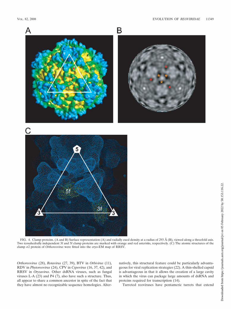
Clamp proteins. The capsid shell of a turreted reovirus isdecorated with 120 or 150 copies of clamp proteins (28, 42). Bycontrast, nonturreted reoviruses, such as rice dwarf virus(RDV), bluetongue virus (BTV), and rotavirus, lack suchclamp proteins. In the Orthoreovirus core, the clamp proteins,namely 150 copies of a 47K 2 protein, bind at three distinctlocations within each icosahedral asymmetric unit, where theyact as molecular clamps between the �1 proteins of the capsidshell below them. Thus, they appear to stabilize the capsidshell. The interactions between the clamp proteins and thecapsid proteins are not equivalent, but the bonding is strongand specific. In RRSV, 120 copies of the clamp protein wereobserved at positions similar to those in members of Orthoreo-virus, although no 2f clamp protein, observed at the icosahe-dral twofold axes in the Orthoreovirus core, was found in RRSV(Fig. 4A and 5). The clamp protein at the twofold axes also wasnot observed in CPV (42). Two kinds of blobs were found at 3fand 5f clamp positions of turreted reoviruses (28, 42). Theywere similar in size and shape. Their dimensions also weresimilar to those of Orthoreovirus. After the positional modifi-cation, the atomic structures of the 2 clamp protein of Or-thoreovirus were well accommodated into the cryo-EM map ofthe RRSV (Fig. 4C). These results suggest that two kinds ofblobs are composed of the same protein and correspond to the3f and 5f clamps of turreted reoviruses; thus, its number is 120in a particle. The positions of the clamp protein around thethreefold axis (3f clamp) in RRSV differed by �20 Å from theposition in Orthoreovirus, while the positions of the clampprotein around the fivefold axis (5f clamp protein) in RRSVwere almost same as those in Orthoreovirus. P8B (42K) ofRRSV, a protein similar in size to that of 2, was tentativelyassigned to the clamp protein. Unlike nonturreted reoviruses,which maintain a second capsid layer (T13 symmetry), tur-reted reoviruses become single-layered particles during tran-scription (22). Therefore, the clamp molecules that tie togetherneighboring capsid shell proteins might be required to increase
capsid stability. In fact, the 2 clamp protein of Orthoreovirusis required for the assembly of �1 capsid shell proteins intoicosahedral particles (28).
Peripheral trimers. Each turret of RRSV is surrounded byfive peripheral trimers, and this structural feature is uniqueamong known turreted reoviruses (Fig. 6). Single-shelled CPVand the Orthoreovirus core do not have trimers around theirturrets. One trimer of RRSV binds three clamp proteins, asshown in Fig. 6A. Thus, the clamp protein might act as molec-ular scaffolding for the trimers as well as acting as a molecularclamp in the capsid shell. In the Orthoreovirus virion, the coreis coated by a layer of trimers that are composed of �1 and 3proteins. The sizes of the sides of the �1333 trimer (�80 Å)are similar to those of RRSV, but the �1333 trimer is muchtaller than the RRSV trimer (Fig. 6C) (18). These proteinsform an incomplete T13l icosahedral lattice interrupted atthe fivefold axes by the �2 turrets. The trimers are designatedQ, R, S, and T trimers according to their positions in theincomplete T13l icosahedral lattice. It is noteworthy in thiscontext that RRSV has only one type of trimer in each icosa-hedral asymmetric unit, and the position of binding to thecapsid shell corresponds to that of the Q trimer in Orthoreo-virus (Fig. 6D). In other words, RRSV lacks the R, S, and Ttrimers, and the innermost capsid layer of RRSV is only par-tially covered with the trimers. Thus, the clamp proteins aticosahedral twofold axes (2f clamp protein) in Orthoreovirus,which act as scaffolding for trimers as described above, mightbe required for sitting the R, S, and T trimers on the capsidshell. However, although aquareovirus virions have only 120copies of the clamp protein (25), they have Q, R, S, and Ttrimers. Therefore, the 2f clamp proteins are not essential forthe assembly of the R, S, and T trimers. The trimer in Or-thoreovirus plays important roles in the viral penetration of thecell membrane (18). By analogy, the trimers in RRSV mighthave a similar function. Indeed, the P9 protein, which has beenputatively identified as the trimer protein, plays an importantrole in transmission via the insect vector (6a, 41). The esti-mated volume for a P9 trimer (117 kDa) is 1.4 � 105 Å3 (1.23Å3/Da) (17, 20), which is sufficiently coincident with the seg-mented volume for a trimer (1.8 � 105 Å3). Based on thesedata, we have tentatively assigned the P9 protein to a compo-nent of the peripheral trimer.
The possibility of the partial degradation of the outer capsidduring the purification, resulting in loss of the R, S, and Ttrimers, has not been excluded completely. However, the pu-rified viruses are infective to rice plants when inoculated viavector insects into which the virus had been injected (26).Furthermore, we have never observed the double-layered par-ticles in the electron microscopy, and the particle morphologyof the dipped materials directly processed from infected plantswas similar to those observed in purified preparations. Thus,we considered that the structure analyzed in this study is theRRSV virion.
Relationships among turreted reoviruses and their evolu-tion. Pentameric turrets at fivefold axes, plus 120 copies of theclamp protein on which peripheral trimers are located, wereattached to the thin-shelled core capsid in particles of RRSV.The thin core capsid shell is a structural feature that is com-mon among those reoviruses for which cryo-EM or X-ray crys-tallographic data are available, namely Aquareovirus (9, 25, 30),
11348 MIYAZAKI ET AL. J. VIROL.
Dow
nloa
ded
from
http
s://j
ourn
als.
asm
.org
/jour
nal/j
vi o
n 05
Feb
ruar
y 20
22 b
y 58
.153
.116
.22.
Orthoreovirus (28), Rotavirus (27, 39), BTV in Orbivirus (11),RDV in Phytoreovirus (24), CPV in Cypovirus (16, 37, 42), andRRSV in Oryzavirus. Other dsRNA viruses, such as fungalviruses L-A (23) and P4 (7), also have such a structure. Thus,all appear to share a common ancestor in spite of the fact thatthey have almost no recognizable sequence homologies. Alter-
natively, this structural feature could be particularly advanta-geous for viral replication strategies (22). A thin-shelled capsidis advantageous in that it allows the creation of a large cavityin which the virus can package large amounts of dsRNA andproteins required for transcription (14).
Turreted reoviruses have pentameric turrets that extend
FIG. 4. Clamp proteins. (A and B) Surface representation (A) and radially cued density at a radius of 293 Å (B), viewed along a threefold axis.Two icosahedrally independent 3f and 5f clamp proteins are marked with orange and red asterisks, respectively. (C) The atomic structures of theclamp 2 protein of Orthoreovirus were fitted into the cryo-EM map of RRSV.
VOL. 82, 2008 EVOLUTION OF REOVIRIDAE 11349
Dow
nloa
ded
from
http
s://j
ourn
als.
asm
.org
/jour
nal/j
vi o
n 05
Feb
ruar
y 20
22 b
y 58
.153
.116
.22.

from the underlying innermost capsid layer around the fivefoldaxes. The present study confirmed that RRSV has a typicalturret structure, as also reported for the Orthoreovirus core(28) and for CPV (16, 42). Thus, our study also confirmed thatthese viruses are appropriately grouped together from a struc-tural perspective (16). By contrast, the nonturreted reoviruses,such as Rotavirus, BTV, and RDV, lack turrets, and the inner-most capsid shell is fully covered with trimers with T13licosahedral symmetry. Thus, the entire innermost capsid iscovered with trimers, allowing the grouping of these viruses
from a structural perspective as well. These groupings by struc-ture reflect similar groupings based on sequence homologiesamong RdRps, the only proteins for which significant homol-ogies have been detected among respective proteins of virusesthat belong to this family (Fig. 7B). The structural similaritiesmentioned above, together with available biochemical data,suggest that turreted and nonturreted reoviruses form an evo-lutionarily related subgroup in the family Reoviridae (1, 21).This grouping does not reflect the host species infected by theviruses. The hosts of turreted reoviruses are plants plus insects,
FIG. 5. Structural comparison between the RRSV virion and the Orthoreovirus core. Surface representations and central cross-sections (40 Åthick) of the RRSV virion and the Orthoreovirus core are shown. The color coding is the same as that described in the legend to Fig. 1.
11350 MIYAZAKI ET AL. J. VIROL.
Dow
nloa
ded
from
http
s://j
ourn
als.
asm
.org
/jour
nal/j
vi o
n 05
Feb
ruar
y 20
22 b
y 58
.153
.116
.22.

insects, and mammals in the case of RRSV, CPV, and Or-thoreovirus, respectively. The hosts of nonturreted reovirusesare plants plus insects for Phytoreovirus, mammals for Rotavi-rus, and mammals plus insects for BTV. Our observationssuggest that reoviruses with a common ancestor separated intoturreted and nonturreted groups, and thereafter they sepa-rated still further to yield the present genera, possibly viaadaptation to their respective hosts.
A characteristic structural feature of turreted viruses is thepresence of clamps that are not found in nonturreted viruses,
such as Rotavirus, BTV, and RDV. The number of suchclamps, either 120 or 150, on the surface of the core of eachviral particle is another criterion for the further grouping of theviruses. The present study showed that RRSV belongs to thegroup with the 120 copies of the clamp protein, as does CPV.
In the turreted Orthoreovirus, the core is fully covered withtrimers with T13l icosahedral symmetry at the Q, R, S, and Tpositions, while the RRSV virion has only peripheral trimersthat were found only at the Q position around turrets. Or-thoreovirus loses all its trimers during the penetration of the
FIG. 6. Peripheral trimers. (A) Close-up view of a trimer. A peripheral trimer is shown bound to three clamps that are marked with asterisks.(B) Radially cued density with fivefold axes at a radius of 333 Å, at which the molecular boundaries of trimers are apparent. (C) Atomic structuresof the Orthoreovirus trimers. CPK models of 33 (left) and �1333 trimers (right) are shown as viewed from the outside of the capsid shell (upper)and after 90° rotation (lower). The 3 molecules are colored pink, magenta, and orange, and the �1 molecules are colored cyan, blue, and green.The 3 molecules are removed from the viral particles during the infection by proteolysis. (D) Surface representation with the icosahedral T13lnet. The positions of trimers that correspond to the Q position in the icosahedral T13l symmetry are shown and labeled Q.
VOL. 82, 2008 EVOLUTION OF REOVIRIDAE 11351
Dow
nloa
ded
from
http
s://j
ourn
als.
asm
.org
/jour
nal/j
vi o
n 05
Feb
ruar
y 20
22 b
y 58
.153
.116
.22.

cell membrane, and only the core particle remains. The core istranscriptionally active and is quite similar to that of CPV, thestructurally simplest member of the Reoviridae, and it lacks anouter capsid layer composed of trimers (16). Thus, RRSVseems to have a novel structure that is architecturally interme-diate between that of CPV and that of Orthoreovirus (Fig. 7A).The structure of RRSV suggests that turreted reoviruses haveacquired different types of secondary layers during the evolu-
tionary steps that led to the ability to infect a wide variety ofhost species.
ACKNOWLEDGMENTS
Naoyuki Miyazaki and Li Xing were supported by grants from theSTINT Foundation and from the NIH HIVRAD program, respec-tively. R.H.C. was supported by the NIH Discovery program. Thisstudy was funded by a grant from the NIH Roadmap Nanomedicine
FIG. 7. Relationships among turreted reoviruses. (A) Schematic representation of the RRSV virion, the CPV virion, the Orthoreovirus core, andthe Orthoreovirus virion. The RdRp, capsid, clamp, trimeric outer capsid, turret, and spike proteins are shown in red, yellow, green, sky blue, blue,and blue-purple, respectively. (B) Neighbor-joining tree constructed from full-length sequences of RdRps of viruses in the family Reoviridae. Thegenera, strains, abbreviations of names of viruses, and accession numbers of sequences are as follows. For Aquareovirus, Chum salmon reovirus(CSRV; AF418295), Striped bass reovirus (SBRV; AF450318), and Golden shiner reovirus (GSRV; AF403399); for Orthoreovirus, Mammalianorthoreovirus subgroup 1, Lang strain (MRV-1; M24734), Jones strain (MRV-2; M31057), and Dearing strain (MRV-3; M31058); for Oryzavirus,Rice ragged stunt virus (RRSV; U66714); for Cypovirus, Bombyx mori cytoplasmic polyhedrosis virus 1 (BmCPV-1; AF323782), Dendrolimus punctatuscypovirus 1 (DpCPV-1; AAN46860), and Lymantria dispar cypovirus 14 (LdCPV-14; AAK73087); for Rotavirus, Rotavirus A and Simian rotavirusstrain SA11 (SiRV-A/SA-11; AF015955); for Phytoreovirus, Rice dwarf virus strain A (RDV-A; D90198); and for Orbivirus, African horse sicknessvirus serotype 9 (AHSV-9; U94887) and Bluetongue virus serotype 2 (BTV-2; L20508) (1).
11352 MIYAZAKI ET AL. J. VIROL.
Dow
nloa
ded
from
http
s://j
ourn
als.
asm
.org
/jour
nal/j
vi o
n 05
Feb
ruar
y 20
22 b
y 58
.153
.116
.22.

Program and by a grant from the Cancer Research Center of theSwedish Research Council (to R.H.C.); by a grant from the NationalProject on Protein Structural and Functional Analysis and by a Grant-in-Aid from the 21st Century Centers of Excellence Program (to A.N.);and by a grant from the National Project on Structures of BiologicalMacromolecular Assemblies and a Grant-in-Aid for Scientific Re-search on Priority Areas (to T.O.) from the Ministry of Education,Culture, Sports, Science, and Technology of Japan.
REFERENCES
1. Attoui, H., F. Mohd Jaafar, M. Belhouchet, P. Biagini, J. F. Cantaloube, P.de Micco, and X. de Lamballerie. 2005. Expansion of family Reoviridae toinclude nine-segmented dsRNA viruses: isolation and characterization of anew virus designated Aedes pseudoscutellaris reovirus assigned to a proposedgenus (Dinovernavirus). Virology 343:212–223.
2. Baker, T. S., and R. H. Cheng. 1996. A model-based approach for determin-ing orientations of biological macromolecules imaged by cryoelectron mi-croscopy. J. Struct. Biol. 116:120–130.
3. Bamford, D. H., J. M. Grimes, and D. I. Stuart. 2005. What does structuretell us about virus evolution? Curr. Opin. Struct. Biol. 15:655–663.
4. Bartlett, N., S. Gillies, S. Bullivant, and A. Bellamy. 1974. Electron micros-copy study of reovirus reaction cores. J. Virol. 14:315–326.
5. Bellamy, A., and J. Harvey. 1976. Biophysical studies of reovirus type 3. III.A laser light-scattering study of the RNA transcriptase reaction. Virology70:28–36.
6. Chandran, K., S. B. Walker, Y. Chen, C. M. Contreras, L. A. Schiff, T. S.Baker, and M. L. Nibert. 1999. In vitro recoating of reovirus cores withbaculovirus-expressed outer-capsid proteins �1 and 3. J. Virol. 73:3941–3950.
6a.Chaogang, S., W. Jianhua, Z. Guoying, S. Gang, P. Baozhen, L. Juanli, J.Dendi, C. Shenxiang, N. M. Upadhyaya, P. Waterhouse, and G. Zuxun. 2003.Ectopic expression of the spike protein of rice ragged stunt Oryzavirus intransgenic rice plants inhibits transmission of the virus to insects. Mol.Breed. 11:295–301.
7. Cheng, R. H., J. R. Caston, G. J. Wang, F. Gu, T. J. Smith, T. S. Baker, R. F.Bozarth, B. L. Trus, N. Cheng, R. B. Wickner, and A. C. Steven. 1994. Fungalvirus capsids, cytoplasmic compartments for the replication of double-stranded RNA, formed as icosahedral shells of asymmetric Gag dimers. J.Mol. Biol. 244:255–258.
8. Dryden, K. A., G. Wang, M. Yeager, M. L. Nibert, K. M. Coombs, D. B.Furlong, B. N. Fields, and T. S. Baker. 1993. Early steps in reovirus infectionare associated with dramatic changes in supramolecular structure and pro-tein conformation: analysis of virions and subviral particles by cryoelectronmicroscopy and image reconstruction. J. Cell Biol. 122:1023–1041.
9. Fang, Q., S. Shah, Y. Liang, and Z. H. Zhou. 2005. 3D reconstruction andcapsid protein characterization of grass carp reovirus. Sci. China C Life Sci.48:593–600.
10. Furuichi, Y. 1974. “Methylation-coupled” transcription by virus associatedtranscriptase of cytoplasmic polyhedrosis virus containing double-strandedRNA. Nucleic Acids Res. 1:809–822.
11. Grimes, J. M., J. N. Burroughs, P. Gouet, J. M. Diprose, R. Malby, S.Zientara, P. P. Mertens, and D. I. Stuart. 1998. The atomic structure of thebluetongue virus core. Nature 395:470–478.
12. Hagiwara, K., and H. Naitow. 2003. Assembly into single-shelled virus-likeparticles by major capsid protein VP1 encoded by genome segment S1 ofBombyx mori cypovirus 1. J. Gen. Virol. 84:2439–2441.
13. Hagiwara, K., S. Rao, S. W. Scott, and G. R. Carner. 2002. Nucleotidesequences of segments 1, 3 and 4 of the genome of Bombyx mori cypovirus 1encoding putative capsid proteins VP1, VP3 and VP4, respectively. J. Gen.Virol. 83:1477–1482.
14. Hagiwara, K., T. Higashi, N. Miyazaki, H. Naitow, R. H. Cheng, A. Naka-gawa, H. Mizuno, T. Tsukihara, and T. Omura. 2004. The amino-terminalregion of major capsid protein P3 is essential for self-assembly of single-shelled core-like particles of Rice dwarf virus. J. Virol. 78:3145–3148.
15. Hagiwara, K., Y. Minobe, Y. Nozu, H. Hibino, I. Kimura, and T. Omura.1986. Component proteins and structures of Rice ragged stunt virus. J. Gen.Virol. 67:1711–1715.
16. Hill, C. L., T. F. Booth, B. V. Prasad, J. M. Grimes, P. P. Mertens, G. C.Sutton, and D. I. Stuart. 1999. The structure of a cypovirus and the func-tional organization of dsRNA viruses. Nat. Struct. Biol. 6:565–568.
17. Kantardjieff, K. A., and B. Rupp. 2003. Matthews coefficient probabilities:improved estimates for unit cell contents of proteins, DNA, and protein-nucleic acid complex crystals. Protein Sci. 12:1865–1871.
18. Liemann, S., K. Chandran, T. S. Baker, M. L. Nibert, and S. C. Harrison.
2002. Structure of the reovirus membrane-penetration protein, �1, in acomplex with its protector protein, 3. Cell 108:283–295.
19. Ling, K. C., E. R. Tiongco, and V. M. Aguiero. 1978. Rice ragged stunt, a newvirus disease. Plant Dis. Rep. 62:701–705.
20. Matthews, B. W. 1968. Solvent content of protein crystals. J. Mol. Biol.33:491–497.
21. McQualter, R. B., G. R. Smith, J. L. Dale, and R. M. Harding. 2003.Molecular analysis of Fiji disease Fijivirus genome segments 1 and 3. VirusGenes 26:283–289.
22. Mertens, P. P. C., H. Attoui, R. Duncan, and T. S. Dermody. 2005. Reoviri-dae, p. 447–454. In C. M. Fauquest, M. A. Mayo, J. Maniloff, U. Dessel-berger, and L. A. Ball (ed.), Virus taxonomy. Eighth report of the Interna-tional Committee on Taxonomy of Viruses. Elsevier Academic Press, SanDiego, CA.
23. Naitow, H., J. Tang, M. Canady, R. B. Wickner, and J. E. Johnson. 2002. L-Avirus at 3.4 Å resolution reveals particle architecture and mRNA decappingmechanism. Nat. Struct. Biol. 9:725–728.
24. Nakagawa, A., N. Miyazaki, J. Taka, H. Naitow, A. Ogawa, Z. Fujimoto, H.Mizuno, T. Higashi, Y. Watanabe, T. Omura, R. H. Cheng, and T. Tsuki-hara. 2003. The atomic structure of Rice dwarf virus reveals the self-assemblymechanism of component proteins. Structure 11:1227–1238.
25. Nason, E. L., S. K. Samal, and B. V. Prasad. 2000. Trypsin-induced structuraltransformation in aquareovirus. J. Virol. 74:6546–6555.
26. Omura, T., Y. Minobe, I. Kimura, H. Hibino, T. Tsuchizaki, and Y. Saito.1983. Improved purification procedure and RNA segments of Rice raggedstunt virus. Ann. Phytopathol. Soc. Jpn. 49:670–675.
27. Prasad, B. V., R. Rothnagel, C. Q. Zeng, J. Jakana, J. A. Lawton, W. Chiu,and M. K. Estes. 1996. Visualization of ordered genomic RNA and local-ization of transcriptional complexes in rotavirus. Nature 382:471–473.
28. Reinisch, K. M., M. L. Nibert, and S. C. Harrison. 2000. Structure of thereovirus core at 3.6 Å resolution. Nature 404:960–967.
29. Reference deleted.30. Shaw, A. L., S. K. Samal, K. Subramanian, and B. V. Prasad. 1996. The
structure of aquareovirus shows how the different geometries of the twolayers of the capsid are reconciled to provide symmetrical interactions andstabilization. Structure 4:957–967.
31. Supyani, S., B. I. Hillman, and N. Suzuki. 2007. Baculovirus expression ofthe 11 mycoreovirus-1 genome segments and identification of the guanylyl-transferase-encoding segment. J. Gen. Virol. 88:342–350.
32. Upadhyaya, N. M., E. Zinkowsky, W. Kositratana, and P. M. Waterhouse.1996. The Mr 43K major capsid protein of rice ragged stunt Oryzavirus is apost-translationally processed product of a Mr 67,348 polypeptide encodedby genome segment 8. Arch. Virol. 141:1689–1701.
33. Upadhyaya, N. M., K. Ramm, J. A. Gellatly, Z. Li, W. Kositratana, and P. M.Waterhouse. 1998. Rice ragged stunt Oryzavirus genome segment S4 couldencode an RNA-dependent RNA polymerase and a second protein of un-known function. Arch. Virol. 143:1815–1822.
34. Upadhyaya, N. M., M. Yang, W. Kositratana, A. Ghosh, and P. M. Water-house. 1995. Molecular analysis of rice ragged stunt Oryzavirus segment 9and sequence conservation among isolates from Thailand and India. Arch.Virol. 140:1945–1956.
35. White, C., and H. Zweerink. 1976. Studies on the structure of reovirus cores:selective removal of polypeptide lambda 2. Virology 70:171–180.
36. Yazaki, K., and K.-I. Miura. 1980. Relation of the structure of cytoplasmicpolyhedrosis viruses and the synthesis of its messenger RNA. Virology 105:467–479.
37. Yu, X., L. Jin, and Z. H. Zhou. 2008. 3.88 Å structure of cytoplasmicpolyhedrosis virus by cryo-electron microscopy. Nature 453:415–419.
38. Zhang, H., X. K. Yu, X. Y. Lu, J. Q. Zhang, and Z. H. Zhou. 2002. Molecularinteractions and viral stability revealed by structural analyses of chemicallytreated cypovirus capsids. Virology 298:45–52.
39. Zhang, X., E. Settembre, C. Xu, P. R. Dormitzer, R. Bellamy, S. C. Harrison,and N. Grigorieff. 2008. Near-atomic resolution using electron cryomicros-copy and single-particle reconstruction. Proc. Natl. Acad. Sci. USA 105:1867–1872.
40. Zhang, X., S. B. Walker, P. R. Chipman, M. L. Nibert, and T. S. Baker. 2003.Reovirus polymerase lambda 3 localized by cryo-electron microscopy ofvirions at a resolution of 7.6 Å. Nat. Struct. Biol. 10:1011–1018.
41. Zhou, G. Y., X. B. Lu, H. J. Lu, J. L. Lei, S. X. Chen, and Z. X. Gong. 1999.Rice ragged stunt Oryzavirus: role of the viral spike protein in transmissionby the insect vector. Ann. Appl. Biol. 135:573–578.
42. Zhou, Z. H., H. Zhang, J. Jakana, X. Y. Lu, and J. Q. Zhang. 2003. Cyto-plasmic polyhedrosis virus structure at 8 Å by electron cryomicroscopy: struc-tural basis of capsid stability and mRNA-processing regulation. Structure11:651–663.
VOL. 82, 2008 EVOLUTION OF REOVIRIDAE 11353
Dow
nloa
ded
from
http
s://j
ourn
als.
asm
.org
/jour
nal/j
vi o
n 05
Feb
ruar
y 20
22 b
y 58
.153
.116
.22.