Embed Size (px)
Citation preview
Proc. Natd. Acad. Sci. USAVol. 82, pp. 45924596, July 1985Biochemistry
Structural invariants of antigen binding: Comparison ofimmunoglobulin VL-VH and VL-VL domain dimers
(antibody/fl-sheet/electrostatlcs/strophoid/x-ray structure)
JnXi NOVOTN.F AND EDGAR HABERMolecular and Cellular Research Laboratory, Massachusetts General Hospital and Harvard Medical School, Boston, MA 02114
Communicated by Elkan R. Blout, March 11, 1985
ABSTRACT Antigen-combining site arises by noncovalentassociation of the variable domain of the immunoglobulinheavy chain (VI,) with that of the light chain (VL). To analyzethe invariant features of the binding region (VL-VH domaininterface), we compared the known immunoglobulin three-di-mensional structures by a variety of methods. The interfaceforms a close-packed, twisted, prism-shaped "fl-barrel" char-acterized by cross-sectional dimensions 1.04 X 0.66 nm and atop-to-bottom twist angle of 2120. The geometry of the interfaceis preserved via invariance of some 15 side chains, both insidethe domains and on their surface. Buried polar residues forma conserved hydrogen-bonding network that has a similartopological connectivity in the two domain types; two hydrogenbonds contributed by invariant side chains extend across theinterface and anchor the fl-sheets in their relative orientation.Invariant aromatic residues close-pack at the bottom of thebinding-site fl-barrel with their ring planes oriented perpen-dicularly in the characteristic "herringbone" packing mode.Electrostatic computations that implicitly include solvent ef-fects show the domains to be stabilized by large electrostaticforces. However, structures that were crystallized at lower pHhave their electrostatic energies appropriately lowered, imply-ing that full ionization of carboxyl side chains is essential forefficient electrostatic stabilization. The unusual mode ofdomain-domain association in the VL-VL dimer RHE corre-lates with its overall repulsive electrostatic energy (+54kJ/mol), as opposed to negative (i.e., stabilizing) energy values(-263 to -543 kJ/mol) found in the domains of the otherstructures. The VL-VL dimer REI mimics closely the interfacegeometry of VL-VH dimers although its domain-domain con-tact area is lower by 18%.
The structural diversity of antibody molecules epitomizesone of the most interesting problems of molecular biology,that of a relationship between molecular shape and biologicalfunction. Although spatial structures of three different anti-body binding sites have been elucidated (1-3), our knowledgeof structural prerequisites of the antigen binding functionremains incomplete. Relative importance ofindividual aminoacid residues that form the antigen binding site is not wellunderstood, nor is it clear how their presence influencesantigen binding. Recent advances in genetic engineering haveput at our disposal a means to modulate antibody specificityat will-e.g., by applying site-directed mutagenesis to genesencoding immunoglobulin polypeptide chains. This possibil-ity, however, can only be realized on the basis of a soundunderstanding of principles that determine protein anatomyin general (4, 5) and that of antibody molecules in particular(6).The antigen-combining site is formed by noncovalent
association of two "variable" (V) domains provided by two
different polypeptide chains, heavy (H) and light (L). TheVL-VH interface consists of two closely packed p3-sheets andits geometry corresponds to a nine-stranded elliptical (4) orprism-shaped (7) barrel. The barrel forms the bottom andsides of the antigen binding site, and amino acid residues thatare part of the domain-domain interface and appear not to beaccessible to solvent or antigen contribute to antibodyspecificity (6).Here we study those conformational features ofthe VL and
VH domains that are conserved in all the antibodies and formthe constant scaffold for the binding site. We do so bycomparing three-dimensional structures with use of a novelprocedure (8, 9): we superimpose, by the least-squaresmethod, only those side chain atoms that are invariant in allthe immunoglobulins. This allows for differences in function-al importance of different parts of the structure and leads tomore meaningful results than the method employed previ-ously (10, 11), namely, a least-squares superposition ofcomplete polypeptide chain backbones (i.e., optimization ofstructural correspondence over the domain as a whole). Wealso analyze the conserved hydrogen-bonding network ex-isting among polar side chains that are buried inside thedomains and discuss the contribution of electrostatic inter-actions to the stability of the binding site.
ATOMIC COORDINATES AND CALCULATIONSCrystallographic coordinates ofhuman Fab fragments NEW,KOL, and MCPC 603; VL-VL dimers RHE and REI; andBence Jones protein (light-chain dimer) MCG were obtainedfrom the Brookhaven Protein Data Bank (12). No attemptswere made to energy-minimize or otherwise improve theoriginal data. Structural manipulations, such as least-squaressuperpositions, generation of hydrogen bonded lists, poten-tial energy evaluations, etc., were performed with the pro-gram CHARMM version 16 (13) as described (6, 14). Theelectrostatic potential was computed by use of a solvent-modified Coulomb formula (14). The effect of solvent wasmodeled by multiplying charges on atoms by a constant thatdepends linearly on the distance of the atom from the surfaceof the protein (15). The potential was evaluated to infinity;i.e., no distance cutoff was applied to evaluations of pair-wise atomic interactions. Stereo drawings were made fromCHARMM-generated files, using previously describedgraphics facilities and software (14). Amino acid alignmentsof immunoglobulin variable domains used as a basis forstructural comparisons and residue numbering were those ofKabat et al. (16).
RESULTS AND DISCUSSIONConservation of the Binding Site Geometry. Although all the
immunoglobulin domains share the same folding scheme-two antiparallel p-sheets packed face-to-face (4)-the num-ber of p-strands, strand orientation, side chain preponder-
Abbreviations: VL, light chain variable; VH, heavy chain variable.
4592
The publication costs of this article were defrayed in part by page chargepayment. This article must therefore be hereby marked "advertisement"in accordance with 18 U.S.C. §1734 solely to indicate this fact.
Dow
nloa
ded
by g
uest
on
June
3, 2
020
Proc. NatL Acad Sci USA 82 (1985) 4593
ance, and other structural characteristics differ widely amongvarious domain types (VL, VH, and constant domains CL,CHi, CH2 and CH3). Only three residues are common to all thedomains: two cysteines that form a disulfide bridge betweenthe /3-sheets and a tryptophan that packs against them (17).Structural diversity of this kind correlates with the fact thatdifferent domain types perform different biological functions,such as antigen binding in VH and VL or complement bindingin CH2. Identical domain types, on the other hand, might beexpected to have many more structural features in common.To compare x-ray structures of VL and VH domains, the
best-resolved crystallographic data-set, the Fab fragmentKOL, was first chosen as a reference structure and orientedmost conveniently in the reference frame of Cartesian coor-dinates ("barrel orientation"' of figure 4 in ref. 6). Second,selected side chains of the VL or VH domains of Fabfragments NEW and MCPC 603 were superimposed, inde-pendently in each domain, on the corresponding side chainsof the reference structure. Because of their invariance andcentral positions in the domain cores (17), residues Cys-23and -88, Trp-35, and Phe-62 were chosen in the VL domain;Cys-22 and -92, Trp-47, and Leu-78 were chosen in the VHdomain.
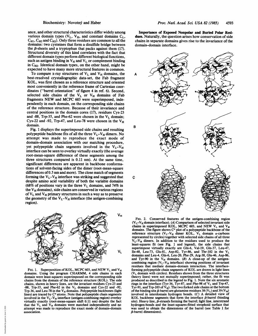
Fig. 1 displays the superimposed side chains and resultingpolypeptide backbone fits of all the three VL-VH dimers. Noattempt was made to reproduce the exact mode ofdomain-domain association with our matching procedure,yet polypeptide chain segments involved in the VL-VHinterface can be seen to overlay virtually exactly (the averageroot-mean-square difference of these segments among thethree structures compared is 0.11 nm). At the same time,significant differences are apparent in backbone conforma-tions of solvent-facing sides of the dimer (root-mean-squaredifferences of0.5 nm and more). The close match of segmentsforming the VL-VH interface was striking and suggested thatdespite amino acid variability of both the variable domains(68% of positions vary in the three VL domains, and 74% inthe VH domains), side chains are conserved in various regionsofVL and VH primary structures in such a way as to preservethe geometry of the VL-VH interface (the antigen-combiningregion).
AX VH
VL
FIG. 1. Superposition ofKOL, MCPC 603, and NEW VL and VHdomains. Using the program CHARMM, 4 side chains in eachdomain were least-squares superimposed on the corresponding sidechains from the domain of the reference structure (KOL). The sidechains, shown in heavy lines, are the invariant residues Cys-23 and-88, Trp-35, and Phe-62 in the VL domains and Cys-22 and -92,Trp-36, and Leu-78 in the VH domains. Polypeptide backbones (lightlines) are traced by Ca atoms. Note that polypeptide chain segmentsinvolved in the VL-VH interface (antigen-combining region) overlayvirtually exactly (root-mean-square shift 0.11 nm) despite the factthat the VL and VH domains were matched independently and noattempt was made to reproduce the exact mode of domain-domainassociation.
Importance of Exposed Nonpolar and Buried Polar Resi-dues. Naturally, the question arises how conservation of sidechains in separate domains gives rise to the invariance of thedomain-domain interface.
A
B
c
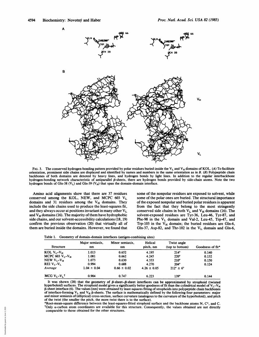
FIG. 2. Conserved features of the antigen-combining region(VL-VH domain interface). (A) Comparison of selected invariant sidechains in superimposed KOL, MCPC 603, and NEW VL and VHdomains. The figure shows Ca plot of a polypeptide backbone of thereference structure (VL-VH dimer KOL, VL domain a-carbonsrepresented by circles) together with selected side chains of all threeVL-VH dimers. In addition to the residues used to produce theleast-squares fit (see Fig. 1 and legend), the side chains thatsuperimpose virtually exactly are Gln-6, Val-19, Gln-37, Leu-47,Ile-48, Leu-73, Glu-81, Asp-82, Tyr-86, and Thr-102 in the VLdomains and Leu-4, Gln-6, Leu-20, Phe-29, Arg-38, Glu-46, Asp-86,and Tyr-90 in the VH domains. (B) A close-up of the antigen-combining region (VL-VH interface) showing positions of invariantresidues that mediate domain-domain interaction. The interface-forming polypeptide chain segments of KOL are drawn in light lines(VL domain with circles). Residues shown from the three structures(heavy lines) were not mutually superimposed; rather, the fit wasproduced as described in the legend to Fig. 1. Note the six aromaticrings in the interface (Tyr-36, Tyr-87, and Phe-98 of VL and Trp-47,Tyr-91, and Trp-103 of VH). The two forked side chains at the bottomofthe binding site 3-barrel are glutaniine residues 38 (VL) and 39 (VH)involved in interdomain hydrogen bonds. (C) A detailed. view ofKOL backbone segments that form the interface P-barrel (bindingsite). Heavy line, 13-strands forming the barrel; light line, interstrandhydrogen-bonds and the least-squares-fitted strophoid surface thatwas used to obtain the dimensions of the barrel (see Table 1 for/3-barrel dimensions).
Biochemistry: Novotn. and Haber
Dow
nloa
ded
by g
uest
on
June
3, 2
020
4594 Biochemistry: Novotnf and Haber
A
LN tE
N 39
A;66
N ff6
IN 39
B
VH
VL
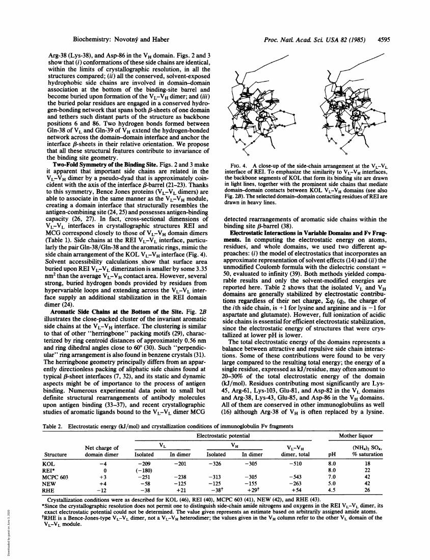
FIG. 3. The conserved hydrogen-bonding pattern provided by polar residues buried inside the VL and VH domains of KOL. (A) To facilitateorientation, prominent side chains are displayed and identified by names and numbers in the same orientation as in B. (B) Polypeptide chainbackbones of both domains are denoted by heavy lines, and hydrogen bonds by light lines. In addition to the regular interbackbonehydrogen-bonding network characteristic of antiparallel 8-sheets, there are hydrogen bonds provided by side-chain atoms. Note the twohydrogen bonds of Gln-38 (VL) and Gln-39 (VH) that span the domain-domain interface.
Amino acid alignments show that there are 37 residuesconserved among the KOL, NEW, and MCPC 603 VLdomains and 31 residues among the VH domains. Theyinclude the side chains used to produce the least-squares fit,and they always occur at positions invariant in many other VLand VH domains (16). The majority ofthem have hydrophobicside chains, and our solvent-accessibility calculations (18, 19)confirm the previous observation (20) that virtually all ofthem are buried inside the domains. However, we found that
some of the nonpolar residues are exposed to solvent, whilesome of the polar ones are buried. The structural importanceofthe exposed nonpolar and buried polar residues is apparentfrom the fact that they belong to the most stringentlyconserved side chains in both VL and VH domains (16). Thesolvent-exposed residues are Tyr-36, Leu-46, Tyr-87, andPhe-98 in the VL domain and Val-2, Leu-45, Trp-47, andTrp-103 in the VH domain; the buried residues are Gln-6,Gln-37, Asp-82, and Thr-102 in the VL domain and Gln-6,
Table 1. Geometry of domain-domain interfaces (antigen-combining sites)Major semiaxis, Minor semiaxis, Helical Twist angle
Structure nm nm pitch, nm (top to bottom) Goodness of fit*KOL VLVH 1.013 0.652 4.195 2140 0.140MCPC 603 VL-VH 1.081 0.662 4.245 2200 0.132NEW VL-VH 1.073 0.630 4.333 2100 0.150REI VL-VL 0.994 0.688 4.270 2040 0.124Average 1.04 ± 0.04 0.66 ± 0.02 4.26 ± 0.05 2120 ± 60
MCG VLVLt 0.904 0.747 6.223 1390 0.144
It was shown (28) that the geometry of 13-sheet-p-sheet interfaces can be approximated by strophoid (twistedhyperboloid) surfaces. The strophoid model gives a significantly better goodness of fit than the cylindrical model ofVL-VH1-sheet interface (6). The values [rm] were obtained by least-squares fitting of strophoids into polypeptide chain backbonesof interface-forming VL and VH P-sheets. The surface is mathematically defined by the following four parameters: majorand minor semiaxis of (elliptical) cross-section, surface curvature (analogous to the curvature of the hyperboloid), and pitchof the twist (the smaller the pitch, the more twist there is to the surface).*Root-mean-square difference between the least-squares-fitted strophoid surface and the backbone atoms N, Ca, and C.tOnly a-carbon atom coordinates are available for this structure. Consequently, the values obtained are not directlycomparable to those obtained for the other structures.
Proc. NatL Acad ScL USA 82 (1985)
Dow
nloa
ded
by g
uest
on
June
3, 2
020
Proc. NatL Acad SCi USA 82 (1985) 4595
Arg-38 (Lys-38), and Asp-86 in the VH domain. Figs. 2 and 3show that (i) conformations ofthese side chains are identical,within the limits of crystallographic resolution, in all thestructures compared; (ii) all the conserved, solvent-exposedhydrophobic side chains are involved in domain-domainassociation at the bottom of the binding-site barrel andbecome buried upon formation of the VL-VH dimer; and (iii)the buried polar residues are engaged in a conserved hydro-gen-bonding network that spans both ,B-sheets of one domainand tethers such distant parts of the structure as backbonepositions 6 and 86. Two hydrogen bonds formed betweenGln-38 of VL and Gln:39 of VH extend the hydrogen-bondednetwork across the domain-domain interface and anchor theinterface (-sheets in their relative orientation. We proposethat all these structural features contribute to invariance ofthe binding site geometry.Two-Fold Symmetry of the Binding Site. Figs. 2 and 3 make
it apparent that important side chains are related in theVL-VH dimer by a pseudo-dyad that is approximately coin-cident with the axis of the interface (3-barrel (21-23). Thanksto this symmetry, Bence Jones proteins (VL-VL dimers) areable to associate in the same manner as the VL-VH module,creating a domain interface that structurally resembles theantigen-combining site (24, 25) and possesses antigen-bindingcapacity (26, 27). In fact, cross-sectional dimensions ofVL-VL interfaces in crystallographic structures REI andMCG correspond closely to those of VL-VH domain dimers(Table 1). Side chains at the REI VL-VL interface, particu-larly the pair Gln-38/Gln-38 and the aromatic rings, mimic theside chain arrangement of the KOL VL-VH interface (Fig. 4).Solvent accessibility calculations show that surface areaburied upon REI VL-VL dimerization is smaller by some 3.35nm2 than the average VL-VH contact area. However, severalstrong, buried hydrogen bonds provided by residues fromhypervariable loops and extending across the VL-VL inter-face supply an additional stabilization in the REI domaindimer (24).Aromatic Side Chains at the Bottom of the Site. Fig. 2B
illustrates the close-packed cluster of the invariant aromaticside chains at the VL-VH interface. The clustering is similarto that of other "herringbone" packing motifs (29), charac-terized by ring centroid distances of approximately 0.56 nmand ring dihedral angles close to 600 (30). Such "perpendic-ular" ring arrangement is also found in benzene crystals (31).The herringbone geometry principally differs from an appar-ently directionless packing of aliphatic side chains found attypical ,83sheet interfaces (7, 32), and its static and dynamicaspects might be of importance to the process of antigenbinding. Numerous experimental data point to small butdefinite structural rearrangements of antibody moleculesupon antigen binding (33-37), and recent crystallographicstudies of aromatic ligands bound to the VL-VL dimer MCG
FIG. 4. A close-up of the side-chain arrangement at the VL-VLinterface of RET. To emphasize the similarity to VL-VH interfaces,the backbone segments of KOL that form its binding site are drawnin light lines, together with the prominent side chains that mediatedomain-domain contacts between KOL VL-VH domains (see alsoFig. 2B). The selected domain-domain contacting residues ofREI aredrawn in heavy lines.
detected rearrangements of aromatic side chains within thebinding site (3-barrel (38).
Electrostatic Interactions in Variable Domains and Fv Frag-ments. In computing the electrostatic energy on atoms,residues, and whole domains, we used two different ap-prQaches: (i) the model of electrostatics that incorporates anapproximate representation of solvent effects (14) and (ii) theunmodified Coulomb formula with the dielectric constant =
50, evaluated to infinity (39). Both methods yielded compa-rable results and only the solvent-modified energies arereported here. Table 2 shows that the isolated VL and VHdomains are generally stabilized by electrostatic contribu-tions regardless of their net charge, Xq1 (qj, the charge ofthe ith side chain, is +1 for lysine and arginine and is -1 foraspartate and glutamate). However, full ionization of acidicside chains is essential for efficient electrostatic stabilization,since the electrostatic energy of structures that were crys-tallized at lower pH is lower.The total electrostatic energy of the domains represents a
balance between attractive and repulsive side chain interac-tions. Some of these contributions were found to be verylarge compared to the resulting total energy; the energy of asingle residue, expressed as kJ/residue, may often amount to20-30%o of the total electrostatic energy of the domain(kJ/mol). Residues contributing most significantly are Lys-45, Arg-61, Lys-103, Glu-81, and Asp-82 in the VL domainsand Arg-38, Lys-43, Glu-85, and Asp-86 in the VH domains.All of them are conserved in other immunoglobulins as well(16) although Arg-38 of VH is often replaced by a lysine.
Table 2. Electrostatic energy (kJ/mol) and crystallization conditions of immunoglobulin Fv fragmentsElectrostatic potential Mother liquor
Net charge of VL VH VL-VH (NH4)2 SO4,Structure domain dimer Isolated In dimer Isolated In dimer dimer, total pH % saturation
KOL -4 -209 -201 -326 -305 -510 8.0 18REP 0 (-180) 8.0 22MCPC 603 +3 -251 -238 -313 -305 -543 7.0 42NEW +4 -58 -125 -125 -155 -263 5.0 42RHE -12 -38 +21 -38t +29t +54 4.5 26
Crystallization conditions were as described for KOL (46), REI (40), MCPC 603 (41), NEW (42), and RHE (43).*Since the crystallographic resolution does not permit one to distinguish side-chain amide nitrogens and oxygens in the REI VL-VL dimer, itsexact electrostatic potential could not be determined. The value given represents an estimate based on arbitrarily assigned amide atoms.tRHE is a Bence-Jones-type VL-VL dimer, not a VL-VH heterodimer; the values given in the VH column refer to the other VL domain of theVL-VL module.
Biochemistry: Novotn. and Haber
Dow
nloa
ded
by g
uest
on
June
3, 2
020
4596 Biochemistry: Novotn5 and Haber
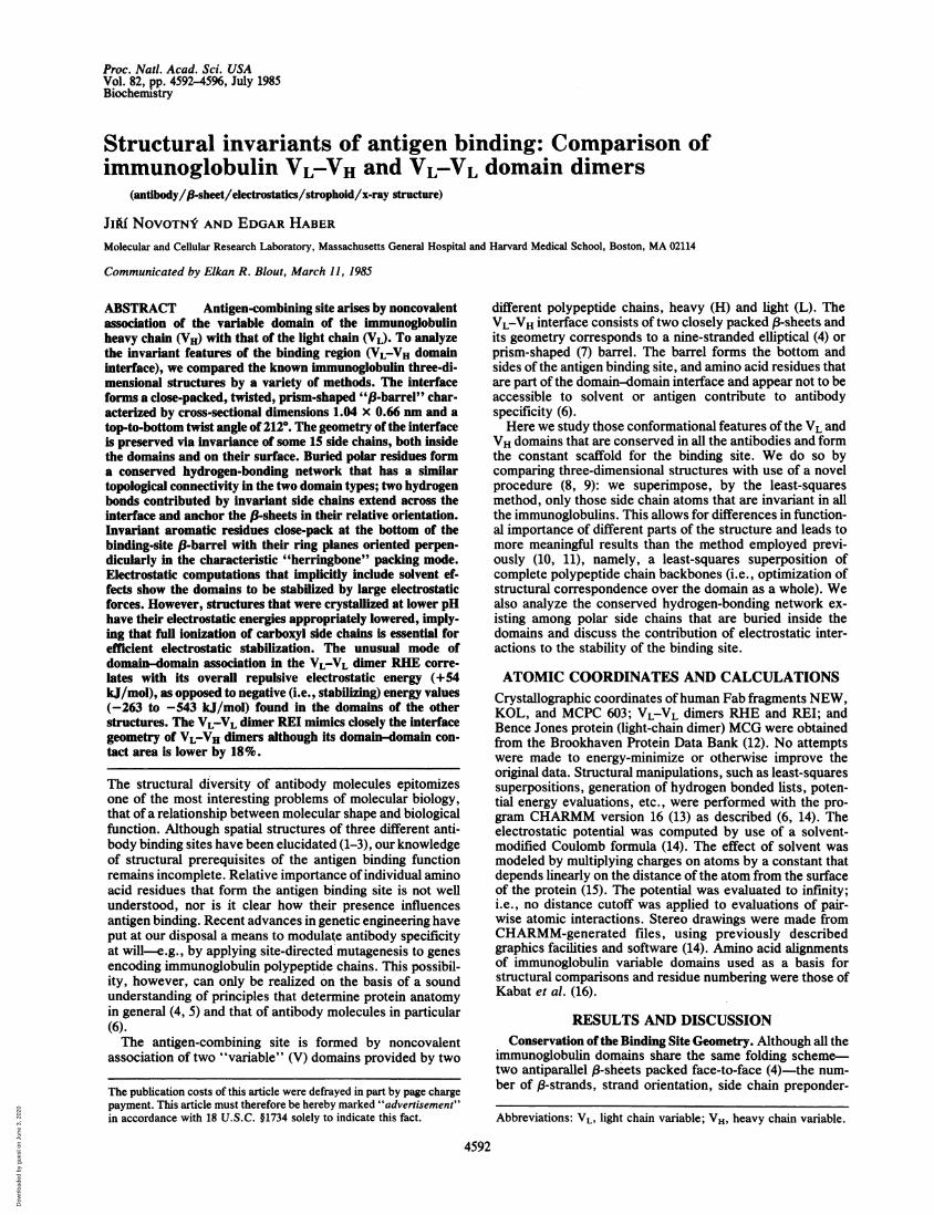
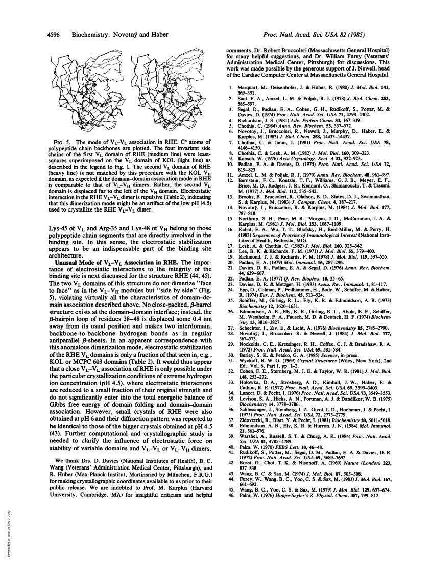
FIG. 5. The mode of VL-VL association in RHE. CA atoms ofpolypeptide chain backbones are plotted. The four invariant sidechains of the first VL domain of RHE (medium line) were least-squares superimposed on the VL domain of KOL (light line) as
described in the legend to Fig. 1. The second VL domain of RHE(heavy line) is not matched by this procedure with the KOL VHdomain, as expected if the domain-domain association mode in RHEis comparable to that of VL-VH dimers. Rather, the second VLdomain is displaced far to the left of the VH domain. Electrostaticinteraction in the RHE VL-VL dimeris repulsive (Table 2), indicatingthat this dimerization mode might be an artifact of the low pH (4.5)used to crystallize the RHE VL-VL dimer.
Lys-45 of VL and Arg-35 and Lys-48 of VH belong to thosepolypeptide chain segments that are directly involved in thebinding site. In this sense, the electrostatic stabilizationappears to be an indispensable part of the binding sitearchitecture.Unusual Mode of VL-VL Association in RHE. The impor-
tance of electrostatic interactions to the integrity of thebinding site is next discussed for the structure RHE (44, 45).The two VL domains of this structure do not dimerize "faceto face" as in the VL-VH modules but "side by side" (Fig.5), violating virtually all the characteristics of domain-do-main association described above. No close-packed, A-barrelstructure exists at the domain-domain interface; instead, the8-hairpin loop of residues 38-48 is displaced some 0.4 nmaway from its usual position and makes two interdomain,backbone-to-backbone hydrogen bonds as in regularantiparallel p-sheets. In an apparent correspondence withthis anomalous dimerization mode, electrostatic stabilizationofthe RHE VL domains is only a fraction of that seen in, e.g.,
KOL or MCPC 603 domains (Table 2), It would thus appear
that a close VL-VL association ofRHE is only possible underthe particular crystallization conditions of extreme hydrogenion concentration (pH 4.5), where electrostatic interactionsare reduced to a small fraction of their original strength anddo not significantly enter into the total energetic balance ofGibbs free energy of domain folding and domain-domainassociation. However, small crystals of RHE were alsoobtained at pH 6 and their diffraction pattern was reported to
be identical to those of the bigger crystals obtained at pH 4.5(43). Further computational and crystallographic study isneeded to clarify the influence of electrostatic force on
stability of variable domains and VL-VL or VL-VH dimers.
We thank Drs. D. Davies (National Institutes of Health), B. C.
Wang (Veterans' Administration Medical Center, Pittsburgh), andR. Huber (Max-Planck-Institut, Martinsried by Munchen, F.R.G.)
for making crystallographic coordinates available to us prior to theirpublic release. We are indebted to Prof. M. Karplus (HarvardUniversity, Cambridge, MA) for insightful criticism and helpful
comments, Dr. Robert Bruccoleri (Massachusetts General Hospital)for many helpful suggestions, and Dr. William Furey (Veterans'Administration Medical Center, Pittsburgh) for discussions. Thiswork was made possible by the generous support of J. Newell, headof the Cardiac Computer Center at Massachusetts General Hospital.
1. Marquart, M., Deisenhofer, J. & Huber, R. (1980) J. Mol. Biol. 141,369-391.
2. Saul, F. A., Amzel, L. M. & Poijak, R. J. (1978) J. Biol. Chem. 253,585-597.
3. Segal, D., Padlan, E. A., Cohen, G. H., Rudikoff, S., Potter, M. &Davies, D. (1974) Proc. Nat!. Acad. Sci. USA 71, 4298-4302.
4. Richardson, J. S. (1981) Adv. Protein Chem. 34, 167-339.5. Chothia, C. (1984) Annu. Rev. Biochem. 53, 537-572.6. Novotn$, J., Bruccoleri, R., Newell, J., Murphy, D., Haber, E. &
Karplus, M. (1983) J. Biol. Chem. 258, 14433-14437.7. Chothia, C. & Janin, J. (1981) Proc. Natl. Acad. Sci. USA 78,
4146-4150.8. Chothia, C. & Lesk, A. M. (1982) J. Mol. Biol. 160, 309-323.9. Kabsch, W. (1976) Acta Crystallogr. Sect. A 32, 922-923.
10. Padlan, E. A. & Davies, D. (1975) Proc. Nat!. Acad. Sci. USA 72,819-823.
11. Amzel, L. M. & Pobak, R. J. (1979) Annu. Rev. Biochem. 48, 961-997.12. Bernstein, F. C., Koetzle, T. F., Williams, G. J. B., Meyer, E. F.,
Brice, M. D., Rodgers, J. R., Kennard, O., Shimanouchi, T. & Tasumi,M. (1977) J. Mol. Biol. 112, 535-542.
13. Brooks, B., Bruccoleri, R., Olafson, B. D., States, D. J., Swaminathan,S. & Karplus, M. (1983) J. Comput. Chent. 4, 187-217.
14. Novotny, J., Bruccoleri, R. & Karplus, M. (1984) J. Mol. Biol. 177,787-818.
15. Northrup, S. H., Pear, M. R., Morgan, J. D., McCammon, J. A. &Karplus, M. (1981) J. Mol. Biol. 153, 1087-1109.
16. Kabat, E. A., Wu, T. T., Bilofsky, H., Reid-Miller, M. & Perry, H.(1983) Sequences of Proteins of Immunological Interest (National Insti-tutes of Health, Bethesda, MD).
17. Lesk, A. & Chothia, C. (1982) J. Mol. Biol. 160, 325-342.18. Lee, B. K. & Richards, F. M. (1971) J. Mol. Biol. 55, 379-400.19. Richmond, T. J. & Richards, F. M. (1978) J. Mol. Biol. 119, 537-555.20. Padlan, E. A. (1979) Mol. Immunol. 16, 287-296.21. Davies, D. R., Padlan, E. A. & Segal, D. (1976) Annu. Rev. Biochem.
44, 639-667.22. Padlan, E. A. (1977) Q. Rev. Biophys. 10, 35-65.23, Davies, D. R. & Metzger, H. (1983) Annu. Rev. Immunol. 1, 81-117.24. Epp, O., Colman, P., Feilhammer, H., Bode, W., Schiffer, M. & Huber,
R. (1974) Eur. J. Biochem. 45, 513-524.25. Schiffer, M., Girling, R. L., Ely, K. R. & Edmundson, A. B. (1973)
Biochemistry 12, 1620-1631.26. Edmundson, A. B., Ely, K. R., Girling, R. L., Abola, E. E., Schiffer,
M., Westholm, F. A., Fausch, M. D. & Deutsch, H. F. (1974) Biochem-istry 13, 3816-3827.
27. Schechter, I., Ziv, E. & Licht, A. (1976) Biochemistry 15, 2785-2790.28. Novotny, J., Bruccoleri, R. & Newell, J. (1984) J. Mol. Biol. 177,
567-573.29. Nockolds, C. E., Kretsinger, R. H., Coffee, C. J. & Bradshaw, R. A.
(1972) Proc. Natl. Acad. Sci. USA 69, 581-584.30. Burley, S. K. & Petsko, G. A. (1985) Science, in press.31. Wyckoff, R. W. G. (1969) Crystal Structures (Wiley, New York), 2nd
Ed., Vol. 6, Part I, pp. 1-2.32. Cohen, F. E., Sternberg, M. J. E. & Taylor, W. R. (1981) J. Mol. Biol.
148, 253-272.33. Holowka, D. A., Strosberg, A. D., Kimball, J. W., Haber, E. &
Cathou, R. E. (1972) Proc. Natl. Acad. Sci. USA 69, 3399-3403.34. Lancet, D. & Pecht, I. (1976) Proc. Natl. Acad. Sci. USA 73, 3549-3553.35. Levison, S. A., Hicks, A. N., Portman, A. J. & Dandliker, W. B. (1975)
Biochemistry 14, 3778-3786.36. Schlessinger, J., Steinberg, I. Z., Givol, I. D., Hochman, J. & Pecht, I.
(1975) Proc. Natl. Acad. Sci. USA 72, 2775-2779.37. Zidovetzki, R., Blatt, Y. & Pecht, I. (1981) Biochemistry 20, 5011-5018.38. Edmundson, A. B., Ely, K. R. & Hurron, J. N. (1984) Mol. Immunol.
21, 561-576.39. Warshel, A., Russell, S. T. & Churg, A. K. (1984) Proc. Nat!. Acad.
Sci. USA 81, 4785-4789.40. Palm, W. (1970) FEBS Lett. 10, 46-48.41. Rudikoff, S., Potter, M., Segal, D. M., Padlan, E. A. & Davies, D. R.
(1972) Proc. Natl. Acad. Sci. USA 69, 3689-3692.42. Rossi, G., Choi, T. K. & Nisonoff, A. (1969) Nature (London) 223,
837-838.43. Wang, B. C. & Sax, M. (1974) J. Mol. Biol. 87, 505-508.44. Furey, W., Wang, B. C., Yoo, C. S. & Sax, M. (1983) J. Mol. Biol. 167,
661-692.45. Wang, B. C., Yoo, C. S. & Sax, M. (1979) J. Mol. Biol. 129, 657-674.46. Palm, W. (1976) Hoppe-Seyler's Z. Physiol. Chem. 357, 799-812.
Proc. Nad Acad Sci. USA 82 (1985)
Dow
nloa
ded
by g
uest
on
June
3, 2
020