Embed Size (px)
Citation preview

©20
05 C
opyr
ight
Eure
kah
/ Lan
des
Bios
cien
ce
Do
Not
Dis
tribu
te
Structural Revelations of TRAF2 Functionin TNF Receptor Signaling PathwayJee Y. Chung, Miao Lu, Qian Yin and Hao Wu*
Abstract
The tumor necrosis factor (TNF) receptor (TNFR) superfamily consists of over 20 type-Itransmembrane proteins with conserved N-terminal cysteine-rich domains (CRDs) in theextracellular ligand binding region, which are specifically activated by the corresponding
superfamily of TNF-like ligands. Members of this receptor superfamily have wide tissue distributionand play important roles in biological processes such as lymphoid and neuronal development, in-nate and adaptive immune response, and cellular homeostasis. A remarkable feature of the TNFRsuperfamily is the ability of these receptors to induce effects either for cell survival or apoptotic celldeath. The downstream intracellular mediators of cell survival signal are a group of proteins knownas TNFR associated factors (TRAFs). There are currently six canonical mammalian TRAFs. Thisreview will focus on the unique structural features of TRAF2 protein and its role in cell survivalsignaling.
Identification of TNF and Its Role in Death and Survival SignalingThe tumor necrosis factor (TNF) receptor and ligand superfamily are widely distributed and are
important for the proper function of the immune system. Currently, over 20 receptors have beenidentified including TNF-R1, TNF-R2, Fas, CD30 and CD40.1 Agents that can manipulate thesignaling of these receptors are currently being used and are showing promise towards the treatmentand prevention of many human diseases.2-4
An interesting dichotomy of the TNFR superfamily is the ability of these receptors to induceboth cell survival (proliferation and differentiation) and apoptotic cell death.1,5,6 The fate of the celldepends on the intracellular region of TNFR members of the superfamily, specifically those with orwithout death domains (DD). Receptors that contain DD, such as Fas, DR4 and DR5, are mostlypro-apoptotic whereas receptors without DDs, such as TNF-R2, CD40, CD30, Ox40, 4-1BB,LTβR and TRANCE-R (also known as RANK), induce mostly survival effects. The functionaldivergence within the receptor superfamily is a consequence of the recruitment and assembly ofdifferent signaling proteins to the intracellular portion of the receptors (Fig. 1).
One of the most thoroughly studied member of the TNF-ligand superfamily is TNF-alpha.Many anecdotal but persuasive observations of tumor necrosis or regression by TNF-alpha were incancer patients who had concurrent bacterial infections. Such stories have been noted throughouthistory and all over the world. In particular, pioneering clinicians in the late 19th century begantreating various kinds of tumors including sarcomas, cancers of the bone and connective tissues,breast cancer, ovarian cancer, Hodgkin’s disease, and melanoma by inducing acute skin infections,
*Corresponding Author: Hao Wu—Department of Biochemistry, Weill Medical Collegeof Cornell University, Room W-206, 1300 York Avenue, New York, New York 10021, U.S.A.Email: [email protected]
TRAFs, edited by Hao Wu. ©2005 Eurekah.com.
Wu(Chung) 9/27/05, 3:36 PM1

©20
05 C
opyr
ight
Eure
kah
/ Lan
des
Bios
cien
ce
Do
Not
Dis
tribu
te
TRAFs2
such as erysipelas.7 The underlying mechanism of this novel “toxin” cancer therapy was attributed toa factor that could be produced and released by immune cells such as macrophages that have beenstimulated by bacterial endotoxins.8 The promise of TNF as a cancer cure led to the molecularidentity of TNF through purification, characterization, and cloning.9-12
Further research with TNF for its anti-tumor activity led to the realization that TNF is a pleio-tropic cytokine important in host defense against pathogens and capable of inducing cell survival,proliferation, and differentiation, as well as cell death, mediated by two TNF receptors, TNF-R1and TNF-R2.13-15
Identification of TRAFs as Major Signal Transducers of the TNFRSuperfamily
The TNFR superfamily members that promote survival signaling are those without DD in theintracellular region leading to the direct recruitment of adapter proteins called TNF receptor associ-ated factors (TRAFs).16-18 Currently, there are six canonical mammalian TRAFs (TRAF1-6) identi-fied, of which all but TRAF4 are involved in the signal transduction of the TNFR superfamily,16,19-29
and a recently identified “noncanonical” member, TRAF7.28,29 Among the TRAF proteins, TRAF1,2, 3 and 5 are considered TRAF2-like because they recognize and associate with TNF receptorfamily members through a conserved sequence motif on these receptors. In contrast, TRAF6 has aunique sequence requirement for its binding sites that does not overlap with TRAF2.17 TRAF7 isalso implicated in a map kinase signal transduction pathway, similar to the functions of other TRAFs.29
However, not much is currently known about the upstream TRAF7 activation mechanism.TRAF signaling activates transcription factors in the nuclear factor-κB (NF-κB) and activator
protein-1 (AP-1) family,30,31 which can turn on numerous genes involved in cellular proliferation,differentiation, and regulation of immune response. Most TRAF proteins can be divided into twodomains, the N- terminal RING and Zinc finger downstream signaling domain and the C-terminalTRAF domain. The TRAF domain can be further divided into a TRAF-N domain and a TRAF-Cdomain, which are important for self-association and receptor interaction, respectively.16 TRAF7does not conform to the canonical TRAF domain organization. It also consists of N-terminal RINGand zinc finger domains, but instead of the TRAF domain, it consists of seven WD40 repeats.29
Recent studies on the activation mechanism of TRAF2 downstream signal transduction has shownthe involvement of a unique lysine-63 linked nondegradative polyubiquitination event, as shown by
Figure 1. Intracellular signaling pathways for the TNFR superfamily and the IL-1R/TLR superfamily. Proteinswith known structures are shown as ribbon drawings. Hypothetical transmembrane helices are built to connectextracellular and intracellular domains of these receptors (shown in orange). The amino terminal domains ofTRAFs are shown as yellow spheres. Reproduced from Wu H.44
Wu(Chung) 9/27/05, 3:36 PM2

©20
05 C
opyr
ight
Eure
kah
/ Lan
des
Bios
cien
ce
Do
Not
Dis
tribu
te
3Structural Revelations of TRAF2 Function in TNF Receptor Signaling Pathway
the negative regulation of NF-κB activity by a TRAF2-interacting deubiquitination enzyme thatwas specific for nondegradative polyubiquitin chains.32-34
Interestingly, TNF-R1 and TNF-R1-like receptors possess the intrinsic capability to induce ei-ther cell death or cell survival. The mechanism by which these opposite cellular fates can coexistwithin one receptor lies on the recruitment of a multifunctional protein, TNF receptor-associatedDD (TRADD), which can interact with both the DD within the receptor, as well as TRAF2.35 Theamino terminal domain of TRADD (TRADD-N) can recruit TRAF2,35 while the carboxyl termi-nal DD of TRADD can recruit a death effector signaling protein called Fas associated DD protein(FADD) and a DD-containing Ser/Thr kinase called receptor-interacting protein (RIP), via DD-DDinteractions.35-37 Therefore, the fate of the cell depends on which proteins associate with TRADD,since both TRAF2 and RIP contribute to survival signaling,38,39 whereas FADD recruitment acti-vates caspases to induce apoptosis. However, the regulation of survival and death pathways fromTNF-R1 is likely to be more complex and may involve cellular inhibitors of apoptosis (cIAPs),FLICE-inhibitory proteins (FLIPs) and c-Jun N-terminal kinase (JNK).40-43
Over the last six years, several TRAF protein structures, including TRAF2, TRAF3, and TRAF6both alone and in complex with receptor peptides, have been determined44 (Table 1). In addition,thermodynamic studies on TRAF2-receptor interactions were conducted and the results are sum-marized in Table 2. This chapter will focus on the structure and signaling mechanism of TRAF2. Fordetailed analyses of TRAF3 and TRAF6 structures, please refer to their respective chapters in thebook.
Domain and Oligomeric Structures of TRAF2
Unique Anti-Parallel β-Sandwich Topology of TRAF2 C-DomainThe unique topology of the TRAF-C domain was first revealed from the crystal structure of the
TRAF2 TRAF domain (Fig. 2A, B), alone and in complex with a receptor peptide from TNF-R2.45
The main structural architecture of the TRAF-C domain features an eight-stranded anti-parallelβ-sandwich. The first sheet of the anti-parallel β-sandwich consists of β1, β8, β5 and β6 strands andβ2, β3, β4 and β7 strands make up the second sheet. The results from the Structure ClassificationOf Protein (SCOP) database46 and the automatic structural similarity search engine, Dali program47
showed that TRAF-C domain represents a novel fold for an eight stranded anti-parallel β-sandwich.A more detailed inspection of the TRAF-C domain reveals additional structural features of the β
strands. The β strands, β2 and β7 of the second sheet, present a bulge due to its highly twisted state.Preceding the β1 strand, there is a short stretch of residues (348-350) labeled β0, which runs parallelto β2, immediately after the β-bulge in this strand. The side chains of β0 residues partly cover oneedge of the β-sandwich. Therefore, the twisting of β2 appears to play a structural role in the TRAF-Cdomain. The structure of the TRAF-C domain in complex with TNFR-2 peptide revealed that β7strand contains the primary receptor peptide interaction site, thus the β-bulge and the twist in thisstrand may also play important structural and biological roles. A three-turn helix is present in thecrossover connection between strands β1 and β2. Comparison and superposition of the 48 indepen-dent copies of the TRAF-C domain of TRAF2 in different crystal forms45,48,49 showed that thestructures are highly conserved with r.m.s.d of around 0.3-0.6 Å, with the exception of the flexibleβ7-β8 loop (up to 3-4Å in Cα distance). Structural comparison of TRAF-C domain structures inthe absence and presence of receptor peptide interactions shows little conformational change, whichindicates that its overall architecture is fairly rigid.
Sequence analysis of the TRAF-C domain showed that a diverse set of proteins with unrelatedfunctions to TRAFs also possesses the TRAF-C domain. These proteins include meprins, a family ofextracellular metalloproteases,50 MUL, a protein involved in Mulibrey Nanism syndrome, USP7(HAUSP), an ubiquitin protease, and SPOP, a POZ (poxvirus and zinc finger) domain-containingprotein.51 Because of its similarities with meprins, TRAF-C domain is also known in literature asmeprin- and TRAF-homology (MATH) domain.50
A recent protein crystal structure of seven in absentia homolog (Siah), revealed that itssubstrate-binding domain (SBD) adopts an eight stranded anti-parallel β strand structure similar to
Wu(Chung) 9/27/05, 3:36 PM3

©20
05 C
opyr
ight
Eure
kah
/ Lan
des
Bios
cien
ce
Do
Not
Dis
tribu
te
TRAFs4
Tabl
e 1.
Expe
rim
enta
l Str
uctu
res
of T
RA
Fs a
nd t
heir
com
plex
es
Prot
ein
Bin
ding
Par
tner
and
Seq
uenc
eaM
etho
dR
esol
utio
n# P
rote
in, # P
artn
erb
PDB
Cod
e, R
efer
ence
TRA
F2 (3
27-5
01)
2.2Å
61C
A4
45
TRA
F2 (3
10-5
01)
TNF-
R2
(420
-428
)C
o-cr
ysta
lliza
tion
2.3Å
6, 2
1CA
9 45
QV
PFSK
EEC
TRA
F2 (3
10-5
01)
CD
40 (2
50-2
66)
Co-
crys
talli
zatio
n2.
7Å3,
21C
ZZ
49
PVQ
ETLH
GC
QPV
TQED
GTR
AF2
(327
-501
)C
D40
(250
-254
)C
o-cr
ysta
lliza
tion
2.0Å
8, 8
1D00
49
PVQ
ETTR
AF2
(327
-501
)C
D40
V25
1I m
utan
t (24
9-25
4)C
o-cr
ysta
lliza
tion
2.0Å
8, 8
1QSC
48
YPI
QET
TRA
F2 (3
27-5
01)
CD
30 (5
76-5
83)
Co-
crys
talli
zatio
n2.
0Å6,
31D
01 49
MLS
VEE
EGTR
AF2
(327
-501
)O
x40
(262
-266
)C
o-cr
ysta
lliza
tion
2.0Å
6, 6
1D0A
49
PIQ
EETR
AF2
(327
-501
)m
4-1B
B (2
31-2
36)
Co-
crys
talli
zatio
n2.
5Å6,
51D
0J 49
GA
AQ
EETR
AF2
(327
-501
)LM
P1 (2
04-2
10)
Co-
crys
talli
zatio
n2.
0Å3,
21C
ZY
49
PQQ
ATD
D
Tabl
e co
ntin
ued
on n
ext
page
Wu(Chung) 9/27/05, 3:36 PM4

©20
05 C
opyr
ight
Eure
kah
/ Lan
des
Bios
cien
ce
Do
Not
Dis
tribu
te
5Structural Revelations of TRAF2 Function in TNF Receptor Signaling Pathway
Tabl
e 1.
Con
tinu
ed
Prot
ein
Bin
ding
Par
tner
and
Seq
uenc
eaM
etho
dR
esol
utio
n# P
rote
in, # P
artn
erb
PDB
Cod
e, R
efer
ence
TRA
F3 (3
41-5
68)
2.8Å
21F
LK 78
TRA
F3 (3
41-5
68)
CD
40 (2
47-2
66)
Soak
ing
3.5Å
2, 2
1FLL
78
TAA
PVQ
ETLH
GC
QPV
TQED
GTR
AF3
(377
-568
)TA
NK
(178
-195
)So
akin
g2.
9Å1,
11L
0A 91
SVPI
QC
TDK
TDK
QEA
LFK
TRA
F3 (3
77-5
68)
TAN
K (1
71-1
91)
Soak
ing
3.5Å
1, 1
1KZ
Z 91
IATD
TQC
SVPI
QC
TDK
TDK
QE
TRA
F3 (3
77-5
68)
LTbR
(385
-408
)So
akin
g3.
5Å1,
11R
F3 92
PYPI
PEEG
DPG
PPG
LSTP
HQ
EDG
KTR
AF6
(346
-504
)2.
4Å1
1LB
4 93
TRA
F6 (3
46-5
04)
CD
40 (2
30-2
38)
Co-
crys
talli
zatio
n1.
8Å1,
11L
B6
93
KQ
EPQ
EID
FTR
AF6
(346
-504
)TR
AN
CE-
R (3
42-3
49)
Co-
crys
talli
zatio
n2.
0Å1,
11L
B5
93
QM
PTED
EYTR
AF2
(327
-501
)TR
AD
D-N
(7-1
63)
Co-
crys
talli
zatio
n2.
0Å1,
11F
3V 75
TRA
DD
-N (1
-169
)N
MR
1F2H
76
a m
: mou
se; o
ther
wis
e fr
om h
uman
. b nu
mbe
r of
pro
tein
and
par
tner
mol
ecul
es p
er c
ryst
allo
grap
hic
asym
met
ric
unit.
Thi
s ta
ble
was
tak
en fr
om W
u H
.44
Wu(Chung) 9/27/05, 3:36 PM5

©20
05 C
opyr
ight
Eure
kah
/ Lan
des
Bios
cien
ce
Do
Not
Dis
tribu
te
TRAFs6
Tabl
e 2.
Ther
mod
ynam
ic c
hara
cter
izat
ions
of T
RA
F2-r
ecep
tor
inte
ract
ions
Rec
epto
r/A
dapt
erTR
AF
and
Sequ
ence
aK
d b
∆H
-T∆
S∆
CP
Met
hod
cR
efer
ence
TRA
F2 (3
10-5
01)
CD
30 (5
73-5
83)
40 µ
M-1
4.0±
0.8
8.03
kca
l/mol
-245
cal
/mol
•K
ITC
Ye
H e
t al73
SDV
MLS
VEE
EGkc
al/m
olC
D40
(250
-266
)60
µM
-9.
5±1.
03.
87 k
cal/m
olN
.D.
PVQ
ETLH
GC
QPV
TQED
Gkc
al/m
olO
x40
(262
-266
)50
µM
-13.
0±0.
97.
22 k
cal
N.D
.PI
QEE
kcal
/mol
/mol
TNF-
R2
(420
-428
)0.
5 µ
MN
.D.
N.D
.N
.D.
QV
PFSK
EEC
m4-
1BB
(231
-236
)1.
0 µ
MN
.D.
N.D
.N
.D.
GA
AQ
EELM
P1 (2
04-2
10)
1.9 µ
MN
.D.
N.D
.N
.D.
PQQ
ATD
DTR
AF2
(327
-501
)TR
AD
D (7
-163
)7.
8 µ
MN
.D.
N.D
.N
.D.
SPR
Park
YC
et a
l75
a m
: mou
se; o
ther
wis
e fr
om h
uman
. b K
d: d
isso
ciat
ion
cons
tant
; DC
P: h
eat c
apac
ity c
hang
e w
ith te
mpe
ratu
re. c
ITC
: iso
ther
mal
titr
atio
n ca
lori
met
ry; S
PR: s
urfa
cepl
asm
on r
eson
ance
. N.D
.: N
ot d
eter
min
ed.
Wu(Chung) 9/27/05, 3:36 PM6

©20
05 C
opyr
ight
Eure
kah
/ Lan
des
Bios
cien
ce
Do
Not
Dis
tribu
te
7Structural Revelations of TRAF2 Function in TNF Receptor Signaling Pathway
the TRAF-C domain structure, despite a lack of significant sequence homology52 (Fig. 2B, C). Inaddition, the SBD is dimeric rather than a trimer. Interestingly, Siah is a member of the E3 ubiquitinligase RING domain proteins and does have sequence similarity in this region to TRAFs. Further-more, it appears that the SBD of Siah enhances TNF-mediated NF-κB activation, which suggests apotential functional similarity between Siah and TRAFs.
The Energetics and Specificity of the Trimeric TRAF DomainThe TRAF domain, consisting of the coiled-coil region and the TRAF-C anti-parallel β sand-
wich domain resembles the shape of a mushroom, in which the TRAF-C forms the cap and thecoiled-coil region forms the stalk45,48,49 (Fig. 3A, B). TRAF domain trimer portrays a perfect or nearperfect three-fold symmetry. The diameter of the mushroom cap ranges between 50 to 80 Å whilethe stalk is approximately 50 Å long. The stalk consists of 5 characteristic coiled-coil heptad repeats(residues 311-347), which are seven amino acid residues denoted as abcdefg, in which the coreresidue positions of a and d are usually occupied by hydrophobic residues,53 as is the case in thisthree-stranded parallel coiled coil structure. Both the coiled-coil domain and the TRAF-C domaincontribute to TRAF domain trimerization.
The trimeric interface of the TRAF-C domain is formed by packing one end of the β-sandwich(the β2-β3, β4-β5 and β6-β7 connections) of one protomer against an edge and a face of theβ-sandwich (β0, β1, and β8 strands, β5-β6 and β7-β8 connections) of the neighboring protomer(Fig. 3C). Both hydrophobic and hydrophilic residues are involved at the interface of the protomers,such as residues I355, Y386, A420, L421 and F491 of one protomer and K357, R385, R458, andD487 of the neighboring protomer.
The calculation of surface area burial upon TRAF domain trimerization reveals the importanceof coiled-coil region in stabilizing the trimer formation. Roughly 640 Å2 surface area is buried uponTRAF-C domain trimerization,45 which is considered small compared to other stable protein-proteininteractions.54 This implies that the TRAF-C domain alone may not be sufficient for trimerization.In support of this analysis, biochemical studies on several TRAF domain constructs of TRAF2
Figure 2. TRAF2 TRAF domain structure. A) Stereo drawing of the TRAF domain of TRAF2 with labeledsecondary structures. B) Topology of TRAF-C domain. C) Ribbon drawing of Siah. Modified from Wu H.44
Wu(Chung) 9/27/05, 3:37 PM7

©20
05 C
opyr
ight
Eure
kah
/ Lan
des
Bios
cien
ce
Do
Not
Dis
tribu
te
TRAFs8
showed that at minimum, three heptad repeats (residues 327-347) which increases the surface areaburial to 1060 Å,2 are required for trimer formation.45 The coiled-coil domain of TRAF2 appears tocontain up to 14 heptad repeats, which could stretch to 140Å long and indicates a strong interac-tion.
Structural and computational analyses suggest that the major specificity determinant for TRAFdomain trimerization lies in the TRAF-C domain residues. The analysis showed that the amino acidresidues contributing to trimerization of the TRAF domain of TRAF2 are largely conserved amongthe TRAF family members.45 This sequence conservation among the different TRAFs suggests thatthey may also be able to form similar homotrimers as well. On the other hand, the coiled-coildomains do not contain conserved sequences characteristic of trimeric coiled-coil, in fact TRAF2coiled-coil was predicted by the Multi-coil program to form a dimeric rather than a trimericcoiled-coil.55,56 Therefore, it appears that the TRAF-C domain, rather than the coiled coil domain,determines the observed specificity of TRAF trimerization, whereas the coiled coil is the majorstability determinant for trimerization.
TRAF2-Receptor Interactions
Conserved Recognition of Diverse ReceptorsThe first glimpse of a TRAF2-receptor interaction provided by the crystal structure of the TRAF
domain of TRAF2 in complex with a receptor peptide from TNF-R245 shows a different mode ofinteraction than that of TNF ligand to its receptor TNF-R1. Each peptide binds symmetrically to ashallow surface depression on the side of the mushroom-shaped trimer, extending from the top tothe bottom rim of the mushroom cap (Fig. 4A, B). The peptide contacts only one protomer of theTRAF domain trimer. Therefore, this type of interaction does not rely structurally on TRAF2trimerization, but relies energetically on avidity-mediated affinity enhancement for the receptorafforded by TRAF2 and receptor trimerization.
A major structural question is how TRAF2 can interact with a diverse group of receptors in theTNF receptor superfamily.17 To go about answering this question, a total of eight crystal structuresof the TRAF domain of TRAF2 in complex with several receptor peptides have been determined,45,48,49
of which three structures are with CD40 receptor peptides (two are not shown) (Fig. 4C). Thesedifferent complex structures include the three TRAF2 binding motifs proposed previously frombiochemical and functional studies, the PxQx(T/S/D) (x = any amino acid) motif in LMP1, CD30,CD40, and CD27,57-64 the φSxEE (φ=large hydrophobe) sequence in TNF-R2 and CD30,16,59 andthe QEE motif in 4-1BB and Ox40.65
Despite the high degree of sequence variability in the receptor peptides, the peptides contain aconserved binding mode at a common site on the TRAF domain. Superposition of seven different
Figure 3. TRAF trimerization. A, B) Trimeric structure of the TRAF domain of TRAF2, shown with the threefoldaxis into the page and vertical, respectively. C) Detailed interaction between the TRAF-C domains in the trimer.Modified from Wu, H.44
Wu(Chung) 9/27/05, 3:37 PM8

©20
05 C
opyr
ight
Eure
kah
/ Lan
des
Bios
cien
ce
Do
Not
Dis
tribu
te
9Structural Revelations of TRAF2 Function in TNF Receptor Signaling Pathway
structures of receptor peptide complexes showed four highly conserved residues with r.m.s.d of lessthan 0.1Å along the main chain atoms of these residues (Fig. 4D). The third residue of this fourresidue core is invariably a Gln or a Glu. This position has the highest degree of conservation and is
Figure 4. TRAF2-receptor interactions. A) Ribbon diagram of a TRAF2-receptor complex, looking down thethreefold axis. The bound receptor chains are shown as stick models. B) Ribbon diagram of a TRAF2-receptorcomplex with the threefold axis vertical. The bound receptor chains are shown as arrows. C) Surface electrostaticrepresentation of TRAF2-peptide complexes. D) Superposition of bound receptor peptides, showing the structuralconservation of the main chain conformations and the side chain conformations at P-2, P0 and P1 positions. Partsof this figure were modified from Ye H et al,49 and Wu H.44
Wu(Chung) 9/27/05, 3:37 PM9

©20
05 C
opyr
ight
Eure
kah
/ Lan
des
Bios
cien
ce
Do
Not
Dis
tribu
te
TRAFs10
denoted P0 or the zero position of the TRAF binding motif. Hence, the labeling scheme for this fourresidue core is P-2, P-1, P0, and P1.
The receptor peptides are extended across four β-strands (β6 of the first sheet, β7, β4, and β3 ofthe second sheet) on one side of the β-sandwich structure of TRAF-C domain. Residues in positionP-1 to P1 runs anti-parallel and adjacent to the latter half of β7 strand (residues 466-468), which isimmediately after the β-bulge. This leads to three anti-parallel β-edge main chain hydrogen bondformation between the peptide and the β7 of TRAF2 and creates an extra β-strand within thesecond sheet (Fig. 5A).
The formation of an extra β-strand by the extension of the peptide on the surface of a protein hasbeen frequently observed in peptide-protein interactions.66,67 Careful analysis of the peptide coreposition (P-2, P0, and P1) revealed a highly twisted β-strand which can also qualify as a polyprolineII (PPII) helix conformation structure. PPII structure has also been observed in peptide-protein
Figure 5. Detailed TRAF2-receptor interactions. A) Interactions seen in the major TRAF2-binding motif. B)Interactions seen in the minor TRAF2-binding motif. TRAF2 structures are shown as magenta worms and whitestick models. The bound receptors are shown as yellow stick models. Modified from Ye H et al.49
Wu(Chung) 9/27/05, 3:38 PM10

©20
05 C
opyr
ight
Eure
kah
/ Lan
des
Bios
cien
ce
Do
Not
Dis
tribu
te
11Structural Revelations of TRAF2 Function in TNF Receptor Signaling Pathway
interactions such as in peptide recognition by SH3 domains68 and class II MHC molecules.69 Theadvantage of a PPII conformation in the peptides is that it maximizes side chain interactions withthe protein surface. This is apparent in the peptide-TRAF2 structures where the twisting of thepeptide allows for the side chains of P-2, P0, and P1 to become buried at the TRAF2 interface.Therefore, the PPII conformation of the receptor peptides on TRAF2 maximizes both main chainand side chain interactions with the TRAF2 surface.
Key Residues of the Universal Major TRAF2 Binding MotifThe side chains of residues P-2, P0, and P1 constitute the major structural determinant for pep-
tide interaction with TRAF2 (Fig. 5A). The residues at these positions occupy distinct pocketswithin the TRAF2 surface. The P-2 residues are frequently Pro or Ser, which make extensive van derWaals contacts with TRAF2. In fact, the side chains of residues at P-2 are completely buried by theTRAF domain surface. In the case of Ser at P-2, additional interaction is observed by hydrogen bondformation between the hydroxyl group and the side chain of S467 in TRAF2. The size and enclosureof P-2 binding pocket indicates only medium sized and nonpolar residues such as Thr, Cys, and Ile,can occupy this space. For example, residues such as Glu or Ala would not fit as well due to its chargeand its small size, respectively. As predicted by the structural study of P-2 binding pocket, the Ala inP-2 position of 4-1BB receptor results in a weaker interaction with TRAF2, as evidenced by weakerbinding affinity and electron density in this region of the complex structure.
The major structural determinants of Gln and Glu at P0 position is the shape and hydrogenbonding interactions afforded by these particular residues. The aliphatic part of these residues packagainst I485 while the hydrophilic region is surrounded by three hydroxyl groups of S453, S454,and S455 in TRAF2. Between the two residues, Gln is in the position to form hydrogen bonds withall three Ser residues of TRAF2, whereas Glu can only form one hydrogen bond. Due to the absenceof charged residues near the vicinity of the P0 site, there appears to be a need for the negative chargein Glu to be more heavily solvated than in Gln.
The P1 position in most TRAF2 binding peptides is occupied by Glu. The carboxylate moiety ofthe Glu residue forms a bi-dentate ion-pair interaction with the side chain guanidinium group ofR393 and a hydrogen bond with Y395 in TRAF2. The size of the P1 binding pocket predicts asubstitution with a smaller residue such as Asp residue, will not be sufficient to form the hydrogenbond that is observed with Glu.
The sequence and structural conservations at the P-2, P0, and P1 positions define the majorTRAF2 binding motif. These positions are occupied by the consensus sequence px(Q/E)E, whereproline is in lower case because it can be substituted by other medium size nonpolar residues (Fig. 6).The major TRAF2 binding motif can also be found on receptors which interact with TRAF1, 3, and5, which explains the overlapping receptor-binding specificity of these TRAFs.
The Minor TRAF2 Binding MotifThe crystal structure of TRAF2 with LMP149 revealed a second TRAF2 binding motif that
utilizes the residue at P3 position rather than at P1. The P1 position is occupied by Ala in LMP1 andcannot make the same interactions as a Glu residue (Fig. 5B). However, the Asp residue of LMP1 atP3 makes the same ion-pair hydrogen bonds with R393 and Y395 that the Glu makes at the P1 ofthe major TRAF2 binding motif. This structural information along with sequence analysis showsthe existence of a minor TRAF2 binding motif, px(Q/E)xxD (Fig. 6). In addition to LMP1, theintracellular protein, TANK (also known as I-TRAF)70,71 possesses the minor TRAF2 binding con-sensus motif (Fig. 6) and may interact with TRAFs similarly as seen in the TRAF2-LMP1 complex.
Extent and Variations of TRAF2 Binding MotifThe next highest degree of structural conservation outside of P-2 to P1 lies at P2 and P-3 positions
of TRAF2 interacting receptor peptides. Beyond P2 and P-3 positions, there are large conformationaldifferences among the various peptides (Fig. 4D). Therefore, the TRAF2 binding motif can incor-porate up to 6 residues (P-3 to P2). However, it should be noted that additional N- and C-terminalcontacts are made with the TRAF domain by the receptors. For example, the TNFR-2 peptide-TRAF2
Wu(Chung) 9/27/05, 3:38 PM11

©20
05 C
opyr
ight
Eure
kah
/ Lan
des
Bios
cien
ce
Do
Not
Dis
tribu
te
TRAFs12
interaction shows ordered residues starting at P-4 position and the CD40-TRAF2 structure showsordered residues up to P6. Incorporating these ordered residues may indicate that a complete TRAF2binding sequence may contain up to eleven residues (P-4 to P6). These additional residues outside ofthe core binding region are most likely exposed on the surface of the TRAF domain, which makesthem tolerant to substitutions by other amino acids. As a final note, the actual TRAF binding regionmay vary from receptor to receptor since the conformations of end residues appear highly dependenton their side chain chemistry.
It should be pointed out that the presence of these motifs is often necessary but may not besufficient for the receptor-TRAF2 interactions, and that other residues at different positions mayalso be important. For example, the P2 residue may also contribute to TRAF2-receptor interactionsince it is in close proximity to D399 of TRAF2 to allow hydrogen bond formations. In the CD40receptor peptide, the P2 is occupied by a Thr and the mutation of this residue to all but Ser elimi-nated association with TRAF2.72 Therefore, it may not be too uncommon to find variations in theTRAF2 binding motifs.
Thermodynamics of TRAF-Receptor Interactions
Weak Affinity and AviditySeveral quantitative studies using isothermal titration calorimetry (ITC) and surface plasma reso-
nance (SPR) on receptor peptide interactions with TRAF2 showed weak affinities between receptorpeptides and TRAF in the absence of ligand. For example, the dissociation constants of CD40,CD30, and Ox40 peptides with TRAF2 range between 40-60µM, and 0.5 to 1.9mM for TNFR-2,4-1BB, and LMP1 (Table 2).73 These quantitative measurements of receptor peptides to TRAF2
Figure 6. Sequence alignment of TRAF2 binding sequences, illustrating the two TRAF2-binding motifs. h:human; m: mouse; b: bovine; r: rat. Modified from Ye H et al.49
Wu(Chung) 9/27/05, 3:38 PM12

©20
05 C
opyr
ight
Eure
kah
/ Lan
des
Bios
cien
ce
Do
Not
Dis
tribu
te
13Structural Revelations of TRAF2 Function in TNF Receptor Signaling Pathway
likely represent the interaction of TRAF2 with actual full length intracellular receptor tails, sincestructural studies showed that only four core residues within receptor peptides act as the majordeterminant for TRAF2 interaction.45,49
The measured binding affinities for TRAF2-receptor interaction is relatively lower than mostobserved protein-protein and protein-peptide interactions.67 This observation indicates that TRAFrecruitment is entirely dependent on affinity enhancement through avidity by receptor trimerization.The exact magnitude of affinity enhancement is difficult to quantify and most likely depends on theconformational state of the trimerized or oligomerized receptors.
Favorable Enthalpy, Unfavorable Entropy and Induced FitITC experiments on TRAF2-receptor peptide interactions consistently showed favorable en-
thalpy gain and unfavorable entropy loss, which indicate that these interactions are energeticallydriven by an exothermic mechanism. The enthalpy of TRAF2-receptor peptide interaction showeda large negative linear dependence with increase in temperature, as measured for TRAF2-CD30interaction at 10, 20, and 30˚C.49 This enthalpy dependence on temperature is indicative of specificinteractions, rather than nonspecific, as shown from other thermodynamic studies involvingprotein-DNA interactions.74
The observed unfavorable entropy despite the burial of significant hydrophobic surfaces uponpeptide binding is likely due to conformational restraints on the receptor peptide by TRAF2 inter-action. Secondary structure prediction of the intracellular domains of most TNFR superfamily mem-bers shows a lack of preformed well-ordered three-dimensional structures. Therefore, this suggeststhat conformational changes and induced fit occur between TRAF2 and receptors.
TRAF2-TRADD Interaction: A Novel Mode of TRAF Signaling
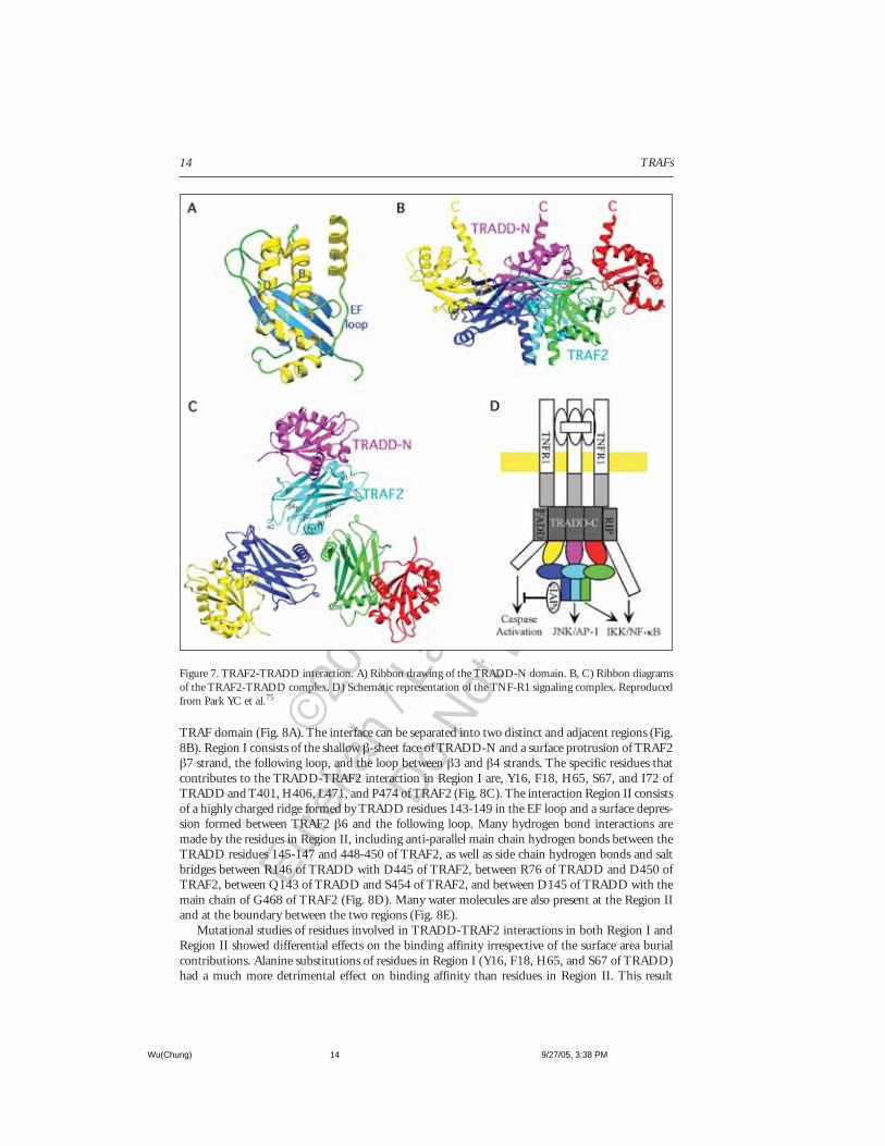
The TRADD-N DomainThe interaction between TRAF2 and TRADD occurs through the TRAF domain of TRAF2 and
the N-terminal domain of TRADD (TRADD-N). The structure of TRADD-N domain shows aα-β sandwich fold with a four-stranded anti-parallel β-sheet and six α helices75,76 (Fig. 7A). Thereare two helices involved at each crossover between β-strands, β1-β2 (helices A and B) and β3-β4(helices C and D). A hairpin-like turn is formed between β2-β3 strands. The remaining E and Fhelices are near the carboxy-terminus of the domain. The EF loop partially covers the exposed faceof the β-sheet.
The α-β sandwich of TRADD-N is most similar to the family of ferredoxin-like α-β sand-wiches.77 Similar α-β sandwich topology has been observed in the structures of the palm domain ofpolymerases and the dimerization domain of carboxypeptidases. However, the extra helices in theβ1-β2 and β3-β4 connections as well as the additional E and F helices makes TRADD-N a moreelaborate structure.
Interactions and Energetics at the TRADD-TRAF2 InterfaceThe trimeric structure of the TRAF domain enforces the threefold symmetry to the stoichio-
metrically bound TRADD-N (Fig. 7B, C). The side view of the TRADD-TRAF2 complex showsTRADD bound to the upper rim of the mushroom cap, which adds a wing-like appearance to thecomplex structure. The carboxyl terminus projects upwards towards the membrane bound receptordirection. The orientation allows TRADD to interact with TNFR1 via the death domains and actsas a platform for other proteins to associate, such as TRAF2, FADD, and RIP (Fig. 7D).
The TRADD-TRAF2 interface partially overlaps with the site of TRAF2-receptor interaction.This indicates a competitive nature of TRAF2-TRADD and TRAF2-receptor interactions. EachTRADD-N molecule contacts one protomer of TRAF2, much like the receptor peptides. The inter-action buries a surface area of 1500Å,2 which leads to small conformational changes in the Cαpositions of TRAF2 (0.5-1.0Å) within or immediately adjacent to the TRADD binding site.
The interface between TRADD-TRAF2 resembles a “ridge into groove” type of contacts, exem-plified by complementary elevations and depressions on the surfaces of TRADD-N and TRAF2
Wu(Chung) 9/27/05, 3:38 PM13
©20
05 C
opyr
ight
Eure
kah
/ Lan
des
Bios
cien
ce
Do
Not
Dis
tribu
te
TRAFs14
TRAF domain (Fig. 8A). The interface can be separated into two distinct and adjacent regions (Fig.8B). Region I consists of the shallow β-sheet face of TRADD-N and a surface protrusion of TRAF2β7 strand, the following loop, and the loop between β3 and β4 strands. The specific residues thatcontributes to the TRADD-TRAF2 interaction in Region I are, Y16, F18, H65, S67, and I72 ofTRADD and T401, H406, L471, and P474 of TRAF2 (Fig. 8C). The interaction Region II consistsof a highly charged ridge formed by TRADD residues 143-149 in the EF loop and a surface depres-sion formed between TRAF2 β6 and the following loop. Many hydrogen bond interactions aremade by the residues in Region II, including anti-parallel main chain hydrogen bonds between theTRADD residues 145-147 and 448-450 of TRAF2, as well as side chain hydrogen bonds and saltbridges between R146 of TRADD with D445 of TRAF2, between R76 of TRADD and D450 ofTRAF2, between Q143 of TRADD and S454 of TRAF2, and between D145 of TRADD with themain chain of G468 of TRAF2 (Fig. 8D). Many water molecules are also present at the Region IIand at the boundary between the two regions (Fig. 8E).
Mutational studies of residues involved in TRADD-TRAF2 interactions in both Region I andRegion II showed differential effects on the binding affinity irrespective of the surface area burialcontributions. Alanine substitutions of residues in Region I (Y16, F18, H65, and S67 of TRADD)had a much more detrimental effect on binding affinity than residues in Region II. This result
Figure 7. TRAF2-TRADD interaction. A) Ribbon drawing of the TRADD-N domain. B, C) Ribbon diagramsof the TRAF2-TRADD complex. D) Schematic representation of the TNF-R1 signaling complex. Reproducedfrom Park YC et al.75
Wu(Chung) 9/27/05, 3:38 PM14

©20
05 C
opyr
ight
Eure
kah
/ Lan
des
Bios
cien
ce
Do
Not
Dis
tribu
te
15Structural Revelations of TRAF2 Function in TNF Receptor Signaling Pathway
Figure 8. Detailed TRAF2-TRADD interaction. A) Interaction surfaces and their locations on the individualstructures (in red). B) Molecular interactions at the two regions of the interactions. C, D, E) Details of region I,region II and water-mediated interactions, respectively. Modified from Park YC et al.75
indicates that despite the larger surface area burial of Region II compared to Region I, the largelyhydrophobic interaction in Region I plays the dominant role in the energetics of the interaction.
Higher Affinity and Distinct Specificity of TRADD-TRAF2 InteractionSurface plasma resonance experiments on TRAF2-TRADD interaction revealed a higher bind-
ing affinity (Kd = 7.8µM) compared to TRAF2-receptor interactions (Kd = 40µM-1.9mM).73 Thehigher affinity between TRADD-TRAF2 suggests that TRADD may be a stronger inducer of TRAF2signaling. This hypothesis was examined in cells expressing exclusively TNF-R1, which signals throughTRADD, and cells that only expressed TNF-R2, which signals through direct TRAF2 recruitment.The strength of TRAF2 recruitment was measured by the activation of a major TRAF2 downstreameffector, JNK protein kinase.38 As predicted from the in vitro binding affinity studies, the JNKactivation was much stronger for TNF-R1 than for TNF-R2 expressing cells.
The TRADD interaction with TRAF proteins appear to be limited to only TRAF2 and TRAF1(Table 2). This selectivity by TRADD is not observed by TNF superfamily receptors lacking theintracellular death domain, since these receptors show similar binding specificities for TRAF1, 2, 3,and 5.45,49,78 The ability of TRADD to associate with both TRAF1 and TRAF2 may have signifi-cance in the prevention of apoptosis by TNF-R1 activation (Fig. 9). Rothe et al has shown in TNF-R2signaling complex, both TRAF1 and TRAF2 are constitutively associated with cellular inhibitors ofapoptosis proteins (cIAPs), cIAP1 and cIAP2, and that this association requires the presence of bothTRAF1 and TRAF2.79 Therefore, as a consequence of the specificity of TRADD for TRAF1 and
Wu(Chung) 9/27/05, 3:39 PM15

©20
05 C
opyr
ight
Eure
kah
/ Lan
des
Bios
cien
ce
Do
Not
Dis
tribu
te
TRAFs16
TRAF2, the cIAPs are brought to TNF-R1 and likely play an important role in blocking the apoptosispathway.40
The predominant outcome of TNF-R1 activation is not apoptosis, as is the case for Fas receptoractivation, but rather cell survival or proliferation. The evidence that TRADD binds specifically andselectively to TRAF1 and TRAF2 strongly supports the survival phenomenon. TRAF2 signaling hasbeen shown in TRAF2 knockout studies to protect cells from apoptosis induced by TNF.38 In addi-tion, mutational studies on TRADD which resulted in reduced affinity for TRAF2 greatly sensitizedcells to cell death.75 These observations implicate TRAF2 as a critical determinant of cellular sur-vival in the TNF-R1 pathway.
Based on these observations, a natural question arises as to when or in what situation does TNF-R1activation lead to apoptosis? One possible answer may be through the mitochondrial release of Smac
Figure 9. Principles of post-receptor signal transduction. A) Receptor activation and TRAF recruitment. B)Competitive TRAF recruitments and regulation of cell survival and death. Reproduced from Wu H.44
Wu(Chung) 9/27/05, 3:39 PM16

©20
05 C
opyr
ight
Eure
kah
/ Lan
des
Bios
cien
ce
Do
Not
Dis
tribu
te
17Structural Revelations of TRAF2 Function in TNF Receptor Signaling Pathway
protein through JNK activation.43 Smac may interact with cIAPs and remove them from TRAF1and TRAF2. Another possible answer may lie on the NF-κB-inducible protein c-FLIP. In the ab-sence of NF-κB activation and c-FLIP, TNF-R1 can induce cell death through a cytoplasmic com-plex containing TRADD, RIP1, FADD, and caspase-8 activation.42
Summary: Emerging Principles of Post-Receptor Signal Transduction
Increased Affinity Through AvidityStructural and biophysical studies on TRAF2-receptor and TRAF2-TRADD interactions showed
that receptor peptides and TRADD contact one protomer of the TRAF domain trimer and that theyinteract with TRAF2 at low affinity. This suggests that receptor oligomerization and affinity en-hancement through avidity is required for TRAF recruitment (Fig. 9). However, because a widerange of affinities between TRAF2 and receptors or TRADD have been observed (Table 2), the issueof whether different receptors would require different avidity contributions for TRAF2 recruitmentis raised.
Interestingly, many TNF-like cytokine ligands, including TNF, are membrane-bound and there-fore may be able to create a higher order of receptor aggregation through membrane-patching orclustering. This membrane receptor aggregation would then increase avidity and thereby enhanceaffinity for TRAF2. In support of this avidity induced affinity hypothesis, both soluble forms ofCD40L and TNF have been shown to be weak inducers of TRAF2 signaling via CD40 and TNF-R2,respectively.80,81 However, this is not the case for TNF-R1 activation by soluble TNF due to a muchstronger TRADD-TRAF2 interaction and recruitment to the receptor.
Based on the TRAF2-receptor structures and the biophysical measurements of binding affinities,the need for receptor aggregation for efficient TRAF2 signaling corresponds well to what is evidentin biology. Many other members of the TNF receptor superfamily such as CD30, Ox40, and 4-1BBligands are membrane bound and mediate signaling in this state. The soluble ligand forms of theseTNF receptor superfamily members are reported to be inefficient in activating the intracellularsignal transduction pathway. In fact, such soluble ligands have been implicated in the role as decoysto down-regulate receptor activity.82,83
Competition Based Regulation of Survival and Death by TRAF2TRAF2 plays a central role in the regulation of cell death and cell survival by TNF receptors,
TNF-R1 and TNF-R2. Studies have shown that overexpression of the survival receptor TNF-R2sensitizes cells to TNF induced apoptosis.84-88 This contradictory outcome can be explained by thecompetitive recruitment hypothesis (Fig. 9). It may be that abundant TNF-R2 levels on the cellmembrane draws all the TRAF2 as well as TRAF1 to its intracellular domain, which then depletescIAPs from TNF-R1 to block caspase activation. Thus, cell survival or death is dependent on intra-cellular pool of cIAPs associated with TRAF1 and TRAF2.
Similar type of TNF-R1 and TNF-R2 interplay may exist among the other members of the TNFreceptor superfamily, such as CD40, CD30, LTβR and CD27. These receptors have also shown theability to induce apoptosis in certain circumstances.89 Activation of any of these receptors could leadto sequestration and or degradation of TRAF1, TRAF2, and cIAP proteins.90 This will then makethe cells vulnerable to TNF induced apoptosis via TNF-R1 signaling.
Remaining QuestionsSignificant amount of structural information on TRAF2 interaction with receptor peptides and
TRADD-N has provided an elegant explanation and agreement with biological observations ofTRAF2 function. However, many questions are left unanswered regarding the molecular mecha-nism of TRAF2 signaling. For example, is TRAF2 in monomeric or in a constitutive trimeric statebefore recruitment to receptors? Is the activation of downstream effectors dependent on oligomer-ization or on conformational changes induced by receptor interaction? What is the exact molecularbasis for this activation? Finally, the ultimate challenge will be in translating the structural and
Wu(Chung) 9/27/05, 3:39 PM17

©20
05 C
opyr
ight
Eure
kah
/ Lan
des
Bios
cien
ce
Do
Not
Dis
tribu
te
TRAFs18
functional studies into potential therapies for many important diseases involving TNF receptorsuperfamily members.
AcknowledgmentThis work was funded by the National Institute of Health (AI45937 and AI47831), the Pew
Charitable Trust and the Rita Allen Foundation. J.Y.C and M.L. were supported by the NIH andKeck post-doctoral fellowships, respectively.
References1. Locksley RM, Killeen N, Lenardo MJ. The TNF and TNF receptor superfamilies: Integrating
mammalian biology. Cell 2001; 104(4):487-501.2. Ashkenazi A, Dixit VM. Death receptors: Signaling and modulation. Science 1998;
281(5381):1305-1308.3. Loenen WA. Cd27 And (TNFR) relatives in the immune system: Their role in health and disease.
Semin Immunol 1998; 10(6):417-422.4. Newton RC, Decicco CP. Therapeutic potential and strategies for inhibiting tumor necrosis
factor-Alpha. J Med Chem 1999; 42(13):2295-2314.5. Smith CA, Farrah T, Goodwin RG. The TNF receptor superfamily of cellular and viral proteins:
Activation, costimulation, and death. Cell 1994; 76(6):959-962.6. Gravestein LA, Borst J. Tumor necrosis factor receptor family members in the immune system.
Semin Immunol 1998; 10(6):423-434.7. Coley WB. The treatment of malignant tumors by repeated inoculations of erysipelas: With a
report of ten original original cases. Am J Med Sci 1893; 105:487-511.8. Carswell EA, Old LJ, Kassel RL et al. An endotoxin-induced serum factor that causes necrosis of
tumors. Proc Natl Acad Sci USA 1975; 72(9):3666-3670.9. Pennica D, Nedwin GE, Hayflick JS et al. Human tumour necrosis factor: Precursor structure,
expression and homology to lymphotoxin. Nature 1985; 312(5996):724-729.10. Wang AM, Creasey AA, Ladner MB et al. Molecular cloning of the complementary DNA for
human tumor necrosis factor. Science 1985; 228(4696):149-154.11. Shirai T, Yamaguchi H, Ito H et al. Cloning and expression in Escherichia coli of the gene for
human tumour necrosis factor. Nature 1985; 313(6005):803-806.12. Beutler B, Cerami A. Cachectin and tumour necrosis factor as two sides of the same biological
coin. Nature 1986; 320(6063):584-588.13. Goeddel DV, Aggarwal BB, Gray PW et al. Tumor necrosis factors: Gene structure and biological
activities. Cold Spring Harb Symp Quant Biol 1986; 51(Pt 1):597-609.14. Fiers W. Tumor necrosis factor. Characterization at the molecular, cellular and in vivo level. Febs
Lett 1991; 285(2):199-212.15. Lewis M, Tartaglia LA, Lee A et al. Cloning and expression of cDNAs for two distinct murine
tumor necrosis factor receptors demonstrate one receptor is species specific. Proc Natl Acad SciUSA 1991; 88(7):2830-2834.
16. Rothe M, Wong SC, Henzel WJ et al. A novel family of putative signal transducers associated withthe cytoplasmic domain of the 75 kDa tumor necrosis factor receptor. Cell 1994; 78(4):681-692.
17. Arch RH, Gedrich RW, Thompson CB. Tumor necrosis factor receptor-associated factors (TRAFs)—A family of adapter proteins that regulates life and death. Genes Dev 1998; 12(18):2821-2830.
18. Chung JY, Park YC, Ye H et al. All Trafs are not created equal: Common and distinct molecularmechanisms of TRAF-mediated signal transduction. Cell Sci 2002; 115(Pt 4):679-688.
19. Cheng G, Cleary AM, Ye ZS et al. Involvement of CRAF1, a relative of TRAF, in CD40 signal-ing. Science 1995; 267(5203):1494-1498.
20. Mosialos G, Birkenbach M, Yalamanchili R et al. The epstein-barr virus transforming protein LMP1engages signaling proteins for the tumor necrosis factor receptor family. Cell 1995; 80(3):389-399.
21. Sato T, Irie S, Reed JC. A novel member of the TRAF family of putative signal transducingproteins binds to the cytosolic domain of CD40. FEBS Lett 1995; 358(2):113-118.
22. Regnier CH, Tomasetto C, Moog-Lutz C et al. Presence of a new conserved domain in Cart1, anovel member of the tumor necrosis factor receptor-associated protein family, which is expressed inbreast carcinoma. J Biol Chem 1995; 270(43):25715-25721.
23. Ishida TK, Tojo T, Aoki T et al. TRAF5, a novel tumor necrosis factor receptor-associated factorfamily protein, mediates CD40 signaling. Proc Natl Acad Sci USA 1996; 93(18):9437-9442.
24. Nakano H, Oshima H, Chung W et al. TRAF5, an activator of NF-Kappab and putative signaltransducer for the lymphotoxin-beta receptor. J Biol Chem 1996; 271(25):14661-14664.
Wu(Chung) 9/27/05, 3:39 PM18

©20
05 C
opyr
ight
Eure
kah
/ Lan
des
Bios
cien
ce
Do
Not
Dis
tribu
te
19Structural Revelations of TRAF2 Function in TNF Receptor Signaling Pathway
25. Mizushima S, Fujita M, Ishida T et al. Cloning and characterization of a cDNA encoding thehuman homolog of tumor necrosis factor receptor-associated factor 5 (TRAF5). Gene 1998;207(2):135-140.
26. Cao Z, Xiong J, Takeuchi M et al. TRAF6 is a signal transducer for Interleukin-1. Nature 1996;383(6599):443-446.
27. Ishida T, Mizushima S, Azuma S et al. Identification of TRAF6, a novel tumor necrosis factorreceptor-associated factor protein that mediates signaling from an amino-terminal domain of theCD40 cytoplasmic region. J Biol Chem 1996; 271(46):28745-28748.
28. Bouwmeester T, Bauch A, Ruffner H et al. A physical and functional map of the human TNF-Alpha/NF-Kappa B signal transduction pathway. Nat Cell Biol 2004; 6(2):97-105.
29. Xu LG, Li LY, Shu HB. TRAF7 potentiates MEKK3-induced AP1 and CHOP activation andinduces apoptosis. J Biol Chem 2004; 279(17):17278-17282.
30. Ghosh S, Karin M. Missing pieces in the NF-KappaB puzzle. Cell 2002; 109(Suppl):S81-96:S81-S96.31. Shaulian E, Karin M. Ap-1 as a regulator of cell life and death. Nat Cell Biol 2002; 4(5):E131-E136.32. Trompouki E, Hatzivassiliou E, Tsichritzis T et al. CYLD is a deubiquitinating enzyme that nega-
tively regulates NF-Kappab activation by TNFR family members. Nature 2003; 424(6950):793-796.33. Brummelkamp TR, Nijman SM, Dirac AM et al. Loss of the cylindromatosis tumour suppressor
inhibits apoptosis by activating NF-KappaB. Nature 2003; 424(6950):797-801.34. Kovalenko A, Chable-Bessia C, Cantarella G et al. The tumour suppressor cyld negatively regulates
NF-KappaB signalling by deubiquitination. Nature 2003; 424(6950):801-805.35. Hsu H, Shu HB, Pan MG et al. TRADD-TRAF2 and TRADD-FADD interactions define two
distinct TNF receptor 1 signal transduction pathways. Cell 1996; 84(2):299-308.36. Stanger BZ, Leder P, Lee TH et al. RIP: A novel protein containing a death domain that interacts
with Fas/APO-1 (CD95) in yeast and causes cell death. Cell 1995; 81(4):513-523.37. Hsu H, Huang J, Shu HB et al. TNF-dependent recruitment of the protein kinase RIP to the
TNF receptor-1 signaling complex. Immunity 1996; 4(4):387-396.38. Yeh WC, Shahinian A, Speiser D et al. Early lethality, functional NF-KappaB activation, and
increased sensitivity to TNF-induced cell death in TRAF2-deficient mice. Immunity 1997;7(5):715-725.
39. Kelliher MA, Grimm S, Ishida Y et al. The death domain kinase RIP mediates the TNF-inducedNF-KappaB signal. Immunity 1998; 8(3):297-303.
40. Wang CY, Mayo MW, Korneluk RG et al. NF-KappaB antiapoptosis: Induction of TRAF1 andTRAF2 and C-IAP1 and c-IAP2 to suppress caspase-8 activation. Science 1998;281(5383):1680-1683.
41. Irmler M, Thome M, Hahne M et al. Inhibition of death receptor signals by cellular FLIP. Nature1997; 388(6638):190-195.
42. Micheau O, Tschopp J. Induction of TNF receptor I-mediated apoptosis via two sequential signal-ing complexes. Cell 2003; 114(2):181-190.
43. Deng Y, Ren X, Yang L et al. A JNK-dependent pathway is required for TNFalpha-inducedapoptosis. Cell 2003; 115(1):61-70.
44. Wu H. Assembly of post-receptor signaling complexes for the tumor necrosis factor receptor super-family. Adv Protein Chem 2004; 68:225-79.
45. Park YC, Burkitt V, Villa AR et al. Structural basis for self-association and receptor recognition ofhuman TRAF2. Nature 1999; 398(6727):533-538.
46. Murzin AG, Brenner SE, Hubbard T et al. SCOP: A structural classification of proteins databasefor the investigation of sequences and structures. J Mol Biol 1995; 247(4):536-540.
47. Holm L, Sander C. Dali: A network tool for protein structure comparison. Trends Biochem Sci1995; 20(11):478-480.
48. Mcwhirter SM, Pullen SS, Holton JM et al. Crystallographic analysis of CD40 recognition andsignaling by human TRAF2. Proc Natl Acad Sci USA 1999; 96(15):8408-8413.
49. Ye H, Park YC, Kreishman M et al. The structural basis for the recognition of diverse receptorsequences by TRAF2. Mol Cell 1999; 4(3):321-330.
50. Uren AG, Vaux DL. Traf proteins and meprins share a conserved domain. Trends Biochem Sci1996; 21(7):244-245.
51. Zapata JM, Pawlowski K, Haas E et al. A diverse family of proteins containing tumor necrosisfactor receptor-associated factor domains. J Biol Chem 2001; 276(26):24242-24252.
52. Polekhina G, House CM, Traficante N et al. Siah ubiquitin ligase is structurally related to TRAFand modulates TNF-alpha signaling. Nat Struct Biol 2002; 9(1):68-75.
53. Gruber M, Lupas AN. Historical review: Another 50th anniversary—New periodicities in coiledcoils. Trends Biochem Sci 2003; 28(12):679-685.
Wu(Chung) 9/27/05, 3:39 PM19

©20
05 C
opyr
ight
Eure
kah
/ Lan
des
Bios
cien
ce
Do
Not
Dis
tribu
te
TRAFs20
54. Janin J, Miller S, Chothia C. Surface, subunit interfaces and interior of oligomeric proteins. J MolBiol 1988; 204(1):155-164.
55. Harbury PB, Zhang T, Kim PS et al. A switch between two-, three-, and four-stranded coiled coilsin GCN4 leucine zipper mutants. Science 1993; 262(5138):1401-1407.
56. Wolf E, Kim PS, Berger B. Multicoil: A program for predicting two- and three-stranded coiledcoils. Protein Sci 1997; 6(6):1179-1189.
57. Aizawa S, Nakano H, Ishida T et al. Tumor necrosis factor receptor-associated factor (TRAF) 5and TRAF2 are involved in CD30-mediated NFkappaB activation. J Biol Chem 1997;272(4):2042-2045.
58. Akiba H, Nakano H, Nishinaka S et al. CD27, a member of the tumor necrosis factor receptorsuperfamily, activates NF-KappaB and stress-activated protein kinase/C-Jun N-terminal kinase viaTRAF2, TRAF5, and NF-KappaB-inducing kinase. J Biol Chem 1998; 273(21):13353-13358.
59. Boucher LM, Marengere LE, Lu Y et al. Binding sites of cytoplasmic effectors TRAF1, 2, and 3on CD30 and other members of the TNF receptor superfamily. Biochem Biophys Res Commun1997; 233(3):592-600.
60. Brodeur SR, Cheng G, Baltimore D et al. Localization of the major NF-KappaB-activating siteand the sole TRAF3 binding site of LMP-1 defines two distinct signaling motifs. J Biol Chem1997; 272(32):19777-19784.
61. Devergne O, Hatzivassiliou E, Izumi KM et al. Association of TRAF1, TRAF2, and TRAF3 withan epstein-barr virus LMP1 domain important for B-lymphocyte transformation: Role in NF-KappaBactivation. Mol Cell Biol 1996; 16(12):7098-7108.
62. Franken M, Devergne O, Rosenzweig M et al. Comparative analysis identifies conserved tumornecrosis factor receptor-associated factor 3 binding sites in the human and simian epstein-barrvirus oncogene LMP1. J Virol 1996; 70(11):7819-7826.
63. Gedrich RW, Gilfillan MC, Duckett CS et al. CD30 contains two binding sites with differentspecificities for members of the tumor necrosis factor receptor-associated factor family of signaltransducing proteins. J Biol Chem 1996; 271(22):12852-12858.
64. Sandberg M, Hammerschmidt W, Sugden B. Characterization of Lmp-1’s association with TRAF1,TRAF2, and TRAF3. J Virol 1997; 71(6):4649-4656.
65. Arch RH, Thompson CB. 4-1bb and Ox40 are members of a tumor necrosis factor (TNF)-nervegrowth factor receptor subfamily that bind TNF receptor-associated factors and activate nuclearfactor KappaB. Mol Cell Biol 1998; 18(1):558-565.
66. Tong L, Qian C, Massariol MJ et al. Conserved mode of peptidomimetic inhibition and substraterecognition of human cytomegalovirus protease. Nat Struct Biol 1998; 5(9):819-826.
67. Kuriyan J, Cowburn D. Modular peptide recognition domains in eukaryotic signaling. Annu RevBiophys Biomol Struct 1997; 26:259-88, (259-288).
68. Lim WA, Richards FM, Fox RO. Structural determinants of peptide-binding orientation and ofsequence specificity in SH3 domains. Nature 1994; 372(6504):375-379.
69. Stern LJ, Brown JH, Jardetzky TS et al. Crystal structure of the human class Ii MHC proteinHLA-DR1 complexed with an influenza virus peptide. Nature 1994; 368(6468):215-221.
70. Cheng G, Baltimore D. Tank, A coinducer with TRAF2 of TNF- and CD 40l-mediated NF-KappaBactivation. Genes Dev 1996; 10(8):963-973.
71. Rothe M, Xiong J, Shu HB et al. I-TRAF is a novel TRAF-interacting protein that regulatesTRAF-mediated signal transduction. Proc Natl Acad Sci USA 1996; 93(16):8241-8246.
72. Pullen SS, Dang TT, Crute JJ et al. Cd40 signaling through tumor necrosis factor receptor-associatedfactors (TRAFs). Binding site specificity and activation of downstream athways by Distinct TRAFs.J Biol Chem 1999; 274(20):14246-14254.
73. Ye H, Wu H. Thermodynamic characterization of the interaction between TRAF2 and tumornecrosis factor receptor peptides by isothermal titration calorimetry. Proc Natl Acad Sci USA 2000;97(16):8961-8966.
74. Ladbury JE. Counting the calories to stay in the groove. Structure 1995; 3(7):635-639.75. Park YC, Ye H, Hsia C et al. A novel mechanism of TRAF signaling revealed by structural and
functional analyses of the TRADD-TRAF2 interaction. Cell 2000; 101(7):777-787.76. Tsao DH, Mcdonagh T, Telliez JB et al. Solution structure of N-TRADD and characterization of
the interaction of N-TRADD and C-TRAF2, a key step in the TNFR1 signaling pathway. MolCell 2000; 5(6):1051-1057.
77. Orengo CA, Thornton JM. Alpha plus beta folds revisited: Some favoured motifs. Structure 1993;1(2):105-120.
78. Ni CZ, Welsh K, Leo E et al. Molecular basis for CD40 signaling mediated by TRAF3. Proc NatlAcad Sci USA 2000; 97(19):10395-10399.
Wu(Chung) 9/27/05, 3:39 PM20

©20
05 C
opyr
ight
Eure
kah
/ Lan
des
Bios
cien
ce
Do
Not
Dis
tribu
te
21Structural Revelations of TRAF2 Function in TNF Receptor Signaling Pathway
79. Rothe M, Pan MG, Henzel WJ et al. The TNFR2-TRAF signaling complex contains two novelproteins related to baculoviral inhibitor of apoptosis proteins. Cell 1995; 83(7):1243-1252.
80. Pullen SS, Labadia ME, Ingraham RH et al. High-affinity interactions of tumor necrosis factorreceptor-associated factors (TRAFs) and CD40 require TRAF trimerization and CD40multimerization. Biochemistry 1999; 38(31):10168-10177.
81. Grell M, Douni E, Wajant H et al. The transmembrane form of tumor necrosis factor is the primeactivating ligand of the 80 kDa tumor necrosis factor receptor. Cell 1995; 83(5):793-802.
82. Hodgkin PD, Chin SH, Bartell G et al. The importance of efficacy and partial agonism in evalu-ating models of b lymphocyte activation. Int Rev Immunol 1997; 15(1-2):101-127.
83. Kehry MR, Castle BE. Regulation of CD40 ligand expression and use of recombinant CD40 ligandfor studying b cell growth and differentiation. Semin Immunol 1994; 6(5):287-294.
84. Heller RA, Song K, Fan N et al. The P70 tumor necrosis factor receptor mediates cytotoxicity.Cell 1992; 70(1):47-56.
85. Vandenabeele P, Declercq W, Vanhaesebroeck B et al. Both TNF receptors are required forTNF-mediated induction of apoptosis in PC60 cells. J Immunol 1995; 154(6):2904-2913.
86. Weiss T, Grell M, Hessabi B et al. Enhancement of TNF receptor P60-mediated cytotoxicity byTNF receptor P80: Requirement of the TNF receptor-associated factor-2 binding site. J Immunol1997; 158(5):2398-2404.
87. Haridas V, Darnay BG, Natarajan K et al. Overexpression of the P80 TNF receptor leads toTNF-dependent apoptosis, nuclear factor-Kappa B activation, and c-jun kinase activation. J Immunol1998; 160(7):3152-3162.
88. Chan FK, Lenardo MJ. A crucial role for P80 TNF-R2 in amplifying P60 TNF-R1 apoptosissignals in T lymphocytes. Eur J Immunol 2000; 30(2):652-660.
89. Grell M, Zimmermann G, Gottfried E et al. Induction of cell death by tumour necrosis factor(TNF) receptor 2, CD40 and CD30: A role for TNF-R1 activation by endogenousmembrane-anchored TNF. Embo J 1999; 18(11):3034-3043.
90. Duckett CS, Thompson CB. CD30-dependent degradation of TRAF2: Implications for negativeregulation of TRAF signaling and the control of cell survival. Genes Dev 1997; 11(21):2810-2821.
91. Li C, Ni CZ, Havert ML et al. Downstream regulator tank binds to the CD40 recognition site onTRAF3. Structure (Camb) 2002; 10(3):403-411.
92. Li C, Norris PS, Ni CZ et al. Structurally distinct recognition motifs in lymphotoxin-beta receptorand CD40 for tumor necrosis factor receptor-associated factor (TRAF)-mediated signaling. J BiolChem 2003; 278(50):50523-50529.
93. Ye H, Arron JR, Lamothe B et al. Distinct molecular mechanism for initiating TRAF6 signalling.Nature 2002; 418(6896):443-447.
Wu(Chung) 9/27/05, 3:39 PM21