Embed Size (px)
Citation preview

Structural Investigation of the Thermostability and ProductSpecificity of Amylosucrase from the Bacterium Deinococcusgeothermalis□S
Received for publication, November 10, 2011, and in revised form, December 20, 2011 Published, JBC Papers in Press, December 30, 2011, DOI 10.1074/jbc.M111.322917
Frederic Guerin‡§¶�**1, Sophie Barbe‡§¶, Sandra Pizzut-Serin‡§¶, Gabrielle Potocki-Veronese‡§¶, David Guieysse‡§¶,Valerie Guillet�**, Pierre Monsan‡§¶‡‡, Lionel Mourey�**, Magali Remaud-Simeon‡§¶, Isabelle Andre‡§¶2,and Samuel Tranier�**3
From the ‡Universite de Toulouse; INSA, UPS, INP, LISBP, 135 Avenue de Rangueil, F-31077 Toulouse, France, the §CNRS,UMR5504, F-31400 Toulouse, France, the ¶INRA, UMR792 Ingenierie des Systemes Biologiques et des Procedes, F-31400Toulouse, France, the �CNRS, IPBS, Departement de Biologie Structurale et Biophysique, 205 Route de Narbonne, BP 64182,F-31077 Toulouse, France, the **Universite de Toulouse, UPS, IPBS, F-31077 Toulouse, and the ‡‡Institut Universitaire deFrance, 103 Boulevard Saint-Michel, F-75005 Paris, France
Background: Amylosucrases (AS) hold great potential for glycodiversification.Results: The first three-dimensional structure of AS from Deinococcus geothermalis solved here revealed an unusual dimerorganization. Structures of complex of AS with turanose were also determined.Conclusion: Dimerization may contribute to thermostability. Turanose versus trehalulose formation is controlled by residuesfrom subsite �1.Significance: This study improves the comprehension of AS properties and provides new insight for AS design.
Amylosucrases are sucrose-utilizing�-transglucosidases thatnaturally catalyze the synthesis of �-glucans, linked exclusivelythrough �1,4-linkages. Side products and in particular sucroseisomers such as turanose and trehalulose are also produced bythese enzymes. Here, we report the first structural and biophys-ical characterization of the most thermostable amylosucraseidentified so far, the amylosucrase from Deinoccocus geother-malis (DgAS). The three-dimensional structure revealed ahomodimeric quaternary organization, never reported beforefor other amylosucrases. A sequence signature of dimerizationwas identified from the analysis of the dimer interface andsequence alignments. By rigidifying the DgAS structure, thequaternary organization is likely to participate in the enhancedthermal stability of the protein. Amylosucrase specificity withrespect to sucrose isomer formation (turanose or trehalulose)was also investigated. We report the first structures of the amy-losucrases from Deinococcus geothermalis and Neisseria poly-saccharea in complex with turanose. In the amylosucrase fromN. polysaccharea (NpAS), key residues were found to force the
fructosyl moiety to bind in an open state with the O3� ideallypositioned to explain the preferential formation of turanose byNpAS. Such residues are either not present or not similarlyplaced in DgAS. As a consequence, DgAS binds the furanoidtautomers of fructose through aweak network of interactions toenable turanose formation. Such topology at subsite �1 is likelyfavoring other possible fructose binding modes in agreementwith the higher amount of trehalulose formed by DgAS. Ourfindings help to understand the inter-relationships betweenamylosucrase structure, flexibility, function, and stability andprovide new insight for amylosucrase design.
Amylosucrases (AS)4 (E.C. number 2.4.1.4) are glucansu-crases belonging to Glycoside-Hydrolase Family 13 of the Car-bohydrate-Active EnZymes (CAZy) classification (1–3). Themain reaction catalyzed by these enzymes is the formation of anamylose-like glucan with a concomitant release of fructosefrom sucrose substrate (Fig. 1) (4). Side reactions includingsucrose hydrolysis and sucrose isomer synthesis (i.e. turanose,�-D-Glcp(133)-�-D-Fru and trehalulose,�-D-Glcp(131)-�-D-Fru) also occur (4, 5) although other GH13 enzymes are knownto be more specific for these types of reaction (6–8). Thesemolecules are known to be less cariogenic than sucrose and arethus of interest for nutritional applications (9). AS are also ableto transfer the glucosyl moieties from sucrose to exogenousacceptors such as maltose, glycogen, maltodextrins, arbutin,and others (10, 11). The ability of AS to convert sucrose (an
□S This article contains supplemental Table S1 and Figs. S1–S5.The atomic coordinates and structure factors (codes 3UCQ, 3UER, and 3UEQ)
have been deposited in the Protein Data Bank, Research Collaboratory forStructural Bioinformatics, Rutgers University, New Brunswick, NJ(http://www.rcsb.org/).
1 Supported by a Ph.D. grant from the Poles de Recherche et d’EnseignementSuperieur de l’Universite de Toulouse and the Region Midi-Pyrenees,France.
2 To whom correspondence may be addressed: Laboratoire d’Ingenierie desSystemes Biologiques et des Procedes, INSA, CNRS UMR5504, UMR INRA792, 135 Avenue de Rangueil, F-31077 Toulouse Cedex 4, France. Tel.:33-561-559-963; Fax: 33-561-559-400; E-mail: [email protected].
3 To whom correspondence may be addressed: Institut de Pharmacologie etde Biologie Structurale, Departement Biologie Structurale et Biophysique,205 Route de Narbonne, BP 64182, F-31077 Toulouse, France. Tel.: 33-561-175-438; E-mail: [email protected].
4 The abbreviations used are: AS, amylosucrase; NpAS, N. polysacchareaamylosucrase; DgAS, D. geothermalis amylosucrase; DrAS, D. radioduransamylosucrase; AmAS, A. macleodii amylosucrase; SEC, size exclusion chro-matography; MALLS, multiangle laser light scattering; MD, moleculardynamics; r.m.s., root mean square; PDB, Protein Data Bank.
THE JOURNAL OF BIOLOGICAL CHEMISTRY VOL. 287, NO. 9, pp. 6642–6654, February 24, 2012© 2012 by The American Society for Biochemistry and Molecular Biology, Inc. Published in the U.S.A.
6642 JOURNAL OF BIOLOGICAL CHEMISTRY VOLUME 287 • NUMBER 9 • FEBRUARY 24, 2012
by guest on May 21, 2018
http://ww
w.jbc.org/
Dow
nloaded from

abundant and inexpensive substrate) into valuable derivatives isa great advantage in comparison to Leloir glucosyltransferases,which use nucleotide-activated sugars as glucosyl donor sub-strates (12). Moreover, protein engineering techniques alsoenabled the creation of novel AS with tailored specificitytoward unnatural acceptor molecules (13).AS that were characterized so far are produced by various
species from the genus Neisseria (4), Deinococcus (14, 15), andAlteromonas (16). However, Neisseria polysaccharea amylosu-crase (NpAS) is the only AS for which several structures, aloneor in complex with sucrose substrate or products, are availableto date (17–20). The three-dimensional structure of NpAS isorganized in five domains, namely A, B, C, N, and B�. B� and Ndomains are only found inAS, whereas the three other domainsare conserved amongGH13 enzymes. Although the potential ofNpAS for glycodiversification is large, this enzyme suffers froma low catalytic efficiency and aweak thermostability, limiting itsindustrial development (15). Directed evolution has beenattempted to improveNpAS catalytic efficiency and thermosta-bility (21). Searching for more thermostable and efficientenzymes in the natural diversity is another alternative that hasmotivated the biochemical characterization of the amylosu-crases from Deinococcus geothermalis (DgAS) (15), Deinococ-cus radiodurans (DrAS) (14), and Alteromonas macleodii(AmAS) (15).With a specific activity of 44 units mg�1 at the optimal tem-
perature of 50 °C, the recombinant DgAS is the most thermo-stable AS characterized to date (15). The size distribution of the�-glucan chains produced by DgAS and NpAS differs and thetwo enzymes do not synthesize equivalent amounts of turanoseand trehalulose. Indeed, DgAS produces significantly higheramounts of trehalulose than NpAS (15). Glucosylation ofunnatural acceptors such as salicin also revealed that DgASonly produces a monoglucosylated form, whereas NpAS is ableto produce a diglucosylated compound (22).Here, we report the first three-dimensional structure of
DgAS in its apo form that reveals an unusual homodimericarrangement, thereby allowing identification of determinantspossibly controlling thermostability. Furthermore, the struc-
tures of both DgAS and NpAS in complex with turanose (afructose acceptor reaction product) were also determined andtheir analysis highlightedmajor differences upon fructose bind-ing, which are discussed with regard to the enzyme productspecificities.
EXPERIMENTAL PROCEDURES
Expression and Purification—Expression and purification ofrecombinant glutathione S-transferase-amylosucrase fusionproteins (GST-DgAS, GST-DrAS, and GST-NpAS) were per-formed as previously described by Emond et al. (15), Pizzut etal. (14), and de Montalk et al. (23), respectively. The GST-ASproteins were purified by affinity chromatography using gluta-thione-Sepharose 4B support (GE Healthcare). The GST tagwas then removed using Prescission protease (GE Healthcare),which left 5 residues (GPLGS) of the cutting site at the N-ter-minal extremity.Thermostability of Amylosucrases—The melting point (Tm)
of amylosucrases was assayed by differential scanning fluori-metry. A mixture of enzyme (2 �M), Sypro-orange (5 �) (Invit-rogen), and 50mMTris, pH 7.0, 150mMNaCl, 1mMDTT, 1mM
EDTAwere incubated using a temperature gradient from 20 to80 °Cwith a 0.3 °C increment. The thermal transitionwasmon-itored using a RTQ-PCR CFX96 Real-time System (Bio-Rad).Tm was given by the inflection point of the curve relative fluo-rescence unit� f(T). Circular dichroism spectra were recordedon a JASCO J815 spectropolarimeter equipped with a Peltiercell temperature controller.Tm valueswere obtained by heatingthe sample (enzyme 2 �M in 50 mM Tris, pH 7.0, 150 mMNaCl,70 �MDTT, 70 �M EDTA) at 1 °C/min and recording the ellip-ticity value at 220 nm from 25 to 80 °C with delays of 30 s.SigmaPlot 10.0 software was used for all graphic analyses, Tmdetermination, and statistics.The half-life (t1⁄2) at 50 °C of DgAS and NpAS preparations
were also determined by incubating AS pure enzymes (300 mgliters�1) in 50mMTris, pH 7.0, 150mMNaCl, 1mMDTT, 1mM
EDTAat 50 °C.At various intervals, aliquotswere taken and theenzyme activity was determined as previously described (22).
FIGURE 1. Reactions catalyzed by amylosucrases from sole sucrose. Glc, glucose; Fru, fructose; sucrose, �-D-glucopyranosyl-1,2-�-D-fructofuranoside;turanose, �-D-glucopyranosyl-1,3-�-D-fructose; trehalulose, �-D-glucopyranosyl-1,1-�-D-fructose.
Structural Investigation of D. geothermalis Amylosucrase
FEBRUARY 24, 2012 • VOLUME 287 • NUMBER 9 JOURNAL OF BIOLOGICAL CHEMISTRY 6643
by guest on May 21, 2018
http://ww
w.jbc.org/
Dow
nloaded from

Size Exclusion ChromatographyMultiangle Laser Light Scat-tering (SEC-MALLS) Experiments—NpAS, DgAS, and DrASprotein samples buffered in 50 mM Tris, pH 7.5, 150 mM NaClwere analyzed on a Shodex KW-803 column (Showa DenkoEurope GmbH, Munich, Germany) with multiangle laser lightscattering (MALLS). The column was equilibrated in a 0.1-�mfiltered 50 mM Tris, pH 7.5, 150 mM NaCl, 0.02% (w/v) NaN3buffer on a Agilent 1260 Infinity LC chromatographic system(AgilentTechnology,Massy, France). Datawere collected usinga DAWN HELEOS-II 18-angle and Optilab T-rEX refractiveindex detector (Wyatt Technology Corp., Toulouse France).Sample concentrations were 1.55, 2.1, and 1.85 g liter�1 forNpAS, DgAS, and DrAS solutions, respectively. Protein sam-pleswere prepared in theTris buffer used as themobile phase toequilibrate the column. 20�l of each protein samplewas loadedon the column and the separation was performed at a flow rateof 0.5 ml min�1 at 22 °C. Results were analyzed using theASTRA V software (Wyatt Technology Corp.).Native-PAGE—Native-PAGE was performed with a two-
phase gel composed of a 4% stacking gel (4% acrylamide, 125mMTris, pH 6.8, 0.1% ammoniumpersulfate, 0.15%N,N,N�,N�-tetramethylethylenediamine) and a 10% separating gel (10%acrylamide, 375 mM Tris, pH 8.8, 0.1% ammonium persulfate,0.15% N,N,N�,N�-tetramethylethylenediamine). 15 �l of theprotein samples (2 g liter�1) mixed with 2 � loading buffer (40mM Tris, pH 6.8, 50% glycerol (v/v), 0.04% bromophenol blue(w/v)) were loaded onto the gel. Electrophoresis was performedat room temperature using a mini-PROTEAN system (Bio-Rad) at a voltage of 100 V with an electrophoresis buffer com-posed of 15 g liter�1 of Tris, pH 8.3, and 72 g liter�1 of glycine.The gels were stained with Coomassie Brilliant Blue.Molecularweight estimation was performed using NativeMark nativeprotein markers (Invitrogen) as standards.
Crystallization—NpAS was crystallized using conditionspreviously described by Skov et al. (17, 18). DgAS crystallizationexperiments were carried out at 12 °C using the hanging dropvapor diffusion method. Best crystals were obtained with a 1:1(v/v) ratio of protein (6 mg ml�1 in 20 mM Tris, pH 8.0) toprecipitant solution (1.5 M sodium acetate, 0.1 M sodium caco-dylate, pH 7.0). Lens shape crystals appeared after 2 weeks andgrew to a maximal size of 140 � 60 � 40 �m3.Soaking Experiments—Crystals of NpAS were soaked for 20
min in the reservoir solution supplemented by 250 mM turan-ose (Sigma). DgAS crystals quickly fractured in the presence ofsuch a high concentration of turanose, so they were soaked fora few seconds in the reservoir solution supplemented with only14 mM turanose.Data Collection and Structure Determination—X-ray exper-
iments were carried out at 100 K. Prior to flash cooling, nativecrystals of DgAS were soaked for a few seconds in the reservoirsolution supplemented with 20% (v/v) glycerol to avoid ice for-mation. Conversely, due to the cryoprotection effect of turan-ose, crystals of AS-turanose complexes were intrinsically cryo-protected. Native DgAS and NpAS-turanose diffractiondatasets were collected to a maximum resolution of 1.97- and1.85-Å, respectively, on beamline ID14-1 at the European Syn-chrotron Radiation Facility (ESRF, Grenoble, France). TheDgAS-turanose complex dataset was collected to 2.10 Å on theESRF beamline ID29. Diffracted intensities were integratedusing iMOSFLM (24) and scaled with SCALA (25) from theCCP4 software suite (26, 27) and 5% of the scaled amplitudeswere randomly selected and excluded from the refinement pro-cedure. Crystals of DgAS in the apo form and in complex withturanose belong to the C2221 space group with 1 molecule perasymmetric unit giving a Matthews coefficient of 2.3 Å3/Da.Crystals of NpAS-turanose belong to the P21212 space group
TABLE 1Data collection and refinement statisticsValues in parentheses are for the outer resolution shell.
NpAS-turanose DgAS DgAS-turanose
Data collectionSpace group P21212 C2221 C2221a, b, c (Å) 96.0, 116.3, 60.5 105.3, 110.2, 115.5 104.7, 110.4, 115.3�, �, � (°) 90.0, 90.0, 90.0 90.0, 90.0, 90.0 90.0, 90.0, 90.0Resolution (Å) 29.27-1.85 (1.95-1.85)* 33.38-1.97 (2.08-1.97) 63.45-2.10 (2.21-2.10)Rsym 0.096 (0.332) 0.086 (0.376) 0.099 (0.324)I/�I 6.5 (2.2) 8.3 (2.0) 4.9 (2.1)Completeness (%) 99.6 (100) 99.6 (99.8) 100 (98.9)Redundancy 3.6 (3.6) 4.0 (3.9) 6.3 (4.2)No. of molecule/AU 1 1 1Matthews coefficient (Å3/Da) 2.4 2.3 2.3
RefinementResolution (Å) 29.10-1.85 33.22-1.97 13-2.10No. of unique reflections 58,309 (8,452) 47,355 (6,837) 39,319 (5,648)Rwork/Rfree (%) 14.7/18.6 14.4/18.7 14.6/20.7Total number of atoms 5,867 5,956 5,712Number of residues in the protein 632 651 651Number of ligand molecules 2 Turanoses, 1 PEG 1 Tris, 13 glycerols 1 TuranoseNumber of water molecules 767 602 394
B-factors (Å2)Protein 11.6 17.9 20.7Ligand 19.9 36.9 26.4Water 21.2 27.5 26.1
R.m.s deviationsBond lengths (Å) 0.022 0.024 0.023Bond angles (°) 1.8 1.8 1.9
Structural Investigation of D. geothermalis Amylosucrase
6644 JOURNAL OF BIOLOGICAL CHEMISTRY VOLUME 287 • NUMBER 9 • FEBRUARY 24, 2012
by guest on May 21, 2018
http://ww
w.jbc.org/
Dow
nloaded from
with 1 molecule per asymmetric unit and a Matthews coeffi-cient of 2.4Å3/Da.Data collection statistics are given inTable 1.The native structure of DgAS was solved by the molecularreplacement method using PHASER (28) and the structure ofNpAS (PDB code 1G5A) (18) as a searchmodel. The translationfunction Z-score was 16.5 and R and Rfree of the refined molec-ular replacement solution were 0.39 and 0.47, respectively.Structures of theNpAS-turanose or DgAS-turanose complexeswere straight refined from their native structures using refmac5(29).Building and Refinement—Structure refinement was per-
formedwith refmac5 from theCCP4GUI (29) andmodels weremanually reconstructed in the SigmaA weighted electron den-sity maps using COOT (30). Water molecules were automati-cally assigned and ligand molecules were manually fitted inresidual maps. Final DgAS structures in the apo form and incomplex with turanose contain 651 residues of the 655 theoret-ical residues with four missing residues at the C-terminalextremity. The final model of NpAS in complex with turanosecontains 628 residues of the 632 theoretical residues with fourmissing residues at the N-terminal extremity. Refinement sta-tistics are given in Table 1.Coordinates—Coordinates have been deposited at the pro-
tein data bank (PDB codes 3UCQ, 3UER, and 3UEQ forDgAS, DgAS-turanose complex, and NpAS-turanose com-plex, respectively).In Vitro Synthesis of Sucrose Isomers—Synthesis of sucrose
isomers (turanose and trehalulose) was performedwith 100mM
sucrose in the presence of 0 or 100 mM fructose at 30 °C or atoptimum enzyme temperature (50 °C for DgAS and 37 °C forNpAs). At the end of the reaction (24 h), samples were centri-fuged at 12,000 � g for 10 min. The concentration of sucroseisomers was determined by HPAEC-PAD using an analyticalCarboPACTM PA100 (4 � 250 mm) column with a CarboPACPA-100 Guard (4 � 50 mm). Detection was performed using aDionex ED40 module with a gold working electrode and anAg/AgCl pH reference electrode (Dionex, Sonnyvale, CA).Molecular Dynamics (MD) Simulations—All MD simula-
tions were carried out using the AMBER 9 suite of programsand the all-atom ff03 force field (31, 32). The starting modelswere derived from the high resolution crystal structures ofDgAS and NpAS (PDB code 1G5A) (18). Fifteen and 20 Na�
cations were added to neutralize the DgAS and NpAS mono-mers, respectively. Each protein together with its counterionswas embedded in a rectangular parallelepipedal solvent boxthat left a space of 0.12 nm around the solute. TIP3P watermolecules (�28,000) were added using the LEaP module inte-grated in the AMBER 9 package (33). Simulation preparationconsisted in different phases of minimization, heating, andequilibration under different type of restraints. The simulationwas carried out at constant temperature (303K) and pressure (1bar) conditions over 60 ns. The temperature and pressure werecontrolled using a Langevin thermostat (34) and Berendsenbarostat (35) with a collision frequency (2 ps�1) and pressurerelaxation time (2 ps). Long-range electrostatic forces werehandled by using the particle-mesh Ewald method (36). Thetime step of the simulations was 2.0 fs and the SHAKE algo-
rithm was used to constrain the lengths of all chemical bondsinvolving hydrogen atoms to their equilibrium values (37).To avoid artifacts,MD simulationswere run three timeswith
different starting velocity distribution. The resulting trajecto-ries were analyzed using the Ptraj module of the AMBER 9package. The root mean square (r.m.s.) deviation was calcu-lated for protein backbone atoms using least squares fitting.Distances between protein loops were calculated with respectto their center of mass. Atomic positional fluctuations (�ri2) ofprotein backbone were calculated using the coordinates of the60-ns trajectories. Amass-weighted average valuewas then cal-culated for each residue. These parameters are related to theB-factors through the following relationship.
Bi �8�2
3� �ri
2 (Eq. 1)
Sequence and Structure Alignments—Multiple sequencealignment was performed using ClustalW2 (38) and repre-sented using ESPript (39). Structural alignments were gener-ated using SwissPDB viewer (40) and subsequently served as abasis to align sequences of homologous proteins of unknownthree-dimensional structures. Multiple sequence alignmentwas then manually corrected. The r.m.s. deviations amongthree-dimensional structures were calculated using Superpose(41) from the CCP4 suite of programs (26, 27).
RESULTS AND DISCUSSION
Overall Structure and Dynamics
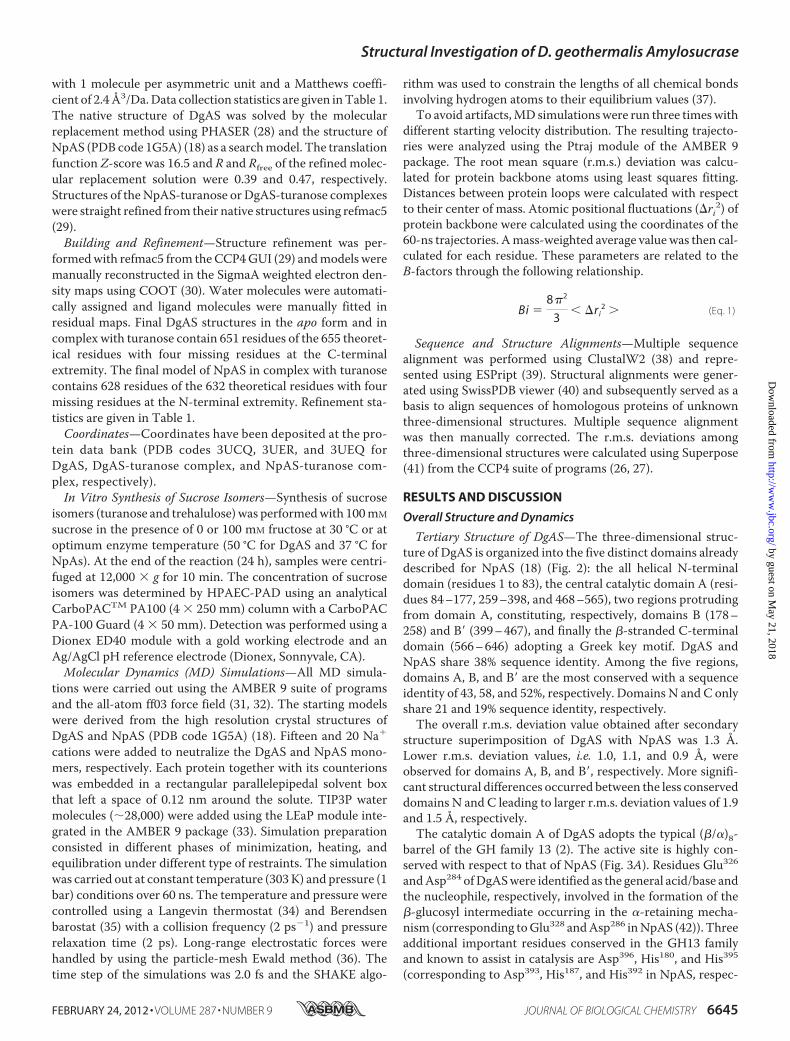
Tertiary Structure of DgAS—The three-dimensional struc-ture of DgAS is organized into the five distinct domains alreadydescribed for NpAS (18) (Fig. 2): the all helical N-terminaldomain (residues 1 to 83), the central catalytic domain A (resi-dues 84–177, 259–398, and 468–565), two regions protrudingfrom domain A, constituting, respectively, domains B (178–258) and B� (399–467), and finally the �-stranded C-terminaldomain (566–646) adopting a Greek key motif. DgAS andNpAS share 38% sequence identity. Among the five regions,domains A, B, and B� are the most conserved with a sequenceidentity of 43, 58, and 52%, respectively. Domains N and C onlyshare 21 and 19% sequence identity, respectively.The overall r.m.s. deviation value obtained after secondary
structure superimposition of DgAS with NpAS was 1.3 Å.Lower r.m.s. deviation values, i.e. 1.0, 1.1, and 0.9 Å, wereobserved for domains A, B, and B�, respectively. More signifi-cant structural differences occurred between the less conserveddomains N and C leading to larger r.m.s. deviation values of 1.9and 1.5 Å, respectively.The catalytic domain A of DgAS adopts the typical (�/�)8-
barrel of the GH family 13 (2). The active site is highly con-served with respect to that of NpAS (Fig. 3A). Residues Glu326andAsp284 ofDgASwere identified as the general acid/base andthe nucleophile, respectively, involved in the formation of the�-glucosyl intermediate occurring in the �-retaining mecha-nism (corresponding toGlu328 andAsp286 inNpAS (42)). Threeadditional important residues conserved in the GH13 familyand known to assist in catalysis are Asp396, His180, and His395(corresponding to Asp393, His187, and His392 in NpAS, respec-
Structural Investigation of D. geothermalis Amylosucrase
FEBRUARY 24, 2012 • VOLUME 287 • NUMBER 9 JOURNAL OF BIOLOGICAL CHEMISTRY 6645
by guest on May 21, 2018
http://ww
w.jbc.org/
Dow
nloaded from

tively). A salt bridge formed by residues Arg520 and Asp137(equivalent toArg509 andAsp144 inNpAS) blocks the bottomofthe catalytic pocket. All the residues defining the catalytic siteare conserved between NpAS and DgAS with the exception ofArg226, which is substituted by Pro219 in DgAS. As a result, theactive site pocket of DgAS shows an enlarged aperture aroundsubsite �2 compared with NpAS that may facilitate ligandaccessibility and binding into the active site (Fig. 3B). Twoligands were found in the active site (Fig. 3A), a glycerol mole-cule (used as cryoprotectant for x-ray experiments) at subsite�1 and a Tris molecule at subsite �1, which are interactingtogether through a polar contact involving glycerol-O2 andTris-O3. Both B and B� domains are found longer in DgAS thanin NpAS. Domain B is composed of 81 amino acid residues inDgAS (versus 76 residues in NpAS), organized in two antipar-allel �-strands and two �-helices. As for domain B�, it is com-posed of four�-helices comprising 69 amino acid residues (ver-sus 65 in NpAS). The domain C of DgAS is composed of 82amino acid residues folded as an eight-stranded �-sandwich aspreviously observed in the NpAS structure. The domains C ofboth enzymes differ in the short �-helix between strands �4and �5 observed only in DgAS. Additionally, the loop intercon-necting strands�2 and�3 is three residues longer inDgAS thanin NpAS. The main difference between the DgAS and NpAShelical domains N resides in the N-terminal extremity, which is
composed by a long �-helix in DgAS and two small �-helices inNpAS (Fig. 3C). The remaining �-helices of the N-terminaldomain (H3, H4, and H5) are structurally conserved in DgASand NpAS.Insight into Enzyme Plasticity through MD Simulations—To
complete our structural comparison of amylosucrases, weundertook the investigation of their dynamic properties. Largescale MD simulations (60 ns) were thus carried out in explicitwater for DgAS and NpAS.As a check for the stability of protein structures during the
course of MD simulations, the time evolution of the backboneatoms r.m.s. deviation was calculated after least square fitting(supplemental Fig. S1A). Variations of backbone r.m.s. devia-tions indicated a distinct conformational behavior of the twoenzymes. The r.m.s. deviation of DgAS rapidly increased by 3 Åwithin the first 12 ns of the simulation and then stabilized forthe rest of the simulation. As for NpAS, the r.m.s. deviationslowly increased by 2.2 Å over the first 30 ns before reaching aplateau (supplemental Fig. S1A). Thus, faster (12 versus 30 ns)and larger amplitude (3 versus 2.2 Å) changes were observed forDgAS compared with NpAS. MD simulations further revealedthat the r.m.s. deviation of DgAS mainly resulted from a highmobility of the N-terminal part of the protein (residues 1–83).As shown in supplemental Fig. S1B, the r.m.s. deviation ofdomain N increased by as much as 5.2 Å over the first 12 ns of
FIGURE 2. Comparison of the three-dimensional architecture of DgAS (PDB code 3UCQ) and NpAS (PDB code 1G5A (18)). Panel A, DgAS. Panel B, NpAS.Panel C, schematic view of DgAS and NpAS primary and secondary structures. The limits of the different domains are given in purple for DgAS and green forNpAS. The same color code is used throughout the different views: catalytic domain A (red), domain N (blue), domain C (cyan), domain B (orange), and domainB� (yellow).
Structural Investigation of D. geothermalis Amylosucrase
6646 JOURNAL OF BIOLOGICAL CHEMISTRY VOLUME 287 • NUMBER 9 • FEBRUARY 24, 2012
by guest on May 21, 2018
http://ww
w.jbc.org/
Dow
nloaded from

the simulation for DgAS, whereas it remained unchanged forNpAS. The high flexibility of the N-extremity was also shownby the large calculated B-factor values (supplemental Fig. S1C).Such large conformational fluctuations of DgAS are due to therearrangement of the first long helix (residues 1–24) into twoshorter helices (composed of residues 1–14 and 15–24, respec-tively). Such conformational changes are not observed forNpAS as its first �-helix is already composed of two short heli-ces in the x-ray structure. These helices are strongly stabilizedthrough a large network of ionic interactions involving chargedresidues (Arg, Lys, Asp, and Glu) from the three first �-helicesbelonging to the N-terminal region of NpAS (Fig. 3C). Note-worthy, there are no equivalent charged residues inDgAS. Evenif other structural regions also appear flexible, as indicated bythe calculated B-factors, differences between both enzymes are,however, less significant. Most flexible regions correspond toloops of the (�/�)8-barrel that confer a pocket topology tothe active site. In particular, loops 7 and 3 (encompassed indomains B� and B, respectively) moved away from each otherduring the MD simulations (supplemental Fig. S1D) result-ing in the opening of the catalytic pocket.With the exceptionof the N domain in DgAS and surface loops (2, 3, 4, 7, and 8)
in both enzymes, the remaining parts of DgAs and NpAS didnot exhibit any significant fluctuation (supplemental Fig. S1,E and F).
Oligomerization and Thermostability
The First Dimeric Structure of Amylosucrase—Whereas theasymmetric unit of DgAS is composed of a single peptide chain,exploration of symmetry related molecular interfaces using thePISA server (43) indicated a stable dimeric organization with aburied interface of 2,950A2 out of a total accessible surface areaof 45,330A2 (Fig. 4A). The buried surface represents 6.1% of thesolvent accessible area. The interface is defined by a set of res-idues located in the catalytic domain (Ala318–Ala320, Leu334–His344, Ala378–Ser385) and also in domains N (Asp21–Gly35,Arg73–Arg88) and C (Thr560–Glu568, Arg584–Gly590) (Fig. 4B).Stabilization between the protomer units of DgAS benefitsfrom strong salt bridge interactions formed between Arg74 andGlu25 and between Arg341 and Asp84, and from a network ofdirect and water-mediated hydrogen-bonding interactions(supplemental Fig. S2). The residues involved in the interfacestabilization of DgAS are not conserved in NpAS. In particular,Arg341, which appears to play a key role in stabilizing the DgAS
FIGURE 3. Comparison of DgAS (PDB code 3UCQ) and NpAS (PDB code 1G5A (18)) crystallographic structures. Panel A, superimposition of the active sitesof DgAS (purple) and NpAS (green). Catalytic residues are shown as sticks. Ligands (Tris and glycerol) were omitted for clarity purpose. Panel B, molecular surfacerepresentation of the DgAS (purple) and NpAS (green) catalytic sites. Panel C, representation of the polar interactions involved in the stabilization of the firsthelix of domain N in DgAS (purple) and NpAS (green).
Structural Investigation of D. geothermalis Amylosucrase
FEBRUARY 24, 2012 • VOLUME 287 • NUMBER 9 JOURNAL OF BIOLOGICAL CHEMISTRY 6647
by guest on May 21, 2018
http://ww
w.jbc.org/
Dow
nloaded from

interface, is one of the five additional residues of the �5–�6interconnecting loop from the (�/�)8-barrel, which is found tobe longer comparedwithNpAS (Figs. 2C and 4B). Of note, suchan insertion region in DgAS is also found to interact throughpolar interactions with the Arg584–Met591 loop from domain Cof the second protomer, which is four residues shorter in NpAS(Fig. 4C).The dimerization interface displays a strong shape comple-
mentarity between the protomers. First, a bundle of hydropho-bic residues from theNdomain involving Leu80 and Leu81 from�-helix H5 of the first protomer fits nicely into the grooveformed by the two �-helices, H3 and H5, from the secondprotomer (supplemental Fig. S2). Residues Leu80, Leu81, andLeu29, belonging to these �-helices and only present in DgAS,are involved in these hydrophobic contacts (supplemental Fig.S2A). In addition, the extremity of the Arg584–Gly590 loop(insertion region situated between strands �3 and �4 of theC-terminal domain inDgAS as described earlier) is found inter-locked between two loops, 339–344 (another insertion regionin DgAS) and 379–385 (highly conserved in the Deinococcus
genus). Interestingly, in such contacting amino acid residues,we observe a bunch of prolines, which are generally known torigidify the polypeptide backbone and may constrain the poly-peptide chain conformation to facilitate dimer formation.Pro587 from the first protomer faces up Pro380, Pro381, andPro383 from the second one. Although oligomeric states havebeen previously observed for other members of the GH13 fam-ily, such as cyclomaltodextrinases (CDase; EC 3.2.1.54) (PDBcode 1H3G (44)), maltogenic amylase (MAase; EC 3.2.1.133(PDB code 1SMA (45)), and neopullulanase (NPase, EC,3.2.1.135) (PDBcode 1J0H (46)), the structure ofDgAS is, to ourknowledge, the first homodimeric amylosucrase reported todate. Comparison of electrophoresis under denaturating (SDS-PAGE) and native conditions argue for a dimeric assembly ofDgAS in solution (Fig. 5, A and B).The dimeric conformation of DgAS in solution was con-
firmed by SEC-MALLS/RI experiments. As shown in Fig. 5C,the elution volume of DgAS differed significantly from that ofNpAS, corresponding to a molecule with a molecular mass of151.7 kDa compatible with a dimeric assembly of DgAS (theo-
FIGURE 4. Quaternary structure organization of DgAS (PDB code 3UCQ). Panel A, visualization of the DgAS dimer. The protomers are symmetry-related following a 2-fold crystallographic symmetry. Panel B, structural superimposition of DgAS (purple) and NpAS (PDB code 1G5A (18)) (green)monomeric units. Structural elements involved in the dimeric interface of DgAS involve 7 regions of domains N, A, and C. The amino acid sequencescomposing the 7 regions are listed. Panel C, sequence alignment of DgAS, DrAS, and NpAS. The 7 regions are shown on the alignment using the samecolor code as in panel B.
Structural Investigation of D. geothermalis Amylosucrase
6648 JOURNAL OF BIOLOGICAL CHEMISTRY VOLUME 287 • NUMBER 9 • FEBRUARY 24, 2012
by guest on May 21, 2018
http://ww
w.jbc.org/
Dow
nloaded from

retical molecular mass of 146.3 kDa). As expected, the NpASmainly eluted as a monomer with a molecular mass of 74.5 kDa(theoretical molecular mass of 71.5 kDa). The monomericNpAS and the dimeric DgAS remained stable over a few weeksat 4 °C.To search for a sequence signature of dimeric forms in other
amylosucrases, we performed a sequence alignment of DgAS,NpAS and DrAS (Fig. 4C). Among the 7 regions found to beinvolved in DgAS dimerization from structural analysis (listedin Fig. 4B), regions 4, 6, and 7 are well conserved in DrAS.Notably, the corresponding loops in NpAS are shorter andcould prevent dimer formation. To get some evidence of DrASdimerization, SEC-MALLS analysis of DrAS was performedand it confirmed the homodimeric organization of this enzymewith amolecularmass of 136.0 kDa (theoreticalmolecularmassof 143.3 kDa) (Fig. 5C). The pattern composed of regions 1–7could thus be proposed as a signature of dimerization. Wescreened the identified pattern against sequences of GH13enzymes (sequence alignment is provided in supplemental Fig.S3). The proposed dimerization patternwas found inDeinococ-cus desertii DdAS, Deinococcus maripensis DmAS, �-amylasesfromMeiothermus rubber,Meiothermus silvanus andTrueperaradiovictrix sequences, suggesting a similar dimeric organiza-tion to DgAS. On the other hand, sequences of amylosucrasesfrom Neisseria and Alteromonas groups did not contain thismolecular pattern. Like NpAS, one could infer that they mightadopt a monomeric form (23).
DgAS Thermostability—The optimum temperature of DgASis 50 °C, whereas an optimal temperature of 37 °Cwas found forNpAS (15). The t1⁄2 of freshly purified DgAS andNpAS was 69 hand 15 min at 50 °C, respectively. The higher stability of DgASwas confirmed by Tm measurements, which gave 58.9 � 0.2 °Cfor DgAS versus 49.6 � 0.4 °C for NpAS when measured bydifferential scanning fluorimetry and 62.0 � 0.2 °C versus51.3 � 0.2 °C when determined by circular dichroism. Closeinspection of the primary sequences of DgAS and NpAS gaverise to the following observation: DgAS contains (i) a higherproportion of charged residues (Lys, Arg, His, Asp, and Glu),which represent 28.8% of the total number of residues com-pared with only 22.9% in NpAS, (ii) a lower number of polaruncharged residues (Gly, Ser, Thr, Asn, Gln, Tyr, and Cys),which represent 24.3% of the total number of residues forDgASand 33.4% forNpAS, (iii)more hydrophobic residues (Leu,Met,Ile, Val, Trp, Pro, Ala, and Phe) representing 46.9% of the totalnumber of residues comparedwith 43.7% forNpAS, and (iv) sixadditional proline residues. All aforementioned features areusually observed in proteins from thermophilic organisms andcould contribute to the higher thermostability of DgAS (47, 48).In addition, we have shownusingMDsimulations onDgAS andNpAS monomers that the N-terminal extremity of DgASexhibits a higher flexibility. DgASdimerizationmight then con-strain the movement of helix H1-H2 at the N terminus of theprotein and one could thus assume that dimerization is likely tocontribute significantly to enzyme stabilization of this enzyme.
FIGURE 5. Biochemical analysis of DgAS and NpAS. Panel A, SDS-PAGE of protein markers (lane 1), DgAS (lane 2), and NpAS (lane 3). Panel B, native PAGE ofprotein markers (lanes 1 and 8), NpAS (lanes 2– 4), and DgAS (lanes 5–7); for both enzymes, no effect of dilution is visible (concentration in proteins are 0.5, 1, and2 g liter�1, respectively). Panel C, SEC-MALLS/RI experiments for NpAS (green line), DgAS (purple line), and DrAS (blue line). Continuous lines represent the lightscattering curve, dashed lines define the variation of refractive index. The experimentally measured molecular weight distribution (as horizontal lines) and theaverage molecular weight are indicated for each elution peaks.
Structural Investigation of D. geothermalis Amylosucrase
FEBRUARY 24, 2012 • VOLUME 287 • NUMBER 9 JOURNAL OF BIOLOGICAL CHEMISTRY 6649
by guest on May 21, 2018
http://ww
w.jbc.org/
Dow
nloaded from
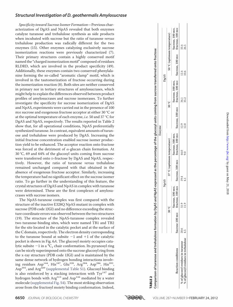
Specificity toward Sucrose Isomer Formation—Previous char-acterization of DgAS and NpAS revealed that both enzymescatalyze turanose and trehalulose synthesis as side productswhen incubated with sucrose but the ratio of turanose versustrehalulose production was radically different for the twoenzymes (15). Other enzymes catalyzing exclusively sucroseisomerization reactions were previously characterized (7).Their primary structures contain a highly conserved motifnamed the “charged isomerizationmotif” composed of residuesRLDRD, which are involved in the product specificity (49).Additionally, these enzymes contain two conserved phenylala-nine forming the so-called “aromatic clamp” motif, which isinvolved in the tautomerization of fructose occurring duringthe isomerization reaction (8). Both sites are neither conservedin primary nor in tertiary structures of amylosucrases, whichmight help to explain the differences observed between productprofiles of amylosucrases and sucrose isomerases. To furtherinvestigate the specificity for sucrose isomerization of DgASandNpAS, experiments were carried out in the presence of 100mM sucrose and exogenous fructose acceptor at either 30 °C orat the optimal temperature of each enzyme, i.e. 50 and 37 °C forDgAS and NpAS, respectively. The results reported in Table 2show that, for all operational conditions, NpAS preferentiallysynthesized turanose. In contrast, equivalent amounts of turan-ose and trehalulose were produced by DgAS. Increasing theinitial fructose concentration enabled sucrose isomer produc-tion yield to be enhanced. The acceptor reaction onto fructosewas forced at the detriment of �-glucan chain formation. At30 °C, 69 and 64% of the glucosyl units coming from sucrosewere transferred onto D-fructose by DgAS and NpAS, respec-tively. However, the ratio of turanose versus trehaluloseremained unchanged compared with that obtained in theabsence of exogenous fructose acceptor. Similarly, increasingthe temperature had no significant effect on the sucrose isomerratio. To go further in the understanding of this feature, thecrystal structures of DgAS andNpAS in complex with turanosewere determined. These are the first complexes of amylosu-crases with sucrose isomers.The NpAS-turanose complex was first compared with the
structure of the inactive E328Q NpAS mutant in complex withsucrose (PDB code 1JGI) and no difference exceeding the struc-ture coordinate errorswas observed between the two structures(19). The structure of the NpAS-turanose complex revealedtwo turanose-binding sites, which were named TB1 and TB2for the site located in the catalytic pocket and at the surface oftheCdomain, respectively. The electron density correspondingto the turanose bound at subsite �1 and �1 of the catalyticpocket is shown in Fig. 6A. The glucosyl moiety occupies cata-lytic subsite �1 in a 4C1 chair conformation. Its pyranosyl ringcan be nicely superimposed onto the sucrose glucosyl ring fromthe x-ray structure (PDB code 1JGI) and is maintained by thesame dense network of hydrogen bonding interactions involv-ing residues Asp144, His187, Glu328, Arg284, Asp286, His392,Asp393, and Arg509 (supplemental Table S1). Glucosyl bindingis also reinforced by a stacking interaction with Tyr147 andhydrogen bonds with Arg513 and Asp144 mediated by a watermolecule (supplemental Fig. S4). Themost striking observationarose from the fructosyl moiety binding conformation. Indeed, T
AB
LE2
Co
mp
aris
on
oft
ura
no
sean
dtr
ehal
ulo
sefo
rmat
ion
by
Dg
AS
and
Np
AS
exp
ress
edas
the
per
cen
tag
eo
fth
eg
luco
sylu
nit
sco
min
gfr
om
sucr
ose
tran
sfer
red
on
tofr
uct
ose
Enzyme
NpA
SDgA
S30
°C37
°C(optim
umtempe
rature)
30°C
50°C
(optim
umtempe
rature)
Reactionco
ndition
sSu
crose10
0m
MSu
crose,10
0m
M,
fruc
tose,1
00m
MSu
crose,10
0m
MSu
crose,10
0m
M,
fruc
tose,1
00m
MSu
crose,10
0m
MSu
crose,10
0m
M,
fruc
tose,1
00m
MSu
crose,10
0m
MSu
crose,10
0m
M,
fruc
tose,1
00m
M
%of
gluc
osylresidu
esin
turano
se20
.651
.919
.444
.012
.934
.112
.433
.4%of
gluc
osylresidu
esin
treh
alulose
4.5
11.9
4.1
9.0
12.8
35.2
10.6
26.5
Structural Investigation of D. geothermalis Amylosucrase
6650 JOURNAL OF BIOLOGICAL CHEMISTRY VOLUME 287 • NUMBER 9 • FEBRUARY 24, 2012
by guest on May 21, 2018
http://ww
w.jbc.org/
Dow
nloaded from

at subsite �1, the electron density of the ligand unequivocallyrevealed that the fructose unit of turanose is bound in the openstate. This is quite surprising, considering that in water solu-tion, turanose adopts an equilibria with a predominant propor-
tion of � pyranoid (47%) and � furanoid (37%) tautomers after20 min at 20 °C as determined by 1H NMR spectroscopy (50).The hydroxyl groups O4�, O5�, and O6� of the open state fruc-tose form direct hydrogen bonds with residues Ala287, Glu328,
FIGURE 6. Stereoview of turanose (orange) conformation bound to NpAS (PDB code 3UEQ) (green) and DgAS (PDB code 3UER) (purple) with a �Aweighted 2Fo � Fc electron density map contoured at 1.0 � around the ligand. Hydrogen bonding interactions are shown as a red dashed line and watermolecules as red spheres. Panel A, TB1 turanose-binding site of NpAS. Panel B, TB1 turanose-binding site of DgAS, the two alternative conformations of theligand corresponding to the two turanose anomers � and � are represented. Panel C, stereoview of the structural superimposition of TB1 turanose binding sitesof NpAS and DgAS using the same color code except for the turanose-NpAS complex where the turanose and water molecules are brown colored for claritypurpose. Residue numbering has been done according to DgAS sequence numbering with the exception of Ile330 and Ala287 (in green) that are only present inthe interaction between NpAS and turanose.
Structural Investigation of D. geothermalis Amylosucrase
FEBRUARY 24, 2012 • VOLUME 287 • NUMBER 9 JOURNAL OF BIOLOGICAL CHEMISTRY 6651
by guest on May 21, 2018
http://ww
w.jbc.org/
Dow
nloaded from

and Ile330, respectively. Noteworthy, a dense network of addi-tional hydrogen bonds mediated by water molecules is alsoobserved around fructose (Table 3). Within this network, thewatermoleculeWat1134mediates twohydrogen bonds betweenArg226 and both O1� and O6�. In addition O6� is also withinhydrogen bond distance of the main chain NH of Ile330 andwater molecule Wat1366. These interactions involving O1� andO6� of the fructose unit probably favor fructose binding in theopen state by preventing hemiacetal formation. Such a bindingmode allows perfect positioning of the O3� for the nucleophilicattack of the �-glucosyl intermediate and the formation ofturanose. It is likely responsible for the predominant turanoseformation byNpAS. In the turanosemolecule found inTB2, thefructose ring adopts a �-D-furanosyl conformation (supple-mental Fig. S5).In DgAS, only one turanose-binding site, equivalent to TB1
of NpAS, was observed (Fig. 6B). The absence of TB2 is likelydue to the fact that residues corresponding to Phe559, Asn560,and Asn562 from domain C of NpAS are substituted by threehydrophobic residues: Lue572, Pro573, and Pro575, respectively.Such substitutions prevent stacking and hydrogen bondinginteractions with turanose. In the TB1 site of DgAS, the gluco-syl moiety of turanose occupies subsite �1 and adopts a 4C1configuration with a very clear electron density. Interactionsbetween the glucopyranosyl ring and the protein are similar tothose described for theNpAS-turanose complex (supplementalTable S1). The electron density of the fructosyl ring is less clearat subsite�1.However, it defines an envelope that fits best with
the �-anomer of fructofuranose (Fig. 6B). The �-anomer alsoprobably bound subsite �1 but to a lesser extent. In addition,we cannot exclude the presence of fructose either in open orpyranoid forms. The O3� glucosylated-�-D-fructofuranose isreported to be one of the less prevalent tautomer of turanose inwater solution (1% at 20 °C (50)). Accommodation of this con-formation in subsite �1 involves H-bonds with the catalyticresidues Glu326 and Asp396 and two water-mediated interac-tions with O1�, O4� and O6�. DgAS binds the furanoid tautom-ers of fructose to form turanose but via a considerably weakernetwork of interactions than that observed in NpAS. Furthercomparisons of DgAS andNpAS subsite�1 revealed structuraltraits that prevent open state conformation of fructose to bindDgAS subsite �1 (Fig. 6C). Indeed, residue Arg226 of NpAS,which interacts via a water molecule with both O1� and O6� ofthe open form of fructose, is replaced by Pro219 in DgAS. Fur-thermore, residue Ile328 corresponding to Ile330 in NpAS,which was directly involved in H-bond interaction with O6� offructose in the open state, cannot play the same role in DgASdue to a slight motion of the loop bearing this residue. Thediscrimination between the various tautomers of turanose isless drastic inDgAS than inNpAS.DgAS subsite�1 accommo-dates various fructose tautomers to optimally arrange O1� orO3� of the fructose unit for nucleophilic attack of the glucosylenzyme intermediate therefore yielding equivalent amounts oftrehalulose and turanose.DgAS- andNpAS-turanose complex analysis thus enabled us
to explain differences observed in DgAS and NpAS product
TABLE 3Interactions of fructose unit from sucrose and turanose with NpAS and DgAS
Structural Investigation of D. geothermalis Amylosucrase
6652 JOURNAL OF BIOLOGICAL CHEMISTRY VOLUME 287 • NUMBER 9 • FEBRUARY 24, 2012
by guest on May 21, 2018
http://ww
w.jbc.org/
Dow
nloaded from

specificity. This work opens new perspectives for the rationaland/or semi-rational redesign of this thermostable amylosu-crase to modulate sucrose isomer synthesis with the view ofreducing side product formation or controlling sucrose isomerprofile for the development of novel syrups enriched in sucrosesubstitutes.
Acknowledgments—We thank the Computing Center of RegionMidi-Pyrenees (CALMIP, Toulouse, France) and the Center for ComputingResources (CRI) of INSA-Toulouse for providing computing resourcesand support. We are grateful to the staff of Synchrotron beamlinesID14-1 and ID29 at the European Synchrotron Radiation Facility(Grenoble, France) for providing assistance in using the beamlines.We are grateful to Javier Perez (Synchrotron SOLEIL) for the fruitfuldiscussions. We also thank Stephanie Terme (Wyatt technologyFrance) for helpful assistance in the SEC-MALLS experiments.
REFERENCES1. Henrissat, B. (1991) A classification of glycosyl hydrolases based on amino
acid sequence similarities. Biochem. J. 280, 309–3162. Henrissat, B., and Davies, G. (1997) Structural and sequence-based classi-
fication of glycoside hydrolases. Curr. Opin. Struct. Biol. 7, 637–6443. Cantarel, B. L., Coutinho, P. M., Rancurel, C., Bernard, T., Lombard, V.,
and Henrissat, B. (2009) The Carbohydrate-Active EnZymes database(CAZy). An expert resource for glycogenomics. Nucleic Acids Res. 37,D233–238
4. Potocki de Montalk, G., Remaud-Simeon, M., Willemot, R. M., Sarcabal,P., Planchot, V., and Monsan, P. (2000) Amylosucrase from Neisseria po-lysaccharea. Novel catalytic properties. FEBS Lett. 471, 219–223
5. Okada, G., and Hehre, E. J. (1974) New studies on amylosucrase, a bacte-rial �-D-glucosylase that directly converts sucrose to a glycogen-like�-glucan. J. Biol. Chem. 249, 126–135
6. Goulter, K. C., Hashimi, S. M., and Birch, R. G. (2012) Microbial sucroseisomerases. Producing organisms, genes, and enzymes. Enzyme Microb.Technol. 50, 57–64
7. Ravaud, S., Robert, X.,Watzlawick, H., Haser, R., Mattes, R., and Aghajari,N. (2007) Trehalulose synthase native and carbohydrate-complexedstructures provide insights into sucrose isomerization. J. Biol. Chem. 282,28126–28136
8. Ravaud, S., Robert, X.,Watzlawick, H., Haser, R., Mattes, R., and Aghajari,N. (2009) Structural determinants of product specificity of sucroseisomerases. FEBS Lett. 583, 1964–1968
9. Thompson, J., and Pikis, A. (2012) Metabolism of sugars by geneticallydiverse species of oral Leptotrichia. Mol. Oral Microbiol. 27, 34–44
10. Seo, D. H., Jung, J.-H., Ha, S.-J., Song, M.-C., Cha, J., Yoo, S. H., Kim, T.-J.,Baek, N. I., and Park, C.-S. (2009) Highly selective biotransformation ofarbutin to arbutin-�-glucoside using amylosucrase fromDeinococcus geo-thermalis DSM 11300. J. Mol. Catal. B 60, 113–118
11. Andre, I., Potocki-Veronese, G., Morel, S., Monsan, P., and Remaud-Si-meon, M. (2010) Sucrose-utilizing transglucosidases for biocatalysis. Top.Curr. Chem. 294, 25–48
12. Chang, A., Singh, S., Phillips, G. N., Jr., and Thorson, J. S. (2011) Glycosyl-transferase structural biology and its role in the design of catalysts forglycosylation. Curr. Opin. Biotechnol 22, 800–808
13. Champion, E., Andre, I., Moulis, C., Boutet, J., Descroix, K., Morel, S.,Monsan, P., Mulard, L. A., and Remaud-Simeon, M. (2009) Design of�-transglucosidases of controlled specificity for programmed chemoen-zymatic synthesis of antigenic oligosaccharides. J. Am. Chem. Soc. 131,7379–7389
14. Pizzut-Serin, S., Potocki-Veronese, G., van der Veen, B. A., Albenne, C.,Monsan, P., and Remaud-Simeon, M. (2005) Characterization of a novelamylosucrase from Deinococcus radiodurans. FEBS Lett. 579, 1405–1410
15. Emond, S., Mondeil, S., Jaziri, K., Andre, I., Monsan, P., Remaud-Simeon,M., and Potocki-Veronese, G. (2008) Cloning, purification and character-ization of a thermostable amylosucrase from Deinococcus geothermalis.
FEMS Microbiol Lett 285, 25–3216. Ha, S. J., Seo, D.H., Jung, J. H., Cha, J., Kim, T. J., Kim, Y.W., and Park, C. S.
(2009) Molecular cloning and functional expression of a new amylosu-crase from Alteromonas macleodii. Biosci. Biotechnol. Biochem. 73,1505–1512
17. Skov, L. K., Mirza, O., Henriksen, A., Potocki de Montalk, G., Remaud-Simeon, M., Sarcabal, P., Willemot, R. M., Monsan, P., and Gajhede, M.(2000) Crystallization and preliminary X-ray studies of recombinant amy-losucrase from Neisseria polysaccharea. Acta Crystallogr. D 56, 203–205
18. Skov, L. K., Mirza, O., Henriksen, A., DeMontalk, G. P., Remaud-Simeon,M., Sarcabal, P., Willemot, R. M., Monsan, P., and Gajhede, M. (2001)Amylosucrase, a glucan-synthesizing enzyme from the �-amylase family.J. Biol. Chem. 276, 25273–25278
19. Skov, L. K., Mirza, O., Sprogøe, D., Dar, I., Remaud-Simeon, M., Albenne,C.,Monsan, P., andGajhede,M. (2002)Oligosaccharide and sucrose com-plexes of amylosucrase. Structural implications for the polymerase activ-ity. J. Biol. Chem. 277, 47741–47747
20. Jensen, M. H., Mirza, O., Albenne, C., Remaud-Simeon, M., Monsan, P.,Gajhede, M., and Skov, L. K. (2004) Crystal structure of the covalent in-termediate of amylosucrase from Neisseria polysaccharea. Biochemistry43, 3104–3110
21. Emond, S., Andre, I., Jaziri, K., Potocki-Veronese, G., Mondon, P.,Bouayadi, K., Kharrat, H., Monsan, P., and Remaud-Simeon, M. (2008)Combinatorial engineering to enhance thermostability of amylosucrase.Protein Sci. 17, 967–976
22. Jung, J. H., Seo, D. H., Ha, S. J., Song, M. C., Cha, J., Yoo, S. H., Kim, T. J.,Baek,N. I., Baik,M. Y., and Park, C. S. (2009) Enzymatic synthesis of salicinglycosides through transglycosylation catalyzed by amylosucrases fromDeinococcus geothermalis and Neisseria polysaccharea. Carbohydr. Res.344, 1612–1619
23. De Montalk, G. P., Remaud-Simeon, M., Willemot, R. M., Planchot, V.,and Monsan, P. (1999) Sequence analysis of the gene encoding amylosu-crase from Neisseria polysaccharea and characterization of the recombi-nant enzyme. J. Bacteriol. 181, 375–381
24. Battye, T. G., Kontogiannis, L., Johnson, O., Powell, H. R., and Leslie, A. G.(2011) iMOSFLM, a new graphical interface for diffraction-image proc-essing with MOSFLM. Acta Crystallogr. D 67, 271–281
25. Evans, P. (2006) Scaling and assessment of data quality.ActaCrystallogr. D62, 72–82
26. Collaborative Computational Project, Number 4 (1994) The CCP4 suite.Programs for protein crystallography. Acta Crystallogr. D 50, 760–763
27. Potterton, E., Briggs, P., Turkenburg, M., and Dodson, E. (2003) A graph-ical user interface to the CCP4 program suite. Acta Crystallogr. D 59,1131–1137
28. McCoy, A. J. (2007) Solving structures of protein complexes by molecularreplacement with Phaser. Acta Crystallogr. D 63, 32–41
29. Murshudov, G. N., Vagin, A. A., and Dodson, E. J. (1997) Refinement ofmacromolecular structures by the maximum-likelihood method. ActaCrystallogr. D 53, 240–255
30. Emsley, P., and Cowtan, K. (2004) COOT, model-building tools for mo-lecular graphics. Acta Crystallogr. D 60, 2126–2132
31. Duan, Y., Wu, C., Chowdhury, S., Lee, M. C., Xiong, G., Zhang, W., Yang,R., Cieplak, P., Luo, R., Lee, T., Caldwell, J., Wang, J., and Kollman, P.(2003) A point-charge force field for molecular mechanics simulations ofproteins based on condensed-phase quantum mechanical calculations.J. Comput. Chem. 24, 1999–2012
32. Lee, M. C., and Duan, Y. (2004) Distinguish protein decoys by using ascoring function based on a new AMBER force field, short moleculardynamics simulations, and the generalized born solvent model. Proteins55, 620–634
33. Case, D. A., Darden, T. E., Cheatham, I. T. E., Simmerling, C. L., Wang, J.,Duke, R. E., Luo, R., Merz, K. M., Pearlman, D. A., Crowley, M., Walker,R. C., Zhang,W.,Wang, B., Hayik, S., Roitberg, A., Seabra, G.,Wong, K. F.,Paesani, F., Wu, X., Brozell, S., Tsui, V., Gohlke, H., Yang, L., Tan, C.,Mongan, J., Hornak, V., Cui, G., Beroza, P., Mathews, D. H., Schafmeister,C., Ross, W. S., and Kollman, P. A. (2006) AMBER 9, University of Cali-fornia, San Francisco, CA
34. Pastor, R. W., Brooks, B. R., and Szabo, A. (1988) An analysis of the accu-
Structural Investigation of D. geothermalis Amylosucrase
FEBRUARY 24, 2012 • VOLUME 287 • NUMBER 9 JOURNAL OF BIOLOGICAL CHEMISTRY 6653
by guest on May 21, 2018
http://ww
w.jbc.org/
Dow
nloaded from

racy of Langevin and molecular dynamics algorithms. Mol. Phys. 65,1409–1419
35. Berendsen, H. J. C., Postma, J. P.M., vanGunsteren,W. F., DiNola, A., andHaak, J. R. (1984) Molecular dynamics with coupling to an external bath.J. Chem. Phys. 81, 3684–3690
36. Essmann, U., Perera, L., Berkowitz, M. L., Darden, T., Lee, H., and Peder-sen, L. G. (1995) A smooth particle mesh Ewald method. J. Chem. Phys.103, 8577–8593
37. Ryckaert, J.-P., Ciccotti, G., and Berendsen, H. J. C. (1977) Numericalintegration of the Cartesian equations of motion of a system with con-straints: molecular dynamics of n-alkanes. J. Comput. Phys. 23, 327–341
38. Larkin, M. A., Blackshields, G., Brown, N. P., Chenna, R., McGettigan,P. A., McWilliam, H., Valentin, F., Wallace, I. M., Wilm, A., Lopez, R.,Thompson, J. D., Gibson, T. J., and Higgins, D. G. (2007) Clustal W andClustal X version 2.0. Bioinformatics 23, 2947–2948
39. Gouet, P., Robert, X., and Courcelle, E. (2003) ESPript/ENDscript. Ex-tracting and rendering sequence and 3D information from atomic struc-tures of proteins. Nucleic Acids Res. 31, 3320–3323
40. Guex, N., and Peitsch, M. C. (1997) SWISS-MODEL and the Swiss-Pdb-Viewer. An environment for comparative protein modeling. Electropho-resis 18, 2714–2723
41. Krissinel, E., andHenrick, K. (2004) Secondary-structurematching (SSM),a new tool for fast protein structure alignment in three dimensions. ActaCrystallogr. D 60, 2256–2268
42. Sarcabal, P., Remaud-Simeon, M., Willemot, R., Potocki de Montalk, G.,
Svensson, B., and Monsan, P. (2000) Identification of key amino acid res-idues in Neisseria polysaccharea amylosucrase. FEBS Lett. 474, 33–37
43. Krissinel, E., and Henrick, K. (2007) Inference of macromolecular assem-blies from crystalline state. J. Mol. Biol. 372, 774–797
44. Fritzsche, H. B., Schwede, T., and Schulz, G. E. (2003) Covalent and three-dimensional structure of the cyclodextrinase from Flavobacterium sp. No.92. Eur. J. Biochem. 270, 2332–2341
45. Kim, J. S., Cha, S. S., Kim, H. J., Kim, T. J., Ha, N. C., Oh, S. T., Cho, H. S.,Cho,M. J., Kim,M. J., Lee,H. S., Kim, J.W., Choi, K. Y., Park, K.H., andOh,B. H. (1999) Crystal structure of a maltogenic amylase provides insightsinto a catalytic versatility. J. Biol. Chem. 274, 26279–26286
46. Hondoh,H., Kuriki, T., andMatsuura, Y. (2003) Three-dimensional struc-ture and substrate binding of Bacillus stearothermophilus neopullulanase.J. Mol. Biol. 326, 177–188
47. Jaenicke, R., and Bohm, G. (1998) The stability of proteins in extremeenvironments. Curr. Opin. Struct. Biol. 8, 738–748
48. Prakash, O., and Jaiswal, N. (2010) �-Amylase, an ideal representative ofthermostable enzymes. Appl. Biochem. Biotechnol. 160, 2401–2414
49. Zhang, D., Li, N., Swaminathan, K., and Zhang, L. H. (2003) Amotif rich incharged residues determines product specificity in isomaltulose synthase.FEBS Lett. 534, 151–155
50. Lichtenthaler, F. W., and Ronninger, S. (1990) Studies on ketoses, 4. -D-glucopyranosyl-D-fructoses: distribution of furanoid and pyranoid tau-tomers in water, dimethyl sulphoxide, and pyridine. J. Chem. Soc. PerkinTrans. 2, 1489–1497
Structural Investigation of D. geothermalis Amylosucrase
6654 JOURNAL OF BIOLOGICAL CHEMISTRY VOLUME 287 • NUMBER 9 • FEBRUARY 24, 2012
by guest on May 21, 2018
http://ww
w.jbc.org/
Dow
nloaded from

Isabelle André and Samuel TranierGuieysse, Valérie Guillet, Pierre Monsan, Lionel Mourey, Magali Remaud-Siméon,
Frédéric Guérin, Sophie Barbe, Sandra Pizzut-Serin, Gabrielle Potocki-Véronèse, DavidDeinococcus geothermalisAmylosucrase from the Bacterium
Structural Investigation of the Thermostability and Product Specificity of
doi: 10.1074/jbc.M111.322917 originally published online December 29, 20112012, 287:6642-6654.J. Biol. Chem.
10.1074/jbc.M111.322917Access the most updated version of this article at doi:
Alerts:
When a correction for this article is posted•
When this article is cited•
to choose from all of JBC's e-mail alertsClick here
Supplemental material:
http://www.jbc.org/content/suppl/2011/12/29/M111.322917.DC1
http://www.jbc.org/content/287/9/6642.full.html#ref-list-1
This article cites 49 references, 7 of which can be accessed free at
by guest on May 21, 2018
http://ww
w.jbc.org/
Dow
nloaded from