Embed Size (px)
Citation preview
Pergamon
Biochemical fharmacology, Vol. JO, No. 7. 1053-1061. 1995. pp. Copyright 8 1995 Elsevier Science Inc.
Printed in Great Britain. All rights reserved OOOG2952(95)00240-5 0006.2952/95 $9.50 + 0.00
STRUCTURE-ACTIVITY RELATIONSHIPS OF PHENOTHIAZINES IN INHIBITING LYMPHOCYTE MOTILITY AS DETERMINED BY
A NOVEL FLOW CYTOMETRIC ASSAY
NICHOLAS MATTHEWS,* RICHARD J. FRANKLINt and DAVID A. KENDRICK? Yamanouchi Research Institute, Littlemore Hospital, Oxford OX4.4XN, U.K. and tFerring Research
Institute, Chilworth Research Centre, Southampton SO1 7NP, U.K.
(Received 8 March 1995; accepted 17 May 1995)
Abstract-Lymphocyte motility is highly dependent on rapid changes in cell shape. The human T-lymphoma cell line, MOLT-4, is constitutively shape-changing and motile, and both of these properties can be inhibited by the phenothiazine, chlorpromazine, as assessed by video analysis and migration across polycarbonate filters. In this paper, the light-scattering facility of a flow cytometer has been used to establish a simpler and more quantitative means of measuring changes in shape. By this method, the structure activity relationship (SAR) of phenothiazines and related compounds has been determined. The most active compounds had the tricyclic phenothiazine nucleus with a constrained dialkylaminoalkyl substituent at the nitrogen. The SAR for inhibition of lymphocyte motility differs from those reported for neuroIeptic effects and for inhibition of PKC or calmod- min. Phenothiazine concentrations that inhibited lymphocyte shape-changing resulted in reduced F-actin con- centrations. This indicates that the probable mode of action is disruption of mechanisms regulating actin polymerisation.
Key words: lymphocyte; motility; phenothiazine; flow cytometry; shape; actin
Initiation and perpetuation of chronic inflammation re- quires extravasation of lymphocytes into inflammatory sites. This is a multistep process involving initial adhe- sion of the lymphocytes to activated endothelium and subsequent induction of motility, so that the lymphocyte can migrate between endothelial cells and through con- nective tissue. Inhibition of either the initial adhesion or the induction of motility would be expected to dampen chronic inflammation. Although there has been a great deal of work on lymphocyte-endothelial adhesion, reg- ulation of lymphocyte motility has been relatively unex- plored.
In a previous study, we found that lymphocyte motil- ity could be inhibited by chelerythrine, calphostin C, sphingosine, H7, and H8; these agents inhibit a variety of serine-threonine kinases, but have in common the ability to inhibit PKCS [ 11. We originally tested pheno- thiazines for inhibition of lymphocyte motility because they too have been reported to inhibit PKC [2]. There is also evidence that phenothiazines have immunosuppres- sive and anti-inflammatory activity in addition to their neuroleptic effects [3, 4, 51.
In preliminary experiments, the prototype phenothi- azine, chlorpromazine, was found to potently inhibit mo- tility of human peripheral blood lymphocytes. Inhibition of motility was seen at concentrations below that re- ported for effects on PKC [2] and calmodulin [6]. As this suggested a potentially novel mode of action, it was of interest to study the SAR for phenothiazine inhibition of lymphocyte motility.
For studies on SAR, we needed a simpler assay than those currently in use. Existing in vitro assays of lym- phocyte motility usually involve migration across filters
* Corresponding author. Tel. 865-747100, FAX 865-748974. $ Abbreviations: PKC, protein kinase C; SAR, structure ac-
tivity relationship.
and are too cumbersome for large numbers of samples. Therefore, we have exploited the fact that ability to shape change rapidly is essential for lymphocyte motility [7,8,9]. Non-motile lymphocytes are spherical, whereas their motile counterparts are irregular, constantly shape- changing cells. The proportion of irregular cells in a lymphocyte population correlates with motility, and can be used as a measure of motility [8, lo]. Although this can be determined by simple microscopic observation, this method is slow and tedious, and we have found that the light-scattering facility of a flow cytometer offers a simple and quantitative means of measuring changes in lymphocyte shape. As a source of motile lymphocytes, we have selected a stable, spontaneously motile variant of a human T lymphocyte line (MOLT-4) [lo].
MATERIALS AND METHODS
Chemical compounds
Compounds are numbered as in Fig. 4. Mepazine (6) was a gift from Byk Gulden, Konstanz, Germany. Most of the other compounds were purchased from commer- cial sources (Aldrich, Sigma, RBI). The structures for 26 and 38 are corrected versions of those shown in the Aldrich Rare Chemicals Collection. Other compounds were prepared by analogy to published methods. Amides (e.g. 34, 50, 51) were prepared from the corresponding acid [l I], and amines (e.g. 37, 43, 52) either from the corresponding amide by reduction or by reductive alky- lation of a less substituted amine. Where these routes were not appropriate (e.g. 42, 44), the IO-unsubstituted phenothiazine was derivatised directly with the relevant electrophile [12]. New compounds showed spectro- scopic and analytical properties consistent with the pro- posed structures. Stock solutions were made at 50 mM in DMSO, although 12.5 mM was used for a minority of compounds with lower solubility. In all cases, stocks
IO.53
1054 N. MATTHEWS, R. J. FRANKLIN and D. A. KENDRICK
were stored in the dark at -2O’C. Other reagents were of round and irregular cells in any sample was deter- purchased from Sigma (Poole, U.K.). mined (see Results section).
Conventional shape-change assay F-actin measurement The T lymphocytes were a spontaneously motile vari-
ant of human MOLT-4 cells grown in 10% FCS in RPM1 1640 medium [lo]. In some experiments, a non-motile variant of the cells [lo] was used as a negative control. Growth medium was used as the diluent in this and other assays. The test compound (50 p.l) was mixed with 50 pl cell suspension (5 x lO?ml) in a well of a 96-well cluster plate. After 1 hr at 37°C a 100~pl volume of 3.7% formaldehyde in PBS was added to fix the cells. The well was then examined at x400 magnification, and be- tween 100 and 200 cells were scored as either “shape- changed” or “non-shape-changed.” The criterion for non-shape-changed was that at least three-quarters of the cell approximated to a circle. Each dilution of test re- agent was tested in triplicate, and results were expressed as the mean % shape-changed cells f SD [l]. Since the test reagents were made up as stock solutions in DMSO, solvent controls were included in all assays. With the data shown in the Results section, the corresponding concentrations of solvent were without effect.
Cells (100 pl, lO?ml) were incubated with 100 pl phenothiazine dilution for 30 min at 37°C before addi- tion of 200 pl prewarmed solution containing 7.4% formaldehyde, 0.2 mg/ml lysophosphatidyl choline, 0.22 pM fluoresceinated phalloidin in PBS. After a further 20 min at 37°C the cells were washed x2 with PBS and resuspended in 400 pl PBS containing 1% formalde- hyde, 1% FCS. The mean fluorescence intensity of 10,000 cells was measured using the FACScan flow cy- tometer.
RESULTS
Validation of the assay
Time lapse video
Cells were suspended in growth medium with 1 mM HEPES, and placed in the wells of 96-well cluster plates, which were then sealed with tape to prevent evaporation. The cluster plate was placed on the stage of a Zeiss Axiovert 35 microscope, and maintained at 37°C by means of a thermostatically controlled fan heater. For video analysis, a Panasonic WV-BL600 camera was used with a Panasonic time lapse video cassette recorder. Recordings were made over 1 hr and replayed at x160.
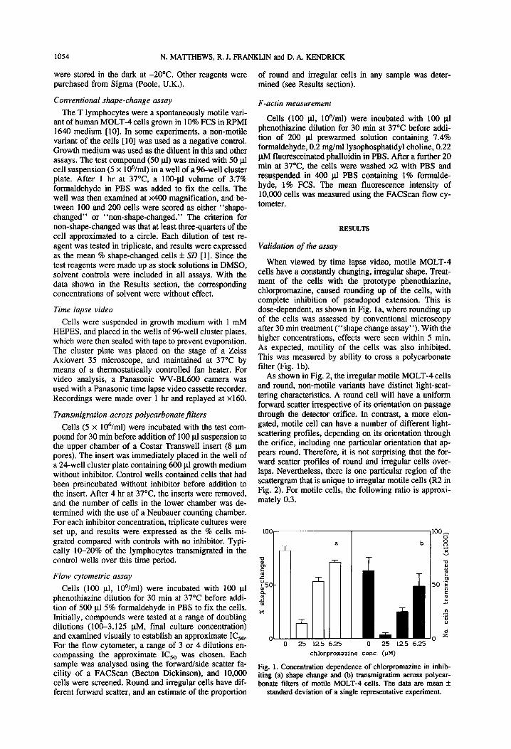
When viewed by time lapse video, motile MOLT-4 cells have a constantly changing, irregular shape. Treat- ment of the cells with the prototype phenothiazine, chlorpromazine, caused rounding up of the cells, with complete inhibition of pseudopod extension. This is dose-dependent, as shown in Fig. la, where rounding up of the cells was assessed by conventional microscopy after 30 min treatment (“shape change assay”). With the higher concentrations, effects were seen within 5 min. As expected, motility of the cells was also inhibited. This was measured by ability to cross a polycarbonate filter (Fig. lb).
Transmigration across polycarbonatejlters
Cells (5 x lO’?ml) were incubated with the test com- pound for 30 min before addition of 100 pl suspension to the upper chamber of a Costar Transwell insert (8 pm pores). The insert was immediately placed in the well of a 24-well cluster plate containing 600 pl growth medium without inhibitor. Control wells contained cells that had been preincubated without inhibitor before addition to the insert. After 4 hr at 37’C, the inserts were removed, and the number of cells in the lower chamber was de- termined with the use of a Neubauer counting chamber. For each inhibitor concentration, triplicate cultures were set up, and results were expressed as the % cells mi- grated compared with controls with no inhibitor. Typi- cally lO-20% of the lymphocytes transmigrated in the control wells over this time period.
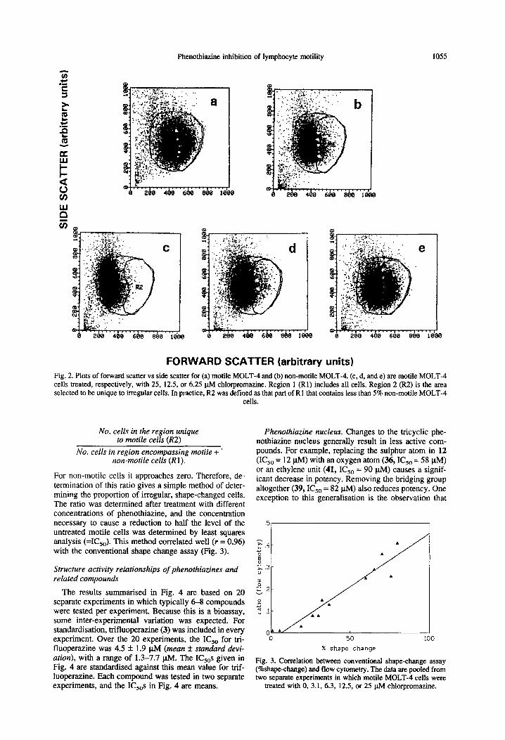
As shown in Fig. 2, the irregular motile MOLT-4 cells and round, non-motile variants have distinct light-scat- tering characteristics. A round cell will have a uniform forward scatter irrespective of its orientation on passage through the detector orifice. In contrast, a more elon- gated, motile cell can have a number of different light- scattering profiles, depending on its orientation through the orifice, including one particular orientation that ap- pears round. Therefore, it is not surprising that the for- ward scatter profiles of round and irregular cells over- laps. Nevertheless, there is one particular region of the scattergram that is unique to irregular motile cells (R2 in Fig. 2). For motile cells, the following ratio is approxi- mately 0.3.
lOOr------ a
Flow cytometric assay
Cells (100 ~1, lO?ml) were incubated with 100 pl phenothiazine dilution for 30 min at 37°C before addi- tion of 500 pl 5% formaldehyde in PBS to fix the cells. Initially, compounds were tested at a range of doubling dilutions (100-3.125 @vi, final culture concentration) and examined visually to establish an approximate IC,,. For the flow cytometer, a range of 3 or 4 dilutions en- compassing the approximate IC,, was chosen. Each sample was analysed using the forward/side scatter fa- cility of a FACScan (Becton Dickinson), and 10,000 cells were screened. Round and irregular cells have dif- ferent forward scatter, and an estimate of the proportion
rl 25
m z 8 d
0 z 0 25 12.5 6.25
chlorpromazine cont. (uM)
Fig. 1. Concentration dependence of chlorpromazine in inhib- iting (a) shape change and (b) transmigration across polycar- bonate filters of motile MOLT-4 cells. The data are mean f
standard deviation of a single representative experiment.
Phenothiazine inhibition of lymphocyte motility 1055
0
FORWARD SCATTER (arbitrary units) Fig. 2. Plots of forward scatter vs side scatter for (a) motile MOLT-4 and (b) non-motile MOLT-4. (c, d, and e) are motile MOLT-4 cells treated, respectively, with 25, 12.5, or 6.25 pM chlorpromazine. Region 1 (Rl) includes all cells. Region 2 (R2) is the area selected to be unique to irregular cells. In practice, R2 was defined as that part of R 1 that contains less than 5% non-motile MOLT-4
cells.
No. cells in the region unique to motile cells (RZ)
No. cells in region encompassing motile + ’ non-motile cells (Rl).
For non-motile cells it approaches zero. Therefore, de- termination of this ratio gives a simple method of deter- mining the proportion of irregular, shape-changed cells. The ratio was determined after treatment with different concentrations of phenothiazine, and the concentration necessary to cause a reduction to half the level of the untreated motile cells was determined by least squares analysis (=I(&,). This method correlated well (r = 0.96) with the conventional shape change assay (Fig. 3).
Structure activity relationships of phenothiazines and related compounds
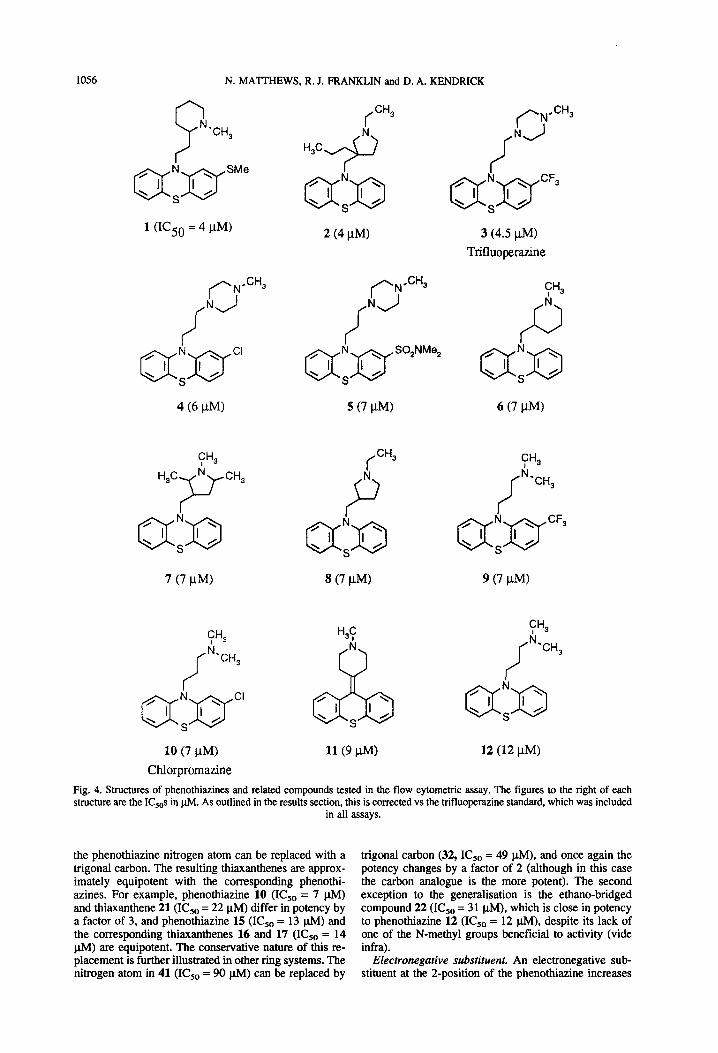
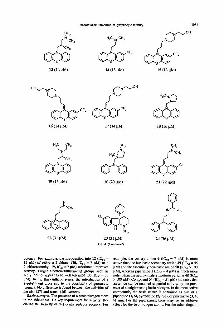
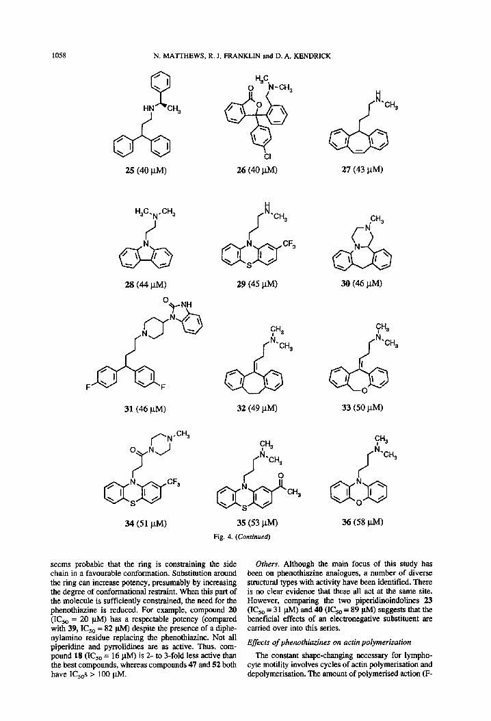
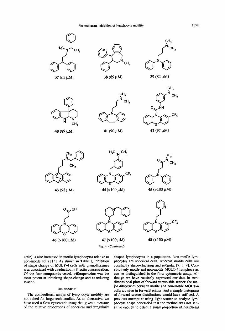
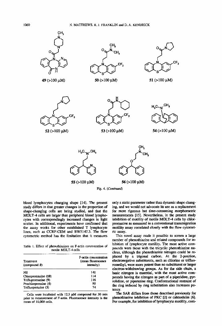
The results summarised in Fig. 4 are based on 20 separate experiments in which typically 68 compounds were tested per experiment. Because this is a bioassay, some inter-experimental variation was expected. For standardisation, trifluoperazine (3) was included in every experiment. Over the 20 experiments, the IC,, for tri- fluoperazine was 4.5 f 1.9 pM (mean f standard devi- ation), with a range of 1.3-7.7 pM. The K&s given in Fig. 4 are standardised against this mean value for trif- luoperazine. Each compound was tested in two separate experiments, and the IC,,s in Fig. 4 are means.
Phenothiazine nucleus. Changes to the tricyclic phe- nothiazine nucleus generally result in less active com- pounds. For example, replacing the sulphur atom in 12 (IC,, = 12 @I) with an oxygen atom (36, IC,, = 58 plvl) or an ethylene unit (41, IC,, = 90 p&l) causes a signif- icant decrease in potency. Removing the bridging group altogether (39, IC,, = 82 p.M) also reduces potency. One exception to this generalisation is the observation that
50
% shape change
0
Fig. 3. Correlation between conventional shape-change assay (%shape-change) and flow cytometry. The data are pooled from two separate experiments in which motile MOLT-4 cells were
treated with 0, 3.1, 6.3, 12.5, or 25 @I chlorpromazine.
1056 N. MATTHEWS, R. J. FRANKLIN and D. A. KENDRICK
1 (IC50 = 4 l.tM)
(--N-CH3
4 (6 PM)
y3 CH,
7 (7 I.W
2 (4 fiM)
,/-NeCH3
NJ
5 (7 PM)
fH3
P Qc)g
8 (7 PM)
CH3
rJJ
H3S:
i
‘CH,
10 (7 PM) Chlorpromazine
110 NW
3 (4.5 /.w Trifluoperazine
6 (7 PM)
9 (7 PM)
CH3
/
k ‘CH,
12 (12 PM)
Fig. 4. Structures of phenothiazines and related compounds tested in the flow cytometric assay. The figures to the right of each structure are the IC,,s in @l. As outlined in the results section, this is corrected vs the trifluoperazine standard, which was included
in all assays.
the phenothiazine nitrogen atom can be replaced with a trigonal carbon. The resulting thiaxanthenes are approx- imately equipotent with the corresponding phenothi- azines. For example, phenothiazine 10 (ICSo = 7 ph4) and thiaxanthene 21 (IC,, = 22 l.tM) differ in potency by a factor of 3, and phenothiazine 15 (IC,, = 13 p.M) and the corresponding thiaxanthenes 16 and 17 (ICSo = 14 @VI) are equipotent. The conservative nature of this re- placement is further illustrated in other ring systems. The nitrogen atom in 41 (IC,, = 90 pM) can be replaced by
trigonal carbon (32, ICSo = 49 pM), and once again the potency changes by a factor of 2 (although in this case the carbon analogue is the more potent). The second exception to the generalisation is the ethano-bridged compound 22 (IC,, = 3 1 @I), which is close in potency to phenothiazine 12 (IC,, = 12 pM), despite its lack of one of the N-methyl groups beneficial to activity (vide infra).
Electronegative substituent. An electronegative sub- stituent at the 2-position of the phenothiazine increases
Phenothiazine inhibition of lymphocyte motility 1057
13 (12 PM) 14 (13 WI 15 (13 l.lM>
16 (14 WI 17 (14 NW 18 (16 I.LM)
19 (16 P-M) 20 (20 l.w
cw ‘CH,
063 ‘I I> \
22 (31 l.tM) 23 (31 /..tM)
Fig. 4. (Conrinued)
21(22 PM)
Q ‘I \
HN ‘G CH 3
24 06 PM)
potency. For example, the introduction into 12 (IC,, = 12 ph4) of either a 2-chloro- (10, IC,, = 7 u.M) or a 2-trifluoromethyl- (9, I&, = 7 @I) substituent improves activity. Larger electron-withdrawing groups such as acetyl do not appear to be well tolerated (35, IC,, = 53 FM). In the thiaxanthene series, the introduction of a 2-substituent gives rise to the possibility of geometric isomers. No difference is found between the activities of the cis- (17) and trans- (16) isomers.
Basic nitrogen. The presence of a basic nitrogen atom in the side-chain is a key requirement for activity. Re- ducing the basicity of this centre reduces potency. For
example, the tertiary amine 9 (IC5, = 7 ph4) is more active than the less basic secondary amine 29 (IC,, = 45 pM) and the essentially non-basic amide 50 (IC,, > 100 l&Q, whereas piperidine 1 (IC5, = 4 pM) is much more potent than the approximately isosteric pyridine 48 (IC,, > 100 ph4). Compound 34 (KS0 = 51 u.A4) indicates that an amide can be restored to partial activity by the pres- ence of a neighbouring basic nitrogen. In the most active compounds, the basic centre is contained as part of a piperidine (1,6), pyrrolidine (2,7,8), or piperazine (3,4, 5) ring. For the piperazines, there may be an additive effect for the two nitrogen atoms. For the other rings, it
1058 N. MATTHEWS, R. J. FRANKLIN and D. A. KENDRICK
HN +H,
J
H,C. SH, N
31 (46 j.tM)
34 (51 J-W
29 (45 @W
CH3
f id
‘CH,
32 (49 PM)
35 (53 WI
Fig. 4. (Continued)
27 (43 pM)
30 (46 PM)
33 (50 PM)
36 (58 po
seems probable that the ring is constraining the side chain in a favourable conformation. Substitution around the ring can increase potency, presumably by increasing the degree of conformational restraint. When this part of the molecule is sufficiently constrained, the need for the phenothiazine is reduced. For example, compound 20 (IC,, = 20 @I) has a respectable potency (compared with 39, I& = 82 @I) despite the presence of a diphe- nylamino residue replacing the phenothiazine. Not all piperidine and pyrrolidines are as active. Thus, com- pound 18 (ICsO = 16 p.M) is 2- to 3-fold less active than the best compounds, whereas compounds 47 and 52 both have I&s z 100 PM.
Others. Although the main focus of this study has been on phenothiazine analagues, a number of diverse structural types with activity have been identified. There is no clear evidence that these all act at the same site. However, comparing the two piperidinoindolines 23 (IC,, = 31 p.M) and40 (I&,= 89 p.M) suggests that the beneficial effects of an electronegative substituent are carried over into this series.
Effects of phenothiazines on actin polymerisation
The constant shape-changing necessary for lympho- cyte motility involves cycles of actin polymerisation and depolymerisation. The amount of polymerisedaction (F-
Phenothiazine inhibition of lymphocyte motility 1059
37 (65 PM)
‘CH,
38 (69 /.LM)
CH3 il
‘CH,
40 (89 W)
43 (98 PM)
46 (>lOO PM)
41(90 PM)
H3C.N.CH3
P c
47 (>lOO PM)
Fig. 4. (Continued)
39 (82 WI
42 (97 W)
45 (>lOO pM)
48 (>lOO p.MI
actin) is also increased in motile lymphocytes relative to non-motile cells [13]. As shown in Table 1, inhibition of shape change of MOLT-4 cells with phenothiazines was associated with a reduction in F-a&in concentration. Of the four compounds tested, trifluoperazine was the most potent at inhibiting shape-change and at reducing F-actin.
DISCUSSION
The conventional assays of lymphocyte motility are not suited for large-scale studies. As an alternative, we have used a flow cytometric assay that gives a measure of the relative proportions of spherical and irregularly
shaped lymphocytes in a population. Non-motile lym- phocytes are spherical cells, whereas motile cells are constantly shape-changing and irregular [7, 8, 91. Con- stitutively motile and non-motile MOLT-4 lymphocytes can be distinguished in the flow cytometric assay. Al- though we have routinely expressed our data in two- dimensional plots of forward versus side scatter, the ma- jor differences between motile and non-motile MOLT-4 cells are seen in forward scatter, and a simple histogram of forward scatter distributions would have sufficed. A previous attempt at using light scatter to analyse lym- phocyte shape concluded that the method was not sen- sitive enough to detect a small proportion of peripheral
1060 N. MATTHEWS, R. J. FRANKLIN and D. A. KENDRICK
fN 3 /
QfJ-yF3 52 (>lOO i.lM)
CHS
0 k.,,
7
a:a’
CFS
50 (>lOO W)
0 N
7 3
51 (>lOO J.LM)
55 (>loo ClM) 56 (>lOO @I)
Fig. 4. (Continued)
blood lymphocytes changing shape [14]. The present study differs in that greater changes in the proportion of shape-changing cells are being studied, and that the MOLT-4 cells are larger than peripheral blood lympho- cytes with correspondingly increased changes in light scatter, In additional, experiments have confirmed that the assay works for other established T lymphocyte lines, such as CCRF-CEM and BW5147.3. The flow cytometric method has the limitation that it measures
Table 1. Effect of phenothiazines on F-actin concentration of motile MOLT-4 cells
Treatment (compound #)
F-actin concentration (mean fluorescence
intensity)
Nil 141 Chlorpromazine (10) 114 Triflupromazine (9) 116 Prochlorperazine (4) 89 Trifluoperazine (3) 74
Cells were incubated with 12.5 @4 compound for 30 min The SAR differs from those described previously for prior to measurement of F-actin. Fhxxxxence intensity is the phenothiazine inhibition of PKC [2] or calmodulin [6]. mean of 10,000 cells. For example, for inhibition of lymphocyte motility, com-
H3c~,,,*CH3
only a static parameter rather than dynamic shape chang- ing, and we would not advocate its use as a replacement for more rigorous but time-consuming morphometric measurements [IS]. Nevertheless, in the present study inhibition of motility of motile MOLT-4 cells by chlor- promazine as measured in a conventional transmigration motility assay correlated closely with the flow cytomet- ric assay.
This novel assay made it possible to screen a large number of phenothiazine and related compounds for in- hibition of lymphocyte motility. The most active com- pounds were those with the tricyclic phenothiazine nu- cleus, although the phenothiaxine nitrogen could be re- placed by a trigonal carbon. At the Zposition, electronegative substitnents. such as chlorine or trifhm- romethyl, were more potent than no substitnent or larger electron-withdrawing groups. As for the side chain, a basic nitrogen is essential, with the most active com- pounds having the nitrogen as part of a piperidine. pyr- rolidine, or piperazine ring. Conformational restraint of the ring induced by ring substitution also increases po- tency.
Phenothiazine inhibition of lymphocyte motility 1061
pound 3 > 9 = 10; but in terms of PKC inhibition tri- fluoperazine (3) is much less active (IC,, = 100 pM) than 9 (IC5o = 28 PM) or 10 (IC5o = 50 p.M) [2]. The discordancies between calmodulin and motility inhibi- tion include: comparable activity of 12 and 41 for cal- modulin but 8-fold difference for lymphocyte motility; high activity of pimozide (31) for calmodulin (IC,, = 7 p.M) but only moderate activity for motility (IC,, = 46 PM). For the most active phenothiazines, IC,,s are in the range 4-10 pM for inhibition of lymphocyte motility, with much higher concentrations needed for inhibition of PKC or calmodulin (>20 FM). This is especially notable because in these latter systems purified proteins were used, whereas for inhibition of lymphocyte mobility in- tact cells were used, and compounds presumably have to cross the plasma membrane.
Is there any relationship between phenothiazine inhi- bition of lymphocyte motility and neuroleptic effects? Some of the most potent inhibitors of motility, such as trifluoperazine (3), mellaril (l), and mepazine (6), are used as anti-psychotic agents. However, cis- and trans- flupenthixol (16, 17), which differ lOOO-fold in terms of neuroleptic activity [ 161, show comparable inhibition of lymphocyte motility. Others have found that immuno- suppression mediated by phenothiazines could be disso- ciated from dopamine antagonism [3].
Phenothiazines have been reported to have a bewil- dering variety of effects in biological systems. Some of these effects, such as inhibition of inositol phosphate metabolism [ 171 and activation of phospholipases [18], are superficially attractive as mechanisms of modulating motility, but in these studies concentrations of >lOO pM were necessary. More recently, chlorpromazine has been found to alter endocytic recycling [ 191 at concentrations more in keeping with those reported in this study. Al- though we do not know the exact mode of action in our system, the ultimate effect is inhibition of actin poly- merisation, a process that is critical for lymphocyte shape-changing and motility [ 131.
REFERENCES
1. Thorp KM, Southern C and Matthews N, Effect of serine/ threonine kinase inhibitors on motility of human lympho- cytes and U937 cells. Immunology 81: 546-550, 1994.
2. Aftab DT, Ballas LM, Loomis CR and Hait WN, Structure- activity relationships of phenothiazines and related drugs for inhibition of protein kinase C. Molec Pharmacol 40: 798-805, 1991.
3. Roudebush RE, Berry PL, Layman NL, Butler LD and Bryant HU, Dissociation of immunosuppression by chlor- promazine and trifluoperazine from pharmacological activ- ities as dopamine antagonists. Int J Immunopharmacol 13: 961-968, 1991.
4. Bertini R, Wang JM, Mengozzi M, Willems J, Joniau M, Van Damme J and Ghezzi P, Effects of chlorpromazine on PMN-mediated activities in vivo and in vitro. Immunology 72: 138-143, 1991.
5. Kasner L, Chan CC, Cordella-Miele E and Gery I, The effect of chlorpromazine on endotoxin-induced uveitis in the Lewis rat. Curr Eye Res 11: 843-848, 1992.
6. Prozialeck WC and Weiss B, Inhibition of calmodulin by phenothiazine and related drugs: Structure-activity rela- tionships. J Pharmacol Exp Ther 222: 509-516, 1982.
7. Haston WS and Shields JM, Contraction waves in lympho- cyte locomotion. J Cell Sci 68: 227-232, 1984.
8. Wilkinson PC. The locomotor caoacitv of human lvmoho-
9.
10.
11.
12.
13.
14.
15.
16.
17.
18.
19.
cytes and its enhancement by celigro&h. Inzmunol&; 57: 281-289, 1986. Verschueren H., De Baetselier P. and Bereiter-Hahn J, Dy- namic morphology of me&static mouse T-lymphoma cells invading through monolayers of lOT’/2 cells. Cell Motil Cytoskeleton 20: 203-2 14, 199 1. Southern C, Wilkinson PC, Thorp KM, Henderson LK, Nemec M and Matthews N, Inhibition of protein kinase C results in a switch from a non-motile to a motile phenotype in diverse human lymphocyte populations. Immunology 84: 326-332, 1995. Harfenist M, Tetracyclic phenothiazines. V. Brominations and dehydrobrominations of some pyrido[3,2,1-kl]phe- nothiazines. J Org Chem 28, 1834-1839, 1963. Habecht E and Feth G, New phenothiazines derivatives and processes for producing such derivatives. U.K. Patent Specification 917, 817, 1963. Verschueren H, Van der Taelen I, Dewit J, De Braekeleer J and De Baetselier P, Metastatic competence of BW5147 T-lymphoma cell lines is correlated with in vivo invasive- ness, motility and F-actin content. J Leuk Biol55: 552-556, 1994.
Eisele S, Lackie JM, Riedwyl H. Zimmermann A and Keller HU, Analysis of lymphocyte shape by visual clari- fication, calculated measures of shape or light scattering. J lmmunol methods 138: 103-109, 1991. Verschueren H, Houben B, De Braekeleer J, De Wit J, Roggen D and De Baetselier P, Methods for computer as- sisted analysis of lymphoid cell shape and motility, includ- ing Fourier analysis of cell outlines. J Immunol Methods 163,99-l 13, 1993. Dollery C, Therapeutic Drugs. Churchill Livingstone, Ed- inburgh, 1989. Fowler CJ and Brannstrom G, Inhibition of inositol-1, 4,5- triphosphate-5-phosphatase by chlorpromazine and related compounds. Methods Find Exp Clin Pharmacol 14: 629- 636, 1992.
Singh IN, Massarelli R and Kanfer JN, Activation of phos- pholipases D and A by amphiphilic cations of cultured LA-N-2 cells is G protein- and protein kinase C-indepen- dent. J Lipid Mediat 7: 85-96, 1993. Wang L-H, Rothberg KG and Anderson RGW, Mis-assem- bly of clathrin lattices on endosomes reveals a regulatory switch for coated pit formation, J Cell &of 123: 1107- 1117, 1993.