Embed Size (px)
Citation preview

Journal of General Virology (1997), 78, 1731–1744. Printed in Great Britain. . . . . . . . . . . . . . . . . . . . . . . . . . . . . . . . . . . . . . . . . . . . . . . . . . . . . . . . . . . . . . . . . . . . . . . . . . . . . . . . . . . . . . . . . . . . . . . . . . . . . . . . . . . . . . . . . . . . . . . . . . . . . . . . . . . . . . . . . . . . . . . . . . . . . . . . . . . . . . . . . . . . . . . . . . . . . . . . . . . . . . . . . . . . . . . . . . . . . . . . . . . . . . . . . . . . . . . . . . . . . . . . . . . . . . . . . . . . . . . . . . . . . . . . . . . . . . . . . . . . . . . . . . . . . . . . . . . . . . . . . . .
Structure and genomic organization of a novel humanendogenous retrovirus family : HERV-K (HML-6)
Patrik Medstrand,1, 2 Dixie L. Mager,3, 4 Hong Yin,1, 2 Ursula Dietrich5 and Jonas Blomberg2
1 Department of Medical Microbiology, Section of Virology, Lund University, So$ lvegatan 23, S-223 62 Lund, Sweden2 Department of Infectious Diseases and Clinical Microbiology, Section of Virology, Uppsala University, Dag Hammarskjo$ lds va$ g 17,S-752 37 Uppsala, Sweden3 Terry Fox Laboratory, British Columbia Cancer Agency, Vancouver, BC, Canada V5Z 1L34 Department of Medical Genetics, University of British Colombia, Vancouver, BC, Canada V6T 1Z15 Georg-Speyer-Haus, Paul-Erlich-Str. 42–44, D-60596 Frankfurt, Germany
Prototypic elements of a novel human endogenousretrovirus (HERV) family were identified and clonedfrom a human genomic library by the use of a polfragment, HML-6, related to type A and type Bretroviruses and class II HERVs. Out of 39 pol-hybridizing clones, five contained structures of full-length retroviral proviruses, with regions showingsimilarity to gag, pol and env, flanked by longterminal repeats (LTRs). Restriction mapping andpartial sequence analysis of each full-length clonerevealed few conserved restriction sites amongHML-6 genomes, and about 20% sequence di-vergence over the reverse transcriptase regionsequenced, suggesting that HML-6 constitutes aheterogeneous, but distinct family of elements
IntroductionThe human genome contains a variety of elements with
structures similar to mammalian retroviruses. Based on simi-larity in the polymerase region, two major classes of elementsare recognized in humans – class I human endogenousretroviruses (HERVs) and class II HERVs (Callahan, 1988).Class II HERVs include the HERV-K family, which is present at
Author for correspondence: Patrik Medstrand. Correspondence to be
sent to the Lund University address. Fax 46 46 189117.
e-mail Patrik.Medstrand!mmb.lu.se
GenBank accession numbers of the sequences reported in this paper
are : U60268 (HML-6.17 5« LTR and downstream sequence), U60269
(HML-6.17 pol–env–3« LTR), U60270 (HML-6.17 solitary LTR),
U60271 (HML-6.15 RT), U60272 (HML-6.17 RT), U60273 (HML-
6.26 RT), U60274 (HML-6.29 RT) and U86698 (HML-6p
gag–pro–pol).
belonging to the HERV-K superfamily. Sequenceanalysis of two clones, HML-6p and HML-6.17,revealed a lysine (K) tRNA UUU primer-binding site,and 40–68% nucleotide sequence similarity to LTR,gag, pro, pol and env regions of type B retrovirusesand class II HERVs. HERV-K (HML-6) elements arepresent at about 30–40 copies per haploid genome.The HML-6 LTRs contain putative progesterone-responsive elements, which may be involved in theregulation of HML-6 expression. Furthermore, thereare about 50 additional solitary HML-6 LTRs perhaploid genome. Such LTRs were integrated withinthe pol region of two clones belonging to thesame HML-6 family, indicating that some site pre-ference may be involved in HERV integration.
about 50 copies per haploid human genome (Ono et al., 1986).The well-characterized HERV-K10 clone is a 9±2 kb full-lengthprovirus and contains a primer-binding site (PBS) for lysinetRNA, but the gag and env regions are disrupted by mutations(Ono et al., 1986). However, other HERV-K elements encodenon-defective full-length gag and env (Lo$ wer et al., 1993 ;Mueller-Lantzsch et al., 1993). HERV-K and related genomeshave been detected in retroviral particles, in some cases inassociation with reverse transcriptase (RT) activity (Boller et al.,1993 ; Lo$ wer et al., 1987, 1993 ; Seifarth et al., 1995 ; Patienceet al., 1996). Indeed, there is ample evidence that HERV-Kencodes the human teratocarcinoma-derived virus particles(HTDV; for a review see Lo$ wer et al., 1996).
Class II HERVs are also represented by a variety of relatedsequences in humans, for example, the NMWV1 to 9 sequences(Franklin et al., 1988), and the HML-1 to -6 sequences(Medstrand & Blomberg, 1993). One of the elements,NMWV4, is a 6 kb provirus containing 430 bp LTRs (May &
0001-4519 # 1997 SGM BHDB

P. Medstrand and othersP. Medstrand and others
Fig. 1. Localization of PCR primers (shown by arrows) used to characterize HML-6 elements. Part of the LTR–gag–pol region ofa putative class II HERV genome is shown. The starting positions of the primer sequences corresponding to HERV-K10 (Ono etal., 1986) are shown above each primer. The black bar represents the position of the HML-6.3 pol fragment, corresponding toposition 4076–4372 of HERV-K10. Primer sequences were as follows: PBSLYS1, ATTTGGCGCCCAACGTGGGGC; PBSLYS3,ATTTGGCGCCCGAACAGGGAC; ABDNC, TG(C/T)TITAATTGTGGTCA, where I indicates an inosine; ABDPOR,CA(AG)TGGAAAGTTTTACCACAAGGAATG (Medstrand et al., 1992). All primers are shown in sense, but a complement ofABDNC and ABDPOR was used.
Westley, 1986). The heterogeneity of this class of HERVs hasbeen illustrated by the differences in the restriction maps of theNMWV1 to 9 elements (Franklin et al., 1988), and of theNMWV4 LTRs, compared to those of the HERV-K family(May & Westley, 1986 ; Ono et al., 1986). A novel HERVfamily was recently discovered to be responsible for the sizevariation of human complement C4 long and short genes(Dangel et al., 1994 ; Tassabehji et al., 1994). This clone, HERV-K (C4), is a 6±4 kb truncated provirus, and represents a distinctclass II HERV family comprising about 10–50 copies perhaploid genome.
Many HERVs are transcriptionally active (Wilkinson et al.,1994), and biosynthesis of retroviral gene products mightinterfere with cellular functions in trans. In addition, HERVscontain cis-acting signals within their LTRs, which mayinfluence the transcription of cellular genes. The involvementof HERVs in such regulation is supported by experimental data(for review see Wilkinson et al., 1994), for example, in thetissue-specific expression of the human salivary amylase gene(Ting et al., 1992). Evidence of particle formation invol-ving HERVs other than HTDV}HERV-K comes from thehuman mammary carcinoma cell line, T47D (Seifarthet al., 1995 ; Patience et al., 1996). These particles containRNA of different HERV classes. However, the nature andorigin of these particles are still obscure. In addition, HERVsor other retroelements such as L1 sequences may be involvedin the ongoing dispersion of Alu sequences (Wallace et al.,1991), and in the formation of processed pseudogenes(Wilkinson et al., 1994), by providing RTs needed for thepropagation of these elements.
In a previous report, retroviral RT-encoding sequenceswere identified (Medstrand & Blomberg, 1993). Here, wefurther characterize one group of these retrovirus-likesequences, HML-6. HML-6 sequences have been described as244 bp pol sequences, displaying 55–60% nucleotide sequenceidentity to HERV-K10. The present study was undertaken togain insight into the structure, phylogeny and possiblebiological significance of these sequences. In this study, weshow that the HML-6 elements display typical provirusstructures, and belong to a distinct class II HERV family.
Methods+ Southern blot analysis. All subsequent standard protocols andsolutions were as described by Ausubel et al. (1987), unless otherwisestated. Genomic DNA was digested to completion with restrictionenzymes, and fragments were separated by electrophoresis on 0±6%agarose gels. Southern hybridizations were done on nylon membranes asdescribed previously ( Medstrand & Blomberg, 1993). Membranes werewashed 2¬20 min in 2¬SSPE, 0±1% (w}v) SDS at room temperature,and 2¬40 min in 1±2¬SSPE, 0±1% SDS at 60 °C (for the pol probe), and0±4¬SSPE, 0±1% SDS at 65 °C for the env and LTR probes (see below).
+ Probes and library screening. The pol probe (clone HML-6.3)was a 298 bp fragment located at the 5« end of the RT domain(Medstrand & Blomberg, 1993) (Fig. 1). Fragments containing HML-6gag, env and LTR regions were isolated during this investigation (seeResults). The gag probe was a 1±8 kb PCR fragment, encompassing aregion between the PBS site and the 3«-located nucleocapsid (NC) regionof gag (Fig. 1), the env probe was a 0±8 kb ApaI–EcoRI fragmentcontaining only the env region of the 1±2 kb pol–env fragment of cloneHML-6.17, and the LTR probe was a 0±8 kb EcoRI fragment of cloneHML-6.17. Two oligonucleotides, LTR1 and LTR2 (Fig. 2), were specifiedas described in Results.
The human genomic library (Goodchild et al., 1995) contains partiallydigested 15–20 kb Sau3A fragments of DNA from a human female inphage vector λ GEM-12 (Promega). The genomic library was lifted usingstandard procedures (Sambrook et al., 1989), screened with the pol probe(see below), and washed at medium stringency in 1±2¬SSPE, 0±1% SDSat 60 °C. Hybridizations with the pol probe to 25 HML-1 to -6 cloneswere used to determine the specific washing conditions (Medstrand &Blomberg, 1993). Only HML-6 pol clones were detected at thisstringency.
Individual clones detected from the library screening were plaque-purified (Sambrook et al., 1989), and λ DNA was digested with restrictionenzymes, separated on 0±7% agarose gels and transferred to nylonmembranes. Southern hybridizations and washings were done as for thegenomic blots (see above), but in 0±4¬SSPE, 0±1% SDS at 65 °C for thegag, env and LTR probes. The two oligonucleotides, LTR1 and LTR2,were washed for 2¬15 min in 5¬SSPE, 0±1% SDS at room temperature,and in an identical solution for 2¬15 min at 55 °C. All fragments werelabelled with [α-$#P]dCTP using a random priming protocol (Sambrooket al., 1989), and oligonucleotides were tailed with [α-$#P]dCTP usingterminal transferase (Eschenfeldt et al., 1987).
+ PCR. PCR reactions were performed as described by Medstrand &Blomberg (1993). Each reaction contained 1 U Taq DNA polymerase(Perkin Elmer Cetus) and 100 ng of each primer, and was carried out with
BHDC

Human endogenous retrovirus HML-6 genomesHuman endogenous retrovirus HML-6 genomes
(a)
(b)
Fig. 2. (a) Nucleotide sequences of the 5« and 3« LTRs of clone HML-6.17 compared with the NMWV4 3« LTR (May & Westley,1986). Dashes indicate residues identical to HML-6.17 3« LTR, and gaps are shown by dots. Cellular direct repeats (DR) areindicated and mismatches between the 5« and 3« DR are indicated in lower-case in boxes. The positions corresponding to theprogesterone-like-responsive elements are underlined and in bold type. Above the first motif are the sequences of a hormone-responsive element (HRE) (Von der Ahe et al., 1985) and HERV-K (Ono et al., 1986) as indicated. A box in the HML-6sequence indicates an insertion, and lower-case letters indicate mismatches to the sequence of HRE. The second motifcorresponds to a partial GRE of MMTV, TGTT(AC)T (Scheidereit & Beato, 1984). The TATA box (TATAAA) and polyadenylationsignal (AATAAA) are boxed. The major deletion of the HML-6.17 5« LTR is indicated by framed dots. Numbers to the right referto positions in the consensus alignment. The locations of the LTR oligonucleotides used are underlined and in italics. Sequencesof the oligonucleotides used were : for LTR1, CCAGA(A/T)GG(A/T)TTGCTGAGGGCAATT; and for LTR2, AG(A/G)A(A/T)-GCA(A/G)TCATGCGAGCCTGCAATT. Both are shown in sense, but a complement of each was used. (b) Nucleotide sequenceimmediately downstream of the HML-6.17 5« LTR. The PBS and a sequence corresponding to a splice donor sequence (SD)[AGGT(AG)AGT; Shapiro & Senapathy, 1987] are underlined. The PBS of HML-6.17 is compared to the downstream sequenceof the 5« LTR of NMWV4, and the last 18 complementary nucleotides of two rat lysine tRNAs with UUU or CUU anticodons(Sprinzl et al., 1985). Identities to HML-6.17 are shown by dashes, and gaps are shown by dots.
the following parameters on a DNA Thermal Cycler (Perkin Elmer
Cetus) : 5 min at 95 °C, followed by 35 cycles of 30 s at 95 °C, annealing
for 30 s at 46 °C, elongation for 2 min at 72 °C and a final extension of
5 min at 72 °C, for primers PBLYS3}ABDNC. For amplification of the
3±4 kb gag–pol fragment (see text), a protocol as described by Kidd et al.
(1990) was used. In this case, PCR was performed with 200 ng of each
primer in combinations of PBLYS1}ABDPOR or PBLYS3}ABDPOR,
2 U of Taq DNA polymerase and 1 µg human DNA with the following
parameters : 5 min at 95 °C, followed by 35 cycles of 1 min at 95 °C,
annealing for 1 min at 60 °C, elongation for 5 min at 72 °C and a finalextension of 10 min at 72 °C. PCR products were analysed on 1–1±5%agarose gels.
+ Sequence analysis. Restriction fragments were subcloned intoplasmid vectors, and sequencing was done on either overlapping ExoIII-deleted subclones, generated according to the protocol of the Erase-a-Base kit (Promega), or on subcloned restriction fragments of 300 bases orfewer, by using the dideoxy termination method with Sequenase version2.0 as per the manufacturer’s protocol (USB). All sequences were
BHDD
P. Medstrand and othersP. Medstrand and others
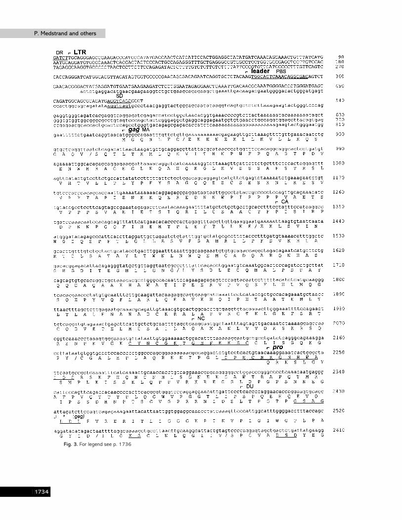
Fig. 3. For legend see p. 1736
BHDE

Human endogenous retrovirus HML-6 genomesHuman endogenous retrovirus HML-6 genomes
Fig. 3. For legend see p. 1736
BHDF

P. Medstrand and othersP. Medstrand and others
Fig. 3. Nucleotide sequence of a composite HERV-K (HML-6) genome. Amino acid sequences were translated from thenucleotide sequence and correspond to sequences encoded by the major retroviral reading frames (Gag, Pro, Pol and Env),where the start of the individual domains was decided from sequence alignments. Insertions/deletions causing a frameshift areindicated with a solidus and stops are shown with asterisks. The nucleotide sequence derived from clone HML-6.17 is shownin upper-case and encompasses positions 1–480 (5« LTR and the leader), and positions 3400–7538 (pol, env and 3« LTR).The nucleotide sequence derived from clone HML-6p is shown in lower-case (positions 369–3663; the leader region, gag, pro
BHDG

Human endogenous retrovirus HML-6 genomesHuman endogenous retrovirus HML-6 genomes
determined on both strands, and analysed by the PC-GENE software(Intelligenetics). The BLAST programs (Altschul et al., 1990) were usedfor searching the GenBank, EMBL and EST (Expressed Sequence Tags)databases.
ResultsCharacterization of HML-6 elements
HML-6 elements have previously only been described forthe pol region (Medstrand & Blomberg, 1993). To facilitate thescreening and characterization of genomic clones, we wantedto isolate a putative HML-6 gag region. A PCR-based screeningof human DNA resulted in expected 3±4 kb products whenamplifications were performed with a pol primer, ABDPOR, incombination with primers complementary to one of twoversions of lysine (CUU or UUU) PBSs, PBSLYS1 or PBSLYS3,respectively (Fig. 1). Cloning of these products, and subsequenthybridization using the HML-6 pol probe, revealed that HML-6 elements had a PBS complementary to lysine UUU. Anexpected 1±8 kb gag fragment was subsequently isolated byusing PCR with primer PBSLYS3 in combination with primerABDNC (Fig. 1). Sequence analysis of one 3±4 kb gag–polclone, HML-6p, revealed a close relationship to previouslydescribed HML-6 sequences in the pol region (see below andFigs 3 and 7).
Thus, HML-6 elements have the same PBS as the previouslyisolated NMWV4 HERV clone (May & Westley, 1986), fromwhich the LTR sequences have been determined. Sequenceswith similarity to the LTRs of NMWV4 were found in fiveEST sequences (accession numbers R27971, R42279,R65589, T11275 and T99775) and two GenBank entries(CHPMHCDRD and MACMHCCDRB) reported by Mayer etal. (1993). Sequence alignment revealed 60–80% identity tothe NMWV4 LTRs. Sequences for two oligonucleotides werechosen from conserved regions, and were used in parallel withthe HML-6 gag probe to characterize HML-6 pol-hybridizinggenomic clones.
Isolation of HML-6 genomic clones
The pol sequence HML-6.3 was used as a probe (Fig. 1) toscreen 8¬10& plaques under specific hybridization conditions.A total of 39 positive clones was identified. Digestion ofplaque-purified DNA from each clone with EcoRI indicated that25 had unique restriction patterns. The screening results werecompatible with the results obtained in a genomic Southernhybridization using the same probe, when about 25–30 discretebands at different hybridizing intensities in the range of0±6–18 kb were recognized (data not shown).
and pol). Overlapping HML-6.17/HML-6p sequences are aligned (positions 369–480 and positions 3400–3663). Underlinedamino acid sequence motifs represent the DNA-binding zinc-finger motifs in NC; the residues of the active site in DU; theactive site of the protease in PR; and the residues of the presumed active site of the polymerase in RT. DR, direct repeat ; PPT,polypurine tract ; MA, matrix ; CA, capsid ; NC, nucleocapsid ; DU, dUTPase; PR, protease; RT, reverse transcriptase; IN,integrase ; SU, surface ; TM, transmembrane.
In an attempt to identify full-length HML-6 clones,oligonucleotides LTR1 and LTR2 were hybridized to the 25immobilized HML-6 clones. One clone with two potential LTRregions was identified by probe LTR1, which recognized the0±8 and 4±1 kb EcoRI fragments of clone HML-6.17. Toelucidate whether this clone represented a full-length HML-6genome, the nucleotide sequences of the LTR-hybridizingfragments were determined.
The sequences of the 3« and 5« LTRs, derived from the 0±8and 4±1 kb EcoRI fragments of clone HML-6.17, respectively,are shown in Fig. 2 (a) in alignment with the NMWV4 3« LTR.The 3« LTR is 468 bp long and the 5« LTR is 329 bp long. Adeletion of 156 bp removed a central part of the 5« LTR, ascompared to the 3« LTR and the NMWV4 LTR. The twoHML-6.17 LTRs contained an additional terminal region of47 bp not present in the NMWV4 LTRs. There is 90% identitybetween the two HML-6.17 LTRs (on either side of the internaldeletion), but only 68% identity between the 3« LTRs of cloneHML-6.17 and NMWV4.
Motifs corresponding to a TATA box and a poly-adenylation signal are present on NMWV4 LTRs (May &Westley, 1986). In the 5« LTR of clone HML-6.17, the 156 bpdeletion spans both regions, and the TATA box was alsodeleted in the HML-6.17 3« LTR. Parts of a glucocorticoid-responsive element (GRE) (Scheidereit & Beato, 1984), fol-lowed directly by a sequence that is similar to an enhancer core,are present on HERV-K LTRs (Ono et al., 1986). Thejuxtaposition of these two elements creates a sequence withsimilarity to the binding site of a progesterone-receptorcomplex (Von der Ahe et al., 1985), which may account for thesteroid-inducible HERV-K transcription seen in T47D cells(Ono et al., 1987). A similar motif was present on the HML-6LTRs (Fig. 2a). A second, partial GRE is located on theNMWV4 LTR (indicated in Fig. 2a).
A PBS sequence motif sharing identity with 15 out of 18 bpcomplementary to a lysine tRNA with a UUU anticodon [thesame as for mouse mammary tumour virus (MMTV) andNMWV4] was present immediately downstream from the 5«LTR (Fig. 2b), whereas the same region was less comp-lementary (13 out of 18 bp) to the lysine tRNA with a CUUanticodon found in HERV-K clones and HERV-K (C4), or toany other known tRNA (Sprinzl et al., 1985). At position 135downstream of the 5« LTR, a sequence motif reminiscent of thesplice donor consensus was found (Fig. 2b).
Identification of full-length clones
To identify other potential full-length HML-6 clones, the0±8 kb LTR fragment of clone HML-6.17 was hybridized
BHDH

P. Medstrand and othersP. Medstrand and others
(a)
(b)
Fig. 4. Schematic representation of amino acid sequence similarity in (a) Gag and Pro, and (b) Pol and Env between HERV-K(HML-6p)/HERV-K (HML-6.17), HERV-K10 (Ono et al., 1986), HERV-K (C4) (Dangel et al., 1994) and MMTV (Moore et al.,1987). Shaded boxes represent stretches of similar amino acids between sequences, whereas open bars indicate regions oflow similarity. Alignments and statistical scorings were done with the MACAW software (National Center for BiotechnologyInformation, Bethseda, Md., USA), implementing the algorithm of Karlin & Altschul (1990). The sections used for calculatingamino acid sequence similarity (Table 1) between sequences are indicated below each alignment. A solid box in HML-6.17indicates the position of the HML-6 solitary LTR. Sequence alignments are available by request from the author. For a list ofabbreviations see Fig. 3.
against the 25 unique HML-6 clones. Five additional cloneswith two hybridizing EcoRI fragments were identified (clonesHML-6.4, -6.15, -6.26, -6.29 and -6.36). The LTR proberecognized, besides the expected 0±8 and 4±1 kb fragments ofclone HML-6.17, an additional EcoRI fragment of 2±3 kb, thesame size as detected by the pol probe. A restriction map ofclone HML-6.17 (see Fig. 5) revealed a 1±2 kb EcoRI fragmentsituated between the 2±3 kb pol LTR and 0±8 kb LTR-hybridizing fragments. Sequence analysis of the 2±3 kb subcloneshowed that an LTR was present within the pol-encodingregion of clone HML-6.17 (see below). This LTR had 78%similarity to the 3« LTR of clone HML-6.17, and 70% similarityto the NMWV4 3« LTR, and had flanking direct repeats oneither side. Such solitary LTRs have been described for severalHERV families (Mager & Goodchild, 1989 ; Leib-Mo$ sch et al.,1993 ; Dangel et al., 1994).
HML-6 sequences and similarity to other retroviruses
A composite HML-6 genome showing the continuoussequences of the 2±3 kb (pol), 1±2 kb (env) and 0±8 kb (3« LTR)EcoRI fragments of clone HML-6.17, preceded by the 3±4 kbgag–pol sequence of clone HML-6p and the 5« LTR of clone
HML-6.17, is shown in Fig. 3. The sequence derived from cloneHML-6p overlaps clone HML-6.17 with 111 bp in the leaderregion and 262 bp in the pol region. The overlapping regionsdisplay 80 and 87% nucleotide sequence identity, respectively,indicating that they belong to the same element family (seebelow and Fig. 7). In this prototype genome, the gag regionstarts 589 bp downstream of the 5« LTR, and is followed bysequences encoding the pro, pol and env regions. Typicalretrovirus motifs are present in the coding regions (underlinedin Fig. 3) : for example, the NC region of gag encodes the twoDNA-binding zinc-fingers ; the pro region encodes conservedmotifs of the active sites of dUTPase (DU) and protease (PR) ;and the pol region encodes the motif corresponding to thepresumed active site of (RT). A schematic representationderived from amino acid sequence alignments of HERV-K(HML-6), HERV-K10, HERV-K (C4) and MMTV shows similarregions in the four retroviral elements (Fig. 4). The genomeorganization is the same as for clone HERV-K10 and type Band D retroviruses, and is probably of the same origin due tothe presence of DU at the same position. HERV-K (HML-6p)shows Gag and Pro domains similar to those in the otherelements, but has a short matrix protein (MA) in comparison to
BHDI

Human endogenous retrovirus HML-6 genomesHuman endogenous retrovirus HML-6 genomes
Table 1. Similarity of HERV-K (HML-6) to other retroviruses
The table shows the percentage amino acid sequence identity of retroviral sequences to HERV-K (HML-6p) inGag and Pro, and to HERV-K (HML-6.17) in Pol and Env. The highest score for each domain is shown inbold type. Identities were calculated over the positions indicated in Fig. 4.
HERV-K10 HERV-K (C4) MMTV* IAPm† MPMV‡
Gag 33±9 – 29±8 29±9 32±5Pro 39±8 – 36±9 29±2 35±7Pol 49±1 41±8 42±6 40±3 44±8Env 30±1 22±7 28±3 – 11±4
* MMTV, mouse mammary tumour virus (Moore et al., 1987).† IAPm, mouse intracisternal A-particle (Mietz et al., 1987).‡ MPMV, Mason–Pfizer monkey virus (Sonigo et al., 1986).
Fig. 5. Restriction maps of HERV-K (HML-6) genomes showing the restriction sites for BamHI (B), EcoRI (E), HindIII (H), NheI(N) and SstI (S). HML clone names are shown to the left of each map. HindIII restriction sites were not determined for clonesHML-6.15 and HML-6.29. Common restriction sites between clones are indicated either with open or solid arrowheads. Theorientation of the clones was determined on the basis of hybridizing fragments using the probes shown below the map of cloneHML-6.17. The bars below each map correspond to restriction fragments that hybridize with the respective probes. The solidboxes on either side of each restriction map indicate the cloning cassette of the λGEM-12 arms.
HERV-K10 and MMTV. HERV-K (HML-6.17) also displaysconserved motifs in Pol, except for a region between theRNase H and integrase (IN) domains, where the HML-6solitary LTR is located. As is evident in Fig. 4, most of the
surface protein (SU) of HERV-K (HML-6.17) is missing,whereas the carboxy-terminal part of SU and the trans-membrane (TM) glycoprotein are similar to the other retroviralelements.
BHDJ

P. Medstrand and othersP. Medstrand and others
(a) (b)
Fig. 6. Southern blot hybridizations of DNA from a human male (lanes 1), DNA from two different females (lanes 2 and 3), andplacental DNA (lanes 4) digested with restriction enzymes indicated and hybridized with (a) the env probe, or (b) the LTRprobe, and exposed to X-ray film for 36 h and 20 h, respectively. Sizes of co-migrated λHindIII restriction fragments (kb) areindicated.
The Gag-, Pro- and Pol-encoding domains show similarityto those of type A, B and D and class II HERVs (29–49% aminoacid identity), whereas the Env domain shows similarity toMMTV (type B) and the HERV-K10 and HERV-K (C4)elements (23–30% amino acid identity), but not to Mason–Pfizer monkey virus (MPMV; 11% identity). In all codingregions, the HML-6 elements share the greatest amino acididentities to clone HERV-K10 (Table 1). Despite the presenceof retroviral structural protein-encoding regions, all containeddeletions, nonsense and frameshift mutations. The TM domainof Env was the least defective, containing only one stop codon.
Genomic organization of HML-6 elements
For a comparison of the HML-6 clones isolated, cloneHML-6.17 and five other clones that had two LTR-hybridizingEcoRI fragments which were identified by the LTR probe ofclone HML-6.17 (HML-6.4, -6.15, -6.26, -6.29 and -6.36) weremapped with restriction enzymes. The restriction map ofHML-6.4 showed that this clone, similar to clone HML-6.17,contained a solitary LTR in the pol region, flanked on eitherside by gag- and env-hybridizing fragments. A 3« LTR was notpresent. Despite an LTR in the pol region of both HML-6.4 andHML-6.17, the restriction maps of the two differed both in theretroviral and flanking cellular DNA. A short region in pol
displayed about 10% nucleotide sequence divergence betweenthe two (not shown). These data indicate that the clonesrepresent unique HML-6 proviruses. From the restriction mapsof the remaining five clones (Fig. 5), the HML-6 elementsevidently make up a rather diverse family of HERVs. Only afew restriction enzyme sites were common among the clones(as indicated in Fig. 5). All full-length elements showed atypical retrovirus structure, with hybridizing fragments in theorder gag, pol and env, flanked by LTR-hybridizing fragments.The HML-6.17 5« LTR made up the last 680 bp of the 4±1 kbEcoRI fragment, and the pol–env–LTR regions encompassed3674 bp (excluding the solitary LTR). Since the interveninggag-hybridizing EcoRI fragment is about 2±6 kb, the size ofHML-6.17 was estimated to be 6±9 kb.
The mapped clones all contained regions of similarity toenv. To see whether this was a general feature of the HML-6elements, Southern hybridization was done with the env probe.In addition, because two of the clones described here containedsolitary LTRs within the pol region, we wanted to see to whatdegree such LTRs were dispersed in human DNA.
The env probe (Fig. 6a) resulted in a similar pattern ofhybridizing fragments for each set of four enzyme-digestedlanes of DNA. In the EcoRI digests, approximately 20–25hybridizing fragments of different intensities were observed,whereas there were about 15 hybridizing HindIII fragments,
BHEA

Human endogenous retrovirus HML-6 genomesHuman endogenous retrovirus HML-6 genomes
Fig. 7. Unrooted tree derived from alignment of the nucleotide sequencesof each full-length HERV-K (HML-6) element. The published sequences ofHERV-K10 (Ono et al., 1986; positions 4102–4345), HERV-K (C4)(Dangel et al., 1994; positions 1459–1679), MMTV (Moore et al., 1987;positions 4274–4517), IAPm (Mietz et al., 1987; positions 3450–3493), HML-6.1, HML-6.2 and HML-6.3 (Medstrand & Blomberg, 1993)were included for comparison. The tree was constructed by using theprograms DNADIST, KITSCH and DRAWTREE of the PHYLIP programpackage. Sequence alignments are available by request from the author.
but these were slightly more intense. The same blot washybridized with the pol probe. The number of bands was aboutthe same as estimated for the env hybridization, and it istherefore likely that most HML-6 elements contain an envgene. The only variation seen, apart from the generalhybridizing pattern with the env probe, was an additionalhybridizing HindIII fragment of about 2±5 kb in DNA from onefemale (Fig. 6a).
The LTR probe was expected to cause a more intensehybridization on the blot due to the presence of two LTRs perHML-6 element. Hybridization with the LTR probe resulted ina more intense hybridization than with the pol and env probes(Fig. 6b), but due to the many hybridizing fragments it was notpossible to determine the LTR copy number. To estimate this,we screened about 2±5¬10& plaques (2¬10& plaques equalabout one human genome) of the human genomic library withthe LTR probe, which after medium-stringency washingconditions resulted in 140 LTR-hybridizing clones. Southernblot analyses with pol and env probes indicated that humansharbour about 30–40 pol–env-hybridizing HML-6 elements.Based on these results, we estimated that about 50 additionalHML-6-related LTRs are present per human haploid genome.
Sequence diversity among HML-6 elements
To estimate the diversity among HML-6 elements, thenucleotide sequence of a region within the RT-encodingdomain of each full-length clone was determined. A treedepicting the nucleotide sequence divergence of the HML-6clones is shown in Fig. 7, together with three previouslydescribed HML-6 RT sequences, HML-6.1 to -6.3 (Medstrand& Blomberg, 1993), and HERV-K10, HERV-K (C4), MMTVand mouse intracisternal A-particle (IAPm), to which the HML-
6 elements displayed 54–63% nucleotide similarity. Althoughthe HML-6 sequences diverged by as much as 20%, they madeup a distinct collection of related sequences compared to therest. The HML-6.36 RT sequence was identical to that ofHML-6.1, and clone HML-6.17 differed by only one nucleotidefrom the HML-6.3 sequence. This single mismatch could havebeen generated during PCR by Taq polymerase (Gelfand &White, 1990). The remaining clones (HML-6p, -6.15, -6.26 and-6.29) were 84–87% similar to HML-6.17. All HML-6 cloneshad either stops or additional insertions}deletions disruptingtheir putative ORFs. In addition, the ORF of clone HML-6.29was disrupted by insertion of an Alu element.
DiscussionWe have in this study characterized a novel class II
HERV family. All class II HERVs identified so far fall intodistinct groups based on sequence similarity (Ono et al., 1986 ;Franklin et al., 1988 ; Medstrand & Blomberg, 1993 ; Dangel etal., 1994 ; Tassabehji et al., 1994). The initial approach used tocharacterize members of this family was a PCR-based screeningmethod with primers corresponding to conserved retroviral polregions (Medstrand & Blomberg, 1993). Here, initial analysisshowed that HML-6 elements contain a PBS complementary tolysine tRNA UUU, and sequence analysis of clone HML-6prevealed retroviral gag, pro and pol regions. The full-lengthclone HML-6.17 revealed a typical proviral DNA structure,with LTR sequences and regions showing similarity to retro-viral pol and env. Sequences of both these clones are related totype A, B and D mammalian retroviruses, showing highest con-servation in the pol region (Table 1). However, terminationcodons and deletions disrupt the reading frames (Fig. 3).Similarity in the env region is a good indicator of relatednessand evolutionary history of retroviruses (McClure et al., 1988).Based upon this criterion, Wilkinson et al. (1994) assignedseveral type-C-related HERVs to the HERV-ERI superfamily.The presence of a sequence motif complementary to a lysinetRNA, a genome structure, and an env region similar to type Bretroviruses suggests that HML-6 elements belong to theHERV-K superfamily.
HERV-K (HML-6.17) was estimated to be a 6±9 kb element.The env region of this clone was truncated in comparison toMMTV and HERV-K10. Restriction mapping of each full-length clone showed that all elements had a typical retrovirusstructure (Fig. 5). This general structure is probably true formost HML-6 elements, due to the presence of approximatelythe same number of hybridizing pol and env fragments in theSouthern blot analyses. Sequence analysis in the RT regionfrom each clone revealed that members within the HML-6family constitute a heterogeneous, but distinct family (Fig. 7).The sequence divergence was as much as 20% in RT amongHML-6 elements, whereas identity to HERV-K10 and HERV-K (C4) did not exceed 65%. Therefore, differences foundbetween the pol and env regions of HML-6 elements and those
BHEB

P. Medstrand and othersP. Medstrand and others
of other HERVs suggest that HML-6 elements are distinct classII HERVs.
Sequencing of the two fragments containing putative LTRsconfirmed the hybridization result. The two HML-6.17 LTRswere 329 and 468 bp long, respectively (Fig. 2a) and displayed90% identity, the same diversity as the two NMWV4 LTRs(May & Westley, 1986). Differences between LTR sequencesof endogenous retroviruses are well documented (Wilkinson etal., 1994), and the base changes observed probably arose afterinsertion of the provirus into the human genome. Based on aneutral divergence rate, base changes have been used toestimate the evolutionary age of proviruses (Mager & Freeman,1995). GenBank searches resulted in the identification ofNMWV4}HML-6 LTRs in the genomes of chimpanzee andrhesus macaque (Macaca mulatta) (Mayer et al., 1993). Mayer etal. (1993) also identified a region upstream of the LTR in thechimpanzee clone, Patr-DRB6 LTR, which showed sequencesimilarity to MMTV env. Sequence comparison between thisregion in Patr-DRB6 LTR and HML-6.17 env displayed asmuch as 90% identity. Evidently, Patr-DRB6 LTR, NMWV4and HML-6.17 belong to the same family of retroviralelements, despite relatively divergent LTRs. The presence ofhomologues of HML-6.17 LTRs in Old World monkeys(Mayer et al., 1993) and the 10% divergence between each LTRof NMWV4 and HML-6.17 (cf. Mager & Freeman, 1995),indicate that HML-6-like elements entered the primate lineagemore than 30 million years ago.
A sequence motif corresponding to the putative GRE ofHERV-K elements was identified in these LTRs. NMWVsequences are abundantly expressed in breast cancer cell lines(e.g. T47D) and in the placenta (Franklin et al., 1988).Expression of these HERV sequences may be influenced byhormones, as has been shown for HERV-K (Ono et al.,1987 ;Franklin et al., 1988). In addition, HML-6 sequences aredifferentially expressed in liver, lung (Medstrand & Blomberg,1993) and PBMCs (Andersson et al., 1996), suggesting thatsome transcriptional control directs the observed differentialtissue-specific expression (Andersson et al., 1996).
Studies on the distribution of retroelements have revealedthat host-genome integration sites are not completely random(Sandmeyer et al., 1990). A preference for integration intotranscribed regions in the genome or in clusters near otherHERVs and transposable elements has been observed (Rogers,1985 ; Taruscio&Manuelidis, 1991). However, different classesof retroelements show specific regional integration patterns(Taruscio & Manuelidis, 1991), suggesting that some in-tegration site or target-specific preferences exist. An interestingexample of non-random integration of retroelements intospecific regions involves the primate haptoglobin locus, wherethree independent HERV-I insertions have occurred during thecourse of evolution (Maeda & Kim, 1990). In our study, wehave found solitary LTRs of the same HML-6 family integratedwithin the genomes of HML-6.4 and HML-6.17, whichsuggests that some site preference, as in the haptoglobin
region for HERV-I, may be involved in integration. The copynumber for different retroelements varies from a few up tothousands per haploid genome (Li & Graur, 1991). Theintegration target-specificity may account for some of the copynumber differences seen for different retroelements. Further-more, insertion into previously integrated proviruses isprobably non-deleterious for the host, and such selectiveintegration may be due to a symbiotic host–virus relationshipestablished over a long period of time.
In the case of the LTRs within the two HML-6 clones, a full-length HML-6 element presumably integrated and wasfollowed by an intrachromosomal recombination event, re-sulting in a solitary LTR. The presence of host-cell-target directrepeats on either side of the solitary LTR in HML-6.17supports this hypothesis (Mager & Goodchild, 1989). HERV-K10-related solitary LTRs are present at about 25000 copies inthe human genome (Leib-Mo$ sch et al., 1993), whereas solitaryLTRs of HERV-K (HML-6) (this study) and HERV-K (C4)(Dangel et al., 1994) families are present at much lower copynumbers.
Insertions of retroelements may alter gene function in thehost by a variety of mechanisms and may result in disordersdue to inactivation or promotion of cellular genes (Favor &Morawetz, 1992). By searching GenBank, we identified asolitary HML-6 LTR present in the 3« end of a human MHCclass I-like cDNA, MICB. MICB is transcribed at low levels incomparison to the related gene product MICA. The down-regulation was suggested to be the result of a disruption of thepolyadenylation site of MICB (Bahram & Spies, 1996). Thesolitary HML-6 LTR is integrated in the polyadenylation siteof this gene, thus explaining the disruption, and possibly thelow abundance of MICB mRNA. However, alternative poly-adenylation signals are located downstream of the original site.These are used but are apparently not as efficiently as thecorresponding site in MICA. Other examples suggest thatretroelements facilitate novel host-organism adaptations,where expression of cellular genes is controlled and modulatedby retroelements (for review see Wilkinson et al., 1994). Onemechanism is represented by the HML-6-related Patr-DRB6LTR (Mayer et al., 1993). Despite a deletion causing a frameshiftin exon 1, DRB6 is transcribed at low levels (Corell et al., 1991).Mayer et al. (1993) showed that transcription was initiated inan LTR located upstream of exon 1. Splicing from the LTRdirectly in-frame into exon 2 of DRB6 led to the expression ofthis gene, where the first amino acids of the presumedpolypeptide are encoded by the LTR. Mayer et al. (1993) alsodemonstrated that transcription initiation took place at aTATA region (corresponding to positions 50–60 in Fig. 2a)that was different to the presumed TATA box on the NMWV4LTR (position 205, Fig. 2a), which indicates that alternativeTATA boxes may direct transcription from deleted LTRs suchas HML-6.17.
Several studies have shown that retroelements may act asmutagens resulting in disorders or as driving forces in genetic
BHEC

Human endogenous retrovirus HML-6 genomesHuman endogenous retrovirus HML-6 genomes
variation. Investigations regarding the potential of HERVs insuch events are emerging, and further studies on the biologyand evolution of these elements will shed light on theirinvolvement in genetical and pathological processes.
We thank Doug Freeman for helpful discussions, Mats Lindeskog forproviding the PBS primers used and for helpful discussions, and Per Almand Jolanta Juraszczyk for technical assistance. This work was in partsupported by the European Commission, project GENE-CT930019 ;funds at the Medical Faculty of Lund ; the Royal PhysiographicFoundation ; the Crafoord Foundation, Lund ; the O> sterlund Foundation,Malmo$ ; by a grant to P.M. from the Medical Research Council, Sweden ;and in part by a grant to D.M. from the Medical Research Council ofCanada.
ReferencesAltschul, S. F., Gish, W., Miller, W., Myers, E. W. & Lipman, D. J.(1990). Basic local alignment search tool. Journal of Molecular Biology215, 403–410.
Andersson, M.-L., Medstrand, P., Yin, H. & Blomberg, J. (1996).Differential expression of human endogenous retroviral sequences similarto mouse mammary tumor virus in normal peripheral blood mononuclearcells. AIDS Research and Human Retroviruses 12, 833–840.
Ausubel, F. M., Brent, R., Kingston, R. E., Moore, D. D., Seidman, J. G.,Smith, J. A. & Struhl, K. (1987). Current Protocols in Molecular Biology.New York : John Wiley & Sons.
Bahram, S. & Spies, T. (1996). Nucleotide sequence of a human MHCclass I MICB cDNA. Immunogenetics 43, 230–233.
Boller, K., Ko$ nig, H., Sauter, M., Mueller-Lantzsch, N., Lo$ wer, R.,Lo$ wer, J. & Kurth, R. (1993). Evidence that HERV-K is the endogenousretrovirus sequence that codes for the human teratocarcinoma-derivedretrovirus HTDV. Virology 196, 349–353.
Callahan, R. (1988). Two families of human endogenous retroviralgenomes. Eukaryotic transposable elements as mutagenic agents. BanburyReport 30, 91–100.
Corell, A., Martin-Villa, J. M., Morales, P., De Juan, M. D., Varela, P.,Vicario, J. L., Martinez-Laso, J. & Arnaiz-Villena, A. (1991). Exon-2nucleotide sequences, polymorphism and haplotype distribution of a newHLA–DRB gene : HLA–DRB sigma. Molecular Immunology 28, 533–543.
Dangel, A. W., Mendoza, A. R., Baker, B. J., Daniel, C. M., Carroll,M. C., Wu, L.-C. & Yu, C. Y. (1994). The dichotomous size variationof human complement C4 genes is mediated by a novel family ofendogenous retroviruses, which also establishes species–species genomicpatterns among Old World primates. Immunogenetics 40, 425–436.
Eschenfeldt, W. H., Puskas, R. S. & Berger, S. L. (1987). Homo-polymeric tailing. Methods in Enzymology 152, 337–342.
Favor, J. & Morawetz, C. (1992). Insertional mutations in mammals andmammalian cells. Mutation Research 284, 53–74.
Franklin, G. C., Chretien, S. C., Hanson, I. M., Rochefort, H., May, F. E.B. & Westley, B. R. (1988). Expression of human sequences related tothose of mouse mammary tumor virus. Journal of Virology 62, 1203–1210.
Gelfand, D. H. & White, T. J. (1990). Thermostable DNA polymerases.In PCR Protocols – A Guide to Methods and Applications, pp. 129–141.Edited by M. A. Innis, D. H. Gelfand, J. J. Sninsky & T. J. White. SanDiego : Academic Press.
Goodchild, N. L., Freeman, J. D. & Mager, D. L. (1995). Spliced HERV-H endogenous retroviral sequences in human genomic DNA: evidencefor amplification via retrotransposition. Virology 206, 164–173.
Karlin, S. & Altschul, S. F. (1990). Methods for assessing the statisticalsignificance of molecular sequence features by using general scoringschemes. Proceedings of the National Academy of Sciences, USA 87,2264–2268.
Kidd, A. H., Erasmus, M. J. & Tiemessen, C. T. (1990). Fiber sequenceheterogeneity in subgroup F adenoviruses. Virology 179, 139–150.
Leib-Mo$ sch, C., Haltmeier, M., Werner, T., Geigl, E.-M., Brack-Werner,R., Francke, U., Erfle, V. & Hehlmann, R. (1993). Genomic distributionand transcription of solitary HERV-K LTRs. Genomics 18, 261–269.
Li, W.-H. & Graur, D. (1991). Evolution by transposition. In Fundamentalsof Molecular Evolution, pp. 172–203. Sunderland, Mass. : SinauerAssociates.
Lo$ wer, J., Wondrak, E. M. & Kurth, R. (1987). Genome analysis andreverse transcriptase activity of human teratocarcinoma-derived retro-viruses. Journal of General Virology 68, 2807–2815.
Lo$ wer, R., Boller, K., Hasenmaier, B., Korbmacher, C., Mu$ ller-Lantzsch, N., Lo$ wer, J. & Kurth, R. (1993). Identification of humanendogenous retroviruses with complex mRNA expression and particleformation. Proceedings of the National Academy of Sciences, USA 90,4480–4484.
Lo$ wer, R., Lo$ wer, J. & Kurth, R. (1996). The virus in all of us :characteristics and biological significance of human endogenous retro-virus sequences. Proceedings of the National Academy of Sciences, USA 93,5177–5184.
McClure, M. A., Johnson, M. S. & Doolittle, R. F. (1988). Sequencecomparison of retroviral proteins : relative rates of change and generalphylogeny. Proceedings of the National Academy of Sciences, USA 85,2469–2473.
Maeda, N. & Kim, H.-S. (1990). Three independent insertions ofretrovirus-like sequences in the haptoglobin gene cluster of primates.Genomics 8, 671–683.
Mager, D. L. & Freeman, J. D. (1995). HERV-H endogenous retrovirus :presence in the New World branch but amplification in the Old Worldprimate lineage. Virology 213, 395–404.
Mager, D. L. & Goodchild, N. (1989). Homologous recombinationbetween the LTRs of a human retrovirus-like element causes a 5 kbdeletion in two siblings. American Journal of Human Genetics 45, 848–854.
May, F. E. B. & Westley, B. R. (1986). Structure of a human retroviralsequence related to mouse mammary tumor virus. Journal of Virology 60,743–749.
Mayer, W. E., O’Huigin, C. & Klein, J. (1993). Resolution of theHLA–DRB6 puzzle : a case of grafting a de novo-generated exon on anexisting gene. Proceedings of the National Academy of Sciences, USA 90,10720–10724.
Medstrand, P. & Blomberg, J. (1993). Characterization of novel reversetranscriptase encoding human endogenous retroviral sequences similar totype A and type B retrovirus : differential transcription in normal humantissues. Journal of Virology 67, 6778–6787.
Medstrand, P., Lindeskog, M. & Blomberg, J. (1992). Expression ofhuman endogenous retroviral sequences in peripheral blood mononuclearcells of healthy individuals. Journal of General Virology 73, 2463–2466.
Mietz, J. A., Grossman, Z., Lueders, K. K. & Kuff, E. L. (1987).Nucleotide sequence of a complete mouse intracisternal A-particlegenome : relationship to known aspects of particle assembly and function.Journal of Virology 61, 3020–3029.
Moore, R., Dixon, M., Smith, R., Peters, G. & Dickson, C. (1987).Complete nucleotide sequence of a milk-transmitted mouse mammarytumor virus : two frameshift suppression events are required fortranslation of gag and pol. Journal of Virology 61, 480–490.
BHED

P. Medstrand and othersP. Medstrand and others
Mueller-Lantzsch, N., Sauter, M., Weiskircher, A., Kramer, K., Best, K.,Buck, M. & Gra$ sser, F. (1993). Human endogenous retroviral elementK10 (HERV-K10) encodes a full-length Gag homologous 73-kDa proteinand a functional protease. AIDS Research and Human Retroviruses 9,343–350.
Ono, M., Yasunaga, T., Miyata, T. & Ushikubo, H. (1986). Nucleotidesequence of human endogenous retrovirus genome related to the mousemammary tumor virus genome. Journal of Virology 60, 589–598.
Ono, M., Kawakami, M. & Ushikubo, H. (1987). Stimulation ofexpression of the human endogenous retrovirus genome by femalesteroid hormones in human breast cancer cell line T47D. Journal ofVirology 61, 2059–2062.
Patience, C., Simpson, G. R., Colletta, A. A., Welch, H. M., Weiss, R. A.& Boyd, M. T. (1996). Human endogenous retrovirus expression andreverse transcriptase activity in the T47D mammary carcinoma cell line.Journal of Virology 70, 2654–2657.
Rogers, J. (1985). The origin and evolution of retrotransposons.International Review of Cytology 93, 187–279.
Sandmeyer, S., Hansen, L. & Chalker, D. (1990). Integration specificityof retrotransposons and retroviruses. Annual Review of Genetics 24,491–518.
Sambrook, J., Fritsch, E. F. & Maniatis, T. (1989). Molecular Cloning : ALaboratory Manual, 2nd edn. Cold Spring Harbor, NY: Cold SpringHarbor Laboratory.
Scheidereit, C. & Beato, M. (1984). Contacts between hormonereceptor and DNA double helix within a glucocorticoid regulatoryelement of mouse mammary tumor virus. Proceedings of the NationalAcademy of Sciences, USA 82, 3029–3033.
Seifarth, W., Skladny, H., Krieg-Schneider, F., Reichert, A., Hehlmann,R. & Leib-Mo$ sch, C. (1995). Retrovirus-like particles released fromhuman breast cancer cell line T47D display type B- and C-relatedendogenous retroviral sequences. Journal of Virology 69, 6408–6416.
Shapiro, M. B. & Senapathy, P. (1987). RNA splice junctions ofdifferent classes of eukaryotes : sequence statistics and functionalimplications in gene expression. Nucleic Acids Research 15, 7155–7174.
Sonigo, P., Barker, C., Hunter, E. & Wain-Hobson, G. (1986).Nucleotide sequence of Mason–Pfizer monkey virus : an immuno-suppressive D-type retrovirus. Cell 45, 377–385.
Sprinzl, M., Moll, J., Messner, F. & Hartmann, T. (1985). Compilationof tRNA sequences. Nucleic Acids Research 13, 1–49.
Taruscio, D. & Manuelidis, L. (1991). Integration site preferences ofendogenous retroviruses. Chromosoma 101, 141–156.
Tassabehji, M., Strachan, T., Anderson, M., Campbell, R. D., Collier, S.& Lako, M. (1994). Identification of a novel family of humanendogenous retroviruses and characterization of one family member,HERV-K (C4), located in the complement C4 gene cluster. Nucleic AcidsResearch 22, 5211–5217.
Ting, C.-N., Rosenberg, M. P., Snow, C. M., Samuelson, L. & Meisler,M. H. (1992). Endogenous retroviral sequences are required for tissue-specific expression of a human salivary amylase gene. Genes &Development 6, 1457–1465.
Von der Ahe, D. S., Janich, S., Scheidereit, C., Renkawitz, G., Schutz,G. & Beato, M. (1985). Glucocorticoid and progesterone receptors bindthe same site in two hormonally regulated promoters. Nature 313,706–709.
Wallace, M. R., Anderson, L. B., Saulino, A. M., Gregory, P. E., Glover,T. W. & Collins, F. S. (1991). A de novo Alu insertion results inneurofibromatosis type 1. Nature 353, 864–866.
Wilkinson, D. A., Mager, D. L. & Leong, J. C. (1994). Endogenoushuman retroviruses. In The Retroviridae, vol. 3, pp. 465–535. Edited byJ. Levy. New York : Plenum Press.
Received 8 November 1996; Accepted 5 February 1997
BHEE