Embed Size (px)
Citation preview

Structure- and sequence-based function predictionfor non-homologous proteins
Lee Sael • Meghana Chitale • Daisuke Kihara
Received: 12 September 2011 / Accepted: 10 January 2012
� Springer Science+Business Media B.V. 2012
Abstract The structural genomics projects have been
accumulating an increasing number of protein structures,
many of which remain functionally unknown. In parallel
effort to experimental methods, computational methods are
expected to make a significant contribution for functional
elucidation of such proteins. However, conventional com-
putational methods that transfer functions from homolo-
gous proteins do not help much for these uncharacterized
protein structures because they do not have apparent
structural or sequence similarity with the known proteins.
Here, we briefly review two avenues of computational
function prediction methods, i.e. structure-based methods
and sequence-based methods. The focus is on our recent
developments of local structure-based and sequence-based
methods, which can effectively extract function informa-
tion from distantly related proteins. Two structure-based
methods, Pocket-Surfer and Patch-Surfer, identify similar
known ligand binding sites for pocket regions in a query
protein without using global protein fold similarity infor-
mation. Two sequence-based methods, protein function
prediction and extended similarity group, make use of
weakly similar sequences that are conventionally discarded
in homology based function annotation. Combined together
with experimental methods we hope that computational
methods will make leading contribution in functional elu-
cidation of the protein structures.
Keywords Computational protein function prediction �Structure-based function prediction � Sequence-based
function prediction � Ligand binding pocket comparison �Protein surface shape comparison � Pocket-Surfer �Patch-Surfer � Weakly similar sequences
Abbreviations
PDB Protein Data Bank
3DZD 3 Dimensional Zernike descriptor
ATP Adenosine triphosphate
HEM Heme
NAD Nicotinamide adenine dinucleotide
FAD Flavin adenine dinucleotide
BTN Biotin
F6P Fructose 6-phosphate
GUN Guanine
PLM Palmitic acid
RTL Retinol
AUC Area under the curve
ROC Receiver operator characteristic
EF Enrichment factor
GO Gene ontology
PFP Protein function prediction
ESG Extended similarity group
AFP-SIG Automatic Function Prediction Special Interest
Group
ISMB Intelligent System in Molecular Biology
CASP Critical Assessment of Techniques for Protein
Structure Prediction
Lee Sael and Meghana Chitale contributed equally to this article.
L. Sael � M. Chitale � D. Kihara (&)
Department of Computer Science, Purdue University, West
Lafayette, IN 47907, USA
e-mail: [email protected]
L. Sael � D. Kihara
Department of Biological Sciences, Purdue University, West
Lafayette, IN 47907, USA
L. Sael � D. Kihara
Markey Center for Structural Biology, Purdue University, West
Lafayette, IN 47907, USA
123
J Struct Funct Genomics
DOI 10.1007/s10969-012-9126-6

Introduction
The structural genomics projects worldwide have deter-
mined an increasing number of protein tertiary structures
over a decade [1–4]. As of writing of this article (September
2011), there are over 9,000 structures in the Protein Data
Bank (PDB) [5, 6] that were deposited from the structural
genomics projects. Among several objectives of these large-
scale efforts, one of the major expectations is that the
determined structures provide clues for elucidating evolu-
tion and function of the proteins [7, 8]. In fact, function of
some targeted proteins in the projects have been elucidated
by global structure similarity to known characterized pro-
teins [9–12] or by a combination of structural comparison
and other sources, such as identified bound cofactors to the
structure [13] or biochemical experimental evidences [14].
Protein function should be ultimately investigated
experimentally. There are indeed efforts towards system-
atic functional screening [15, 16] and also efforts to com-
bine computational and experimental function assignments
in a genome-scale [7, 17]. In parallel to such experimental
method developments, computational function prediction
methods are expected to play an important role in post-
structural genomics functional elucidation given the fact
that computational methods can quickly screen existing
data, which is the basis of transferring function from
known proteins. Computational methods alone can often
sufficient evidences for inferring function of proteins [9–
12]. Also Experiments can be greatly benefited if possible
functions of uncharacterized proteins are suggested by
computational methods [17].
However, conventional computational methods which
transfer function from obvious homologous proteins, such
as BLAST [18, 19] or FASTA [20] or domain searches
[21], leave many protein structures with unknown function,
as evidenced in many structures with unknown function
deposited to PDB from the structural genomics projects.
Methods that predict function from globally similar pro-
teins work well when highly similar known proteins exist
in the database. Protein structures solved by the structural
genomics efforts, which are left behind in functional
annotation, do not have apparent global similarity to any of
known proteins. Therefore, to increase the annotation
coverage, it is crucial to develop methods which can use
local structure similarity or distantly related proteins.
In this article, we briefly review two avenues of com-
putational function prediction methods, i.e. structure-based
and sequence-based methods. The focus is on our recently
developed function prediction methods, local structure-
based methods and sequence-based methods, which can
effectively extract function information from distantly
related proteins that are discarded by conventional meth-
ods. For more general review of computational function
prediction, readers are referred to recent comprehensive
reviews [22–24].
Approaches for structure-based function prediction
Computational structure-based function prediction methods
transfer function of known proteins to a query protein if the
known proteins have global or local structural similarity to
the query. Structure-based function prediction methods can
be divided into global and local approaches. In principle,
global methods aim to find distantly related proteins of the
same fold to a query protein that are not detectable by
considering the sequence similarity [25–27]. Any methods
developed for protein structure comparison can be used for
this task, e.g. the Combinatorial Extension method [28],
Dali [29], SSAP [30], VAST [31], and COSEC [32]. More
recently, moment-based methods, the spherical harmonics
and the 3D Zernike descriptors (3DZD), have been applied
to describe protein surfaces [33–38]. Moment-based
methods describe a protein surface as a series expansion of
a 3D mathematical function that represents the protein
surface shape. Thus, a structure is compactly represented as
a vector of coefficients of the series function, which allows
a fast real-time structure database search. We showed that
some functional class of proteins, e.g. DNA binding pro-
teins, can be detected by surface comparison using the
3DZD, because the descriptors capture the surface shape
similarity that is required for the function of the proteins
(e.g. saddle like shape of DNA binding regions in DNA
binding proteins) [35]. Although global structure similarity
indicates functional similarity of proteins in most of the
cases, one needs to keep in mind that there are notable
exceptions of ‘‘superfolds’’ [39], which are commonly
occurring protein folds that are adopted by various protein
families. Thus, conservation of functional residues needs to
be confirmed before transferring function to a query protein
from a known protein of the same global fold.
In contrast to the global structure comparison methods,
local structure-based approaches compare local regions of
a query protein to a database of known functional sites, e.g.
active sites of enzymes. The Catalytic Site Atlas [40], AFT
[41], ASSAM [42], SPASM [43], SURFACE [44], FLORA
[45], CavBase [46], and SitesBase [47] search local sites
with a set of residues/atoms in a query protein that match
with known functional sites. Another method, eF-seek [48],
represents a protein surface as a graph with nodes char-
acterized with local geometry and electrostatic potentials,
and local regions in a query protein that are similar to
known functional sites are sought by a sub-graph matching
algorithm. Thornton and her colleagues explored the use of
the spherical harmonics in representing and comparing
protein pockets [49]. Binkowski et al. developed a method
L. Sael et al.
123

that characterize a pocket with conserved residues at the
pocket [50, 51] and also with its pairwise atom distances
[52]. The ProFunc server performs various structure- and
sequence-based function predictions for a query protein
structure ranging from sequence and structural motif
searches, active site identification, and global fold com-
parison [53]. Proknow is another method that integrates
multiple evidences for function prediction, such as
sequence and structure similarity and protein–protein
interactions [54]. The advantage of the local structure-
based methods over the global structure-based methods is
that the former could identify functional sites in a query
protein even when the query does not have evolutionary
closely related proteins in a database. To meet the urgent
task of annotating protein structures solved by the struc-
tural genomics projects that do not have apparent homol-
ogy to known proteins, we have recently developed local
structure-based methods, Pocket-Surfer [55] and Patch-
Surfer [56, 57]. These two methods will be overviewed in
the next section.
Pocket-Surfer and Patch-Surfer
Pocket-Surfer [55] and Patch-Surfer [56, 57] predict ligand
molecules that bind to a query protein by comparing the
geometrical shape and the physicochemical properties of a
pocket region of the query protein to a database of known
binding pockets. Both methods allow a quick real-time
scan of the database of binding pockets since they use a
rotational invariant pocket representation, which does not
require time consuming pre-alignment of pockets upon
comparison. Technically, the rotational invariant repre-
sentation is achieved using the 3DZD, a rotation invariant
series expansion of a 3D function [58, 59]. The difference
between Pocket-Surfer and Patch-Surfer is that the former
represents a pocket as a single object while the latter rep-
resents a pocket with a set of surface patches, each of
which captures features of local regions of the pocket
surface. Figure 1 illustrates the query process of Pocket-
Surfer (Fig. 1a) and Patch-Surfer (Fig. 1b).
The first step of the binding ligand prediction for a query
protein by Pocket-Surfer and Patch-Surfer is to generate
the surface of the protein from its PDB file and to extract
binding pockets from the surface. The surface of the pro-
teins is determined by the boundary of solvent accessible
and solvent excluded regions generated by the Adaptive
Poisson-Boltzmann Solver (APBS) program [60]. If the
center location of binding pockets in the query protein is
not known, it can be predicted with an external pocket
finding program, such as VisGrid [61] or LIGSITE [62].
The extent of a pocket surface is computed by casting rays
from the predetermined pocket centers and selecting the
surface positions that are encountered first by the rays as
the pocket surface. Once a pocket is determined, Pocket-
Surfer encodes the geometrical shape and the surface
electrostatic potential of the whole pocket with 3DZDs. On
the other hand, Patch-Surfer segments the pocket surface
into patches. The segmentation is done by spreading seed
points on the pocket surface and extracting local surface
regions that are included within a sphere of 5 A radius
centered at each seed point. For example, adenosine
Fig. 1 The flow chart of pocket search with Pocket-Surfer and Patch-
Surfer. a Pocket-Surfer. Pockets are encoded by the 3DZD. Examples
of the 3DZDs are shown in graphs on the right hand side. The x axis
of the plot indicates the position of terms in the series expansion and
the y axis is the value of the coefficients of the terms. The similarity of
two 3DZDs is computed as the Euclidean distance of the two vectors
of the 3DZDs. b Patch-Surfer. Since a pocket is represented by a set
of patches, first, similar patches from the two pockets are matched
using a bipartite matching algorithm. The similarity of two pockets
reflects the average similarity of the matched patches, the relative
position (distance) of patches within each pocket, and the size of the
pockets. The weighting factors, which are trained on the known
pockets in the database, are used for normalizing scores of different
properties at different patches by considering the distribution of the
scores
Structure- and sequence-based function prediction
123

triphosphate (ATP) binding pocket is represented with, on
average, 29.5 overlapping patches. Then, the shape, the
electrostatic potential, and the hydrophobicity of each
patch are mapped on a 3D grid, each of which is considered
as a 3D function and encoded with the 3DZD. Since the
3DZD is a vector of coefficients of each term in the series
function, the similarity of two 3DZDs can be efficiently
quantified by computing the Euclidean distance of the two
vectors. Another advantage of the 3DZD is that the series
expansion does not change with rotations of the target 3D
object (rotationally invariant). Thus, time-consuming pre-
alignment of pockets is not needed for comparison. For
mathematical details of the 3DZD, refer to the original
papers [35, 56, 58, 59].
Once the query pocket is encoded with the 3DZDs, it is
compared to the known pockets stored in the database.
Pocket-Surfer simply computes the Euclidean distance
between the 3DZD of the query pocket and the 3DZD of
the database pockets. The comparison of a pair of pockets
is more complex for Patch-Surfer since a pocket is repre-
sented by a set of patches. Patch-Surfer first identifies pairs
of similar patches from the two pockets by employing a
modified bipartite matching algorithm [63]. Then, the
similarity of the two pockets is quantified by a linear
combination of three terms, the average similarity of
matched pairs of patches, the relative distance of patches
within each pocket, and the size of the pockets. Next, the
existing pockets in the database are sorted by the distance
to the query pocket. Finally, the top k most similar pockets
are considered to make the final prediction of the binding
ligand for the query pocket using a type of the k-nearest
neighbour algorithm (essentially the algorithm takes
weighted consensus within the k highest ranking ligands).
Please refer to the original papers for the details of the
algorithm [56, 57].
Performance of Pocket-Surfer on benchmark datasets
We benchmarked the accuracy of pocket retrieval of
Pocket-Surfer on two datasets of ligand binding pockets
selected from PDB. The first dataset contains 100 proteins
that bind either one of nine different ligand molecules
including ATP, nicotinamide adenine dinucleotide (NAD),
flavin adenine dinucleotide (FAD), and glucose. The sec-
ond dataset contains 175 proteins that bind one of twelve
different ligand molecules. There are no overlap between
the ligand types and proteins in the two datasets. Pocket-
Surfer identified correct ligand types within top 3 ranks
75.6% of the cases for the first dataset and 61.5% for the
second dataset. These results were superior to other similar
moment-based methods compared in the study [55]. In
addition, comparison with existing binding ligand
prediction servers showed that Pocket-Surfer achieved the
highest value for the area under the Receiver Operator
Characteristic curve (AUC-ROC) [33]. Please refer to the
original papers [33, 55] for more detailed results of the
benchmark studies.
Patch-Surfer results on the representative binding
pocket database
Binding pockets of the same ligand type do not always
have similar global shape and physicochemical properties
at the corresponding location in the pockets [64]. This
divergence of properties of pockets can occur due to sev-
eral reasons: For example, some ligand molecules can take
different conformations upon binding. Also occasionally
water molecules or additional ligand molecules bind at the
same pocket, which results in the change of overall pocket
shape, size, and properties.
The intention of the patch-representation by Patch-Sur-
fer is to identify local surface regions that are consistent in
shape and/or physicochemical properties in pockets of the
same ligand type that do not have globally similar shape
and properties. Overall the performance of Patch-Surfer is
better than Pocket-Surfer in a benchmark study we con-
ducted on the dataset of 100 proteins that bind to one of
nine different ligand molecules [55, 56]. Pocket-Surfer
made correct binding ligand prediction for 36.1 and 82.7%
of the cases within top-1 and top-3 predictions (i.e. correct
ligand is predicted within top-1/top-3 highest scoring
ligands ranked by Pocket-Surfer), whereas Patch-Surfer’s
results were 45.0 and 86.0% for the top-1 and top-3 pre-
dictions, respectively. The area under the curve (AUC)
value of the receiver operator characteristic (ROC), a
metric to evaluate the overall database retrieval perfor-
mance [65], was 0.81 for Pocket-Surfer while 0.82 is
achieved by Patch-Surfer [56].
We have recently developed a larger database of rep-
resentative ligand binding pockets selected from PDB for
practical use of Patch-Surfer [66]. The representative
pockets were selected from the Protein-Small-Molecule
DataBase (PSMDB) [67]. Among several non-redundant
datasets of structures of protein–ligand complexes provided
in PSMDB, we chose the list available at http://compbio.
cs.toronto.edu/psmdb/downloads/CPLX_25_0.85_7HA.list
, where proteins were pruned with 25% sequence identity
and redundant ligands that have a Tanimoto coefficient of
0.85 or higher to other ligand molecules were filtered out.
Small ligands with less than 7 heavy atoms were not
included in this list. From this list, we further removed
ligands that are too distant from the protein (more distant
than 3.5 A to any heavy atom in the protein) and also
covalently bound ligands (ligands that are closer than 1.4 A
L. Sael et al.
123

to the protein). This procedure remains 9,393 pockets
(protein–ligand pairs) with 2,707 ligand types.
On this representative pocket database, we benchmarked
the retrieval performance of Patch-Surfer using a diverse
test set of query pockets that bind either FAD (10), HEM
(16), NAD (15), biotin (BTN) (8), fructose 6-phosphate
(F6P) (8), guanine (GUN) (10), palmitic acid (PLM) (24),
or retinol (RTL) (5) (Fig. 2). In the second parenthesis, the
number of query pockets of that type is shown (in total 96
query pockets). For each of these query pockets, pockets in
the database were ranked according to the similarity to the
query. Then, the retrieval was evaluated in terms of the
enrichment factor (EF), which describes the ratio of cor-
rectly retrieved pockets relative to the percentage of the
database scanned [68, 69]:
EFx ¼ NP=Nx
� �.TP=TDB
� �; ð1Þ
where TP is the total number of pockets that bind the same
ligand type P as the query in the database, TDB is the size of
the database, NP is the number of pocket for the ligand type
P ranked within the top X% by the database search method
(Patch-Surfer) and Nx is the total number of retrieved
pockets ranked in the top X% of the database. EF is a
commonly used metric for evaluating the database retrieval
for a large database, for example, in evaluation of methods
for drug database search in the cheminformatics domain.
The results are shown in Fig. 2a. At 0.1% retrieval (i.e.
considering top 9 pockets), all of the ligands except for two
smallest ligand types, F6P and GUN, have high EF values,
ranging from around 16.31 (NAD) to over 84.84 (RTL). At
1% retrieval, all of the ligand has EF over 5.0, from 5.13
(F6P) to 49.26 (RTL). The search against this large data-
base was, on average 5–6 min for a query. Having a high
EF at a low percentage of retrieval, as we achieved here, is
crucial when further computational or experimental vali-
dation of binding ligands are to be performed. It is entirely
feasible to perform around 100 computational ligand
protein docking with consideration of full ligand flexibility
[70] or experimental ligand screening in a realistic time.
Together with the other types of structure-based function
methods, Patch-Surfer as well as Pocket-Surfer will be
valuable tools for elucidating function of proteins whose
structure are solved by structural genomics efforts. We are
currently in the process of making Patch-Surfer available
for academic users [66] as a new component of the protein
surface comparison server http://kiharalab.org/3d-surfer/
[34]. Users will be able to submit a protein structure, from
which pockets regions will be identified and compared
against the above-mentioned representative known ligand
binding pockets.
Sequence-based function prediction methods that use
weakly similar sequences
Sequence-based function prediction methods are applicable
for a larger number of proteins than structure-based
methods. This is obviously because sequence information
is available for the majority of proteins and also because
most of function information is stored in sequence
databases.
As mentioned in Introduction, conventional methods
that are based on homology [18–20] or high sequence-
conservation (domains, motifs) [21, 71–73], cover only a
small portion of proteins in a genome in terms of function
annotation [22, 74]. In recent years, to meet the need of
assisting systems biology approaches that deals with a
large number of proteins, several novel sequence-based
methods have been developed that employ not only highly
similar but also weakly similar sequences as the source of
function information. The development of such new gen-
eration sequence-based approaches is supported by the
realization that weakly similar sequences still share func-
tional similarity in many cases [75–77] especially when a
Fig. 2 The enrichment factor
for eight types of ligand
molecules using Patch-Surfer
scanned against the
representative binding pocket
database. In total 96 query
pockets from eight ligand types
were used. a EF is shown
relative to the percentage of top
ranking pockets. The EF is
averaged for the same types of
ligand molecules. b The
structures of eight ligands used
as queries
Structure- and sequence-based function prediction
123

general level functional category is concerned [78, 79].
Such methods include those which use BLAST or PSI-
BLAST search results systematically by applying algo-
rithmic techniques and making use of the Gene Ontology
(GO) vocabulary structure [80] (e.g. Gotcha [81], GoFigure
[82], OntoBlast [83], PFP [78, 79, 84, 85], ESG [86], and
ConFunc [87]). Another direction of recent development is
to consider phylogenetic trees aiming more specific func-
tion prediction among protein subfamilies (e.g. SIFTER
[88], and FlowerPower [89]). JAFA is a meta-server which
combines predictions from different servers [90]. A list of
more methods for sequence-based function prediction can
be found in our recent articles [22, 23, 91].
In the later section we overview two sequence based
function prediction methods, protein function prediction
(PFP) [78, 79, 84] and extended similarity group (ESG)
[86] developed in our group as the examples of these recent
methods. PFP makes use of strongly as well as weakly
similar sequences to the query sequence and shows
improved sensitivity and coverage, whereas ESG draws
consensus from the multiple level neighborhoods of similar
sequences to improve the precision of predicted GO
annotations. Examples of function predictions by PFP
using weakly similar sequences are provided.
The protein function prediction algorithm
The PFP algorithm extracts function information (GO
terms) from sequences retrieved by PSI-BLAST including
very weakly similar sequences with an E-value of up to
100. This enables it to predict low resolution terms when
there are no homologous sequences available in the
database. GO terms are ranked by a raw score computed
using Eq. 2. The score for GO term fa is defined as
sðfaÞ ¼XN
i¼1
XNfuncðiÞ
j¼1
� logðEvalueðiÞÞ þ bð ÞPðfajfjÞ� �
ð2Þ
where N is the number of PSI-BLAST hits obtained for a
query sequence, Nfunc(i) is the number of GO annotations
for the sequence hit i, E-value(i) is the PSI-BLAST E-value
for the sequence hit i, fj is the jth annotation of the
sequence hit i, and constant b takes value 2 (= log10100) to
keep the score positive. The conditional probability P(fa|fj)
indicates the likelihood of having function fa as an anno-
tation for the query sequence given that fj is used to
annotate the sequence (function association). This function
association is computed as the ratio of co-occurrences of
terms fa and fj in annotations of the same proteins in the
UniProt sequence database [92] relative to the number of
times term fj is used to annotate proteins. Since the score
for a GO term is basically the sum of weights, -log(E-
value), of all sequences up to very weakly similar ones,
consensus annotations from the weakly similar sequence
hits can have a high score even if the annotations do not
exist in the top sequence hits. Figure 3 illustrates this
computation of the raw score for a GO term fa.
Along with this raw score computation, PFP also
transfers the scores of a GO term partially to its less spe-
cific parent GO terms in the GO hierarchy in proportion to
the ratio of the number of genes annotated by the child and
the parent terms. The raw scores are then converted into
p values using the background score distributions for each
term and are further translated into an expected accuracy
based on the benchmark dataset.
Fig. 3 An example of raw
score computation for GO term
fa by the PFP algorithm
L. Sael et al.
123
The extended similarity group algorithm
Extended similarity group (ESG) runs PSI-BLAST itera-
tively and advocates functional terms that occur consis-
tently in the series of PSI-BLAST database searches. The
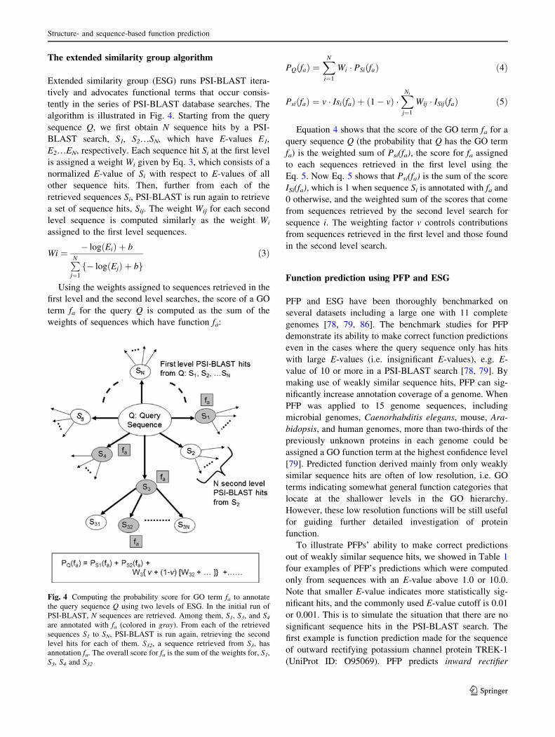
algorithm is illustrated in Fig. 4. Starting from the query
sequence Q, we first obtain N sequence hits by a PSI-
BLAST search, S1, S2…SN, which have E-values E1,
E2…EN, respectively. Each sequence hit Si at the first level
is assigned a weight Wi given by Eq. 3, which consists of a
normalized E-value of Si with respect to E-values of all
other sequence hits. Then, further from each of the
retrieved sequences Si, PSI-BLAST is run again to retrieve
a set of sequence hits, Sij. The weight Wij for each second
level sequence is computed similarly as the weight Wi
assigned to the first level sequences.
Wi ¼ � logðEiÞ þ b
PNj¼1
f� logðEjÞ þ bgð3Þ
Using the weights assigned to sequences retrieved in the
first level and the second level searches, the score of a GO
term fa for the query Q is computed as the sum of the
weights of sequences which have function fa:
PQðfaÞ ¼XN
i¼1
Wi � PSiðfaÞ ð4Þ
PsiðfaÞ ¼ v � IsiðfaÞ þ ð1� vÞ �XNi
j¼1
Wij � ISijðfaÞ ð5Þ
Equation 4 shows that the score of the GO term fa for a
query sequence Q (the probability that Q has the GO term
fa) is the weighted sum of Psi(fa), the score for fa assigned
to each sequences retrieved in the first level using the
Eq. 5. Now Eq. 5 shows that Psi(fa) is the sum of the score
ISi(fa), which is 1 when sequence Si is annotated with fa and
0 otherwise, and the weighted sum of the scores that come
from sequences retrieved by the second level search for
sequence i. The weighting factor v controls contributions
from sequences retrieved in the first level and those found
in the second level search.
Function prediction using PFP and ESG
PFP and ESG have been thoroughly benchmarked on
several datasets including a large one with 11 complete
genomes [78, 79, 86]. The benchmark studies for PFP
demonstrate its ability to make correct function predictions
even in the cases where the query sequence only has hits
with large E-values (i.e. insignificant E-values), e.g. E-
value of 10 or more in a PSI-BLAST search [78, 79]. By
making use of weakly similar sequence hits, PFP can sig-
nificantly increase annotation coverage of a genome. When
PFP was applied to 15 genome sequences, including
microbial genomes, Caenorhabditis elegans, mouse, Ara-
bidopsis, and human genomes, more than two-thirds of the
previously unknown proteins in each genome could be
assigned a GO function term at the highest confidence level
[79]. Predicted function derived mainly from only weakly
similar sequence hits are often of low resolution, i.e. GO
terms indicating somewhat general function categories that
locate at the shallower levels in the GO hierarchy.
However, these low resolution functions will be still useful
for guiding further detailed investigation of protein
function.
To illustrate PFPs’ ability to make correct predictions
out of weakly similar sequence hits, we showed in Table 1
four examples of PFP’s predictions which were computed
only from sequences with an E-value above 1.0 or 10.0.
Note that smaller E-value indicates more statistically sig-
nificant hits, and the commonly used E-value cutoff is 0.01
or 0.001. This is to simulate the situation that there are no
significant sequence hits in the PSI-BLAST search. The
first example is function prediction made for the sequence
of outward rectifying potassium channel protein TREK-1
(UniProt ID: O95069). PFP predicts inward rectifier
Fig. 4 Computing the probability score for GO term fa to annotate
the query sequence Q using two levels of ESG. In the initial run of
PSI-BLAST, N sequences are retrieved. Among them, S1, S3, and S4
are annotated with fa (colored in gray). From each of the retrieved
sequences S1 to SN, PSI-BLAST is run again, retrieving the second
level hits for each of them. S32, a sequence retrieved from S3, has
annotation fa. The overall score for fa is the sum of the weights for, S1,
S3, S4 and S32
Structure- and sequence-based function prediction
123
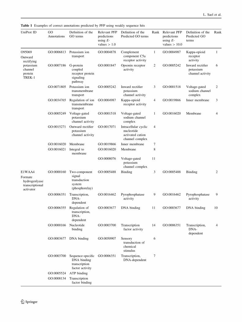
Table 1 Examples of correct annotations predicted by PFP using weakly sequence hits
UniProt ID GO
Annotations
Definition of the
GO terms
Relevant PFP
predictions
using E-
values [ 1.0
Definition of the
Predicted GO terms
Rank Relevant PFP
predictions
using E-
values [ 10.0
Definition of the
Predicted GO
terms
Rank
O95069
Outward
rectifying
potassium
channel
protein
TREK-1
GO:0006813 Potassium ion
transport
GO:0004878 Complement
component C5a
receptor activity
1 GO:0004987 Kappa-opioid
receptor
activity
1
GO:0007186 G-protein
coupled
receptor protein
signaling
pathway
GO:0001847 Opsonin receptor
activity
2 GO:0005242 Inward rectifier
potassium
channel activity
6
GO:0071805 Potassium ion
transmembrane
transport
GO:0005242 Inward rectifier
potassium
channel activity
3 GO:0001518 Voltage-gated
sodium channel
complex
2
GO:0034765 Regulation of ion
transmembrane
transport
GO:0004987 Kappa-opioid
receptor activity
4 GO:0019866 Inner membrane 3
GO:0005249 Voltage-gated
potassium
channel activity
GO:0001518 Voltage-gated
sodium channel
complex
1 GO:0016020 Membrane 4
GO:0015271 Outward rectifier
potassium
channel activity
GO:0017071 Intracellular cyclic
nucleotide
activated cation
channel complex
4
GO:0016020 Membrane GO:0019866 Inner membrane 7
GO:0016021 Integral to
membrane
GO:0016020 Membrane 8
GO:0008076 Voltage-gated
potassium
channel complex
11
E1WAA4
Formate
hydrogenlyase
transcriptional
activator
GO:0000160 Two-component
signal
transduction
system
(phosphorelay)
GO:0005488 Binding 3 GO:0005488 Binding 2
GO:0006351 Transcription,
DNA-
dependent
GO:0016462 Pyrophosphatase
activity
9 GO:0016462 Pyrophosphatase
activity
9
GO:0006355 Regulation of
transcription,
DNA-
dependent
GO:0003677 DNA binding 11 GO:0003677 DNA binding 10
GO:0000166 Nucleotide
binding
GO:0003700 Transcription
factor activity
14 GO:0006351 Transcription,
DNA-
dependent
4
GO:0003677 DNA binding GO:0050907 Sensory
transduction of
chemical
stimulus
6
GO:0003700 Sequence-specific
DNA binding
transcription
factor activity
GO:0006351 Transcription,
DNA-dependent
7
GO:0005524 ATP binding
GO:0008134 Transcription
factor binding
L. Sael et al.
123

Table 1 continued
UniProt ID GO
Annotations
Definition of the
GO terms
Relevant PFP
predictions
using E-
values [ 1.0
Definition of the
Predicted GO terms
Rank Relevant PFP
predictions
using E-
values [ 10.0
Definition of the
Predicted GO
terms
Rank
GO:0017111 Nucleoside-
triphosphatase
activity
Q8UVE6
Transcription
factor AP2
alpha 1
GO:0001501 Skeletal system
development
GO:0003705 RNA polymerase II
transcription
factor activity,
enhancer binding
6 GO:0008134 Transcription
factor binding
7
GO:0006351 Transcription,
DNA-
dependent
GO:0003677 DNA binding 7 GO:0003700 Transcription
factor activity
8
GO:0006355 Regulation of
transcription,
DNA-
dependent
GO:0003700 Transcription
factor activity
13 GO:0006351 Transcription,
DNA-
dependent
2
GO:0007422 Peripheral
nervous system
development
GO:0008134 Transcription
factor binding
14
GO:0014036 Neural crest cell
fate
specification
GO:0006351 Transcription,
DNA-dependent
3
GO:0030318 Melanocyte
differentiation
GO:0060041 Retina
development in
camera-type
eye
GO:0003700 Sequence-specific
DNA binding
transcription
factor activity
GO:0005634 Nucleus
Q12386
Actin-like
protein ARP8
GO:0006312 Mitotic
recombination
GO:0005488 Binding 1 GO:0003676 Nucleic acid
binding
4
GO:0006338 Chromatin
remodeling
GO:0003676 Nucleic acid
binding
4 GO:0003682 Chromatin
binding
7
GO:0006974 Response to DNA
damage
stimulus
GO:0003682 Chromatin binding 7 GO:0008135 Translation
factor activity,
nucleic acid
binding
13
GO:0006355 Regulation of
transcription,
DNA-
dependent
GO:0003677 DNA binding 12 GO:0003723 RNA binding 16
GO:0003729 mRNA binding GO:0003697 Single-stranded
DNA binding
13 GO:0046034 ATP metabolism 3
GO:0043140 ATP-dependent
30-50 DNA
helicase activity
GO:0003700 Transcription
factor activity
15 GO:0009199 Ribonucleoside
triphosphate
metabolism
9
GO:0005634 Nucleus GO:0006351 Transcription,
DNA-dependent
2 GO:0006351 Transcription,
DNA-
dependent
13
GO:0005856 Cytoskeleton GO:0046034 ATP metabolism 6
Structure- and sequence-based function prediction
123

potassium channel activity (GO:0005242) with E-value
cutoffs of both 1.0 and 10.0. Although this prediction does
not exactly match with this protein’s annotation, it is close
in the GO hierarchy to the correct annotation, outward
rectifier potassium channel activity (GO:0015271). Both
terms have a common immediate parent terms, voltage-
gated potassium channel activity (GO:0005249). This
query protein is involved in the G-protein coupled receptor
protein signaling pathway (GO:0007186), for which PFP
using the E-value cutoff of 1.0 and 10.0 has captured more
specialized child terms of GO:0004888 trans membrane
signaling receptor activity and GO:0004930 G-protein
coupled receptor activity (e.g. kappa-opioid receptor
activity, GO: 0004987). Overall in this example, even using
weak sequence hits, which are conventionally discarded in
the homology search, PFP still managed to indicate that
this protein is potassium channel that locate inner mem-
brane (transmembrane).
The second example of formate hydrogenlyase tran-
scriptional activator (Uniprot ID: E1WAA4) is involved in
transcription, DNA-dependent (GO:0006351). This GO
term was predicted by PFP within the top 10 ranks when
using the E-value cutoff of 1.0 and 10.0. Also this protein is
annotated with GO:0017111 nucleoside-triphosphatase
activity, where PFP predicts a less specific parental term,
GO:0016462 pyrophosphatase activity as an annotation.
Similar results can be seen in the last two examples for
Q8UVE6 and Q12386. Using only sequence hits of E-value
above 1.0/10.0, PFP correctly predicted their functional
class, transcription factor. More examples can be found in
the original paper [79].
PFP’s superior performance has been also demonstrated
in the community-wide computational function prediction
assessments. In Automatic Function Prediction Special
Interest Group (AFP-SIG) meeting held at the Intelligent
System in Molecular Biology (ISMB) AFP-SIG 2005
[93] and the function prediction category at the Critical
Assessment of Techniques for Protein Structure Prediction
7 (CASP7) [94], PFP has shown best overall performance
among the participants.
In contrast to PFP whose aim is to increase the sensi-
tivity to enlarge annotation coverage, ESG is intended to
make more precise prediction by iterative database sear-
ches. In the thorough benchmark study [86], ESG was
found to have a higher precision than PFP and the other
existing methods with a comparable sensitivity to PFP.
ESG was found to have more accurate prediction for multi-
domain proteins since the second round of PSI-BLAST
searches are often initiated from different local regions of
the query sequence.
Availability of PFP and ESG
PFP and ESG are available freely for academic users as web
servers at http://kiharalab.org/web/pfp.php and http://kiharalab.
org/web/esg.php. The users can submit sequences and receive
predicted GO terms for the sequences. The stand-alone PFP
program is available upon request.
Conclusion
Many protein structures determined by the structural
genomics projects remain functionally unknown since they
are not homologous to or do not have the global sequence
or structural similarity to characterized proteins. In this
article, we have discussed structure-based methods and
sequence-based methods developed in our group to cope
with such proteins with unknown function. Two structure-
based methods, Pocket-Surfer and Patch-Surfer, detect
similar known binding pockets for pocket regions in a
query protein without using global protein fold similarity.
Two sequence-based methods, PFP and ESG, make use of
weakly similar sequences that are conventionally discarded
in homology based function annotation. Combined together
Table 1 continued
UniProt ID GO
Annotations
Definition of the
GO terms
Relevant PFP
predictions
using E-
values [ 1.0
Definition of the
Predicted GO terms
Rank Relevant PFP
predictions
using E-
values [ 10.0
Definition of the
Predicted GO
terms
Rank
GO:0031011 Ino80 complex GO:0009199 Ribonucleoside
triphosphate
metabolism
9
Function predictions by PFP for four proteins are shown. Annotations from the UniProt database are shown in the first three columns from left.
The next three columns (from the 4th to the 6th column) show prediction by PFP that are derived only from weak sequence hits with an E-value
of 1.0 or larger. Only the predictions relevant to the correct annotations are shown. ‘‘Rank’’ is the rank of the prediction based on the PFP’s
confidence score. Since the predicted GO terms are ranked for each of the three GO categories separately, there are multiple (up to 3) predictions
with the same rank. The last three columns (the 7th–9th column) are predictions by PFP using weak sequence hits with an E-value of 10.0 or
larger
L. Sael et al.
123
with experimental methods we hope that computational
methods will make a leading contribution in functional
elucidation of the protein structures.
Acknowledgments This work is supported in part by the National
Institute of General Medical Sciences of the National Institutes of
Health (R01GM075004, R01GM097528), the National Science
Foundation (DMS0800568, EF0850009, IIS0915801) and Showalter
Trust. MC is supported by Bilsland Dissertation Fellowship from
College of Science, Purdue University.
References
1. Chandonia JM, Brenner SE (2006) The impact of structural
genomics: expectations and outcomes. Science 311:347–351
2. Norvell JC, Berg JM (2007) Update on the protein structure
initiative. Structure 15:1519–1522
3. Terwilliger TC, Stuart D, Yokoyama S (2009) Lessons from
structural genomics. Annu Rev Biophys 38:371–383
4. Todd AE, Marsden RL, Thornton JM, Orengo CA (2005) Pro-
gress of structural genomics initiatives: an analysis of solved
target structures. J Mol Biol 348:1235–1260
5. Berman HM, Westbrook J, Feng Z, Gilliland G, Bhat TN,
Weissig H, Shindyalov IN, Bourne PE (2000) The protein data
bank. Nucleic Acids Res 28:235–242
6. Westbrook J, Feng Z, Chen L, Yang H, Berman HM (2003) The
protein data bank and structural genomics. Nucleic Acids Res
31:489–491
7. Ellrott K, Zmasek CM, Weekes D, Sri KS, Bakolitsa C, Godzik
A, Wooley J (2011) TOPSAN: a dynamic web database for
structural genomics. Nucleic Acids Res 39:D494–D496
8. Shin DH, Hou J, Chandonia JM, Das D, Choi IG, Kim R, Kim SH
(2007) Structure-based inference of molecular functions of pro-
teins of unknown function from Berkeley Structural Genomics
Center. J Struct Funct Genomics 8:99–105
9. Teplyakov A, Pullalarevu S, Obmolova G, Doseeva V, Galkin A,
Herzberg O, Dauter M, Dauter Z, Gilliland GL (2004) Crystal
structure of the YffB protein from Pseudomonas aeruginosa
suggests a glutathione-dependent thiol reductase function. BMC
Struct Biol 4:5
10. Teplyakov A, Obmolova G, Sarikaya E, Pullalarevu S, Krajewski
W, Galkin A, Howard AJ, Herzberg O, Gilliland GL (2004)
Crystal structure of the YgfZ protein from Escherichia colisuggests a folate-dependent regulatory role in one-carbon
metabolism. J Bacteriol 186:7134–7140
11. Li De La Sierra-Gallay I, Collinet B, Graille M, Quevillon-
Cheruel S, Liger D, Minard P, Blondeau K, Henckes G, Aufrere
R, Leulliot N, Zhou CZ, Sorel I, Ferrer JL, Poupon A, Janin J, van
Tilbeurgh H (2004) Crystal structure of the YGR205w protein
from Saccharomyces cerevisiae: close structural resemblance to
E. coli pantothenate kinase. Proteins 54:776–783
12. Graille M, Quevillon-Cheruel S, Leulliot N, Zhou CZ, Gallay
ILD, Jacquamet L, Ferrer JL, Liger D, Poupon A, Janin J,
van Tilbeurgh H (2004) Crystal structure of the YDR533c S. ce-
revisiae protein, a class II member of the Hsp31 family.
Structure 12:839–847
13. Liger D, Graille M, Zhou CZ, Leulliot N, Quevillon-Cheruel S,
Blondeau K, Janin J, van Tilbeurgh T (2004) Crystal structure
and functional characterization of yeast YLR011wp, an enzyme
with NAD(P)H-FMN and ferric iron reductase activities. J Biol
Chem 279:34890–34897
14. Sanishvili R, Yakunin AF, Laskowski RA, Skarina T, Evd-
okimova E, Doherty-Kirby A, Lajoie GA, Thornton JM,
Arrowsmith CH, Savchenko A, Joachimiak A, Edwards AM
(2003) Integrating structure, bioinformatics, and enzymology to
discover function: BioH, a new carboxylesterase from Esche-richia coli. J Biol Chem 278:26039–26045
15. Kuznetsova E, Proudfoot M, Sanders SA, Reinking J, Savchenko
A, Arrowsmith CH, Edwards AM, Yakunin AF (2005) Enzyme
genomics: application of general enzymatic screens to discover
new enzymes. FEMS Microbiol Rev 29:263–279
16. Fridman E, Pichersky E (2005) Metabolomics, genomics, pro-
teomics, and the identification of enzymes and their substrates
and products. Curr Opin Plant Biol 8:242–248
17. Roberts RJ (2011) COMBREX: COMputational BRidge to
EXperiments. Biochem Soc Trans 39:581–583
18. Altschul SF, Gish W, Miller W, Myers EW, Lipman DJ (1990)
Basic local alignment search tool. J Mol Biol 215:403–410
19. Altschul SF, Madden TL, Schaffer AA, Zhang J, Zhang Z, Miller
W, Lipman DJ (1997) Gapped BLAST and PSI-BLAST: a new
generation of protein database search programs. Nucleic Acids
Res 25:3389–3402
20. Pearson WR, Lipman DJ (1988) Improved tools for biological
sequence comparison. Proc Natl Acad Sci USA 85:2444–2448
21. Finn RD, Mistry J, Tate J, Coggill P, Heger A, Pollington JE,
Gavin OL, Gunasekaran P, Ceric G, Forslund K, Holm L,
Sonnhammer EL, Eddy SR, Bateman A (2010) The Pfam protein
families database. Nucleic Acids Res 38:D211–D222
22. Hawkins T, Kihara D (2007) Function prediction of uncharac-
terized proteins. J Bioinform Comput Biol 5:1–30
23. Hawkins T, Chitale M, Kihara D (2008) New paradigm in protein
function prediction for large scale omics analysis. Mol Biosyst
4:223–231
24. Kihara D (2011) Protein function prediction for omics era.
Springer, London
25. Gherardini PF, Helmer-Citterich M (2008) Structure-based
function prediction: approaches and applications. Brief Funct
Genomic Proteomic 7:291–302
26. Martin AC, Orengo CA, Hutchinson EG, Jones S, Karmirantzou
M, Laskowski RA, Mitchell JB, Taroni C, Thornton JM (1998)
Protein folds and functions. Structure 6:875–884
27. Thornton JM, Todd AE, Milburn D, Borkakoti N, Orengo CA
(2000) From structure to function: approaches and limitations.
Nat Struct Biol 7(Suppl):991–994
28. Shindyalov IN, Bourne PE (1998) Protein structure alignment by
incremental combinatorial extension (CE) of the optimal path.
Protein Eng 11:739–747
29. Holm L, Sander C (1993) Protein structure comparison by
alignment of distance matrices. J Mol Biol 233:123–138
30. Orengo CA, Taylor WR (1996) SSAP: sequential structure
alignment program for protein structure comparison. Methods
Enzymol 266:617–635
31. Thompson KE, Wang Y, Madej T, Bryant SH (2009) Improving
protein structure similarity searches using domain boundaries
based on conserved sequence information. BMC Struct Biol 9:33
32. Mizuguchi K, Go N (1995) Comparison of spatial arrangements
of secondary structural elements in proteins. Protein Eng 8:
353–362
33. Kihara D, Sael L, Chikhi R, Esquivel-Rodriguez J (2011)
Molecular surface representation using 3D Zernike descriptors
for protein shape comparison and docking. Curr Protein Pept Sci
12:520–530
34. La D, Esquivel-Rodriguez J, Venkatraman V, Li B, Sael L, Ueng
S, Ahrendt S, Kihara D (2009) 3D-SURFER: software for high-
throughput protein surface comparison and analysis. Bioinfor-
matics 25:2843–2844
35. Sael L, Li B, La D, Fang Y, Ramani K, Rustamov R, Kihara D
(2008) Fast protein tertiary structure retrieval based on global
surface shape similarity. Proteins 72:1259–1273
Structure- and sequence-based function prediction
123
36. Sael L, Kihara D (2009) Protein surface representation and
comparison: new approaches in structural proteomics. In: Chen J,
Lonardi S (eds) Biological data mining. Chapman & Hall/CRC
Press, Boca Raton, pp 89–109
37. Venkatraman V, Sael L, Kihara D (2009) Potential for protein
surface shape analysis using spherical harmonics and 3D Zernike
descriptors. Cell Biochem Biophys 54:23–32
38. Ritchie DW, Graham J (1999) Fast computation, rotation, and
comparison of low resolution spherical harmonic molecular sur-
faces. J Comp Chem 20:383–395
39. Orengo CA, Jones DT, Thornton JM (1994) Protein superfamilies
and domain superfolds. Nature 372:631–634
40. Porter CT, Bartlett GJ, Thornton JM (2004) The catalytic site
atlas: a resource of catalytic sites and residues identified in
enzymes using structural data. Nucleic Acids Res 32:D129–D133
41. Arakaki AK, Zhang Y, Skolnick J (2004) Large-scale assessment
of the utility of low-resolution protein structures for biochemical
function assignment. Bioinformatics 20:1087–1096
42. Artymiuk PJ, Poirrette AR, Grindley HM, Rice DW, Willett P
(1994) A graph-theoretic approach to the identification of three-
dimensional patterns of amino acid side-chains in protein struc-
tures. J Mol Biol 243:327–344
43. Kleywegt GJ (1999) Recognition of spatial motifs in protein
structures. J Mol Biol 285:1887–1897
44. Ferre F, Ausiello G, Zanzoni A, Helmer-Citterich M (2004)
SURFACE: a database of protein surface regions for functional
annotation. Nucleic Acids Res 32:D240–D244
45. Redfern OC, Dessailly BH, Dallman TJ, Sillitoe I, Orengo CA
(2009) FLORA: a novel method to predict protein function from
structure in diverse superfamilies. PLoS Comput Biol 5:e1000485
46. Schmitt S, Kuhn D, Klebe G (2002) A new method to detect
related function among proteins independent of sequence and fold
homology. J Mol Biol 323:387–406
47. Gold ND, Jackson RM (2006) Fold independent structural com-
parisons of protein-ligand binding sites for exploring functional
relationships. J Mol Biol 355:1112–1124
48. Kinoshita K, Nakamura H (2005) Identification of the ligand
binding sites on the molecular surface of proteins. Protein Sci
14:711–718
49. Morris RJ, Najmanovich RJ, Kahraman A, Thornton JM (2005)
Real spherical harmonic expansion coefficients as 3D shape
descriptors for protein binding pocket and ligand comparisons.
Bioinformatics 21:2347–2355
50. Binkowski TA, Adamian L, Liang J (2003) Inferring functional
relationships of proteins from local sequence and spatial surface
patterns. J Mol Biol 332:505–526
51. Binkowski TA, Freeman P, Liang J (2004) pvSOAR: detecting
similar surface patterns of pocket and void surfaces of amino acid
residues on proteins. Nucleic Acids Res 32:W555–W558
52. Binkowski TA, Joachimiak A (2008) Protein functional surfaces:
global shape matching and local spatial alignments of ligand
binding sites. BMC Struct Biol 8:45
53. Laskowski RA, Watson JD, Thornton JM (2005) ProFunc: a
server for predicting protein function from 3D structure. Nucleic
Acids Res 33:W89–W93
54. Pal D, Eisenberg D (2005) Inference of protein function from
protein structure. Structure (Camb) 13:121–130
55. Chikhi R, Sael L, Kihara D (2010) Real-time ligand binding
pocket database search using local surface descriptors. Proteins
78:2007–2028
56. Sael L, Kihara D (2011) Binding ligand prediction for proteins
using partial matching of local surface patches. Int J Mol Sci
11:5009–5026
57. Sael L, Kihara D (2012) Detecting local ligand-binding site
similarity in non-homologous proteins by surface patch com-
parison. Proteins (in press)
58. Novotni M, Klein R (2003) 3D Zernike descriptors for content
based shape retrieval. In: ACM symposium on solid and physical
modeling, proceedings of the eighth ACM symposium on solid
modeling and applications pp 216–225
59. Canterakis N (1999) 3D Zernike moments and Zernike affine
invariants for 3D image analysis and recognition. In: Proceedings
of 11th scandinavian conference on image analysis, pp 85–93
60. Baker NA, Sept D, Joseph S, Holst MJ, McCammon JA (2001)
Electrostatics of nanosystems: application to microtubules and
the ribosome. Proc Natl Acad Sci USA 98:10037–10041
61. Li B, Turuvekere S, Agrawal M, La D, Ramani K, Kihara D
(2007) Characterization of local geometry of protein surfaces
with the visibility criterion. Proteins 71:670–683
62. Huang B, Schroeder M (2006) LIGSITEcsc: predicting ligand
binding sites using the Connolly surface and degree of conser-
vation. BMC Struct Biol 6:19
63. Demange G, Gale D, Stomayor M (1986) Multi-item auctions.
J Polit Econ 94:863–872
64. Kahraman A, Morris RJ, Laskowski RA, Favia AD, Thornton JM
(2010) On the diversity of physicochemical environments expe-
rienced by identical ligands in binding pockets of unrelated
proteins. Proteins 78:1120–1136
65. Gribskov M, Robinson NL (1996) Use of receiver operating
characteristic (ROC) analysis to evaluate sequence matching.
Comput Chem 20:25–33
66. Sael L, Kihara D (2012) Constructing patch-based ligand-binding
pocket database for predicting function of proteins. BMC Bio-
inform (in press)
67. Wallach I, Lilien R (2009) The protein-small-molecule database,
a non-redundant structural resource for the analysis of protein-
ligand binding. Bioinformatics 25:615–620
68. Bender A, Glen RC (2005) A discussion of measures of enrich-
ment in virtual screening: comparing the information content of
descriptors with increasing levels of sophistication. J Chem Inf
Model 45:1369–1375
69. Venkatraman V, Chakravarthy PR, Kihara D (2009) Application
of 3D Zernike descriptors to shape-based ligand similarity
searching. J Cheminform 1:19
70. Huang SY, Zou X (2010) Advances and challenges in protein-
ligand docking. Int J Mol Sci 11:3016–3034
71. Hulo N, Bairoch A, Bulliard V, Cerutti L, De CE, Langendijk-
Genevaux PS, Pagni M, Sigrist CJ (2006) The PROSITE data-
base. Nucleic Acids Res 34:D227–D230
72. Mulder NJ, Apweiler R, Attwood TK, Bairoch A, Bateman A,
Binns D, Bradley P, Bork P, Bucher P, Cerutti L, Copley R,
Courcelle E, Das U, Durbin R, Fleischmann W, Gough J, Haft D,
Harte N, Hulo N, Kahn D, Kanapin A, Krestyaninova M, Lons-
dale D, Lopez R, Letunic I, Madera M, Maslen J, McDowall J,
Mitchell A, Nikolskaya AN, Orchard S, Pagni M, Ponting CP,
Quevillon E, Selengut J, Sigrist CJ, Silventoinen V, Studholme
DJ, Vaughan R, Wu CH (2005) InterPro, progress and status in
2005. Nucleic Acids Res 33:D201–D205
73. Bru C, Courcelle E, Carrere S, Beausse Y, Dalmar S, Kahn D
(2005) The ProDom database of protein domain families: more
emphasis on 3D. Nucleic Acids Res 33:D212–D215
74. Chitale M, Kihara D (2011) Computational protein functionprediction: framework and challenges. In: Kihara D (ed) Protein
function prediction for omis era. Springer, London, pp 1–17
75. John B, Sali A (2004) Detection of homologous proteins by an
intermediate sequence search. Protein Sci 13:54–62
76. Salamov AA, Suwa M, Orengo CA, Swindells MB (1999)
Combining sensitive database searches with multiple intermedi-
ates to detect distant homologues. Protein Eng 12:95–100
77. Park J, Teichmann SA, Hubbard T, Chothia C (1997) Interme-
diate sequences increase the detection of homology between
sequences. J Mol Biol 273:349–354
L. Sael et al.
123

78. Hawkins T, Luban S, Kihara D (2006) Enhanced automated
function prediction using distantly related sequences and con-
textual association by PFP. Protein Sci 15:1550–1556
79. Hawkins T, Chitale M, Luban S, Kihara D (2009) PFP: auto-
mated prediction of gene ontology functional annotations with
confidence scores using protein sequence data. Proteins 74:
566–582
80. Harris MA, Clark J, Ireland A, Lomax J, Ashburner M, Foulger
R, Eilbeck K, Lewis S, Marshall B, Mungall C, Richter J, Rubin
GM, Blake JA, Bult C, Dolan M, Drabkin H, Eppig JT, Hill DP,
Ni L, Ringwald M, Balakrishnan R, Cherry JM, Christie KR,
Costanzo MC, Dwight SS, Engel S, Fisk DG, Hirschman JE,
Hong EL, Nash RS, Sethuraman A, Theesfeld CL, Botstein D,
Dolinski K, Feierbach B, Berardini T, Mundodi S, Rhee SY,
Apweiler R, Barrell D, Camon E, Dimmer E, Lee V, Chisholm R,
Gaudet P, Kibbe W, Kishore R, Schwarz EM, Sternberg P, Gwinn
M, Hannick L, Wortman J, Berriman M, Wood V, de la Cruz N,
Tonellato P, Jaiswal P, Seigfried T, White R (2004) The Gene
Ontology (GO) database and informatics resource. Nucleic Acids
Res 32:D258–D261
81. Martin DM, Berriman M, Barton GJ (2004) GOtcha: a new
method for prediction of protein function assessed by the anno-
tation of seven genomes. BMC Bioinform 5:178
82. Khan S, Situ G, Decker K, Schmidt CJ (2003) GoFigure: auto-
mated gene ontology annotation. Bioinformatics 19:2484–2485
83. Zehetner G (2003) OntoBlast function: from sequence similarities
directly to potential functional annotations by ontology terms.
Nucleic Acids Res 31:3799–3803
84. Hawkins T, Chitale M, Kihara D (2010) Functional enrichment
analyses and construction of functional similarity networks with
high confidence function prediction by PFP. BMC Bioinform
11:265
85. Si L, Yu D, Kihara D, Yi F (2008) Combining sequence simi-
larity scores and textual information for gene function annotation
in the literature. Inf Retr 11:389–404
86. Chitale M, Hawkins T, Park C, Kihara D (2009) ESG: extended
similarity group method for automated protein function predic-
tion. Bioinformatics 25:1739–1745
87. Wass MN, Sternberg MJ (2008) ConFunc–functional annotation
in the twilight zone. Bioinformatics 24:798–806
88. Engelhardt BE, Jordan MI, Muratore KE, Brenner SE (2005)
Protein molecular function prediction by Bayesian phylogenom-
ics. PLoS Comput Biol 1:e45
89. Krishnamurthy N, Brown D, Sjolander K (2007) FlowerPower:
clustering proteins into domain architecture classes for phyloge-
nomic inference of protein function. BMC Evol Biol 7(Suppl
1):S12
90. Friedberg I, Harder T, Godzik A (2006) JAFA: a protein function
annotation meta-server. Nucleic Acids Res 34:W379–W381
91. Chitale M, Hawkins T, Kihara D (2009) Automated prediction of
protein function from sequence. In: Bujnicki J (ed) Prediction of
protein structure, functions, and interactions. Wiley, London,
pp 63–86
92. Uniprot Consortium (2010) The Universal Protein Resource
(UniProt) in 2010. Nucleic Acids Res 38:D142–D148
93. Friedberg I, Jambon M, Godzik A (2006) New avenues in protein
function prediction. Protein Sci 15:1527–1529
94. Lopez G, Rojas A, Tress M, Valencia A (2007) Assessment of
predictions submitted for the CASP7 function prediction cate-
gory. Proteins 69:165–174
Structure- and sequence-based function prediction
123