Embed Size (px)
Citation preview

1
Structure-guided Development of Specific Pyruvate Dehydrogenase Kinase Inhibitors Targeting the ATP-
binding Pocket*
Shih-Chia Tsoǂ1, , Xiangbing Qi
ǂ1 ,Wen-Jun Guiǂ, Cheng-Yang Wu
ǂ, Jacinta L. Chuang
ǂ, Ingrid
Wernstedt-Asterholm§, Lorraine K. Morlock
ǂ, Kyle R. Owensǂ, Philipp E. Scherer
§, Noelle S.
Williamsǂ, Uttam K. Tambar
ǂ2, R. Max Wynnǂ§, and David T. Chuang
ǂ§3
From Departments of Biochemistryǂ and Internal Medicine
§
University of Texas Southwestern Medical Center, Dallas, Texas 75390
Running title: Pyruvate Dehydrogenase Kinase Inhibitors
Keywords: Pyruvate dehydrogenase kinase; Pyruvate dehydrogenase complex; Enzyme inhibitors; Drug
development; Diabetes; Glucose metabolism; Structure-based inhibitor design; Mitochondrial protein
kinase; Hepatic steatosis
Background: Up-regulated pyruvate
dehydrogenase kinase isoforms (PDKs) are
associated with impaired glucose homeostasis in
diabetes.
Results: Novel PDK inhibitors were developed
using structure-based design, which improve
glucose tolerance with reduced hepatic steatosis in
diet-induced obese mice.
Conclusion: Obesity phenotypes are effectively
treated by chemical intervention with PDK
inhibitors.
Significance: PDKs are potential drug targets for
obesity and type 2 diabetes.
ABSTRACT
Pyruvate dehydrogenase kinase isoforms
(PDKs 1 - 4) negatively regulate activity of the
mitochondrial pyruvate dehydrogenase
complex (PDC) by reversible phosphorylation.
PDK isoforms are up-regulated in obesity,
diabetes, heart failure and cancer and are
potential therapeutic targets for these
important human diseases. Here, we employed
structure-guided design to convert a known
Hsp90 inhibitor to a series of highly specific
PDK inhibitors, based on structural
conservation in the ATP-binding pocket. The
key step involved the substitution of a carbonyl
group in the parent compound with a sulfonyl
in the PDK inhibitors. The final compound of
this series, 2-[(2,4-dihydroxyphenyl)sulfonyl]
isoindoline-4,6-diol, designated PS10, inhibits
all four PDK isoforms with IC50 = 0.8 µM for
PDK2. The administration of PS10 (70 mg/kg)
to diet-induced obese mice significantly
augments PDC activity with reduced
phosphorylation in different tissues. Prolonged
PS10 treatments result in improved glucose
tolerance and notably lessened hepatic steatosis
in the mouse model. The results support the
pharmacological approach of targeting PDK to
control both glucose and fat levels in obesity
and type 2 diabetes.
The mitochondrial pyruvate dehydrogenase
complex (PDC)4 catalyzes the oxidative
decarboxylation of pyruvate to give rise to acetyl-
CoA, and is the gate-keeping enzyme linking
glycolysis and the Krebs cycle. The mammalian
PDC is a 9.5 million-dalton protein machine
organized about a 60-meric core consisting of
dihydrolipoyl transacetylase (E2) and the E3-
binding protein (E3BP), to which multiple copies
of pyruvate dehydrogenase (E1) and dihydrolipoyl
transacetylase (E2), dihydrolipoamide
dehydrogenase (E3), as well as isofroms of
pyruvate dehydrogenase kinase (PDKs 1-4) and
pyruvate dehydrogenase phosphatase (PDPs 1-2)
are attached through ionic interactions (1). Due to
its strategic location, the regulation of PDC
activity is critical for glucose homeostasis and fuel
selection in the glucose-fatty acid cycle (2). The
mammalian PDC is acutely regulated by reversible
phosphorylation (3). The phosphorylation of PDC
by PDK results in inactivation; and
dephosphorylation by PDP restores PDC activity.
http://www.jbc.org/cgi/doi/10.1074/jbc.M113.533885The latest version is at JBC Papers in Press. Published on December 19, 2013 as Manuscript M113.533885
Copyright 2013 by The American Society for Biochemistry and Molecular Biology, Inc.
by guest on Novem
ber 16, 2018http://w
ww
.jbc.org/D
ownloaded from

2
When glucose levels are low during fasting, PDC
is highly phosphorylated and inactive, so as to
preserve the substrates (pyruvate, lactate and
alanine) for gluconeogenesis (2).
The PDKs are potential therapeutic targets
because of increased PDK expression in disease
states such as diabetes, cancer and heart failure.
PDK4, but not PDK2, is drastically induced in
muscle and heart in streptozotocin-induced
diabetes (4), obesity (5) and type 2 diabetes (6),
which attenuates PDC activity leading to reduced
glucose oxidation. The accumulated evidence has
established that the upregulation of PDK4 is
mediated through the PPAR-FOXO3-PGC-1
complex (4). The PDK2/PDK4 double knockout
mice fed a high–fat diet show marked
improvements in glucose tolerance and insulin
sensitivity over wild-type mice on the same diet
(7). The expression of PDK1 (8-10), PDK2 (11),
and PDK3 (12) is significantly elevated in certain
cancers. Tyrosine phosphorylation of PDK1 with
increased kinase activity is essential for tumor cell
proliferation and hypoxia (13). Inhibition of PDK
activity with dichloroacetate (DCA) or siRNA
promotes apoptosis in cancer cells and impedes
tumor growth (14).
The classic PDK inhibitor DCA, an
analogue of the PDC substrate pyruvate, has been
used since early 1970 to inhibit PDK activity and
increase the PDC flux, with concomitant reduction
in glucose levels in animals (15). DCA exerts its
inhibitory effects by binding to an allosteric site in
the N-terminal domain of PDK isoforms (16,17).
However, DCA is a non-specific low-potency
PDK inhibitor and requires high doses for its
therapeutic effects (18), which leads to peripheral
neurological toxicity and tumor growth (19). R-
lipoic acid in mM concentrations abates PDK
activity in vitro (20), but its function as a PDK
inhibitor in vivo is uncertain. Phenylbutyrate
enhances PDC activity in vitro and in vivo (21);
but the compound is a modest PDK inhibitor (Ki =
0.3 mM) with multiple targets and diverse clinical
applications (22). Dihydrolipoamide mimetics
including AZD7545 (23) and secondary amides of
SDZ048-619 (24) have also been developed. This
family of compounds inhibits PDK2 activity by
impeding PDK binding to the E2/E3BP core of
PDC (25). Paradoxically, these dihydrolipoamide
mimetics strongly stimulates PDC core-free PDK4
activity in vitro, which precludes these compounds
as bona fide PDK inhibitors (26). To date, there
have been no effective PDK inhibitors for novel
therapeutic approaches to cancer, obesity and type
2 diabetes as well as heart disease.
Mitochondrial PDK isoforms are members
of the GHKL ATPase/kinase superfamily that
includes DNA gyrase B, heat-shock protein 90
(Hsp90), histidine kinases CheA and EnvZ as well
as the DNA-repair enzyme MutL (27). Members
of this superfamily share four conserved motifs
(N-, G1-, G2- and G3-boxes) that build a unique
Bergerat ATP-binding fold consisting of a four-
stranded mixed -sheet and three α helices, and is
located in the C-terminal domains of PDK
isoforms (28,29). This signature fold also contains
a unique structural element known as the “ATP
lid”, whose conformational change is coupled to
ATP hydrolysis and protein-protein interactions
(29).
In this study, we sought to develop robust
PDK inhibitors that can be used to improve
glucose metabolism and correct metabolic
dysfunction in vivo. Based on the unique structural
features present in the ATP-binding pocket of
PDK2, a single functional-group change was made
in a known Hsp90 inhibitor that binds to the
corresponding pocket of the latter protein (30,31)
from the GHKL family. This approach efficiently
converted the Hsp90 inhibitor to a highly specific
inhibitor for all PDK isoforms. These final PDK
inhibitors of this series robustly augments PDC
activity with reduced phosphorylation in tissues,
which leads to improved glucose tolerance and
reduced hepatic steatosis in diet-induced obese
(DIO) mice. These findings demonstrate the utility
of structure-based inhibitor design and support the
pharmacological approach of targeting PDK to
control glucose and fat levels in obesity and type 2
diabetes.
EXPERIMENTAL PROCEDURES
Chemicals - All reagents and chemicals were
obtained from Sigma-Aldrich unless otherwise
indicated. Synthesis of novel PDK inhibitors PA1,
PA7, PS2, PS8, and PS10 were described in the
Supplemental Data.
Proteins - Recombinant human PDK2 was
expressed and purified as a N terminal His6-tagged
SUMO fusion protein with a tobacco-etch-virus
protease (TEV) cleavage site in front of the N-
terminal PDK2 sequence (26), and was used
by guest on Novem
ber 16, 2018http://w
ww
.jbc.org/D
ownloaded from

3
directly for the activity assay and binding affinity
analyses. For crystallization, the protein was
subjected to a TEV-protease digestion, and the
untagged PDK2 protein was purified on a
Superdex 200 column in 20 mM Tris-HCl (pH
8.0), 150 mM NaCl and 5 mM DTT. The purified
protein was concentrated to 35-40 mg/ml and
stored at -80 ºC in small aliquots. Recombinant
human PDK1, PDK3 and PDK4 were expressed
and purified as described previously (26).
To express the N-terminal domain (residue
1-236) of human Hsp90, the first strand cDNA
was synthesized with the human total RNA as
template using the Omniscript Reverse
Transcriptase from Qiagen (Gaithersburg, MD).
The sequence encoding the N-terminal domain
was amplified and cloned into the pSUMO
expression vector (Lifesensors, Malvern, PA). The
fusion protein of His6-tagged SUMO-Hsp90 N-
terminal domain was expressed in E.coli BL21
cells and purified with Ni-NTA resin (Qiagen) and
on Superdex-200 column in 20 mM Tris.HCl, pH
7.5 and 500 mM NaCl.
Assay for inhibition of PDK activity - To
determine the IC50 for PDK inhibitors, a mixture
containing 0.05-0.2 µM PDK, 6 µM E1, with or
without 0.5 µM of the PDC core E2/E3BP, and
various amounts of inhibitor was incubated at
25°C for 10 min in a buffer of 20 mM Tris-Cl (pH
7.5), 10 mM KCl, 5 mM MgCl2, 2 mM DTT,
0.02% (v/v) Tween-20, and 0.1 mg/ml bovine
serum albumin before the addition of 50 μM ATP
to initiate the reaction. All inhibition titrations
were performed at 10 dose-points ranging from
31.6 nM to 1 mM in a 3.162-fold dilution series,
with each inhibitor concentration tested in
duplicate. The remaining steps were described
previously (26). IC50 values were obtained by the
curve fitting of inhibition isotherms using Prism 6
(GraphPad software, Inc.).
The kinase-profiling of PS8 on 21 human
protein kinases were performed at Reaction
Biology Corp. (Malvern, PA). IC50 values were
determined by 10-dose titration of PS8 from 15
nM to 300 μM in presence of 10 μM ATP. Each
protein kinase was also tested against its known
inhibitor as a positive control.
Isothermal titration calorimetry (ITC) - The
PDK2 or Hsp90 N-terminal domain protein was
dialyzed against one liter of the dialysis buffer
containing 50 mM Tris-Cl, pH 7.5, 50 mM KCl, 1
mM MgCl2, and 0.5 mM β-mercaptoethanol.
Known or novel PDK inhibitor solutions (150-
1500 µM) were placed in the titration syringe and
injected in 8-µl increments into the reaction cell
containing 1.4 ml of 18-70 µM PDK2 or Hsp90 N-
terminal domain at 15°C in a VP-ITC
microcalorimeter (GE Healthcare, Piscataway, NJ).
All of the ITC data were initially analyzed by the
NITPIC program (32) to construct the baseline,
followed by curve-fitting in Origin 7 to obtain
binding parameters. The concentrations of PDK2
and Hsp90 N-terminal domain proteins were
determined by measuring A280 and using calculated
molar extinction coefficients (M-1
∙cm-1
) of 49,530
and 18,910, respectively.
Crystallization of PDK2 and PDK2 -
inhibitor complexes - Crystals of human PDK2
were obtained by the hanging-drop vapor-
diffusion method. Two µl of protein solution was
mixed with 2 µl of the well solution (0.9 M
ammonium tartrate, 0.1 M sodium acetate pH 4.6)
and kept in a 20ºC incubator. Crystals were
developed in one week and reached the size of 500
µm in two weeks. Mature crystals were transferred
to a fresh soaking solution (0.75 M ammonium
tartrate, 0.1M sodium acetate pH 4.6 and 5%
glycerol with various indicated inhibitors). After
overnight incubation, crystals were serially
transferred to a cryo-solution containing 20%
glycerol and snap frozen in liquid nitrogen.
Structure determination and refinements -
All X-ray diffraction data for PDK2 and PDK2-
inhibitor complexes were collected at beamline
19-ID at the Advanced Photon Source, Argonne
National Laboratories. Diffraction data for each
PDK2-inhibitor complex were collected from a
single crystal. All crystals share the same space
group of I4122, and the highest resolution of
diffraction ranged from 1.70 Å to 1.95 Å. The
molecular replacement, structure modeling and
refinement were performed as described
previously (33). The crystal structure of inhibitor-
free human PDK (PDB code 2BTZ) was used as
the search model.
Pharmacokinetic Studies - Twenty-one male
C57BL/6J mice were dosed IP with 70 mg/kg PS-
10, 0.2 ml/mouse formulated as 10% DMSO/20%
water/70% of 25% (2-hydroxypropyl)-β-
cyclodextrin for determination of PS-10 PK.
Twenty-one female CD-1 mice were dosed IP with
20 mg/kg PS-8 , 0.2 ml/mouse formulated as 5%
by guest on Novem
ber 16, 2018http://w
ww
.jbc.org/D
ownloaded from

4
ethanol and 95% of 0.1 M sodium bicarbonate pH
9.0 for determination of PS-8 PK. Animal (n=3)
were sacrificed and whole blood was harvested for
each time point. Plasma was processed from
whole blood by centrifugation of the ACD treated
blood for 10' at 10,000 rpm in a standard
centrifuge. The analytical processing of blood
samples and pharmacokinetics studies using
LC/MS/MS were as described previously with
LC/MS/MS methods optimized for detection of
PS-10 and PS-8 (33).
Treatments of mice with PDK inhibitors -
Six- to eight-week old C57BL/6J male mice were
obtained from the local campus breeding colony at
UT Southwestern Medical Center (Dallas, TX)
and randomized into two groups: vehicle- and
PS10-treated. Prior to the treatment, mice were
fed with a 60% high-fat diet, which contained
32% saturated and 68% unsaturated fat
(catalog number: D12492, Research Diet Inc.
New Brunswick, NJ), for eight to ten weeks to
produce DIO animals. PS-10 was dissolved in
100% DMSO and then diluted to make a 10%
DMSO aqueous solution containing 17.5% (w/v)
(2-hydroxypropyl)-β-cyclodextrin for delivery.
Animals were dosed at mid-day by intraperitoneal
(IP) injections at 70 mg/kg using 1-ml syringe and
30-gauge needle. The length of the treatment is
indicted in each experiment. At 10 h after the last
injection, animals were euthanized using carbon
dioxide asphyxiation followed by cervical
dislocation and dissection. Blood was harvested
by cardiac puncture and stored on ice. Acidified
citrate dextrose (ACD) was used as an
anticoagulant. Immediately after blood collection,
heart, liver, kidneys and both hind-leg quadriceps
muscles were removed and snap frozen in liquid
nitrogen. Average ischemia time before organ
harvest was about 2 to 3 min. Blood was
centrifuged in an Eppendorf 5415R refrigerated
microcentrifuge at 9,300 x g for 5 min to isolate
plasma, which was subsequently stored at -80ºC. Assay for PDC activity in mouse tissues -
Liquid nitrogen-stored tissue samples were
removed and thawed on ice. Individual kidneys
(200-250 mg), hearts (200-300 mg), muscle (200-
300 mg) and liver (250-400 mg) tissues samples
were manually homogenized in an ice-chilled
glass homogenizer containing 1 ml of the
homogenization buffer. The homogenization
buffer contained 30 mM KPi, pH 7.5, 3 mM
EDTA, 5 mM DTT, 1 mM benzamidine, 3% fetal
bovine serum, 5% Triton X-100 and 1 mM
leupeptin. Samples were transferred to ice-cold 10
ml polycarbonate tubes and spun in an
ultracentrifuge at 25,000 x g for 10 min to pellet
cell and tissue debris. Supernatants were removed
and stored on ice until diluted (1:3 for muscle, 1:5
for liver and 1:20 for kidneys and heart tissues)
with a dilution buffer containing 50 mM HEPES,
pH 7.5, 1.0 mM DTT, 0.1% Triton X-100, 5 mM
DCA, 50 mM NaF, 3% fetal bovine serum and 1
mM leupeptin. The diluted samples (50 µl) were
placed in each well of a 24-well plate containing
310 μl of the reaction mixture. A micro-bridge
(Hampton Research) was pre-set into each well
holding one piece of filter wick pre-soaked with 2
M NaOH. The reaction mixture contained 30 mM
KPi, pH 7.5, 0.4 mM CoA, 3 mM NAD+, 5% fetal
bovine serum, 2 mM thiamine diphosphate, 2 mM
MgCl2 and 65 µg of recombinant human E3. [1-14
C] pyruvate (PerkinElmer, Boston, MA) was
added to each well to initiate the reaction, with the
wells sealed with a clear mylar adhesive film. The
assay plates were incubated at 37°C for 10 min.
Fifty µl of a 20% TCA solution was added to each
well to stop the reaction. Assay plates were
incubated further at 37°C for 45 min. 14
CO2-
trapped on 2 M NaOH soaked filter wicks were
counted in a liquid scintillation counter. Total
protein concentrations in the samples were
determined by using BCA protein assay kit
(Thermo fisher Scientific, Rockford, IL).
Western blotting - SDS-PAGE gels were run
using 15-20 µg of protein lysate per lane. Western
blots were transferred to PVDF membranes for 2
hrs at 200 mV. PVDF membranes were blocked
with 5% non-fat dried milk and then probed using
polyclonal antibodies to pyruvate
dehydrogenase/decarboxylase E1-α and to
phosphorylated E1α (pE1α). The E1α antibody
was obtained from MitoSciences/Abcam
(Cambridge, MA). Antibodies against the
phosphorylated serine (pSer293) residue of the
E1α subunit were purchased from EMD
Millipore/Calbiochem Biochemical (Billerica,
MA). One milliliter of Luminata Forte western
HRP (Millipore Corporation, Billerica, MA)
substrate reagent was pipetted across the
membrane for signal detection in a FluorChem E
system (Cell Biosciences, Santa Clara, CA).
by guest on Novem
ber 16, 2018http://w
ww
.jbc.org/D
ownloaded from

5
Glucose Tolerance Test - Mice were fasted
for 6 hours after compound treatment. Ten hours
after compound administration, 1.5 g/kg of
glucose was delivered intraperitoneally to mice.
Tail vein serum samples were collected
immediately before and 15, 30, 60 and 120
minutes after the glucose challenge. The glucose
levels in serum samples were determined by a
glucose meter.
Blood Biochemistry - Glucose levels were
determined with Sigma Diagnostics Glucose
(Sigma Aldrich, St. Louis, MO). The levels of
lactate, cholesterol, and triglyceride were
measured by Vitros 250 blood chemistry analyzer
(Johnson & Johnson Inc.) in the Metabolic
Phenotyping Core in UT Southwestern Medical
Center.
Histochemistry of the liver - Histological
examination of the liver was performed in the
institutional Immunohistochemistry Laboratory.
Liver tissue was dissected, grossly trim then fixed
by immersion for 48 hrs in 4% Formalin/PBS (4%
formic acid, 137 mM NaCl, 2.7 mM KCl and 10
mM phosphate buffer, pH 7.5) at 4C. Liver
samples were then transferred to 10% (w/v)
sucrose in PBS and incubate at 4C for 24 hrs.
Tissues were incubated in 18% sucrose in PBS at
4C for 24 hours. Finally, samples were
transferred to a fresh 18% sucrose solution and
embedded in OCT (Optimal cutting temperature
compound), cryo-sectioned and stained with Oil
Red O.
Statistical analysis - Data are shown as
mean ± standard deviation. Prism 6.0 (GraphPad
Inc.) was used to perform the two-tailed Student t
test for comparison between groups, and non-
linear regression to fit inhibition curves. *p <0.05
is consider significant. **p <0.01 and ***p <0.001.
RESULTS
In vitro potencies of known PDK inhibitors -
As shown in Fig. 1A, PDK is a homodimer with
each monomer consisting of an N-terminal
regulatory domain (pink) and a C-terminal
nucleotide-binding domain (green). The active-site
cleft is formed between sidewalls of these two
domains. Based on the PDK-inhibitor structures,
the known PDK inhibitors DCA (IC50= 290 µM)
(34) and AZD7545 (IC50 = 87 nM-600 nM) bind to
the pyruvate-binding site and the lipoyl-binding
pocket, respectively, in the N-terminal domain of
PDK (16). The SDZ048-619 derivative, (+)-1-N-
[2,5-(S,R)-dimethyl-4-N-(4-cyanobenzoyl)
piperazine]-(R)- 3,3, 3-trifluoro-2-hydroxy-2-
methylpropan-amide (compound 3), with IC50 = 16
nM is an analog of AZD7545, and likely also
binds to the lipoyl-binding pocket (24). All the
above compounds are allosteric PDK inhibitors,
since their binding sites in the N-terminal domain
are distant from the active-site cleft. In contrast,
antibiotic radicicol (Fig. 1B) (IC50 = 230-400 µM)
(16) and M77976 (IC50 = 648 µM) (35) dock to the
ATP-binding pocket in the C-terminal domains of
PDK3 and PDK4, respectively; and are ATP-
competitive inhibitors. Except for AZD7545 and
compound 3, the above known PDK inhibitors
show IC50 in the sub-mM range. PDKs and Hsp90
of the GHKL family show conserved chain-folds
in the ATP-binding pocket (27); however,
radicicol shows a far better binding affinity for
Hsp90 (Kd = 46.3 nM) than PDK2 (Kd =18,600
nM). Similarly, M77976 also inhibits Hsp90
significantly better than PDK4, with IC50 of 4.4
µM for Hsp90 (36) compared with 648 µM for
PDK4 (see above).
A single functional-group substitution
converts an Hsp90 inhibitor to a PDK-specific
inhibitor - Compound DC23 identified by high-
throughput screens performed in this laboratory
shows a good potency for inhibition of both PDK4
(IC50 = 0.8 µM) and PDK2 (IC50 = 3.82 µM)
(Table 1). However, similar to M77976, DC23 is
also an inhibitor for Hsp90 with IC50 = 0.3 µM
(37). DC23 shows a much higher binding affinity
for Hsp90 than PDK2 with Kd values of 25 nM
and 6,760 nM, respectively (Table 1). Radicicol,
M77976, and DC23 share a common resorcinol
moiety (highlighted in red) in their respective
structures (Fig. 1B). In light of the significant
conservation in the ATP-binding pocket between
PDKs and Hsp90, compounds PA1 and PA7,
which were reported as Hsp90 inhibitors (30,31),
were synthesized. PA1 inhibits both PDK2 (IC50 =
6.78 µM) and PDK4 (IC50 = 1.86 µM) (Table 1).
Similarly, PA7 shows IC50 values of 5.68 µM and
1.05 µM for PDK2 and PDK4, respectively. Both
PA1 and PA7 contain a carbonyl group
sandwiched between an isoindoline ring
(highlighted in blue) and a resorcinol moiety (in
red). As expected, PA1 preferentially binds to
Hsp90 (Kd = 9.0 nM) over PDK2 (Kd = 3,570 nM).
PA7, with the 5-bromo-group removed from the
by guest on Novem
ber 16, 2018http://w
ww
.jbc.org/D
ownloaded from
6
resorcinol ring, is also a far more potent ligand for
Hsp90 (Kd = 27.3 nM) than PDK2 (Kd = 1,827 nM).
Remarkably, a single substitution of the carbonyl
group in PA7 with a sulfonyl group practically
converts the potent Hsp90 inhibitor PA7 to a
PDK-specific inhibitor in PS2. The IC50 is 2.11
µM for PDK2; more significantly, the Kd values
are 711 nM for PDK2 and 50,900 nM for Hsp90.
Based on these new Kd values, the change from the
carbonyl group in PA7 to the sulfonyl group as in
PS2 represents 4,791-fold shift in binding
affinities in favor of the PDK2. The addition of
the 5-hydroxyl group to the isoindoline ring in PS8
results in improved IC50 values for both PDK2
(1.07 µM) and PDK4 (1.10 µM) (Table 1). The
relative binding affinities for PDK2 (Kd = 426 nM)
and Hsp90 (Kd = 60,100 nM) become further more
favorable for PDK2. The introduction of a second
hydroxyl group to the isoindoline ring generates
PS10 with significantly better IC50 and Kd values
than those for PS8 for PDK2 and PDK4. The Kd
value of 239 nM for PS10 binding to PDK2 is the
lowest among the ATP-competitive PDK
inhibitors.
Structures of PDK2-inhibitor complexes
reveal a distinct ligand-binding mode - PDK2
crystals were soaked with 0.25 mM - 0.5 mM
concentrations of various PDK inhibitors. Crystals
of PDK2-inhibitor complexes diffracted to 1.70-
1.95 Å resolutions. All residues were in the most
favorable and allowed regions of the
Ramachandran plot. The final models show
excellent geometry and residual statistics (Table 2).
Fig. 2A shows a high degree of conservation
in the nucleotide-binding domain between PDK2
and Hsp90, when the PDK2-PA7 structure (in
green, this study) is superimposed with the
published Hsp90-PA7structure (in orange) (30).
However, the size and contour of the ATP-binding
pocket in Hsp90 significantly differ from those of
the corresponding pocket in PDK2. In Hsp90, the
ATP-binding pocket shows a narrow opening of
5.2 Å leading to a deep tunnel-like surface (Fig.
2B). By comparison, the ATP-binding pocket in
PDK2 shows a wider opening of 7.5 Å with a
shallow cavity (Fig. 2C). These differences form
the basis for the structure-based design of PDK-
specific inhibitors. PA7 binds to the ATP-binding
pocket of Hsp90 with the isoindoline ring in a
planar conformation (Fig. 2B). In contrast, the
same ring in PDK2-bound PA7 is tilted toward the
α10 helix (Fig. 2C). The different conformations
in the isoindoline ring become apparent when the
Hsp90-bound PA7 structure is superimposed with
the PDK2-bound PA7 structure (Fig. 2D). The
more relaxed planar orientation of the isoindoline
ring in Hsp90 explains, in part, the drastically
higher binding affinity of Hsp90 for PA7 than
PDK2 (Table 1). In PDK2, PA7 interacts with
conserved Leu252 in the N box, Asp290 and
Gly294 in the G1 box, and Thr354 in the G3 box
(Fig. 3C). Equivalent contacts are observed in the
Hsp90-PA7 structure (Fig. 3A). Similar
interactions are also present in the PDK2-PA1 and
PS2 structures (Fig. 3B and D). The substitution of
a carbonyl group in PA7 with a sulfonyl group in
PS2 retains the favorable position of the
isoindoline ring in PDK2-bound PS2 (Fig. 2D).
On the other hand, the tetrahedral bond angles of
the sulfonyl group in PS2, when bound to Hsp90,
can conceivably cause the isoindoline ring to clash
with the α2 helix in Hsp90, resulting in the
markedly reduced affinity of Hsp90 for PS2
compared to PA7 (Table 1). The incorporation of
5-OH group to the isoindoline ring in PS8
promotes interactions of the hydroxyl group with
Glu262 (Fig. 3E), which is unique for PDK
isoforms, making PS8 a better PDK inhibitor than
PS2 (Table 1). The presence of two OH groups in
the isoindoline ring in PS10 permits the second
OH group to contact, through a water molecule,
Asn255 of the N box while maintaining the
contact with Glu262 (Fig. 3F). The additional
interactions with Asn255 through the second OH
group likely foster the better IC50 and Kd values of
PS10 compared to PS8.
The above structures of PDK2-inhibitor
complexes establish that the PA- and PS-series of
PDK inhibitors occupy the adenine region of ATP-
binding pocket in PDK2. These synthetic
inhibitors do not extend into the space normally
accommodated by the phosphoryl groups of ATP,
i.e. the phosphate region. Recent studies have
shown that K+ ions, which coordinate with the α-
phosphoryl group of bound ATP in the ATP-
binding pocket, are critical for high-affinity
nucleotide binding to PDK2 (38,39).The presence
or absence of 50 mM KCl has no effect on the
binding affinity (Kd) of PS10 for PDK2 measured
by ITC (data not shown). The results are
consistent with the lack of PS10 interactions with
by guest on Novem
ber 16, 2018http://w
ww
.jbc.org/D
ownloaded from

7
residues in the phosphate region of the ATP-
binding pocket.
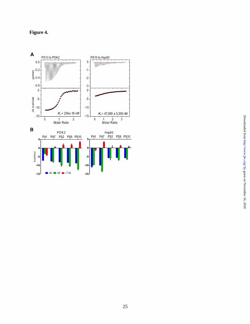
PS-series inhibitors show favorable binding
enthalpies for PDK2 - The binding of PDK
inhibitors to PDK2 or Hsp90 was measured by
isothermal titration calorimetry (ITC). The fitting
of binding isotherms (Fig. 4A) showed a distinctly
higher affinity of PS10 for PDK2 (Kd = 239 nM)
than for Hsp90 (Kd = 47,000 nM). The binding
enthalpy (ΔH in kcal/mol) of PS10 for Hsp90 is
also shown much smaller than that for PDK2 (Fig.
4A). The development of compounds from PA1
toward PS10 on the same chemical scaffold was
accompanied by the steadily more favorable (i.e.
more negative) thermodynamic signatures in terms
of binding enthalpies (ΔH) and Gibbs binding
energies (ΔG), when titrated into PDK2, although
the term of binding entropies (-TΔS) become less
favorable (or more positive) (Fig. 4B, left panel).
These gains in binding enthalpy indicate the
progressively more favorable interactions between
PDK2 and the inhibitors as PA1 is evolved into
PS10, despite the accompanied relatively small
entropic penalties (40). In contrast, the conversion
from carbonyl group-containing compounds (PA1
and PA7) to the sulfonyl group-harboring
counterparts (PS2, PS8, and PS10) results in
significant losses of binding enthalpies for Hsp90
(Fig, 3B, right panel). The favorable binding
enthalpies further support the vastly improved
selectivity of PS8 and PS10 as PDK inhibitors
over the parental compound PA1.
PS-series inhibitors show high selectivity for
PDK isoforms - The selectivity of PS8 was studied
by determining IC50 for the inhibition of a 21-
kinase panel including PDK2. PS8 shows the
lowest IC50 of 70 nM for PDK2 under the assay
conditions with myelin-binding protein as an
artificial susbstrate (Fig. 5). All other kinases on
the panel show at least 3 orders of magnitude
higher IC50 values for inhibition by PS8. The
results established the specificity for PS8 as a
PDK2 inhibitor. To dissect the specificity of PS-
series inhibitors against the four PDK isoforms, in
vitro kinase assays were performed with or
without the E2/E3BP core of PDC. The E3
component is not included in the assay mixture,
since E3 is not required for our kinase activity
assay that measures incorporation of the 32
P-
phosphoryl group into the E1 protein. PS8 inhibits
all four isoforms at sub-micromolar to low
micromolar range (Table 3). Except PDK4, PDK
isoforms anchor to the E2/E3BP of PDC for
optimal kinase activity in vivo. In the presence of
E2/E3BP, PS8 is a more effective inhibitor for all
PDK isoforms than in the absence of E2/E3BP,
particularly for PDK3. The most improved IC50 for
PDK3 in the presence of E2/E3BP among the four
PDK isoforms may be explained by the markedly
reduced affinity of PDK3 for ATP/ADP, when this
PDK isoform is bound to the PDC core (29). PS10
shows the similar IC50 values for the inhibition of
four PDK isoforms when assayed in absence of
E2/E3BP (Table 3). The above results, taking
together, indicate that both PS8 and PS10 are pan-
PDK inhibitors.
To assess possible toxicity of PS-series
compounds due to non-specific interactions, HeLa
and HBEC30 cells were titrated with PS8. The
IC50 values for the growth inhibition of HeLa and
HBEC30 cells by PS8 are 223 μM and 253 μM,
respectively. The toxicity of PS8 is 100-fold less
potent than cycloheximide in both cell lines.
Similarly, PS10 shows an IC50 of 284 μM for the
growth inhibition of HeLa cells. These results
suggest that the toxicity of PS8 and PS10 is
minimal in vivo.
Pharmacokinetic properties of PS8 and
PS10 - PS8 and PS10 both show half-lives of
greater than 240 min in vitro in hepatic S9
fractions (data not shown), which suggest that
neither is extensively metabolized by phase I
oxidative or reductive metabolism. In vivo, both
compounds show a rapid distribution phase,
followed by a slower terminal elimination phase
after IP delivery. The pharmacokinetic parameters
on Table 4 show that the distribution and
elimination of PS8 was slightly more rapid than
PS10, possibly due to its somewhat more
hydrophobic nature. Both compounds show good
plasma exposure (AUClast) as well as a volume of
distribution, which is suggestive of modest tissue
penetration (Table 4).
PS10 stimulates PDC activity in tissues of
DIO mice-Both PS8 and PS10 show good IC50 for
the four PDK isoforms (Table 3); however, PS10
was chosen for in vivo studies because of its better
solubility in DMSO used in the formulation. Male
C57BL/6J mice were fed a high-fat diet for three
weeks to produce DIO model with impaired
glucose tolerance. These DIO mice were initially
treated with a single dose of either vehicle or PS10
by guest on Novem
ber 16, 2018http://w
ww
.jbc.org/D
ownloaded from

8
(70 mg/kg) by IP injection. The animals were
sacrificed 10 h later in the early morning in the fed
state. Maximal enhancement of PDC activity by
PS10 in tissues was achieved under these
conditions. Tissues (heart, liver, kidneys and
quadriceps muscle) were harvested and analyzed
for PDC activity by the radiochemical assay with
[1-14
C]pyruvate as a substrate. Fig. 6A (top) shows
that PDC activity was low in the heart and liver
from vehicle-treated DIO mice. PS10 treatments
result in 11-fold and 23-fold higher PDC activity
in heart and liver, respectively than the vehicle-
treated. There is a 1.4-fold enhancement of PDC
activity in PS10-treated kidneys compared with
vehicle-treated. In contrast, there is no difference
of PDC activity in quadriceps muscle between
PS10-treated and vehicle-treated DIO mice. The
elevated PDC activity correlates with significantly
decreased amounts of the phosphorylated E1α
subunit in heart and liver of PS10 treated DIO
mice compared with vehicle-treated (Fig. 6A,
bottom). These results corroborate that PS10
functions as a PDK inhibitor in vivo to attenuate
phosphorylation levels of the E1α subunit, leading
to stimulated PDC activity in DIO mice. The
increased PDC activity is not due to enhanced
phosphatase activity, since PS10 at up to 1 mM is
without effect on PDP1 activity in vitro. In the
next series of experiments, DIO mice were treated
with vehicle or PS10 (70 mg/kg/day) for three
days and tissues were collected for biochemical
studies. As shown in Fig. 6B, except in the heart,
PDC activity profiles and the phospho-E1α
subunit level are similar between the single-dose
and multiple-dose treatments of DIO mice with
PS10. In the heart, the prolonged PS10 treatment
appears to attenuate the enhancement of PDK
activity compared to the single administration of
the compound.
PS10 increases glucose tolerance and
lessens hepatic steatosis in DIO mice - DIO mice
on a high fat diet for 10 weeks were treated with
vehicle or PS10 (70 mg/kg/day) by IP injections
for four additional weeks and subjected to a
glucose tolerance test. The vehicle and PS10
treatments were continued for 2 more days and,
animals were sacrificed in the early morning while
in the fed state, and tissues and blood were
collected for biochemical studies. Results from the
glucose tolerance tests (Fig. 7A) show that when
challenged with 1.5 g/kg of glucose, the plasma
glucose level in the vehicle-treated control was at
200 mg/dl at 0 min, peaked at 482 mg/dl at 30 min
and reduced to 210 mg/dl at 120 min. In PS10-
treated DIO mice, the glucose level at 168 mg/dl at
0 min was lower than that in vehicle-treated
animals, reached 312 mg/dl at 30 min and returned
to 163 mg/dl at 120 min. The two groups of
animals show significant differences in the glucose
levels at 30, 60, and 120 min, with lower glucose
levels uniformly observed in the PS10-treated DIO
mice. The data therefore suggest that the PS10
treatment increases glucose tolerance over vehicle-
treated DIO mice. Notably, there are no significant
differences in food intake (Fig. 7B) and body
weight (Fig. 7C) between the vehicle- and PS10-
treated animals. DIO mice treated with PS10 also
showed significantly lower plasma lactate (Fig.
7D), cholesterol (Fig. 7E) and triglycerides (Fig.
7F) levels and a reduction in fat-mass (Fig. 7G),
compared with the mice treated with vehicle.
Moreover, larger amounts of fat were present in
the liver of the vehicle-treated DIO mice
compared with PS10-treated, when the liver slices
were stained with Oil Red O (Fig. 7H). The
accumulated hepatic fat was primarily
macrovesicular in vehicle-treated DIO mice and
became microvesicular in the PS10-treated
counterpart.
DISCUSSION
With the increased understanding that PDKs
play a pivotal role in controlling glucose oxidation
in disease states such as diabetes (3-5), cancer
(8,9,11-14) and congestive heart failure, there is
growing need for effective PDK inhibitors. The
classic PDK inhibitor DCA binds to an enclosed
allosteric site in the N-terminal domain, which is
the binding site of PDC’s substrate pyruvate for
the physiological feedback inhibition (16,17).
However, this allosteric site is relatively small
(volume = 211Å3), also buried and can only
accommodate small ligands such as pyruvate and
DCA (Fig. 8). The space limitation in the PDK
allosteric site precludes the structure-based
modification of DCA to improve its potency as a
PDK inhibitor. The strategy of developing PDK
inhibitors by targeting the lipoyl-binding pocket
was unsuccessful in vivo (24). PDK2 and PDK3
are anchored to lipoyl-bearing domains on the
E2/E3BP core of PDC for optimal kinase activity
(29,41). Dihydrolipoamide mimetics attenuate
by guest on Novem
ber 16, 2018http://w
ww
.jbc.org/D
ownloaded from

9
PDK2 and PDK3 activities by impeding binding
of these PDK isoforms to the inner lipoyl-bearing
domain of the E3/E3BP core (42). However,
PDK4, which is up-regulated in obesity and
diabetes (3-5) shows robust kinase activity without
binding to inner lipoyl- bearing domain of the
E2E3BP core. The binding of the
dihydrolipoamide mimetic AZD7545 to PDK4
stimulates rather than inhibits its kinase activity
considerably (26).
In the present study, we undertook a
different approach to develop a new generation of
PDK inhibitors that dock to the ATP-binding
pocket (volume = 865 Å3) of PDK2, which is open
and four times larger than the allosteric DCA-
binding pocket (Fig. 8). The conservation in the
ATP-binding pocket between Hsp90 and PDK2,
both belonging to the GHKL ATPase/kinase
superfamily (27), makes it possible to utilize the
chemical scaffold in Hsp90 inhibitors PA1 and
PA7 as the starting point for designing the PDK-
specific inhibitors. The distinct conformations of
the bound PA7 between the Hsp90-PA7 and
PDK2-PA7 structures (Fig. 2D) provided the first
clue for utilizing structure-based design to develop
PDK-specific inhibitors. It is remarkable that a
single replacement of the carbonyl group in PA7
with a sulfonyl group in PS2 results in a drastic
conversion of an Hsp90 inhibitor (PA7) to a PDK
inhibitor (PS2). Our results epitomize the
feasibility of designing a highly selective kinase
inhibitor by taking advantage of the unique
structural features in the ATP-binding pocket.
The in vivo efficacy of PDK inhibitor PS10
was evaluated in DIO mice. DIO mice fed high-fat
diet develop symptoms characteristic of the
metabolic syndrome and if left on the diet long
enough will eventually develop type 2 diabetes
(43). The modest selectivity of PS10 for the four
PDK isoforms is desirable, since PDK2/PDK4
double knockout mice showed far more robust
improvement in glucose tolerance and insulin
sensitivity than the single PDK2 or PDK4
knockout mice (7). PDC activity is low in heart
and liver from DIO mice fed a high-fat diet for
three weeks (Fig. 6A and 6B). The effectiveness of
PS10 as a PDK inhibitor in vivo is established by
higher PDC activity in most tissues from PS10-
treated over vehicle-treated DIO mice. The
stimulation of PDC activity in liver and heart is
compelling, given that PS10 is competitive with
ATP, which is usually present at 1-10 mM in the
mitochondria matrix (44,45). The results may be
explained by the accumulation of PS10 in the
mitochondria matrix. It is equally plausible that
the modest inhibition of PDKs by PS10 in vivo is
sufficient to tip the balance between PDKs and
PDPs in favor of the latter, resulting in the
significant dephosphorylation of PDC.
The increased PDC flux in liver promotes
glucose disposal, leading to improved glucose
tolerance in PS10-treated DIO mice (Fig. 7A). The
reduced plasma level of the gluconeogenic
substrate lactate (Fig. 7D) explains in part the
lower glucose concentrations in PS10-treated DIO
mice (Fig. 7A). In the heart, a single dose of PS10
treatment results in drastic enhancement of PDC
activity (Fig. 6A); however, the prolonged PS10
treatment causes a reduction in the fold increase of
PDC activity (Fig. 6B). The results suggest a reset
of the cardiac PDC flux during the long-term PS10
dosing. PDK4 expression is up-regulated in right
ventricular hypertrophy causing an increase in
glycolysis over glucose oxidation (46). The
increased cardiac PDC flux by PS10 may offer an
approach to mitigating impaired glucose oxidation
in congenital heart failure.
The above increased glucose disposal
through enhanced PDC flux is coupled with
decreased lipogenesis in PS10-treated DIO mice,
as demonstrated by the lessened hepatic steatosis,
lower fat mass and attenuated plasma cholesterol
as well as triglycerides levels (Fig. 7D-H). The
combination of reduced lipogenesis and increased
glucose oxidation has been reported in acetyl-CoA
carboxylase 2 (47) or PDK4 (48) knockout mice
on high-fat diets. In the liver of PDK4-deficient
mice, the expression of both fatty acid synthase
and acetyl-CoA carboxylase is reduced, which
likely results in reduced lipogenesis with improved
hepatic steatosis (43). Interestingly, the two
transcription factors that promote fatty acid
oxidation, i.e. PPARα and PGC-1α (49) in PDK4-
deficient mice fed the high-fat diet are restored to
the levels of the wild-type animals on chow diet
(48). The excess acetyl-CoA from both glucose
and fatty acid oxidation is converted to ketone
bodies, since plasma concentrations of both β-
hydroxybutyrate and acetoacetate were
considerably elevated in PDK2/PDK4 double
knock-out mice compared to the wild-type (7).
Taken together, the present results illustrate the
by guest on Novem
ber 16, 2018http://w
ww
.jbc.org/D
ownloaded from
10
therapeutic potentials of PDK inhibitors in
increasing hepatic glucose oxidation through PDC
flux while suppressing lipogenesis in the liver of
diet-induced obesity.
by guest on Novem
ber 16, 2018http://w
ww
.jbc.org/D
ownloaded from

11
REFERENCES
1. Reed, L. J. (2001) A trail of research from lipoic acid to alpha-keto acid dehydrogenase
complexes. The Journal of biological chemistry 276, 38329-38336
2. Randle, P. J. (1995) Metabolic fuel selection: general integration at the whole-body level.
The Proceedings of the Nutrition Society 54, 317-327
3. Harris, R. A., Hawes, J. W., Popov, K. M., Zhao, Y., Shimomura, Y., Sato, J., Jaskiewicz,
J., and Hurley, T. D. (1997) Studies on the regulation of the mitochondrial alpha-ketoacid
dehydrogenase complexes and their kinases. Adv Enzyme Regul 37, 271-293
4. Wu, P., Inskeep, K., Bowker-Kinley, M. M., Popov, K. M., and Harris, R. A. (1999)
Mechanism responsible for inactivation of skeletal muscle pyruvate dehydrogenase
complex in starvation and diabetes. Diabetes 48, 1593-1599
5. Rosa, G., Di Rocco, P., Manco, M., Greco, A. V., Castagneto, M., Vidal, H., and
Mingrone, G. (2003) Reduced PDK4 expression associates with increased insulin
sensitivity in postobese patients. Obesity research 11, 176-182
6. Kuzuya, T., Katano, Y., Nakano, I., Hirooka, Y., Itoh, A., Ishigami, M., Hayashi, K.,
Honda, T., Goto, H., Fujita, Y., Shikano, R., Muramatsu, Y., Bajotto, G., Tamura, T.,
Tamura, N., and Shimomura, Y. (2008) Regulation of branched-chain amino acid
catabolism in rat models for spontaneous type 2 diabetes mellitus. Biochemical and
biophysical research communications 373, 94-98
7. Jeoung, N. H., Rahimi, Y., Wu, P., Lee, W. N., and Harris, R. A. (2012) Fasting induces
ketoacidosis and hypothermia in PDHK2/PDHK4-double-knockout mice. Biochem J 443,
829-839
8. Papandreou, I., Cairns, R. A., Fontana, L., Lim, A. L., and Denko, N. C. (2006) HIF-1
mediates adaptation to hypoxia by actively downregulating mitochondrial oxygen
consumption. Cell metabolism 3, 187-197
9. Kim, J. W., Tchernyshyov, I., Semenza, G. L., and Dang, C. V. (2006) HIF-1-mediated
expression of pyruvate dehydrogenase kinase: a metabolic switch required for cellular
adaptation to hypoxia. Cell metabolism 3, 177-185
10. Kaplon, J., Zheng, L., Meissl, K., Chaneton, B., Selivanov, V. A., Mackay, G., van der
Burg, S. H., Verdegaal, E. M., Cascante, M., Shlomi, T., Gottlieb, E., and Peeper, D. S.
(2013) A key role for mitochondrial gatekeeper pyruvate dehydrogenase in oncogene-
induced senescence. Nature 498, 109-112
11. Michelakis, E. D., Sutendra, G., Dromparis, P., Webster, L., Haromy, A., Niven, E.,
Maguire, C., Gammer, T. L., Mackey, J. R., Fulton, D., Abdulkarim, B., McMurtry, M. S.,
and Petruk, K. C. (2010) Metabolic modulation of glioblastoma with dichloroacetate.
Science translational medicine 2, 31ra34
12. Lu, C. W., Lin, S. C., Chen, K. F., Lai, Y. Y., and Tsai, S. J. (2008) Induction of pyruvate
dehydrogenase kinase-3 by hypoxia-inducible factor-1 promotes metabolic switch and
drug resistance. The Journal of biological chemistry 283, 28106-28114
13. Hitosugi, T., Fan, J., Chung, T. W., Lythgoe, K., Wang, X., Xie, J., Ge, Q., Gu, T. L.,
Polakiewicz, R. D., Roesel, J. L., Chen, G. Z., Boggon, T. J., Lonial, S., Fu, H., Khuri, F.
R., Kang, S., and Chen, J. (2011) Tyrosine phosphorylation of mitochondrial pyruvate
dehydrogenase kinase 1 is important for cancer metabolism. Molecular cell 44, 864-877
14. Bonnet, S., Archer, S. L., Allalunis-Turner, J., Haromy, A., Beaulieu, C., Thompson, R.,
Lee, C. T., Lopaschuk, G. D., Puttagunta, L., Bonnet, S., Harry, G., Hashimoto, K.,
by guest on Novem
ber 16, 2018http://w
ww
.jbc.org/D
ownloaded from

12
Porter, C. J., Andrade, M. A., Thebaud, B., and Michelakis, E. D. (2007) A mitochondria-
K+ channel axis is suppressed in cancer and its normalization promotes apoptosis and
inhibits cancer growth. Cancer cell 11, 37-51
15. Whitehouse, S., and Randle, P. J. (1973) Activation of pyruvate dehydrogenase in
perfused rat heart by dichloroacetate (Short Communication). Biochem J 134, 651-653
16. Kato, M., Li, J., Chuang, J. L., and Chuang, D. T. (2007) Distinct structural mechanisms
for inhibition of pyruvate dehydrogenase kinase isoforms by AZD7545, dichloroacetate,
and radicicol. Structure 15, 992-1004
17. Knoechel, T. R., Tucker, A. D., Robinson, C. M., Phillips, C., Taylor, W., Bungay, P. J.,
Kasten, S. A., Roche, T. E., and Brown, D. G. (2006) Regulatory roles of the N-terminal
domain based on crystal structures of human pyruvate dehydrogenase kinase 2 containing
physiological and synthetic ligands. Biochemistry 45, 402-415
18. Jiang, D. K., Sun, J., Cao, G., Liu, Y., Lin, D., Gao, Y. Z., Ren, W. H., Long, X. D.,
Zhang, H., Ma, X. P., Wang, Z., Jiang, W., Chen, T. Y., Gao, Y., Sun, L. D., Long, J. R.,
Huang, H. X., Wang, D., Yu, H., Zhang, P., Tang, L. S., Peng, B., Cai, H., Liu, T. T.,
Zhou, P., Liu, F., Lin, X., Tao, S., Wan, B., Sai-Yin, H. X., Qin, L. X., Yin, J., Liu, L.,
Wu, C., Pei, Y., Zhou, Y. F., Zhai, Y., Lu, P. X., Tan, A., Zuo, X. B., Fan, J., Chang, J.,
Gu, X., Wang, N. J., Li, Y., Liu, Y. K., Zhai, K., Zhang, H., Hu, Z., Liu, J., Yi, Q., Xiang,
Y., Shi, R., Ding, Q., Zheng, W., Shu, X. O., Mo, Z., Shugart, Y. Y., Zhang, X. J., Zhou,
G., Shen, H., Zheng, S. L., Xu, J., and Yu, L. (2013) Genetic variants in STAT4 and
HLA-DQ genes confer risk of hepatitis B virus-related hepatocellular carcinoma. Nature
genetics 45, 72-75
19. Stacpoole, P. W., Barnes, C. L., Hurbanis, M. D., Cannon, S. L., and Kerr, D. S. (1997)
Treatment of congenital lactic acidosis with dichloroacetate. Archives of disease in
childhood 77, 535-541
20. Korotchkina, L. G., Sidhu, S., and Patel, M. S. (2004) R-lipoic acid inhibits mammalian
pyruvate dehydrogenase kinase. Free radical research 38, 1083-1092
21. Ferriero, R., Manco, G., Lamantea, E., Nusco, E., Ferrante, M. I., Sordino, P., Stacpoole,
P. W., Lee, B., Zeviani, M., and Brunetti-Pierri, N. (2013) Phenylbutyrate therapy for
pyruvate dehydrogenase complex deficiency and lactic acidosis. Science translational
medicine 5, 175ra131
22. Iannitti, T., and Palmieri, B. (2011) Clinical and experimental applications of sodium
phenylbutyrate. Drugs in R&D 11, 227-249
23. Mayers, R. M., Butlin, R. J., Kilgour, E., Leighton, B., Martin, D., Myatt, J., Orme, J. P.,
and Holloway, B. R. (2003) AZD7545, a novel inhibitor of pyruvate dehydrogenase
kinase 2 (PDHK2), activates pyruvate dehydrogenase in vivo and improves blood glucose
control in obese (fa/fa) Zucker rats. Biochemical Society transactions 31, 1165-1167
24. Aicher, T. D., Anderson, R. C., Gao, J., Shetty, S. S., Coppola, G. M., Stanton, J. L.,
Knorr, D. C., Sperbeck, D. M., Brand, L. J., Vinluan, C. C., Kaplan, E. L., Dragland, C. J.,
Tomaselli, H. C., Islam, A., Lozito, R. J., Liu, X., Maniara, W. M., Fillers, W. S.,
DelGrande, D., Walter, R. E., and Mann, W. R. (2000) Secondary amides of (R)-3,3,3-
trifluoro-2-hydroxy-2-methylpropionic acid as inhibitors of pyruvate dehydrogenase
kinase. Journal of medicinal chemistry 43, 236-249
25. Kato, M., Wynn, R. M., Chuang, J. L., Tso, S. C., Machius, M., Li, J., and Chuang, D. T.
(2008) Structural basis for inactivation of the human pyruvate dehydrogenase complex by
phosphorylation: role of disordered phosphorylation loops. Structure 16, 1849-1859
by guest on Novem
ber 16, 2018http://w
ww
.jbc.org/D
ownloaded from

13
26. Wynn, R. M., Kato, M., Chuang, J. L., Tso, S. C., Li, J., and Chuang, D. T. (2008)
Pyruvate dehydrogenase kinase-4 structures reveal a metastable open conformation
fostering robust core-free basal activity. The Journal of biological chemistry 283, 25305-
25315:PMC2533096
27. Dutta, R., and Inouye, M. (2000) GHKL, an emergent ATPase/kinase superfamily.
Trends in biochemical sciences 25, 24-28
28. Steussy, C. N., Popov, K. M., Bowker-Kinley, M. M., Sloan, R. B., Jr., Harris, R. A., and
Hamilton, J. A. (2001) Structure of pyruvate dehydrogenase kinase. Novel folding pattern
for a serine protein kinase. The Journal of biological chemistry 276, 37443-37450
29. Kato, M., Chuang, J. L., Tso, S. C., Wynn, R. M., and Chuang, D. T. (2005) Crystal
structure of pyruvate dehydrogenase kinase 3 bound to lipoyl domain 2 of human
pyruvate dehydrogenase complex. The EMBO journal 24, 1763-1774
30. Kung, P. P., Huang, B., Zhang, G., Zhou, J. Z., Wang, J., Digits, J. A., Skaptason, J.,
Yamazaki, S., Neul, D., Zientek, M., Elleraas, J., Mehta, P., Yin, M. J., Hickey, M. J.,
Gajiwala, K. S., Rodgers, C., Davies, J. F., and Gehring, M. R. (2010)
Dihydroxyphenylisoindoline amides as orally bioavailable inhibitors of the heat shock
protein 90 (hsp90) molecular chaperone. Journal of medicinal chemistry 53, 499-503
31. Murray, C. W., Carr, M. G., Callaghan, O., Chessari, G., Congreve, M., Cowan, S., Coyle,
J. E., Downham, R., Figueroa, E., Frederickson, M., Graham, B., McMenamin, R.,
O'Brien, M. A., Patel, S., Phillips, T. R., Williams, G., Woodhead, A. J., and Woolford,
A. J. (2010) Fragment-based drug discovery applied to Hsp90. Discovery of two lead
series with high ligand efficiency. Journal of medicinal chemistry 53, 5942-5955
32. Keller, S., Vargas, C., Zhao, H., Piszczek, G., Brautigam, C. A., and Schuck, P. (2012)
High-precision isothermal titration calorimetry with automated peak-shape analysis.
Analytical chemistry 84, 5066-5073
33. Tso, S. C., Qi, X., Gui, W. J., Chuang, J. L., Morlock, L. K., Wallace, A. L., Ahmed, K.,
Laxman, S., Campeau, P. M., Lee, B. H., Hutson, S. M., Tu, B. P., Williams, N. S.,
Tambar, U. K., Wynn, R. M., and Chuang, D. T. (2013) Structure-based design and
mechanisms of allosteric inhibitors for mitochondrial branched-chain alpha-ketoacid
dehydrogenase kinase. Proceedings of the National Academy of Sciences of the United
States of America 110, 9728-9733
34. Li, J., Kato, M., and Chuang, D. T. (2009) Pivotal role of the C-terminal DW-motif in
mediating inhibition of pyruvate dehydrogenase kinase 2 by dichloroacetate. The Journal
of biological chemistry 284, 34458-34467
35. Kukimoto-Niino, M., Tokmakov, A., Terada, T., Ohbayashi, N., Fujimoto, T., Gomi, S.,
Shiromizu, I., Kawamoto, M., Matsusue, T., Shirouzu, M., and Yokoyama, S. (2011)
Inhibitor-bound structures of human pyruvate dehydrogenase kinase 4. Acta
crystallographica. Section D, Biological crystallography 67, 763-773
36. Dymock, B. W., Barril, X., Brough, P. A., Cansfield, J. E., Massey, A., McDonald, E.,
Hubbard, R. E., Surgenor, A., Roughley, S. D., Webb, P., Workman, P., Wright, L., and
Drysdale, M. J. (2005) Novel, potent small-molecule inhibitors of the molecular
chaperone Hsp90 discovered through structure-based design. Journal of medicinal
chemistry 48, 4212-4215
37. Feldman, R. I., Mintzer, B., Zhu, D., Wu, J. M., Biroc, S. L., Yuan, S., Emayan, K.,
Chang, Z., Chen, D., Arnaiz, D. O., Bryant, J., Ge, X. S., Whitlow, M., Adler, M.,
Polokoff, M. A., Li, W. W., Ferrer, M., Sato, T., Gu, J. M., Shen, J., Tseng, J. L., Dinter,
by guest on Novem
ber 16, 2018http://w
ww
.jbc.org/D
ownloaded from

14
H., and Buckman, B. (2009) Potent triazolothione inhibitor of heat-shock protein-90.
Chemical biology & drug design 74, 43-50
38. Hiromasa, Y., and Roche, T. E. (2008) Critical role of specific ions for ligand-induced
changes regulating pyruvate dehydrogenase kinase isoform 2. Biochemistry 47, 2298-
2311
39. Green, T., Grigorian, A., Klyuyeva, A., Tuganova, A., Luo, M., and Popov, K. M. (2008)
Structural and functional insights into the molecular mechanisms responsible for the
regulation of pyruvate dehydrogenase kinase 2. The Journal of biological chemistry 283,
15789-15798
40. Freire, E. (2008) Do enthalpy and entropy distinguish first in class from best in class?
Drug discovery today 13, 869-874
41. Baker, J. C., Yan, X., Peng, T., Kasten, S., and Roche, T. E. (2000) Marked differences
between two isoforms of human pyruvate dehydrogenase kinase. The Journal of
biological chemistry 275, 15773-15781
42. Tuganova, A., Klyuyeva, A., and Popov, K. M. (2007) Recognition of the inner lipoyl-
bearing domain of dihydrolipoyl transacetylase and of the blood glucose-lowering
compound AZD7545 by pyruvate dehydrogenase kinase 2. Biochemistry 46, 8592-8602
43. Wang, C. Y., and Liao, J. K. (2012) A mouse model of diet-induced obesity and insulin
resistance. Methods Mol Biol 821, 421-433
44. Kennedy, H. J., Pouli, A. E., Ainscow, E. K., Jouaville, L. S., Rizzuto, R., and Rutter, G.
A. (1999) Glucose generates sub-plasma membrane ATP microdomains in single islet
beta-cells. Potential role for strategically located mitochondria. The Journal of biological
chemistry 274, 13281-13291
45. Metelkin, E., Demin, O., Kovacs, Z., and Chinopoulos, C. (2009) Modeling of ATP-ADP
steady-state exchange rate mediated by the adenine nucleotide translocase in isolated
mitochondria. The FEBS journal 276, 6942-6955
46. Piao, L., Sidhu, V. K., Fang, Y. H., Ryan, J. J., Parikh, K. S., Hong, Z., Toth, P. T.,
Morrow, E., Kutty, S., Lopaschuk, G. D., and Archer, S. L. (2013) FOXO1-mediated
upregulation of pyruvate dehydrogenase kinase-4 (PDK4) decreases glucose oxidation
and impairs right ventricular function in pulmonary hypertension: therapeutic benefits of
dichloroacetate. J Mol Med (Berl) 91, 333-346
47. Choi, C. S., Savage, D. B., Abu-Elheiga, L., Liu, Z. X., Kim, S., Kulkarni, A., Distefano,
A., Hwang, Y. J., Reznick, R. M., Codella, R., Zhang, D., Cline, G. W., Wakil, S. J., and
Shulman, G. I. (2007) Continuous fat oxidation in acetyl-CoA carboxylase 2 knockout
mice increases total energy expenditure, reduces fat mass, and improves insulin
sensitivity. Proceedings of the National Academy of Sciences of the United States of
America 104, 16480-16485
48. Hwang, B., Jeoung, N. H., and Harris, R. A. (2009) Pyruvate dehydrogenase kinase
isoenzyme 4 (PDHK4) deficiency attenuates the long-term negative effects of a high-
saturated fat diet. Biochem J 423, 243-252
49. Lin, J., Handschin, C., and Spiegelman, B. M. (2005) Metabolic control through the
PGC-1 family of transcription coactivators. Cell metabolism 1, 361-370
50. Dundas, J., Ouyang, Z., Tseng, J., Binkowski, A., Turpaz, Y., and Liang, J. (2006)
CASTp: computed atlas of surface topography of proteins with structural and
topographical mapping of functionally annotated residues. Nucleic acids research 34,
W116-118
by guest on Novem
ber 16, 2018http://w
ww
.jbc.org/D
ownloaded from

15
Acknowledgments - Crystal structures presented in this report are derived from work performed at
Argonne National Laboratory, Structural Biology Center at the Advanced Photon Source, operated under
Department of Energy contract DE-AC02-06CH11357
FOOTNOTES
*This work was supported by grants DK62306, DK26758, DK55758, DK92921 and GM102604 from the
National Institutes of Health and grants I-1286 and I-1748 from the Welch Foundation and the Sloan
Research Fellowship. I.W.A. was supported with a fellowship from the Throne-Host Foundation, the
Swedish Research Council (2006-3931) and from VINNOVA (Marie Curie Qualification).
□S This article contains Supplemental Data.
The atomic coordinates and structure factors (codes 4MP2, 4MP7, 4MPC, 4MPE and 4MPN) have been
deposited in the Protein Data Bank (http://wwpdb.org/).
1These authors contributed equally to the study.
2Correspondence may be addressed to U.K.T. ([email protected]).
3Correspondence may be addressed to D.T.C ([email protected]).
4 The abbreviations used are; PDC, pyruvate dehydrogenase complex; E1, pyruvate dehydrogenase; E2,
dihydrolipoyl transacetylase; E3, dihydrolipoamide dehydrogenase; E3BP, E3-binding protein; PDK,
pyruvate dehydrogenase kinase; PDP, pyruvate dehydrogenase phosphatase; PPAR, peroxisome
proliferator-activate receptor; FOXO, fork-head box O; PGC-1, PPAR γ coactivator-1; DCA,
dichloroacetate; Hsp90, heat shock protein 90; DIO, diet-induced obese; TEV, tobacco-etch-virus
protease, ITC, isothermal titration calorimetry; ACD, acidified citrate dextrose; IP, intraperitoneal; OCT,
optimal cutting temperature compound.
FIGURE LEGENDS
Figure 1. Structure of PDK2 and known and novel inhibitors. (A) The PDK dimer showing AZD7545,
and dichloroacetate- binding sites in N-terminal domain (pink); and radicicol bound to the ATP-binding
pocket in the C-terminal domain (green). (B) Chemical structures of known PDK inhibitors: DCA,
AZD7545, compound 3, radicicol, and M77976; and novel PDK inhibitors: DC23, PA1, PA7, PS2, PS8,
and PS10. The resorcinol ring is indicated in red and isoindoline moiety in blue.
Figure 2. Comparison of inhibitor-binding pockets in PDK2 and Hsp90. (A) Superimposition of the
C-terminal domain of PDK2 (green) harboring PA7 (pink) with the N-terminal domain of Hsp90 (orange)
with bound PA7 (cyan). (B) PA7 in Hsp90 (C) PA7 in PDK2, (D) superimposition of Hsp90-bound
(cyan) and PDK2-bound PA7 (pink) (left), and the structure of PDK2-bound PS2 (right).
Figure 3. Stereo views of inhibitor-binding pockets in PDK2 and Hsp90. (A) The Hsp90-PA7
structure (30). (B) The PDK2-PA1 structure with Fo-Fc density map (green mesh) contoured to 4 σ. (C)
PDK2-PA7 structure contoured to 4 σ. (D) The PDK2-PS2 structure contoured to 4 σ. (E) The PDK2-PS8
structure contoured to 3 σ. (F) The PDK2-PS10 structure contoured to 3 σ. w, ordered water molecule
Figure 4. Thermodynamics analysis of inhibitor binding to PDK2 and Hsp90. (A) Thermograms of
PS10 binding to PDK2 and Hsp90 obtained by ITC. (B) Thermodynamic signatures of inhibitor bindings
by guest on Novem
ber 16, 2018http://w
ww
.jbc.org/D
ownloaded from

16
to PDK2 (left panel) and Hsp90 (right panel), ΔG, Gibbs binding energy; ΔH, binding enthalpy; ΔS,
binding entropy; T, absolute temperature.
Figure 5. Kinase profiling of compound PS8. Inhibition of the 21 representative kinases, including
PDK2, in the Human Kinome by PS8 were measured in the concentration range of 15 nM to 300 µM.
IC50 values for each kinase were derived from individual inhibition curves. The IC50 for PDK2 is at least 3
order magnitudes lower than next lowest value for CDK1/cyclin B.
Figure 6. Enhanced PDC activity with reduced phosphorylation level in PS10-treated DIO mice. (A)
Short-term response. C57BL/6J male mice were fed high-fat diet for 3 weeks and treated with vehicle (V,
n=4) or PS10 at 70 mg/kg (T, n=4) by a single IP injection while they had free access to food. Animals
were sacrificed at 10 AM, i.e.10 h after the injection. Tissues were harvested and analyzed for PDC
activity and phosphorylation levels of E1α subunit. Upper panel, PDC activity in heart, liver, kidney and
muscle. Lower panel, amounts of the phosphorylated (p-E1) and total (E1) E1α subunit in different tissues
determined by Western blotting analysis. (B) Long-term response. C57BL/6J male mice were fed high-fat
diet for 10 weeks and then treated with vehicle (n=3) or PS10 at 70 mg/kg/day (n=3) for 3 days. The
remaining procedures and result presentation are as in (A). **, P<0.01; *, P<0.05.
Figure 7. Glucose- and lipid-controlling properties of PS10. (A) Glucose tolerance test. C57BL/6J
male mice were fed a high-fat diet for 10 weeks and treated with vehicle (n=4) or PS10 at 70 mg/kg/day
(n=6) for 4 weeks and were fasted for 6 h followed by injection of 1.5 g glucose/kg by IP injection. Blood
glucose levels were monitored at 0-2 h after the glucose injection. (B) Food intake of DIO mice fed the
high-fat diet for 10 weeks followed by treatments with vehicle (n=5) or PS10 at 70 mg/kg/day (n=5) for
one week. (C) Body weight change in DIO mice from (A) after 6 week of treatments with vehicle or PS10.
(D) Plasma lactate concentrations in DIO mice from (B). Plasma lactate concentrations were determined
as described in the Methods. (E) Plasma cholesterol concentration in DIO mice from (C). (F) Plasma
triglycerides concentrations in DIO mice also from (C). (G) Change in the fat mass. DIO mice were
treated as in (B). Fat mass was determined as described in Methods. (H) Oil Red O stains of liver slices
from vehicle- and PS10-treated DIO mice as in (B). ***, P<0.001; **, P<0.01; *, P<0.05; ns, not
significant statistically.
Figure 8. Calculated volumes of the DCA-binding and ATP-binding pockets in PDK2. The N-terminal
domain of the PDK2 monomer with the allosteric site occupied by DCA is derived from coordinates of
PDB code: 2BU8. PDK inhibitor PS10 was modeled into the ATP-binding pocket in the C-terminal
domain of the same monomer, according to the PS10 coordinates of PDB code: 4MPN from this study.
The volumes of the DCA- binding (211 Å3) and ATP-binding (865 Å
3) pockets, as represented by blue
meshes, were computed using program CASTp (50).
by guest on Novem
ber 16, 2018http://w
ww
.jbc.org/D
ownloaded from

17
Table 1. IC50 and dissociation constants of known and novel PDK inhibitors
Compound IC50, μM
for PDK2
IC50, μM
for PDK4
Kd, nM
for PDK2
Kd, nM
for Hsp 90
DCA* 183 80.0 -- --
Radicicol * 77.8 -- 18,600 ± 3,200 46.3 ± 7.2
DC23 3.82 0.28 6,760 ± 2,040 25.0 ± 10.1
PA1 6.78 1.86 3,570 ± 560 6.0 ± 2.6
PA7 5.68 1.05 1,827 ± 179 27.3 ± 2.5
PS2 2.11 2.20 711 ± 33 50,900 ± 9,200
PS8 1.07 1.10 426 ± 32 60,100 ± 1,300
PS10 0.80 0.76 239 ± 16 47,000 ± 5,200
PDK activity was assayed with increasing concentrations (31.6 nM to 1 mM) of the inhibitor as
described in Experimental procedures. IC50 values were obtained by the curve fitting of
inhibition isotherms using program Prism 6 (GraphPad software, Inc.). Dissociation constants
(Kd) were determined by ITC as also described in Experimental procedures. * Known PDK
inhibitor.
by guest on Novem
ber 16, 2018http://w
ww
.jbc.org/D
ownloaded from

18
Table 2. Crystallographic data collection and refinement statistics (molecular replacement)
PDK2+PA1 PDK2+PA7 PDK2+PS2 PDK2+PS8 PDK2+PS10
PDB ID 4MP2 4MP7 4MPC 4MPE 4MPN
Data collection
Space group I4122, 1 molecular per
asymmetric unit, ~70% solvent
content
Cell dimensions
a, b (Å)
c (Å)
110.32
229.52
110.01
227.74
110.63
228.57
110.42
228.62
110.62
228.74
() Resolution (Å) 50-1.75 50-1.80 50-1.70 50-1.95 50-1.75
(1.78-
1.75)* (1.83-1.80) (1.73-1.70) (1.98-1.95)
(1.78-1.75)
Rmerge 4.6 (71.7) 6.7 (91.1) 4.9 (84.5) 6.9 (82.7) 5.0 (75.6)
I / I 40.3 (3.1) 30.9 (2.6) 42.1 (2.1) 29.8 (2.4) 53.3 (3.5)
Completeness (%) 99.1
(100.0)
99.9
(100.0)
99.5
(100.0)
99.9
(100.0)
99.9 (100.0)
Redundancy 7.9 (8.0) 10.3 (10.3) 7.8 (7.1) 9.3 (9.4) 14.1 (14.2)
Refinement
Resolution (Å) 1.75 1.80 1.70 1.95 1.75
No. reflections 70461 64713 77555 51386 118280
Rwork / Rfree (%) 20.4/21.9 19.1/20.5 19.9/21.8 19.2/20.4 18.7/20.2
No. atoms
Protein 3000 2965 3040 2958 3011
Inhibitor 20 19 20 21 22
Tratrate 10 10 10 10 10
Water 212 213 197 184 179
B-factors
Protein 36.4 35.3 36.4 41.2 27.0
Inhibitor 47.7 22.8 24.4 29.7 14.7
Tartrate 55.5 53.6 52.9 62.0 48.8
Water 44.0 41.9 41.5 44.3 29.2
R.m.s. deviations
Bond lengths (Å) 0.006 0.006 0.006 0.007 0.007
Bond angles () 1.093 1.018 1.099 1.037 1.043
*Values in parentheses are for highest-resolution shell.
by guest on Novem
ber 16, 2018http://w
ww
.jbc.org/D
ownloaded from

19
Table 3. IC50 value for the inhibition of the four PDK isoforms by PS8 and PS10
IC50 of PS8, μM IC50 of PS10, μM
E2/E3BP - + -
PDK1 2.50 ± 0.14 2.14 ± 0.33 2.07
PDK2 1.07 ± 0.14 0.71 ± 0.14 0.80
PDK3 13.72 ± 2.08 2.55 ± 0.38 21.30
PDK4 1.10 ± 0.21 0.84 ± 0.08 0.77
PDK activity was assayed with increasing concentrations (31.6 nM to 1 mM) of the inhibitor in
the presence or absence of the E2/E3BP core of PDC as described in Experimental
procedures. IC50 values were obtained by the curve fitting of inhibition isotherms using program
Prism 6 (GraphPad Software, Inc.).
by guest on Novem
ber 16, 2018http://w
ww
.jbc.org/D
ownloaded from

20
Table 4. Pharmacokinetic parameters for PS8 and PS10.
PS8 PS10
Dose 20 mg/kg IP 70 mg/kg IP
Terminal T1/2 93.3 min 161 min
Cmax 7,600 ng/ml 32,400ng/ml
Tmax 10 min 10 min
AUClast 310,035 min∙ng/ml 1,905,136 min∙ng/ml
Vz/F 209 ml 172 ml
CL/F 1.55 ml/min 0.741 ml/min
Terminal T1/2, half-life of the terminal phase; Cmax, observed maximum plasma concentration;
Tmax, time to reach Cmax; AUClast, area under the concentration-time curve from 0 to the last
measured point; Vz/F, apparent volume of distribution during terminal phase; and CL/F, volume
of plasma cleared of the drug per unit time, where F is the fraction bioavailable as compared to
an IV dose, which is not known.
by guest on Novem
ber 16, 2018http://w
ww
.jbc.org/D
ownloaded from

21
Figure 1.
by guest on Novem
ber 16, 2018http://w
ww
.jbc.org/D
ownloaded from

22
Figure 2.
by guest on Novem
ber 16, 2018http://w
ww
.jbc.org/D
ownloaded from

23
Figure 3.
by guest on Novem
ber 16, 2018http://w
ww
.jbc.org/D
ownloaded from

24
Figure 3. (continued)
by guest on Novem
ber 16, 2018http://w
ww
.jbc.org/D
ownloaded from
25
Figure 4.
by guest on Novem
ber 16, 2018http://w
ww
.jbc.org/D
ownloaded from

26
Figure 5.
by guest on Novem
ber 16, 2018http://w
ww
.jbc.org/D
ownloaded from

27
Figure 6.
by guest on Novem
ber 16, 2018http://w
ww
.jbc.org/D
ownloaded from

28
Figure 7.
by guest on Novem
ber 16, 2018http://w
ww
.jbc.org/D
ownloaded from

29
Figure 8.
by guest on Novem
ber 16, 2018http://w
ww
.jbc.org/D
ownloaded from

Williams, Uttam K. Tambar, R. Max Wynn and David T. ChuangWernstedt-Asterholm, Lorraine K. Morlock, Kyle R. Owens, Philipp E. Scherer, Noelle S. Shih-Chia Tso, Xiangbing Qi, Wen-Jun Gui, Cheng-Yang Wu, Jacinta L. Chuang, Ingrid
Targeting the ATP-binding PocketStructure-guided Development of Specific Pyruvate Dehydrogenase Kinase Inhibitors
published online December 19, 2013J. Biol. Chem.
10.1074/jbc.M113.533885Access the most updated version of this article at doi:
Alerts:
When a correction for this article is posted•
When this article is cited•
to choose from all of JBC's e-mail alertsClick here
Supplemental material:
http://www.jbc.org/content/suppl/2013/12/19/M113.533885.DC1
by guest on Novem
ber 16, 2018http://w
ww
.jbc.org/D
ownloaded from