Embed Size (px)
Citation preview
feature articles
52 doi:10.1107/S0108767307064252 Acta Cryst. (2008). A64, 52–64
Acta Crystallographica Section A
Foundations ofCrystallography
ISSN 0108-7673
Received 3 October 2007
Accepted 29 November 2007
# 2008 International Union of Crystallography
Printed in Singapore – all rights reserved
Structure determination from powder diffractiondata
W. I. F. David* and K. Shankland
ISIS Facility, Rutherford Appleton Laboratory,Chilton, Oxon OX11 0QX, UK. Correspondence
e-mail: [email protected]
Advances made over the past decade in structure determination from powder
diffraction data are reviewed with particular emphasis on algorithmic
developments and the successes and limitations of the technique. While global
optimization methods have been successful in the solution of molecular crystal
structures, new methods are required to make the solution of inorganic crystal
structures more routine. The use of complementary techniques such as NMR to
assist structure solution is discussed and the potential for the combined use of
X-ray and neutron diffraction data for structure verification is explored.
Structures that have proved difficult to solve from powder diffraction data are
reviewed and the limitations of structure determination from powder diffraction
data are discussed. Furthermore, the prospects of solving small protein crystal
structures over the next decade are assessed.
1. Introduction
Over the past decade, structure determination from powder
diffraction data (SDPD) has matured into a technique that,
although not completely routine, is widely and successfully
used in the context of organic, inorganic and organometallic
compounds (David et al., 2002). Although inorganic crystal
structures generally have simpler chemical formulae and
smaller unit-cell dimensions than organic materials, they are
often more complicated to solve than their organic counter-
parts and many new inorganic structures are determined from
powder diffraction data by analogy with chemically similar
materials. However, for new and more complex systems, such
as molecular sieves and a new generation of mixed metal
oxides, chalcogenides and hydrides, crystal-structure solution
from first principles can be a challenge. There are a number of
reasons that this may occur. Firstly, the structural symmetry of
inorganic materials is often significantly higher than their
organic counterparts and thus the degree of complete reflec-
tion overlap is higher. Secondly, the topology of organic
materials is generally straightforward to comprehend –
isolated molecules of known connectivity pack together,
leading to an easy parameterization for global optimization.
Inorganic materials often consist of connected polyhedra and
the topology of this connectivity is not generally known a
priori and thus parameterization for global optimization is
often less straightforward. Of course, these restrictions do not
apply to direct methods and Patterson methods, which
explains why they are still currently dominant in this field.
Although crystal structures have been solved from powder
diffraction data from the earliest days of X-ray crystal-
lography, an important marker for SDPD occurred a decade
ago in 1998. Over the previous few years, global optimization
methods for SDPD had begun to show significant potential
(see, for example, Harris et al., 1994; Ramprasad et al., 1995)
and a ‘blind test’ involving two unknown crystal structures
(one inorganic, one organic) was organized by Le Bail and
Cranswick in order to assess the maturity of the field (http://
www.cristal.org/SDPDRR). Although the organizers allowed
six weeks for the structure solution and there were 70 down-
loads of the diffraction data, only two successful solutions
were reported for the molecular organic test structure, tetra-
cycline hydrochloride, C22H24N2O8 .HCl (Fig. 1).
The organizers justifiably concluded that SDPD was not a
routine procedure for the majority of researchers. However,
the two successful solutions indicated that algorithms did exist
to determine pharmaceutical structures from powder diffrac-
tion data apparently quite routinely. Interestingly, the two
methods of solution were very different; one solution involved
the use of traditional Patterson methods to first locate the Cl�
atom and subsequent cycles of Fourier synthesis to reveal the
remaining atoms; the other employed a simulated-annealing
global optimization technique (David et al., 1998) and was
Figure 1The molecular formula of tetracycline hydrochloride.
found to be the most accurate answer supplied with reference
to a subsequently determined single-crystal structure.
Over the ten years since this round-robin challenge, it has
been the latter strategy (and other similar strategies) that has
proven to be the most effective in the generation of new
crystal structures from powder data. The availability of easy-
to-use computer programs, coupled with continual innovation
in the area of algorithms, has meant that the many rather than
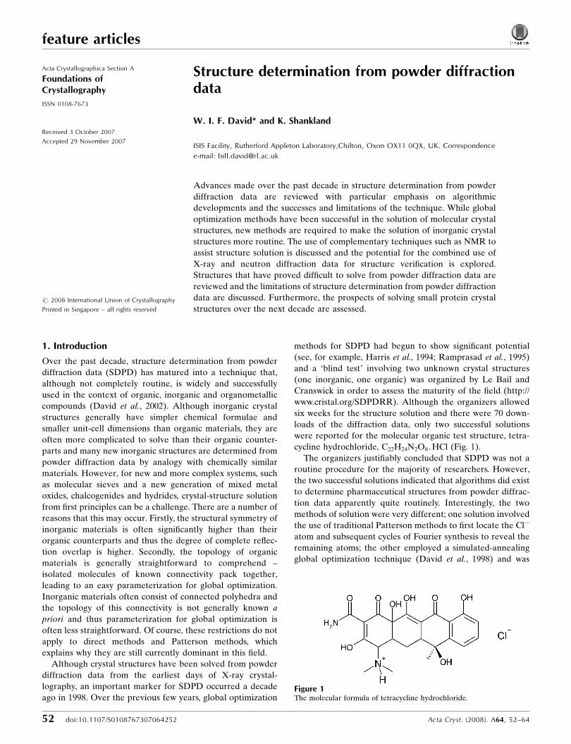
the few can now take advantage of the power of SDPD. Fig. 2
underlines this, showing the steady rise in results obtained
from global optimization methods, but also the regular stream
of results from direct methods.
In this paper, we discuss a number of recent developments
and present examples of the determination of organic, inor-
ganic and biological crystal structures. Our main focus is,
however, on molecular organic materials, as this is the area
that has seen the most significant expansion in the last decade
in terms of published crystal structures.
2. Algorithm developments
2.1. Introduction
SDPD is a sequential process with clearly defined stages
and at each stage there can be problems that make it impos-
sible to proceed (David et al., 2002). The majority of problems
are caused by the collapse of the three dimensions of recip-
rocal space to the single dimension of a powder diffraction
pattern, with the resultant Bragg-peak overlap being parti-
cularly severe at shorter d spacings. This places some funda-
mental restrictions upon the amount of information that can
be derived from the pattern; these restrictions are discussed in
considerable detail in Appendix A and the reader’s attention
is drawn to this section, as an understanding of the restrictions
is key to assessing future developments in SDPD.
It is apparent that, in such a sequential process, care has to
be taken at all stages; even at the sample preparation stage,
recrystallization to improve sample crystallinity or light
grinding to improve powder averaging can lead to significantly
better data. The traditional bottlenecks of indexing and
structure solution turn out to be intimately linked; advances in
structure-solution methods have stretched the capabilities of
SDPD to the extent that the study of relatively large structures
(with correspondingly large unit cells and sometimes very long
axes) is now quite common. This size increase has exposed
some of the limitations of well established powder-indexing
programs that were written at a time when looking at struc-
tures (largely inorganic) with much smaller unit cells was in
fact the norm. As such, it was to a large extent success in
structure solution that suggested it was time to develop new
algorithms and strategies for powder indexing.
2.2. Indexing
Unit-cell determination is an essential first step in structure
solution. In most methods, peak positions are extracted and
then trial unit cells are assessed in order to determine the
correct lattice parameters. With high-resolution data, this
process is often straightforward. However, with poorer data
and particularly when the sample contains more than one
crystalline phase, indexing can become a serious bottleneck. If
the data appear reasonable and indexing does fail, then the
most likely cause is the existence of more than one phase.
Identification of known phases should be attempted and the
corresponding peak positions removed from the indexing
process. If, however, no known phases are identified, then
allowance must be made for the possibility of ‘impurity’
phases such as starting materials – dividing peaks into groups
of ‘sharp’ and ‘broad’ may help. If this fails, then two
experimental approaches can also be used: (i) resynthesis (or
recrystallization) of the material under different experimental
conditions – for example, in the case of two polymorphic
forms, this may lead to different proportions of the two
polymorphs which can then aid in the identification of the two
Acta Cryst. (2008). A64, 52–64 David and Shankland � Structure determination from powder diffraction data 53
feature articles
Figure 2Approximate numbers of publications involving direct methods andpowder diffraction (upper graph) and global optimization and powderdiffraction (lower graph) as a function of time since 1990. Source: Web ofKnowledge search, September 2007.
distinct sets of Bragg peak positions; and (ii) heating the
sample – one phase may disappear at elevated temperatures.
Differential scanning calorimetry is particularly useful in pre-
screening for this effect.
Perhaps the two most significant algorithmic developments
in indexing over the past decade have been (i) the incor-
poration of the possibility of impurity phases into the
exhaustive successive dichotomy algorithms of DICVOL04
(Louer & Boultif, 2006) and (ii) the development of a singular-
value-decomposition-based algorithm in TOPAS (Coelho,
2003). The X-Cell program (Neumann, 2003), which uses an
extinction-specific dichotomy procedure to perform an
exhaustive search of parameter space, is also capable of
handling impurity phases and zero-point errors. Whilst it is
difficult to assess the impact that these relatively recent
developments have had upon the success or otherwise of
indexing in general, it is certainly true to say that these
programs, being of recent origin, are well suited to the
indexing of materials with large unit cells and their increased
use is likely to decrease the reliance on the older strategy of
trying several distinct indexing programs in order to obtain a
likely solution.
2.3. Space-group determination
Whereas the inability to determine the correct unit cell
makes structure solution impossible, some uncertainty in the
correct space group is not an intractable problem as each
potential space group may be separately tested. Space groups
are determined by examining diffraction patterns for system-
atically absent reflections. For example, in a monoclinic
system, if all 0k0 reflections (k odd) are absent, then it is
probable that there is a 21 screw axis and that space group P21
is more likely than P2. Determining absences for long
d-spacing reflections is normally straightforward, but at
shorter d spacings reflection overlap makes it a much more
subjective process. For example, a 010 reflection will typically
be well separated from other reflections, making accurate
determination of its intensity easy. In contrast, the 030/050/070
reflections are much more likely to lie in clumps of overlapped
reflections, leading to difficulties in accurate intensity esti-
mation. Accordingly, conclusions about contributing space-
group-symmetry elements are generally drawn on the basis of
a very small number of clear intensity observations. While
observing lattice-centring extinctions is usually relatively easy,
the determination of the correct space-group-symmetry
elements is generally more challenging. Choosing the space
group with the fewest number of contributing reflections, the
process of parsimony, is a good pragmatic approach to resol-
ving this problem and, in the case of molecular organic
materials, considerable help in space-group selection comes
from the well known frequency distribution of space groups,
where some 80% of compounds crystallize in one of the
following: P21/c, P�11, P212121, P21 and C2/c. For relatively
simple systems with a small number of atoms in the unit cell,
the structure may be solved in P1 and the space group
subsequently determined from a search for the appropriate
symmetry elements – this is a useful alternative strategy
particularly for small inorganic structures. However, over the
past decade, probabilistic approaches to space-group deter-
mination have been developed that remove the need for
subjective judgements about the presence or absence of
classes of reflections throughout the pattern (Markvardsen et
al., 2001; Altomare, Caliandro, Camalli, Cuocci, da Silva et al.,
2004). Following a model-independent fit (Pawley or Le Bail)
to the diffraction data in the holosymmetric space group
consistent with the observed crystal class, the user is presented
with a list of possible extinction symbols ranked in order of
probability. Armed with the most likely extinction symbol, it is
usually a straightforward matter to pick the most likely space
group (though see x3.2 for an example of a more difficult
case). As such, these methods are particularly useful when
dealing with systems of orthorhombic symmetry or higher,
where the number of possible space groups (and settings) is
relatively large.
2.4. Direct methods, Patterson methods and charge flipping
Direct methods of structure determination dominate the
field of single-crystal diffraction because of their incredible
success rate, range of applicability, speed, ease of use and
reliability. However, in general, they expect to work on large
numbers of well determined reflection intensities collected to
atomic resolution. For a powder diffraction measurement, this
situation is rarely the case; see the detailed discussion in
Appendix A for further details. Even if ‘good’ data can be
obtained to 1 A (this is often the case with inorganic struc-
tures, particularly with neutron powder diffraction data), then,
for all but the simplest materials, observed diffraction features
can contain contributions from several overlapped reflections
meaning that the condition of ‘well determined’ reflection
intensities is not met. Experimental methods, such as texture
analysis (Wessels et al., 1999) and differential lattice expansion
through multiple temperature measurements (Shankland,
David & Sivia, 1997; Fernandes, 2006) can create almost
single-crystal-like data sets which have been successfully used
to solve crystal structures using direct methods. However, it is
adaptations of traditional direct methods, specifically tailored
to the analysis of powder diffraction data, which have been
used most successfully in recent years. The best known
package is EXPO2004 (Altomare, Caliandro, Camalli, Cuocci,
Giacovazzo et al., 2004) which has evolved constantly and now
incorporates indexing, space-group determination, structure
solution and refinement capabilities (Altomare et al., 2006). It
can be applied to organic (Brunelli et al., 2007; Altomare et al.,
2007), organometallic (Masciocchi & Sironi, 2005) and inor-
ganic crystal structures (Fukuda et al., 2007), but the majority
of reported structures solved using EXPO (in its various
versions) falls into the latter two groups, reflecting both the
particular strengths of the package and its traditional user
base.
As illustrated by one of the contributors to the first SDPD
round robin, Patterson methods, even in a standard form, can
be a powerful tool for structure determination in cases where
feature articles
54 David and Shankland � Structure determination from powder diffraction data Acta Cryst. (2008). A64, 52–64
there are strongly scattering atoms. Indeed, some years ago it
had been shown that they can also be applied where large
fragments of known geometry are involved (Rius & Mira-
vitlles, 1988). In an important series of papers (see, for
example, Rius et al., 2000, 2007), Rius and his colleagues have
described modifications to traditional direct methods based
around Patterson-function arguments that have permitted the
solution of many very complex materials (see, for example,
Corma et al., 2003), whilst the usefulness of the Patterson
function in decomposing overlapping Bragg peaks has also
been shown (Estermann & David, 2002). Perhaps the most
comprehensive approach is based upon hyperphase permu-
tation algorithms (Bricogne, 1991) but their use remains to be
fully exploited.
Recently, the charge-flipping method (Oszlanyi & Suto��,
2008) has been adapted to powder diffraction data with some
very promising results (Baerlocher, Gramm et al., 2007;
Baerlocher, McCusker & Palatinus, 2007). As yet, it appears
that it is still subject to the requirement for near atomic
resolution data.
2.5. Global optimization methods
Global optimization methods of SDPD involve moving a
molecular model of the molecule under study around a known
unit cell, constantly adjusting its conformation, position and
orientation until the best agreement with the observed
diffraction data is obtained. Of course, there is no guarantee
that the best minimum obtained for the agreement function
will be the global one (corresponding to the correct crystal
structure) but we are fortunate in diffraction that the value
obtained can be compared with a value obtained for a
corresponding Pawley- or Le Bail-type fit to the data, in order
to inform us how close we in fact are to the ‘best’ fit obtain-
able.
That these methods have been so successful in the context
of SDPD is almost entirely due to the fact that they incor-
porate a massive amount of prior chemical information in the
form of the known molecular topology of the material under
study; typically, all known bond lengths and angles for the
molecule are fixed, leaving only its conformation, plus its
position and orientation within the unit cell to be determined.
It is this information that compensates for the reduced infor-
mation content of the powder pattern.
2.5.1. Grid search. Grid-type searches represent the
simplest form of the global optimization method available,
where every parameter that defines the search space is
explored on a systematic grid. Their one significant advantage
is that, given a fine enough grid, one is guaranteed to find the
global minimum. However, naıve grid-type searches are
computationally intractable for problems of even relatively
low complexity (Shankland & David, 2002) and are not at all
competitive with other search methods of the type described
below. That said, with some suitable modification, they can
prove extremely useful – see, for example, Ivashevskaja et al.
(2003) and Mazina et al. (2004).
2.5.2. Stochastic search algorithms. The most popular
global optimization methods for the SDPD of organic
compounds in recent years have been stochastic in nature.
These methods can be described colloquially as a ‘random
walk through the good solutions’. Many different stochastic
methods have been explored, including simple Monte Carlo
(Harris et al., 1994), genetic algorithms (Shankland, David &
Csoka, 1997; Harris et al., 2004; Feng & Dong, 2007), evolu-
tionary strategies (Chong & Tremayne, 2006) and particle
swarm (Csoka & David, 1999) but it is simulated annealing
(Andreev et al., 1997; Engel et al., 1999; Putz et al., 1999; Pagola
& Stephens, 2000; Coelho, 2000; Favre-Nicolin & Cerny, 2004;
David et al., 2006) that is most widely used and that has had
the largest impact. Its high level of success is undoubtedly
attributable to the fact that it is an extremely effective algor-
ithm that is easy to use (it has relatively few control variables,
all of which can be set automatically) and, as such, is suitable
for use by typical practitioners of powder diffraction. Various
modifications to the basic annealing scheme, such as parallel
tempering (Earl & Deem, 2005), have been implemented
(Favre-Nicolin & Cerny, 2004) in order to further improve the
ability of the algorithm to sample the search space efficiently.
Of the other stochastic algorithms, genetic algorithms in
particular have been shown to be effective and capable of
delivering solutions of considerable complexity (Albesa-Jove
et al., 2004; Pan et al., 2006). Their usage has, to date, been
limited by program availability though very recently a freely
available program GEST has been released (Feng & Dong,
2007).
2.5.3. Deterministic algorithms. Whilst stochastic tech-
niques have been shown to be effective global optimization
methods, many other algorithms from different research areas
remain to be evaluated in the context of SDPD and it is likely
some of these will be more efficient and successful than
the techniques currently in use. One example is the hybrid
Monte Carlo (HMC) algorithm which combines the key
components of Monte Carlo (MC) and molecular dynamics
(MD) approaches into a single algorithm. An extensive
discussion of HMC in the context of SDPD may be found in
the paper of Johnston et al. (2002). In essence, HMC may be
considered in terms of a particle that follows a trajectory
determined by Hamilton’s equations of motion in a hyper-
space defined by a set of structural variables. The total energy
of the particle at any point is equal to the sum of the kinetic
energy and potential energy given by the goodness-of-fit
target function. Whilst in theory the total energy is conserved,
the use of finite time step sizes in the numerical evaluation of
the equations of motion means that this is not the case. To
control this effect, a Metropolis acceptance criterion is used to
determine whether to accept or reject the configuration at the
end of a given MD trajectory. The trajectory either continues
from the end point if it is accepted or returns to the previous
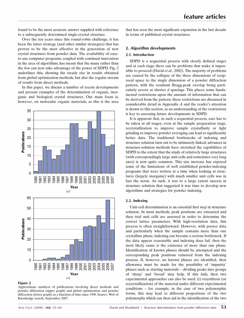
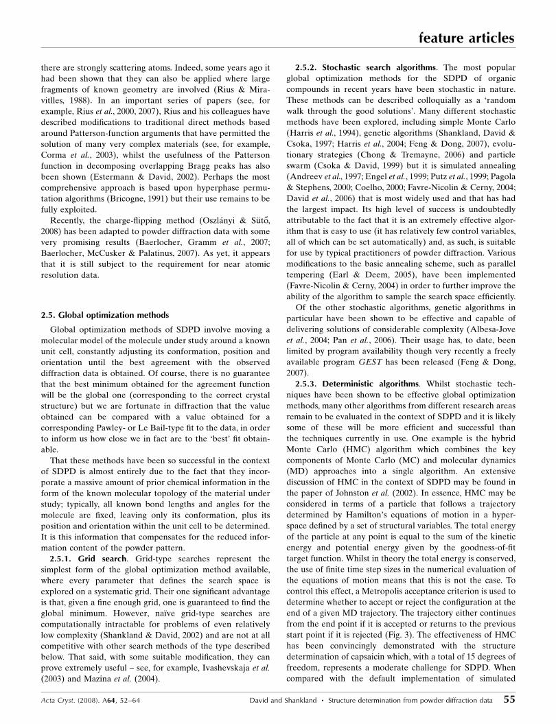
start point if it is rejected (Fig. 3). The effectiveness of HMC
has been convincingly demonstrated with the structure
determination of capsaicin which, with a total of 15 degrees of
freedom, represents a moderate challenge for SDPD. When
compared with the default implementation of simulated
Acta Cryst. (2008). A64, 52–64 David and Shankland � Structure determination from powder diffraction data 55
feature articles
annealing in DASH, HMC is a factor of two more successful in
locating the global minimum over a series of 20 repeat runs.
Significantly, the HMC algorithm required considerably fewer
�2 evaluations than simulated annealing to achieve this level
of success. Remarkably, given the discussion in Appendix A,
the quickest solution required less than 20000 evaluations to
locate the radius of convergence corresponding to 10�11 of the
total parameter space.
2.5.4. Semi-global and local searches. The rate of conver-
gence of stochastic algorithms can sometimes be improved by
the incorporation of elements of local searching, such as
steepest descent, into the overall minimization strategy. For
example, the program DASH (David et al., 2006) uses a semi-
global simplex-type algorithm to further minimize the cost
function at the end of each simulated annealing run; the
program Organa (Brodski et al., 2005) uses simple gradient
minimization when appropriate and applies it to all of its cost
functions; a sequential quadratic programming subroutine
from the NAG library has been used to implement
Larmackian-type evolution in the context of a genetic algor-
ithm-based approach (Turner et al., 2000). Some indication of
the potential of the simplex algorithm is given by the fact that,
in a simple test, of 500 simplex runs started from random
points in DASH, one was actually successful in accurately
solving the crystal structure of the form B polymorph of
famotidine (C8H15N7O2S3, P21/c, Z0 = 1, 13 degrees of
freedom).
2.5.5. Maximum-likelihood techniques. As mentioned at
the start of x2.5, the principal reason for the success of global
optimization methods in SDPD is the incorporation of the
molecular geometry into the solution process. However, this
strength is also the principal limitation of the technique – the
complete (and correct!) molecular structure must be incor-
porated if the global least-squares minimum is to be reached.
This limitation may, however, be relaxed if more generalized
maximum-likelihood methods are used. This approach has
been widely adopted by the macromolecular crystallography
community and has recently been applied successfully to solve
structures from powder diffraction data (Markvardsen et al.,
2002; Favre-Nicolin & Cerny, 2004). Consider that the
majority (but not all) of the structural contents is to be
determined in the optimization process. This can occur, for
example, when there are disordered solvent molecules present
in the structure in addition to the main molecule of interest. It
might also occur if structural fragments are omitted from the
optimization process in order to decrease the complexity of
the global search. In such cases, maximum-likelihood optimi-
zation still allows the majority of the structure to be correctly
located. Use of this approach has been illustrated with the
examples of the nitrate and acetate salts of the anticonvulsant
agent remacemide. If the nitrate and acetate ions are excluded
from a standard least-squares global optimization then the
structures cannot be solved; the best solutions, whilst infor-
mative in that they show parts of the remacemide molecule
located at the positions of the acetate and nitrate ions in an
attempt to account for their scattering contribution, are not
sufficiently close to the true structure to allow structure
completion. In contrast to this, if the nitrate and acetate ions
are not explicitly considered in the maximum-likelihood
optimization, the remacemide ion is quickly and correctly
located for both structures with a very high success rate. It is
then a trivial matter to subsequently fix the remacemide ion
within the unit cell and then locate and orient the nitrate and
acetate molecules by global optimization. This approach
relaxes the constraint that the correct molecular contents are
included from the outset of the global optimization process –
for materials such as hydrates and solvates or zeolites and
molecular sieves with guest molecules, this is an important
consideration.
2.5.6. Incorporating additional chemical information.
Constraints form a fundamental part of most global optimi-
zation approaches, with bond lengths, bond angles and fixed
dihedral angles in the material under investigation typically
being held at known standard values during the optimization
process. Note, however, that some practitioners advocate the
use of ‘loose restraints’ (Favre-Nicolin & Cerny, 2004) in order
to allow faster convergence to a minimum. That said, struc-
tural variables are typically restricted to the external mol-
ecular degrees of freedom plus those internal torsion angles
whose values cannot be assigned a priori. Use of the
Cambridge Structural Database (Allen, 2002) can help to
provide likely bounds for torsion angles and the concept can
be further extended to non-bonded contacts. While database
mining can place bounds on likely torsion-angle values, the
direct use of additional structural information from other
techniques to determine torsion-angle values is more effective.
For example, if the complete molecular conformation can be
determined in advance of the diffraction experiment, global
optimization is reduced to a problem of determining the
position and orientation of a rigid molecule. Middleton and
colleagues (Middleton et al., 2002) outlined such a procedure
in which a set of interatomic distances is measured by
rotational-echodouble resonance (REDOR) SS-NMR. The
feature articles
56 David and Shankland � Structure determination from powder diffraction data Acta Cryst. (2008). A64, 52–64
Figure 3The potential energy (correlated integrated intensities �2) and totalenergy (kinetic energy plus potential energy) evaluated over a single MDtrajectory during the crystal-structure solution of capsaicin. The initialtotal energy is shown as a dotted line in order to highlight the total energyfluctuations arising from the finite MD step size.
molecular conformation is then derived from a restrained
molecular-dynamics optimization in which the use of high
harmonic force constants ensures that all conformations in the
simulation have interatomic distances that satisfy the input
distances. The best conformation is then optimized against the
X-ray powder diffraction data by global optimization. By way
of example, the anti-ulcer drug cimetidine, in polymorphic
form A, was solved from X-ray powder diffraction data using
DASH with a MD optimized model derived from four SS-
NMR-determined C—15N distances. Each torsion angle in the
MD-optimized starting model was allowed to vary �20� from
its input value. In terms of structure-solution performance, this
model delivers a speed and reliability approaching that of a
rigid-body optimization. However, routine application will
probably only be possible when the SS-NMR methodology
develops to a stage where isotopic labelling is no longer a pre-
requisite and when the specialized SS-NMR instrumentation
required is more commonly available.
An alternative way of biasing the search towards favourable
molecular conformations and packing motifs is the incor-
poration of potential energy as an additional term in the
overall cost function (Putz et al., 1999; Coelho, 2000; Lanning
et al., 2000; Brodski et al., 2005). The overhead in calculating
such energies for simple van der Waals type interactions is
small, though a suitable force field is required and a weighting
factor is needed to balance the diffraction and energy
contributions in the calculation of the overall cost function.
Another approach (Brenner et al., 1997, 2002) utilizes a
periodic nodal surface calculated from a few phased strong
low-index reflections to divide the unit cell into regions of high
and low electron density. In the case of molecular organic
materials, the resultant ‘structure envelope’ can be used as a
boundary within which to restrict the possible position/
orientation/conformation of the molecule within the unit cell,
leading to a significant reduction in the search space that
needs to be explored.
2.5.7. Parallel computing. In the absence of a fine-grained
grid search (a prohibitively slow method as mentioned
earlier), none of the global optimization methods mentioned
above can guarantee finding the global minimum in the rele-
vant parameter space in a finite time frame. As such, it is
prudent to perform multiple global optimization runs in order
to improve the chances of locating the global minimum or
some point sufficiently close to it to permit final refinement of
the structure. Indeed, for complex structures with a large
number of parameters, where the success rate in finding the
global minimum can fall to a very small number (perhaps 1%
or less), it is a necessity to perform multiple runs. This can turn
SDPD into a highly CPU intensive process, where one might
have to wait days for an answer, even when using highly
efficient cost functions such as the method of correlated
integrated intensities (David et al., 1998). Fortunately, each
run is independent of any other, and a simple and attractive
option is to distribute the individual runs across a number of
different computers/CPUs/cores in order to return the answer
more quickly. Such a ‘grid-type’ computing approach to SDPD
using both simulated annealing (as implemented in DASH)
and HMC has been described previously (Markvardsen et al.,
2005) and the speed gains to be had are, to a first approxi-
mation, proportional to the number of CPU cores contributing
to the grid system. As such, speed gains of two orders of
magnitude or more, over the already highly efficient execution
speeds for DASH and HMC, can be expected from a modest
grid of non-dedicated PCs. The importance of such speed gains
is twofold: firstly, it allows results to be obtained on time scales
that are more commensurate with the expectations of crys-
tallographers for structure determination; secondly, it allows
the parallel exploration of alternative strategies for solving the
problem in hand, such as the use of multiple different starting
models (e.g. cis and trans isomers), different diffraction data
ranges, the inclusion of preferred-orientation corrections and
the use of lower cooling rates in the annealing process.
Of course, there is nothing new in the parallel execution of
large optimization problems, even in the context of SDPD –
see, for example, Shankland, David & Csoka (1997) and
Habershon et al. (2003). What is most significant about the
work described above is the utilization of systems (such as
Condor and GridMP) to harness non-dedicated PC resources;
in this, crystallographers are following the trend set in other
scientific areas such as protein–ligand docking, demonstrating
that dedicated hardware (such as a Beowulf cluster) is not a
pre-requisite to accessing massive computing power.
3. Examples
3.1. Organic crystal structures
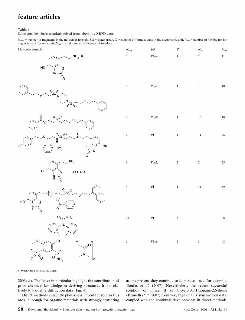
Some state-of-the-art results from SDPD from pharma-
ceuticals and organic crystal structures can be found in recent
doctoral work (Docherty, 2004; Fernandes, 2006), where the
structure determination (using DASH) and refinement (using
TOPAS) of numerous compounds of pharmaceutical interest
(see Table 1) from mainly laboratory X-ray powder diffraction
is reported. As can be seen from the molecular formulas,
degrees of freedom and number of independent fragments in
the asymmetric unit, these compounds span a wide range of
chemical and crystallographic complexity, yet all were solved
relatively straightforwardly. The success rate in finding the
global minimum fell to only a few percent for the most
complex examples and this indicates that tackling still more
complex examples will require further algorithmic develop-
ments. Of particular note are: (a) the benzoate structure,
where the anion is in fact disordered and where the location of
this disordered fragment was determined directly by global
optimization of two 50% occupancy benzoates (Johnston et al.,
2004), (b) the � form of carbamazepine, where Z0 = 4 and
there are 120 atoms in the asymmetric unit (Fernandes et al.,
2007) and (c) the dimethyl formamide solvate of chloro-
thiazide, where there are six independent fragments in the
asymmetric unit and a total of 42 degrees of freedom
(Fernandes et al., 2007).
Other good examples include the crystal structures of a
series of novel cyclic molecules (Terent’ev et al., 2007) and of
some mono-unsaturated triacylglycerols (van Mechelen et al.,
Acta Cryst. (2008). A64, 52–64 David and Shankland � Structure determination from powder diffraction data 57
feature articles
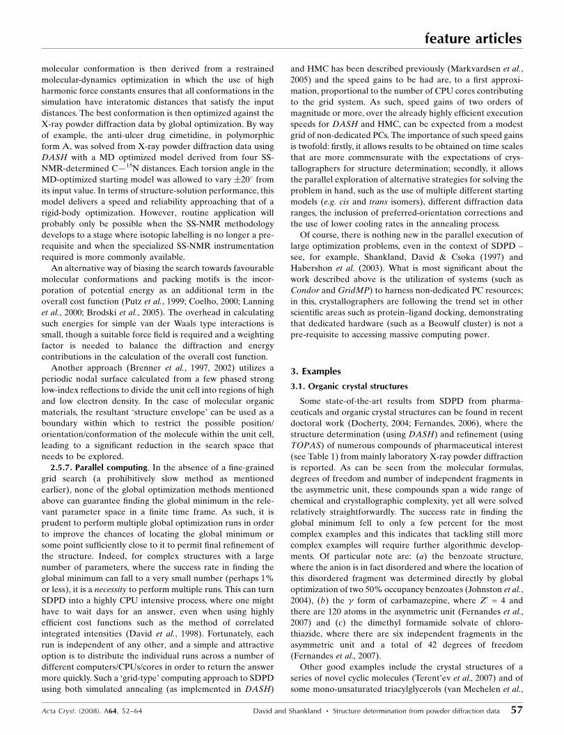
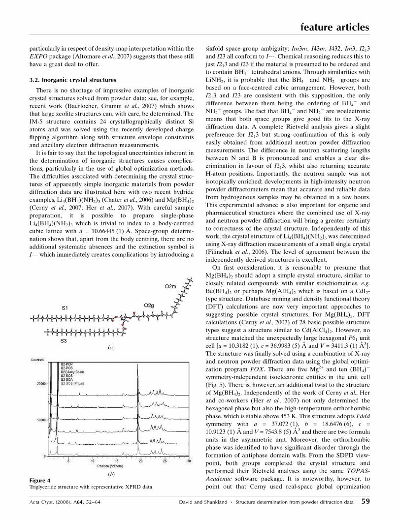
2006a,b). The latter in particular highlight the contribution of
prior chemical knowledge in deriving structures from rela-
tively low quality diffraction data (Fig. 4).
Direct methods currently play a less important role in this
area, although for organic materials with strongly scattering
atoms present they continue to dominate – see, for example,
Boufas et al. (2007). Nevertheless, the recent successful
solution of phase II of bicyclo[3.3.1]nonane-2,6-dione
(Brunelli et al., 2007) from very high quality synchrotron data,
coupled with the continual developments in direct methods,
feature articles
58 David and Shankland � Structure determination from powder diffraction data Acta Cryst. (2008). A64, 52–64
Table 1Some complex pharmaceuticals solved from laboratory XRPD data.
Nfrag = number of fragments in the molecular formula, SG = space group, Z0 = number of formula units in the asymmetric unit, Ntor = number of flexible torsionangles in each formula unit, Ndof = total number of degrees of freedom.
Molecular formula Nfrag SG Z0 Ntor Ndof
2 P21/a 1 2 11
1 P21/a 1 7 10
1 P21/n 1 12 18
2 P�11 1 14 26
3 Pca21 2 2 28
2 P�11 1 14 27
1† P�11 4 1 28
3 P21/c 2 3 42
† Synchrotron data, ID31, ESRF.
particularly in respect of density-map interpretation within the
EXPO package (Altomare et al., 2007) suggests that these still
have a great deal to offer.
3.2. Inorganic crystal structures
There is no shortage of impressive examples of inorganic
crystal structures solved from powder data; see, for example,
recent work (Baerlocher, Gramm et al., 2007) which shows
that large zeolite structures can, with care, be determined. The
IM-5 structure contains 24 crystallographically distinct Si
atoms and was solved using the recently developed charge
flipping algorithm along with structure envelope constraints
and ancillary electron diffraction measurements.
It is fair to say that the topological uncertainties inherent in
the determination of inorganic structures causes complica-
tions, particularly in the use of global optimization methods.
The difficulties associated with determining the crystal struc-
tures of apparently simple inorganic materials from powder
diffraction data are illustrated here with two recent hydride
examples, Li4(BH4)(NH2)3 (Chater et al., 2006) and Mg(BH4)2
(Cerny et al., 2007; Her et al., 2007). With careful sample
preparation, it is possible to prepare single-phase
Li4(BH4)(NH2)3 which is trivial to index to a body-centred
cubic lattice with a = 10.66445 (1) A. Space-group determi-
nation shows that, apart from the body centring, there are no
additional systematic absences and the extinction symbol is
I--- which immediately creates complications by introducing a
sixfold space-group ambiguity; Im3m, I�443m, I432, Im3, I213
and I23 all conform to I---. Chemical reasoning reduces this to
just I213 and I23 if the material is presumed to be ordered and
to contain BH4� tetrahedral anions. Through similarities with
LiNH2, it is probable that the BH4� and NH2
� groups are
based on a face-centred cubic arrangement. However, both
I213 and I23 are consistent with this supposition, the only
difference between them being the ordering of BH4� and
NH2� groups. The fact that BH4
� and NH2� are isoelectronic
means that both space groups give good fits to the X-ray
diffraction data. A complete Rietveld analysis gives a slight
preference for I213 but strong confirmation of this is only
easily obtained from additional neutron powder diffraction
measurements. The difference in neutron scattering lengths
between N and B is pronounced and enables a clear dis-
crimination in favour of I213, whilst also returning accurate
H-atom positions. Importantly, the neutron sample was not
isotopically enriched; developments in high-intensity neutron
powder diffractometers mean that accurate and reliable data
from hydrogenous samples may be obtained in a few hours.
This experimental advance is also important for organic and
pharmaceutical structures where the combined use of X-ray
and neutron powder diffraction will bring a greater certainty
to correctness of the crystal structure. Independently of this
work, the crystal structure of Li4(BH4)(NH2)3 was determined
using X-ray diffraction measurements of a small single crystal
(Filinchuk et al., 2006). The level of agreement between the
independently derived structures is excellent.
On first consideration, it is reasonable to presume that
Mg(BH4)2 should adopt a simple crystal structure, similar to
closely related compounds with similar stoichiometries, e.g.
Be(BH4)2 or perhaps Mg(AlH4)2 which is based on a CdI2-
type structure. Database mining and density functional theory
(DFT) calculations are now very important approaches to
suggesting possible crystal structures. For Mg(BH4)2, DFT
calculations (Cerny et al., 2007) of 28 basic possible structure
types suggest a structure similar to Cd(AlCl4)2. However, no
structure matched the unexpectedly large hexagonal P61 unit
cell [a = 10.3182 (1), c = 36.9983 (5) A and V = 3411.3 (1) A3].
The structure was finally solved using a combination of X-ray
and neutron powder diffraction data using the global optimi-
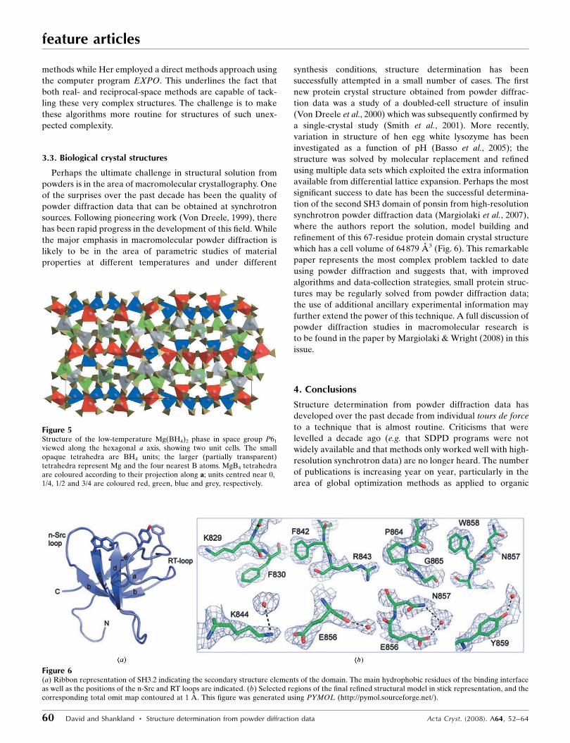
zation program FOX. There are five Mg2+ and ten (BH4)�
symmetry-independent isoelectronic entities in the unit cell
(Fig. 5). There is, however, an additional twist to the structure
of Mg(BH4)2. Independently of the work of Cerny et al., Her
and co-workers (Her et al., 2007) not only determined the
hexagonal phase but also the high-temperature orthorhombic
phase, which is stable above 453 K. This structure adopts Fddd
symmetry with a = 37.072 (1), b = 18.6476 (6), c =
10.9123 (1) A and V = 7543.8 (5) A3 and there are two formula
units in the asymmetric unit. Moreover, the orthorhombic
phase was identified to have significant disorder through the
formation of antiphase domain walls. From the SDPD view-
point, both groups completed the crystal structure and
performed their Rietveld analyses using the same TOPAS-
Academic software package. It is noteworthy, however, to
point out that Cerny used real-space global optimization
Acta Cryst. (2008). A64, 52–64 David and Shankland � Structure determination from powder diffraction data 59
feature articles
Figure 4Triglyceride structure with representative XPRD data.
methods while Her employed a direct methods approach using
the computer program EXPO. This underlines the fact that
both real- and reciprocal-space methods are capable of tack-
ling these very complex structures. The challenge is to make
these algorithms more routine for structures of such unex-
pected complexity.
3.3. Biological crystal structures
Perhaps the ultimate challenge in structural solution from
powders is in the area of macromolecular crystallography. One
of the surprises over the past decade has been the quality of
powder diffraction data that can be obtained at synchrotron
sources. Following pioneering work (Von Dreele, 1999), there
has been rapid progress in the development of this field. While
the major emphasis in macromolecular powder diffraction is
likely to be in the area of parametric studies of material
properties at different temperatures and under different
synthesis conditions, structure determination has been
successfully attempted in a small number of cases. The first
new protein crystal structure obtained from powder diffrac-
tion data was a study of a doubled-cell structure of insulin
(Von Dreele et al., 2000) which was subsequently confirmed by
a single-crystal study (Smith et al., 2001). More recently,
variation in structure of hen egg white lysozyme has been
investigated as a function of pH (Basso et al., 2005); the
structure was solved by molecular replacement and refined
using multiple data sets which exploited the extra information
available from differential lattice expansion. Perhaps the most
significant success to date has been the successful determina-
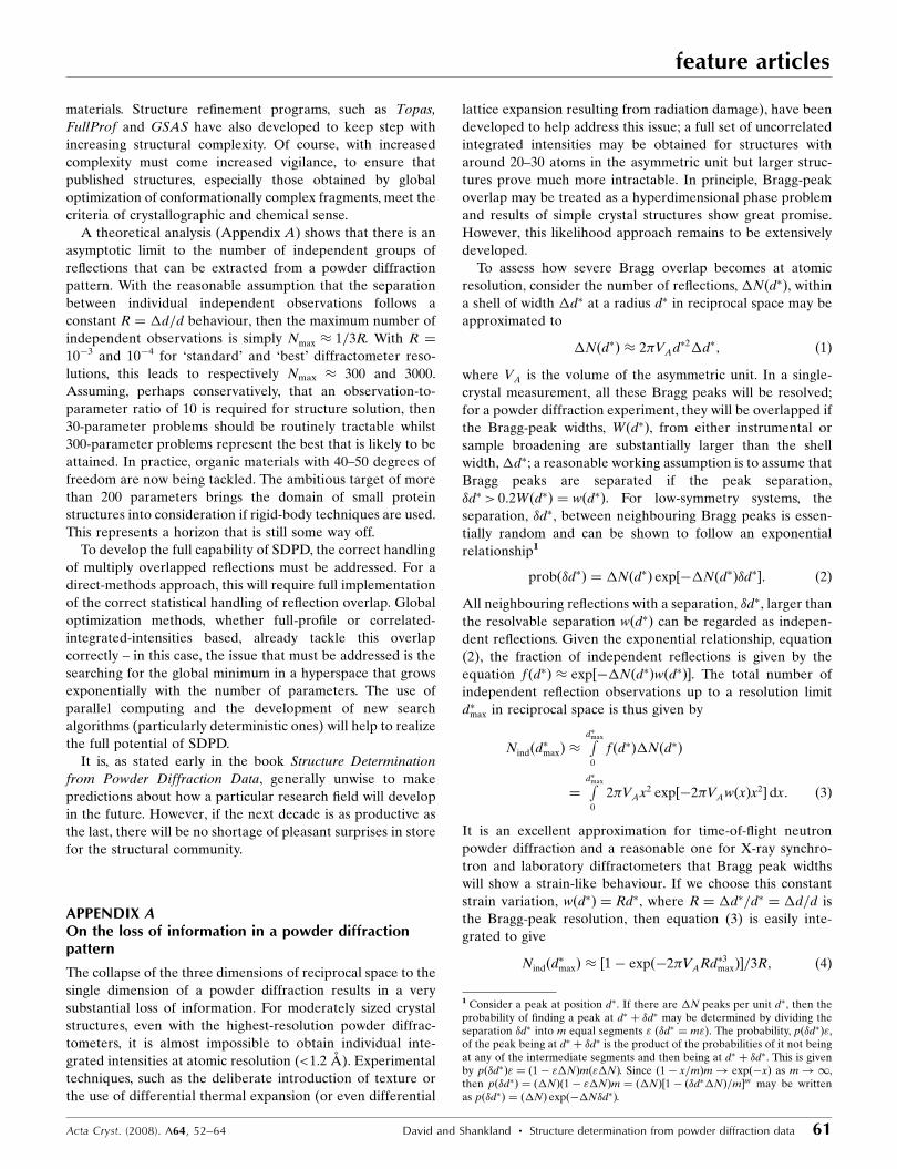
tion of the second SH3 domain of ponsin from high-resolution
synchrotron powder diffraction data (Margiolaki et al., 2007),
where the authors report the solution, model building and
refinement of this 67-residue protein domain crystal structure
which has a cell volume of 64879 A3 (Fig. 6). This remarkable
paper represents the most complex problem tackled to date
using powder diffraction and suggests that, with improved
algorithms and data-collection strategies, small protein struc-
tures may be regularly solved from powder diffraction data;
the use of additional ancillary experimental information may
further extend the power of this technique. A full discussion of
powder diffraction studies in macromolecular research is
to be found in the paper by Margiolaki & Wright (2008) in this
issue.
4. Conclusions
Structure determination from powder diffraction data has
developed over the past decade from individual tours de force
to a technique that is almost routine. Criticisms that were
levelled a decade ago (e.g. that SDPD programs were not
widely available and that methods only worked well with high-
resolution synchrotron data) are no longer heard. The number
of publications is increasing year on year, particularly in the
area of global optimization methods as applied to organic
feature articles
60 David and Shankland � Structure determination from powder diffraction data Acta Cryst. (2008). A64, 52–64
Figure 6(a) Ribbon representation of SH3.2 indicating the secondary structure elements of the domain. The main hydrophobic residues of the binding interfaceas well as the positions of the n-Src and RT loops are indicated. (b) Selected regions of the final refined structural model in stick representation, and thecorresponding total omit map contoured at 1 A. This figure was generated using PYMOL (http://pymol.sourceforge.net/).
Figure 5Structure of the low-temperature Mg(BH4)2 phase in space group P61
viewed along the hexagonal a axis, showing two unit cells. The smallopaque tetrahedra are BH4 units; the larger (partially transparent)tetrahedra represent Mg and the four nearest B atoms. MgB4 tetrahedraare coloured according to their projection along a; units centred near 0,1/4, 1/2 and 3/4 are coloured red, green, blue and grey, respectively.
materials. Structure refinement programs, such as Topas,
FullProf and GSAS have also developed to keep step with
increasing structural complexity. Of course, with increased
complexity must come increased vigilance, to ensure that
published structures, especially those obtained by global
optimization of conformationally complex fragments, meet the
criteria of crystallographic and chemical sense.
A theoretical analysis (Appendix A) shows that there is an
asymptotic limit to the number of independent groups of
reflections that can be extracted from a powder diffraction
pattern. With the reasonable assumption that the separation
between individual independent observations follows a
constant R ¼ �d=d behaviour, then the maximum number of
independent observations is simply Nmax � 1=3R. With R ¼
10�3 and 10�4 for ‘standard’ and ‘best’ diffractometer reso-
lutions, this leads to respectively Nmax � 300 and 3000.
Assuming, perhaps conservatively, that an observation-to-
parameter ratio of 10 is required for structure solution, then
30-parameter problems should be routinely tractable whilst
300-parameter problems represent the best that is likely to be
attained. In practice, organic materials with 40–50 degrees of
freedom are now being tackled. The ambitious target of more
than 200 parameters brings the domain of small protein
structures into consideration if rigid-body techniques are used.
This represents a horizon that is still some way off.
To develop the full capability of SDPD, the correct handling
of multiply overlapped reflections must be addressed. For a
direct-methods approach, this will require full implementation
of the correct statistical handling of reflection overlap. Global
optimization methods, whether full-profile or correlated-
integrated-intensities based, already tackle this overlap
correctly – in this case, the issue that must be addressed is the
searching for the global minimum in a hyperspace that grows
exponentially with the number of parameters. The use of
parallel computing and the development of new search
algorithms (particularly deterministic ones) will help to realize
the full potential of SDPD.
It is, as stated early in the book Structure Determination
from Powder Diffraction Data, generally unwise to make
predictions about how a particular research field will develop
in the future. However, if the next decade is as productive as
the last, there will be no shortage of pleasant surprises in store
for the structural community.
APPENDIX AOn the loss of information in a powder diffractionpattern
The collapse of the three dimensions of reciprocal space to the
single dimension of a powder diffraction results in a very
substantial loss of information. For moderately sized crystal
structures, even with the highest-resolution powder diffrac-
tometers, it is almost impossible to obtain individual inte-
grated intensities at atomic resolution (<1.2 A). Experimental
techniques, such as the deliberate introduction of texture or
the use of differential thermal expansion (or even differential
lattice expansion resulting from radiation damage), have been
developed to help address this issue; a full set of uncorrelated
integrated intensities may be obtained for structures with
around 20–30 atoms in the asymmetric unit but larger struc-
tures prove much more intractable. In principle, Bragg-peak
overlap may be treated as a hyperdimensional phase problem
and results of simple crystal structures show great promise.
However, this likelihood approach remains to be extensively
developed.
To assess how severe Bragg overlap becomes at atomic
resolution, consider the number of reflections, �Nðd�Þ, within
a shell of width �d� at a radius d� in reciprocal space may be
approximated to
�Nðd�Þ � 2�VAd�2�d�; ð1Þ
where VA is the volume of the asymmetric unit. In a single-
crystal measurement, all these Bragg peaks will be resolved;
for a powder diffraction experiment, they will be overlapped if
the Bragg-peak widths, Wðd�Þ, from either instrumental or
sample broadening are substantially larger than the shell
width, �d�; a reasonable working assumption is to assume that
Bragg peaks are separated if the peak separation,
�d�> 0:2Wðd�Þ ¼ wðd�Þ. For low-symmetry systems, the
separation, �d�, between neighbouring Bragg peaks is essen-
tially random and can be shown to follow an exponential
relationship1
probð�d�Þ ¼ �Nðd�Þ exp½��Nðd�Þ�d��: ð2Þ
All neighbouring reflections with a separation, �d�, larger than
the resolvable separation wðd�Þ can be regarded as indepen-
dent reflections. Given the exponential relationship, equation
(2), the fraction of independent reflections is given by the
equation f ðd�Þ � exp½��Nðd�Þwðd�Þ�. The total number of
independent reflection observations up to a resolution limit
d�max in reciprocal space is thus given by
Nindðd�maxÞ �
Rd�max
0
f ðd�Þ�Nðd�Þ
¼Rd�max
0
2�VAx2 exp½�2�VAwðxÞx2� dx: ð3Þ
It is an excellent approximation for time-of-flight neutron
powder diffraction and a reasonable one for X-ray synchro-
tron and laboratory diffractometers that Bragg peak widths
will show a strain-like behaviour. If we choose this constant
strain variation, wðd�Þ ¼ Rd�, where R ¼ �d�=d� ¼ �d=d is
the Bragg-peak resolution, then equation (3) is easily inte-
grated to give
Nindðd�maxÞ � ½1� expð�2�VARd�3maxÞ�=3R; ð4Þ
Acta Cryst. (2008). A64, 52–64 David and Shankland � Structure determination from powder diffraction data 61
feature articles
1 Consider a peak at position d�. If there are �N peaks per unit d�, then theprobability of finding a peak at d� þ �d� may be determined by dividing theseparation �d� into m equal segments " (�d� ¼ m"). The probability, pð�d�Þ",of the peak being at d� þ �d� is the product of the probabilities of it not beingat any of the intermediate segments and then being at d� þ �d�. This is givenby pð�d�Þ" ¼ ð1� "�NÞmð"�NÞ. Since ð1� x=mÞm! expð�xÞ as m!1,then pð�d�Þ ¼ ð�NÞð1� "�NÞm ¼ ð�NÞ½1� ð�d��NÞ=m�m may be writtenas pð�d�Þ ¼ ð�NÞ expð��N�d�Þ.
which is to be compared with the total number of independent
reflections observed in a single-crystal measurement:
Ntotðd�maxÞ �
Rd�max
0
�Nðd�Þ ¼Rd�max
0
2�VAx2 dx ¼ 23�VAd�3max: ð5Þ
The reduction in the number of independent reflections
relative to the total number of reflections is illustrated in
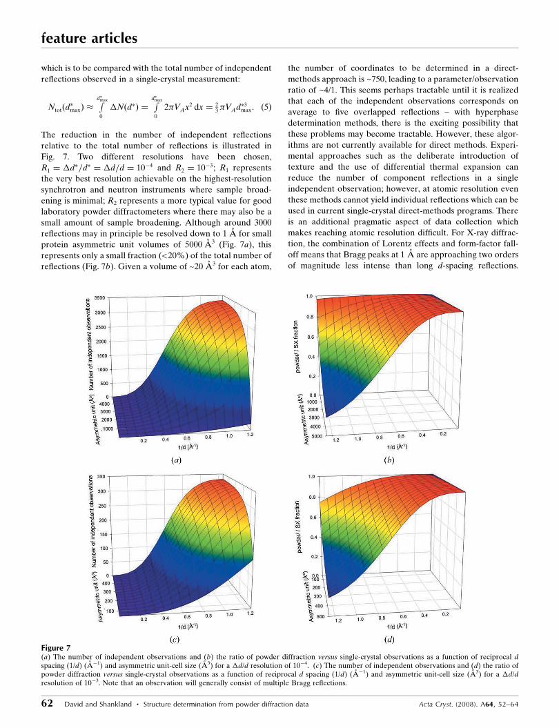
Fig. 7. Two different resolutions have been chosen,
R1 ¼ �d�=d� ¼ �d=d ¼ 10�4 and R2 ¼ 10�3; R1 represents
the very best resolution achievable on the highest-resolution
synchrotron and neutron instruments where sample broad-
ening is minimal; R2 represents a more typical value for good
laboratory powder diffractometers where there may also be a
small amount of sample broadening. Although around 3000
reflections may in principle be resolved down to 1 A for small
protein asymmetric unit volumes of 5000 A3 (Fig. 7a), this
represents only a small fraction (<20%) of the total number of
reflections (Fig. 7b). Given a volume of ~20 A3 for each atom,
the number of coordinates to be determined in a direct-
methods approach is ~750, leading to a parameter/observation
ratio of ~4/1. This seems perhaps tractable until it is realized
that each of the independent observations corresponds on
average to five overlapped reflections – with hyperphase
determination methods, there is the exciting possibility that
these problems may become tractable. However, these algor-
ithms are not currently available for direct methods. Experi-
mental approaches such as the deliberate introduction of
texture and the use of differential thermal expansion can
reduce the number of component reflections in a single
independent observation; however, at atomic resolution even
these methods cannot yield individual reflections which can be
used in current single-crystal direct-methods programs. There
is an additional pragmatic aspect of data collection which
makes reaching atomic resolution difficult. For X-ray diffrac-
tion, the combination of Lorentz effects and form-factor fall-
off means that Bragg peaks at 1 A are approaching two orders
of magnitude less intense than long d-spacing reflections.
feature articles
62 David and Shankland � Structure determination from powder diffraction data Acta Cryst. (2008). A64, 52–64
Figure 7(a) The number of independent observations and (b) the ratio of powder diffraction versus single-crystal observations as a function of reciprocal dspacing (1/d) (A�1) and asymmetric unit-cell size (A3) for a �d/d resolution of 10�4. (c) The number of independent observations and (d) the ratio ofpowder diffraction versus single-crystal observations as a function of reciprocal d spacing (1/d) (A�1) and asymmetric unit-cell size (A3) for a �d/dresolution of 10�3. Note that an observation will generally consist of multiple Bragg reflections.
Larger solid angles and longer counting times are required to
offset this intensity reduction. In practice, in the absence of
significant sample broadening, 1 A atomic resolution may be
obtained for moderately sized asymmetric units (ca 700 A3);
however, for small protein unit cells the best resolution
obtainable is probably nearer 2 A. From Fig. 7(a), this suggests
that the maximum number of useful observations is around
1500–2000. Of course, global optimization methods that use
full profile fitting or the equivalent correlated integrated
intensities approach do not need to disentangle a priori the
individual intensities. Working on the assumption that an
observation/parameter ratio of 10 should enable structures to
be solved if the appropriate algorithm exists, then, with
appropriate parameterization, small protein structures may
yet be solved from powder diffraction data. In many materials,
however, the very highest resolutions are not attainable
and R2 ¼ �d=d ¼ 10�3 is more realistic. Fig. 7(c) shows the
number of independent observations at this resolution. The
highest values that can be expected at this resolution are
around 300–350, suggesting that 30–35 parameters may be
straightforwardly obtained. This is the experience of global
optimization methods where the parameterization is in terms
of external degrees of freedom and internal torsion angles.
Fig. 7(d) again shows that the information loss in a powder
measurement at atomic resolution is substantial compared to
single-crystal measurements for moderately complex struc-
tures (VA > 300 A3 corresponding to ~15 independent atoms
in the asymmetric unit). This leads to the pragmatic experi-
mental consideration that, if a small single crystal can be
found, then it is best to perform a single-crystal experiment; an
ancillary powder diffraction measurement is, of course,
essential to verify that the single crystal is representative of
the bulk.
The authors would like to thank the following people for
providing information about recent developments and chal-
lenging examples encountered in their research into structure
determination from powder diffraction data: Jon Wright and
Irena Margoliaki (ESRF); Kenneth Harris (University of
Cardiff); Vincent Favre-Nicolin (CEA) and Radovan Cerny
(University of Geneva); Alastair Florence and Norman
Shankland (University of Strathclyde); Carmelo Giacovazzo
and Rosanna Rizzi (IC Bari); Rene Peschar (University of
Amsterdam) and Vladimir Chernyshev (Moscow State
University); Holger Putz (Crystal Impact); Jordi Rius
(ICMAB-CSIC); Alan Coelho (Brisbane). We are also
grateful to the manuscript referees for helpful comments.
References
Albesa-Jove, D., Kariuki, B. M., Kitchin, S. J., Grice, L., Cheung, E. Y.& Harris, K. D. M. (2004). Chemphyschem, 5, 414–418.
Allen, F. H. (2002). Acta Cryst. B58, 380–388.Altomare, A., Caliandro, R., Camalli, M., Cuocci, C., Giacovazzo, C.,
Moliterni, A. G. G. & Rizzi, R. (2004). J. Appl. Cryst. 37, 1025–1028.Altomare, A., Caliandro, R., Camalli, M., Cuocci, C., da Silva, I.,
Giacovazzo, C., Moliterni, A. G. G. & Spagna, R. (2004). J. Appl.Cryst. 37, 957–966.
Altomare, A., Camalli, M., Cuocci, C., Giacovazzo, C., Moliterni,A. G. G. & Rizzi, R. (2007). J. Appl. Cryst. 40, 344–348.
Altomare, A., Cuocci, C., Giacovazzo, C., Moliterni, A. G. G. & Rizzi,R. (2006). J. Appl. Cryst. 39, 145–150.
Andreev, Yu. G., Lightfoot, P. & Bruce, P. G. (1997). J. Appl. Cryst. 30,294–305.
Baerlocher, C., Gramm, F., Massuger, L., McCusker, L. B., He, Z. B.,Hovmoller, S. & Zou, X. D. (2007). Science. 315, 1113–1116.
Baerlocher, C., McCusker, L. B. & Palatinus, L. (2007). Z. Kristallogr.222, 47–53.
Basso, S., Fitch, A. N., Fox, G. C., Margiolaki, I. & Wright, J. P. (2005).Acta Cryst. D61, 1612–1625.
Boufas, S., Merazig, H., Moliterni, A. G. & Altomare, A. (2007). ActaCryst. C63, m315–m317.
Brenner, S., McCusker, L. B. & Baerlocher, C. (1997). J. Appl. Cryst.30, 1167–1172.
Brenner, S., McCusker, L. B. & Baerlocher, C. (2002). J. Appl. Cryst.35, 243–252.
Bricogne, G. (1991). Acta Cryst. A47, 803–829.Brodski, V., Peschar, R. & Schenk, H. (2005). J. Appl. Cryst. 38,
688–693.Brunelli, M., Neumann, M. A., Fitch, A. N. & Mora, A. J. (2007). J.
Appl. Cryst. 40, 702–709.Cerny, R., Filinchuk, Y., Hagemann, H. & Yvon, K. (2007). Angew.
Chem. Int. Ed. 46, 5765–5767.Chater, P. A., David, W. I. F., Johnson, S. R., Edwards, P. P. &
Anderson, P. A. (2006). Chem. Commun. pp. 2439–2441.Chong, S. Y. & Tremayne, M. (2006). Chem. Commun. pp. 4078–4080.Coelho, A. A. (2000). J. Appl. Cryst. 33, 899–908.Coelho, A. A. (2003). J. Appl. Cryst. 36, 86–95.Corma, A., Rey, F., Valencia, S., Jorda, J. L. & Rius, J. (2003). Nature
Materials, 2, 493–497.Csoka, T. & David, W. I. F. (1999) Acta Cryst. A55, Supplement,
Abstract No. P08.03.012.David, W. I. F., Shankland, K., McCusker, L. B. & Baerlocher, Ch.
(2002). Structure Determination from Powder Diffraction Data,edited by W. I. F. David, K. Shankland, L. B. McCusker & Ch.Baerlocher, pp. 1–11. Oxford University Press.
David, W. I. F., Shankland, K. & Shankland, N. (1998). Chem.Commun. pp. 931–932.
David, W. I. F., Shankland, K., van de Streek, J., Pidcock, E.,Motherwell, W. D. S. & Cole, J. C. (2006). J. Appl. Cryst. 39,910–915.
Docherty, A. (2004). PhD thesis, University of Strathclyde, Glasgow,Scotland.
Earl, D. J. & Deem, M. W. (2005). Phys. Chem. Chem. Phys. 7,3910–3916.
Engel, G. E., Wilke, S., Konig, O., Harris, K. D. M. & Leusen, F. J. J.(1999). J. Appl. Cryst. 32, 1169–1179.
Estermann, M. A. & David, W. I. F. (2002). Structure Determinationfrom Powder Diffraction Data, edited by W. I. F. David, K.Shankland, L. B. McCusker & Ch. Baerlocher, pp. 202–218. OxfordUniversity Press.
Favre-Nicolin, V. & Cerny, R. (2004). Z. Kristallogr. 219,847–856.
Feng, Z. J. & Dong, C. (2007). J. Appl. Cryst. 40, 583–588.Fernandes, P. (2006). PhD thesis, University of Strathclyde, Glasgow,
Scotland.Fernandes, P., Shankland, K., Florence, A. J., Shankland, N. &
Johnston, A. (2007). J. Pharm. Sci. 96, 1192–1202.Filinchuk, Y. E., Yvon, K., Meisner, G. P., Pinkerton, F. E. & Balogh,
M. P. (2006). Inorg. Chem. 45, 1433–1435.Fukuda, K., Ito, M. & Iwata, T. (2007). J. Solid State Chem. 180,
2305–2309.Habershon, S., Harris, K. D. M. & Johnston, R. L. (2003). J. Comput.
Chem. 24, 1766–1774.Harris, K. D. M., Habershon, S., Cheung, E. Y. & Johnston, R. L.
(2004). Z. Kristallogr. 219, 838–846.
Acta Cryst. (2008). A64, 52–64 David and Shankland � Structure determination from powder diffraction data 63
feature articles
Harris, K. D. M., Tremayne, M., Lightfoot, P. & Bruce, P. G. (1994). J.Am. Chem. Soc. 116, 3543–3547.
Her, J.-H., Stephens, P. W., Gao, Y., Soloveichik, G. L., Rijssenbeek, J.,Andrus, M. & Zhao, J.-C. (2007). Acta Cryst. B63, 561–568.
Ivashevskaja, S. N., Aleshina, L. A., Andreev, V. P., Nizhnik, Y. P.,Chernyshev, V. V. & Schenk, H. (2003). Acta Cryst. E59,o1006–o1008.
Johnston, A., Florence, A. J., Shankland, K., Markvardsen, A.,Shankland, N., Steele, G. & Cosgrove, S. D. (2004). Acta Cryst. E60,o1751–o1753.
Johnston, J. C., David, W. I. F., Markvardsen, A. J. & Shankland, K.(2002). Acta Cryst. A58, 441–447.
Lanning, O. J., Habershon, S., Harris, K. D. M., Johnston, R. L.,Kariuki, B. M., Tedesco, E. & Turner, G. W. (2000). Chem. Phys.Lett. 317, 296–303.
Louer, D. & Boultif, A. (2006). Z. Kristallogr. Suppl. 23, 225–230.Markvardsen, A. J., David, W. I. F., Johnson, J. C. & Shankland, K.
(2001). Acta Cryst. A57, 47–54.Markvardsen, A. J., David, W. I. F. & Shankland, K. (2002). Acta
Cryst. A58, 316–326.Markvardsen, A. J., Shankland, K., David, W. I. F. & Didlick, G.
(2005). J. Appl. Cryst. 38, 107–111.Margiolaki, I. & Wright, J. P. (2008). Acta Cryst. A64, 169–180.Margiolaki, I., Wright, J. P., Fitch, A. N., Wilmanns, M. & Pinotsis, N.
(2007). J. Am. Chem. Soc. 129, 11865–11871.Masciocchi, N. & Sironi, A. (2005). C. R. Chim. 8, 1617–1630.Mazina, O. S., Rybakov, V. B., Chernyshev, V. V., Babaev, E. V. &
Aslanov, L. A. (2004). Crystallogr. Rep. 49, 998–1009.Mechelen, J. B. van, Peschar, R. & Schenk, H. (2006a). Acta Cryst.
B62, 1121–1130.Mechelen, J. B. van, Peschar, R. & Schenk, H. (2006b). Acta Cryst.
B62, 1131–1138.Middleton, D. A., Peng, X., Saunders, D., Shankland, K., David,
W. I. F. & Markvardsen, A. J. (2002). Chem. Commun. pp.1976–1977.
Neumann, M. A. (2003). J. Appl. Cryst. 36, 356–365.Oszlanyi, G. & Suto��, A. (2008). Acta Cryst. A64, 123–134.Pagola, S. & Stephens, P. W. (2000). Mater. Sci. Forum, 321–3,
40–45.Pan, Z. G., Xu, M. C., Cheung, E. Y., Harris, K. D. M., Constable, E. C.
& Housecroft, C. E. (2006). J. Phys. Chem. B, 110, 11620–11623.Putz, H., Schon, J. C. & Jansen, M. (1999). J. Appl. Cryst. 32,
864–870.Ramprasad, D., Pez, G. P., Toby, B. H., Markley, T. J. & Pearlstein,
R. M. (1995). J. Am. Chem. Soc. 117, 10694–10701.Rius, J., Crespi, A. & Torrelles, X. (2007). Acta Cryst. A63,
131–134.Rius, J. & Miravitlles, C. (1988). J. Appl. Cryst. 21, 224–227.Rius, J., Torrelles, X., Miravitlles, C., Ochando, L. E., Reventos, M. M.
& Amigo, J. M. (2000). J. Appl. Cryst. 33, 1208–1211.Shankland, K. & David, W. I. F. (2002). Structure Determination from
Powder Diffraction Data, edited by W. I. F. David, K. Shankland,L. B. McCusker & Ch. Baerlocher, pp. 252–283. Oxford UniversityPress.
Shankland, K., David, W. I. F. & Csoka, T. (1997). Z. Kristallogr. 212,550–552.
Shankland, K., David, W. I. F. & Sivia, D. S. (1997). J. Mater. Chem. 7,569–572.
Smith, G. D., Pangborn, W. & Blessing, R. H. (2001). Acta Cryst. D57,1091–1100.
Terent’ev, A. O., Platonov, M. M., Sonneveld, E. J., Peschar, R.,Chernyshev, V. V., Starikova, Z. A. & Nikishin, G. I. (2007). J. Org.Chem. 72, 7237–7243.
Turner, G. W., Tedesco, E., Harris, K. D. M., Johnston, R. L. &Kariuki, B. M. (2000). Chem. Phys. Lett. 321, 183–190.
Von Dreele, R. B. (1999). J. Appl. Cryst. 32, 1084–1089.Von Dreele, R. B., Stephens, P. W., Smith, G. D. & Blessing, R. H.
(2000). Acta Cryst. D56, 1549–1553.Wessels, T., Baerlocher, C. & McCusker, L. B. (1999). Science, 284,
477–479.
feature articles
64 David and Shankland � Structure determination from powder diffraction data Acta Cryst. (2008). A64, 52–64