Embed Size (px)
Citation preview

i
Structure-function Study of PchB, an Isochorismate-Pyruvate Lyase from Pseudomonas
aeruginosa
By
Andrew N. Ouellette
Submitted to the Department of Molecular Biosciences and the Faculty of the Graduate School
of the University of Kansas in partial fulfillment of the requirements for the degree of Master of
Arts in Biochemistry & Biophysics
_______________________________
Chairperson Audrey Lamb
_______________________________
Committee Members Roberto De Guzman
_______________________________
Emily Scott
Date Defended: 06/06/2011

ii
The Thesis Committee for Andrew N. Ouellette
certifies that this is the approved version of the following thesis:
Structure-function Study of PchB, an Isochorismate-Pyruvate Lyase from Pseudomonas
aeruginosa
________________________________
Chairperson Audrey Lamb
_______________________________
Committee Members Roberto De Guzman
_______________________________
Emily Scott
Date approved: 06/13/2011

iii
Abstract
Enzymes act as biological catalysts for chemical reactions that would otherwise occur at
rates insufficient for the survival and growth of an organism. Understanding the fundamental
forces that drive catalysis in enzymes is applicable in the production of novel antibiotics through
rational drug design and advancing our understanding of how enzymes achieve incredible rate
enhancements over their uncatalyzed reactions. A body of work to understand these forces has
been established in the chorismate mutases and the pericyclic reaction they catalyze in the
production of prephenate from chorismate.
In Pseudomonas aeruginosa a structural homologue of the Escherichia coli chorismate
mutase EcCM has been found in the isochorismate-pyruvate lyase PchB. While the physiological
role of PchB is that of an isochorismate-pyruvate lyase (IPL) in the production of salicylate and
pyruvate from isochorismate, PchB also exhibits an adventitious chorismate mutase (CM)
activity. The contribution of forces that drive IPL and CM activity in PchB is somewhat
controversial with differing hypothesises of transition state stabilization through electrostatic
interactions and the formation of a reactive substrate through conformational destabilization.
Crystallization and mutational studies have shown the importance of a positive charge at position
42 for efficient catalysis within a dynamic active site loop region of PchB. The charge swap
PchB mutant K42E showed no detectable activity and PchB mutant K42A retaining one percent
of WT activity.
The WT PchB and PchB mutant K42E crystal structures with the products of the IPL
activity (salicylate and pyruvate) bound in the active site show a conserved active site
architecture among each other and that of previously solved PchB mutant K42A structure with

iv
salicylate and pyruvate and WT PchB structure with two pyruvate molecules bound in the active
site. The conservation of the active site architecture strongly suggests that the differences in the
catalytic activities are due to the change in chemical nature of the residue at position 42 within
the dynamic active site loop region.
The initial steps into the exploration of the contributions of the dynamic active site loop
region to IPL and CM activity in PchB through the current nuclear magnetic resonance
spectroscopy assignment of 32 of the 101 backbone resonances of PchB set the foundation for
further assignment and collection of relaxation data that will provide loop movement analysis on
the picosecond to millisecond time scale.
v
Acknowledgments
I would like to thank everyone who has helped me throughout the course of my academic
career. I would like to thank Qianyi Lou for generating the plasmids for the PchB mutants used
in this study and also for the generation of the substrates chorismate and isochorismate which
were extensively used throughout the crystallization sparse matrix screening. I would also like to
thank Kelli Olechoski for generating the original clone of PchB and also Jingping Lu for the
production of the non-his tagged clone of PchB. I would like to acknowledge those that helped in
the setup of many PchB crystallization trays: Katie Waugh, Surya Lakhanpal, Allison Ho and
Bob Wiggin.
I would also like to thank Roberto De Guzman and Asokan Anbanandam for their help
and guidance in setting up and recommendations for specific NMR analysis studies. I would like
to extend a special thank you to Fernando Estrada for his patience and guidance during many
conversations over the PchB backbone resonance assignments. I would like to acknowledge the
resonance assignments previously done on the PchB mutant K42E by Mike Connor and the use
of reagents and equipment from the De Guzman lab.
Finally I would like to thank my committee members Audrey Lamb, Roberto De Guzman
and Emily Scott for all the advice and guidance they have given me. I would also like to thank
Richard Schowen for his many wonderful discussion and lectures over enzyme catalysis and
theory and I would like to extend a very special thank you to Audrey Lamb for her support and
guidance over the years.

vi
Table of Contents
Abstract iii
Acknowledgements v
Table of Contents vi
List of Figures viii
List of Tables ix
CHAPTER 1
Enzyme Catalysis in Biological Systems 1
1.1 Introduction 1
The Function of Enzymes 1
Enzyme Facilitated Catalysis 2
The Active Site of Enzymes 2
Transition State Theory 3
General Acid General Base 5
Covalent Catalysis 6
Metal Ion Catalysis 7
Proximity and Orientation Effects 8
Pericyclic Reactions 8
Selective Binding of the Transition State by Enzymes 9
1.2 PchB 11
Physiological Role of PchB 11
Initial Structural Studies of PchB 11
Mutational Studies of PchB 12
References 18
CHAPTER 2
Protein X-ray Crystallography of PchB, an Isochorismate-Pyruvate Lyase from
Pseudomonas aeruginosa 20
2.1 Introduction 20
PchB, an Isochorismate-Pyruvate Lyase from Pseudomonas aeruginosa 20
2.2 Materials and Methods 25
Expression and Purification of Recombinant and WT PchB 25
Rationale of Crystallization Screens of WT PchB and Recombinant Proteins 26
Screening and Optimization 27
Optimization of Cryoprotectants for PchB Crystals 30
Crystallization Conditions for WT PchB and K42E Structures 30
Collection of Crystallographic Data and Structure Determination for PchB 32
2.3 Results 35
Crystal Structure of WT PchB with Salicylate and Pyruvate Bound 35

vii
Crystal Structure of PchB mutant K42E with Salicylate and Pyruvate Bound 38
2.4 Discussion 40
Comparison of WT PchB Structures (2 Pyr and Sal & Pyr) 40
Comparison of WT PchB (Sal & Pyr) & K42E Structures 42
Comparison of K42E (Sal & Pyr) with K42A (Sal & Pyr) 42
Comparison of WT PchB and Mutant Active Site Surface Area 45
The Conserved Active Site Architecture of PchB 47
References 50
CHAPTER 3
Nuclear Magnetic Relaxation Studies of PchB Mutant K42H 52
3.1 Introduction 52
Dynamics of the Active Site Loop in PchB 53
3.2 Materials and Methods 55
Expression and Purification of 13
C-,15
N- and 15
N-labeled recombinant 55
and WT PchB
Expression and Purification of Selectively 15
N-labeled Ala and Leu PchB 57
NMR Spectroscopy 57
Optimization of NMR Buffers 58
3.3 Results 59
3.4 Discussion 66
References 69
CHAPTER 4
Conclusion 70
References 74
viii
List of Figures
Figure Page
1-1 Comparison of reaction mechanisms 4
1-2 Secondary structure of PchB and EcCM 14
1-3 Active site overlay of WT PchB and mutant K42A crystal structures 17
2-1 Overview of reactions catalyzed by PchA and PchB 21
2-2 Active site view of chorismate mutases 24
2-3 Time lapse images of WT PchB crystal growth 29
2-4 Diffraction and crystal images of WT PchB and mutant K42E 31
2-5 Structure of WT PchB with salicylate and pyruvate bound and structural homologue 36
EcCM with a transition state analog bound
2-6 Structure of PchB mutant K42E with salicylate and pyruvate bound with comparative 39
WT PchB
2-7 Stereo overlay comparing the active sites of two WT PchB crystal structures 41
2-8 Stereo overlay comparing the active sites of WT PchB and K42E crystal structures 43
2-9 Stereo overlay comparing the active sites of PchB mutants K42E and K42A 44
crystal structures
2-10 Interior view of the PchB active site 46
2-11 Overlay comparing the active sites of WT PchB and PchB mutants K42E and K42A 48
3-1 View of the active site loop region in crystal structures of apo WT PchB and 54
WT PchB with pyruvate bound
3-2 Assigned 1H-
15N HSQC Spectra of
PchB K42H 60
3-3 1H-
13C HNCA Spectra of PchB K42H 61
3-4 1H-
13C CACB(CO)NH Spectra of PchB K42H 64
3-5 1H-
15N HSQC Spectra of
PchB K42H with
15N-labeled Ala and Leu Overlays 65
3-6 Secondary Structure of PchB 67

ix
List of Tables
Table Page
1-1 WT PchB and selected mutant steady state kinetic parameters 15
2-1 Table of sparse matrix crystallization screens for WT PchB and mutant K42E 28
2-2 Crystallographic statistics 34
2-3 Summary of WT and K42E Structures 37

1
CHAPTER 1
Enzyme Catalysis in Biological Systems
1.1 Introduction
The Functions of Enzymes
Enzymes function to accelerate chemical reactions within biological systems that
otherwise would not occur on a time scale sufficient to support the metabolism of organisms.
The upper limit to the rate at which enzymes enhance reaction rates can reach as high as 1020
-
fold.1
This is in stark contrast to some uncatalyzed reactions that have half-lives themselves
which are billions of years.2 Over the course of evolution some enzymes have become so
efficient at catalysis that their rate constants approach the rate of diffusion of a substrate in
solution.3 In other words every encounter between the substrate and enzyme progresses towards
production of products. Not all enzymes achieve this level of efficiency but this serves to
highlight the important concept that enzymes are specific for a specific substrate within a
biological system. Enzymes can be thought of as vessels that bind specific substrates thereby
limiting the substrates interaction with the environment of the solvent while at the same time
promoting specific chemical reactions based on the structure and composition of the enzyme’s
active site. This specificity and efficiency of enzymes supports the massive rate enhancements of
chemical reactions and serves to accomplish a host of chemical reactions within biological
systems that support the continued metabolism of living organisms.4
2
Enzyme Facilitated Catalysis
Reactions catalyzed by enzymes progress through a series of steps until product
formation is complete and released, regenerating the enzyme. First the substrate binds to the
active site of the enzyme creating the enzyme substrate complex (ES). The enzyme substrate
complex then progresses toward the high energy transition state ([ESǂ]) through molecular
“breathing” motions.5,6
When the transition state is reached, bonds are broken and formed
thereby transitioning the ES complex to the enzyme product complex (EP) which dissociates into
free enzyme and product in solution (1).
(1) E + S ↔ ES ↔ [ESǂ] ↔ EP ↔ E + P
This progression from substrate to product in an enzyme is completely mediated by the binding
interactions of the enzyme active site with the substrate.7
The Active Site of Enzymes
The enzyme active site is usually a pocketed region of the enzyme where accelerated
chemical reactions take place in the presence of bound substrate resulting in the production of
products. One way enzyme active sites accomplish massive reaction rate enhancements is by
having a surface area composed of various constituent amino acid side chain groups that are able
to interact with the substrate through hydrogen bonding, electrostatic interactions and van der
Waals forces.7 Enzymes can also incorporate cofactors such as divalent metal ions (Fe
2+, Mg
2+,
Mn2+
, Zn
2+) or coenzymes which are metalloorganic or complex organic molecules such as
Coenzyme A.4 This being said, binding of a substrate within the active site of an enzyme alone in
a classical “lock and key”8 sense, where both substrate and enzyme are complimentary to each

3
other without strain on the substrate, is not sufficient to induce catalysis (Figure 1-1,B). The
enzyme must bind the substrate in such a way as to induce strain on the bonds of the substrate
thereby supporting the high energy transition state of the chemical reaction over the ground state
(E + S) of the substrate free in solution (Figure 1-1,C).9
Transition State Theory
Enzymes affect the rate at which a reaction will occur and do not affect reaction
equilibria. There is specific activation energy (ΔGǂ
cat) that must be overcome in the transition
state by an ES complex for the reaction to proceed (Figure 1-1,C).4 The transition state can be
described as a high energy, unstable form of the substrate that neither wholly resembles the
substrate nor that of the product. The chemical bonds of the transition state molecule are in a
quasi-state of simultaneously bond breaking and bond forming. The actual time that a substrate
stays in the transition state is very small and has been estimated to be equal to a single bond
vibration.6 The transition state itself represents the highest barrier of free energy that must be
crossed for a given substrate molecule to transition to product (Figure 1-1,C).10
Linus Pauling
first popularized Polanyi’s idea that for the activation energy to be decreased and a reaction
proceed the enzyme must preferentially stabilize the transition state of the substrate and thereby
enhance the rate of catalysis.9 This theory of transition state stabilization is the foundation for
drug development that inhibits enzymes by designing transitional state analogues that mimic the
transition state of a particular substrate and as a result have a greater affinity for the enzymes
active site inhibiting enzyme function.6

4
Figure 1-1. Comparison of reaction mechanisms. Overview of uncatalyzed, “lock and key”, and
transition state stabilization views of reaction mechanisms. The enzyme is depicted as a blue “pac-
man” figure with the substrate depicted as blue and grey balls connected by a blue stick. Panel A,
Shows the progression of an uncatalyzed reaction showing a large barrier in free energy necessary
to produce products. Panel B, Shows the classical “lock and key” theory of enzyme catalysis
resulting in an even higher free energy barrier than the uncatalyzed reaction. Panel C, Shows the
theory of transition state stabilization whereas binding lowers the free energy barrier need to cross
over to the formation of products. This figured was adapted from Nelson et al. 2005.

5
When a substrate binds in the active site of an enzyme it does so through the interactions
previously mentioned above (hydrogen bonding, electrostatic interactions and van der Waals
forces). These weak interactions are then optimized in the transition state. The weak interactions
in the initial binding event in the active site of an enzyme serve multiple purposes in bringing the
substrate closer to the transition along the reaction coordinate. This includes decreasing the
entropy of the substrate by aligning reactive groups into high energy conformations thereby
lowering the freedom of the molecule.11
This preorganization of the substrate alone has been
shown to have rate enhancements of 108.4 Also as the substrate binds, the solvation shell of water
molecules that stabilize the substrate in solution are lost and hydrogen bonding is replaced by
functional groups within the active site resulting in what is called an “induced fit” (Figure 1-
1,C). This induced fit of the enzyme substrate complex brought on by binding of the substrate
results in a conformation change in the enzyme that aligns the reactive groups in the active site
necessary for catalysis and optimizes their interaction with the substrate.12
While binding
interactions account for a majority of rate enhancement for an enzyme9,13
the specific
interactions of the aligned reactive groups contribute to the catalytic mechanism through three
main mechanisms: general acid base catalysis, covalent catalysis and metal ion catalysis.4,14
General Acid-Base Catalysis
The transfer of protons (H+) to and from a substrate intermediate in an enzyme’s active
site helps direct the substrate intermediate towards the transition state and rate enhancements
over the uncatalyzed reaction on the order of 102
to 105 are observed.
4 When catalysis is
dependent on H+ (H3O
+) or OH
- ions from water alone within the active site it is referred to as

6
specific acid-base catalysis. The term general acid-base catalysis describes the transfer of protons
mediated by a BrØnsted acid (species that can donate a proton) or BrØnsted base (species that can
accept a proton), these include many of the amino acids such as lysine, arginine, cysteine,
histidine, serine, tyrosine and glutamic and aspartic acid.4,14
Many times enzymes incorporate
both general acids and bases within their reaction mechanisms or in what is known as a
concerted general acid-base mechanism. General acid-base catalysis is the most common form
catalysis in biological systems.4,13,14
Covalent Catalysis
The enzymatic mechanism utilizing covalent catalysis is a stepwise mechanism that first
involves a transient intermediate form of the substrate covalently bonded to the enzyme. This
interaction lowers the free energy of activation and aligns reactive groups bringing the substrate
closer to the transition state. This creates a step wise reaction mechanism that is subsequently
dependent on the breakdown of the covalent bonded transient intermediate to produce to
products and to regenerate free enzyme. The mechanism of covalent catalysis proceeds through
nucelophilic and electrophilic processes.14
All of amino acids mentioned above for general acid-
base catalysis are capable of interacting with substrate as nucleophiles, generating the formation
of covalent bonds with the substrate.4
The catalyzed reaction proceeds through an initial
nucleophilic attack on the substrate that generates a covalent bond between the now transient
intermediate and enzyme and produces an unstable charged group within the intermediate. The
electrons from the charged intermediate can be/are then shuttled to a now electrophilic group
within the enzyme. To eliminate the covalently bound product and regenerate the enzyme, a

7
nucelophilic attack must occur between the covalently bound intermediate and enzyme resulting
in again a charged intermediate that then breaks down and shuttles electrons to an electrophile
within the enzyme thereby breaking the covalent bond and releasing the product. Many proteases
such as chymotrypsin use covalent catalysis to carry out their reaction mechanisms.4,14,15
Metal Ion Catalysis
The weak interactions found within the active site of an enzyme can include divalent
metal ions (Fe2+
, Mg
2+, Mn
2+, Zn
2+) tightly bound to enzymes active site or other ions that come
directly from solution itself (Na+, K
+, Mg
2+, Ca
2+). Metal ions are required for catalysis in a third
of all known enzymes.4,14
The use of metal ions help to move the substrate towards the transition
state by a number of mechanisms. A metal ion can provide charge stabilization for the transition
state by acting as a Lewis acid. The major advantage of metal ions in this regard is that metal ion
concentration is independent of the pH, meaning that there can be high levels of metal ions
available for catalysis, independent of the concentration of hydrogen ions available in solution. A
metal ion can generate OH- ions by effecting bound water molecules promoting nucelophilic
catalysis. The position of metal ions with the active site themselves can also lead to charge
shielding of certain chemical groups within the substrate, negating repulsion effects that would
otherwise inhibit further binding of the substrate or enhance selective charges formed during the
transition state.14
8
Proximity and Orientation Effects
All of the above interactions serve to lower the activation energy of catalysis by
preferentially binding the transition state of a substrate over that of the ground level energy
conformation. The various methods incorporated thereafter (general acid-base, covalent and
metal ion catalysis) account for a relatively small portion of lowering the activation energy
relative to the initial binding of the substrate.4,9,14
It is important to note that the reaction
mechanisms incorporated by enzymes mimic those used in uncatalyzed reactions through what
are referred to as proximity and orientation effects. This is to say that the reactive groups must
come within the proper distance and alignment to allow catalysis to proceed. This preordering of
the substrate into a high energy conformation through alignment and positioning of the reactive
groups allow for a substantial decrease in the activation energy needed to achieve catalysis.9,14
In
this way proximity and orientation effects can be thought of as supporting a reactive substrate
conformation.
Pericyclic Reactions
If the alignment and orientation of reactive groups is a driving force towards the
transition state in a multi-substrate reaction then one could hypothesize that within a single
substrate reaction these intramolecular forces would be an even greater driving force along the
reaction coordinate. In essence enzymes use proximity and orientation effects to create a
subpopulation of reactive substrate conformations higher in energy than a ground state
experienced in solution. This leads to the transition state thereby enhancing the rate of catalysis
over the uncatalyzed reaction.9 This idea of producing a reactive substrate conformation and the

9
resultant rate enhancement is apparent in pericyclic reactions, which are seldom found in
biology.16
Pericyclic are concerted reactions that have a cyclic transition state where bond
breaking and formation is dependent on interacting orbitals within the cyclic intermediate.17
The
ordering of the substrate into a reactive substrate conformation is sufficient to lead to catalysis.18
Enzymes that catalyze these reactions do not utilize general acid-base, covalent or metal ion
catalytic mechanisms. Understanding pericyclic reactions has become a major area of interest in
the field of enzymology because pericyclic reactions represent a window into understanding the
fundamental forces that drive catalysis.
Work done on enzymes that catalyze pericyclic reactions has led to the introduction of
the term “NAC” from in silico findings from Bruce and co-workers.18
The NAC stands for Near
Attack Conformation and refers to a required conformation of the substrate in which the reactive
groups are aligned for the reaction. The NAC has been proposed to be considered a ground state
because the aligned reactive groups neither participate in bond breaking or bond formation while
in the state of a NAC and therefore the NAC is not equivalent to the transition state.18
While the
idea of the NAC as a true ground state is debatable1 the idea of what a NAC represents is not
hard to envision and provides a viewpoint into what is required of enzymes that catalyze
pericyclic reactions.
Selective Binding of the Transition State by Enzymes
We have already discussed how the ability of an enzyme to catalyze a reaction is
dependent on the ability of the enzyme to bind the transition state. What was not discussed before
is how the rate of enhancement for a catalytic enzyme is proportional to the affinity of the

10
enzyme for the transition state as shown below (3). This is under the assumption that the
chemical step is the rate limiting with eq 1,2 and 3 representing a single substrate system.19
Equation 1 shows
KUNǂ
E+S ↔ E+Sǂ → E+P
(1) KS ↕ KCATǂ ↕ KTS
ES ↔ ESǂ → E+P
(2) KTS KS
------- = -------
KUNǂ KCAT
ǂ
(3) KUNǂ
KTS = --------- KS
KCATǂ
the thermodynamic relationship between binding of the ground state and transition state for an
enzyme and single substrate. This leads to the relative comparison of the dissociation constants
(KS,KTS) with the uncatalyzed (KUNǂ) and catalyzed (KCAT
ǂ) pseudo equilibrium constants (2).
Rearrangement of eq 2 results in eq 3, showing that the dissociation constant (KTS) of the
enzyme transition state complex (ESǂ) is equal to the relative rate enhancement of the enzyme
(KUNǂ/ KCAT
ǂ) multiplied by the dissociation constant (KS) of the enzyme substrate complex (ES).
If the affinity for the transition state is directly proportional to the rate enhancement then the
greater the rate at which an enzyme enhances a reaction should be evident by that enzyme’s
ability to bind the transition state of the substrate.19
The catalytic power of enzymes is therefore
contingent upon the interactions the enzyme makes with the substrate that correlate to binding of
the transition state and the dynamic forces that drive these interactions.
11
This thesis focuses on understanding the structural and dynamic forces that add in the
pericyclic reactions catalyzed by PchB, an isochorismate pyruvate-lyase from Pseudomonas
aeruginosa with adventitious chorismate mutase activity. Protein X-ray crystallography and
nuclear magnetic resonance studies were carried out to ascertain the contributions of charge
stabilization and loop dynamics to the efficiency of PchB’s activity. The goal of this work is to
provide structural and dynamic information that can be used to enhance our understanding of the
fundamental concepts of enzyme catalysis and contribute to the future development of novel
inhibitors against PchB.
The work presented within would not be possible without previous structural and
mechanistic studies of PchB. Here we review the major contributions of our group that began to
elucidate the fundamental forces that drive catalysis in PchB.
1.2 PchB
Physiological Role of PchB
The physiological role of PchB in Pseudomonas aeruginosa is that of an isochorismate
pyruvate-lyase catalyzing the production of salicylate and pyruvate from isochorismate.20
The
salicylate is then ultimately incorporated into the siderophore pyochelin. Siderophores are low
molecular weight iron chelators that serve as bacterial virulence factors.21
Pyochelin scavenges
iron from the bacterial host allowing for the continued colonization and growth of P. aeruginosa.
Initial Structural Studies of PchB
Originally structural studies were carried out on WT PchB yielding two crystal structures
of PchB, apo (PDB code: 2H9C) and pyruvate (PDB code: 2H9D) bound structures.22
The

12
crystal structures showed that PchB is a structural homologue of the Escherichia coli chorismate
mutase EcCM consisting of an intertwined dimer (Figure 1-2). Major findings of the first PchB
crystallization study included that the active site loop becomes ordered in the presence of two
pyruvate molecules bound in the active site with five of the nine side chain interactions in the
active site of EcCM conserved among PchB.22
This reduction in side chain interactions within
the active site of PchB, relative to EcCM, likely serves as the basis for the catalytic promiscuity
of PchB as an isochorismate-pyruvate lyase (IPL) and chorismate mutase (CM).22
This work
established the foundation for further structural and mechanistic studies.
Mutational Studies of PchB
In the second published study from our group a variety of PchB mutants were made at
K42, A43 and I87.23
These mutations served to test various aspects of the proposed mechanism
of PchB’s catalytic activity. Mutations of residue K42 included K42A, K42E, K42Q and K42H.
These mutations addressed the ability of K42 to stabilize the transition state through electrostatic
interactions. The alanine mutation provided the absence of any charge at position 42 while the
glutamic acid mutation provided a negative charge relative to the WT positive charge found with
lysine. The K42Q mutation produced a polar uncharged side chain while the K42H mutation
allowed for a titratable positive charge based on the pH of the solution. The A43P mutation was
introduced to slow down the active site loop dynamics by inserting a rigid proline into the
flexible loop region. The I87T variant was based on a previous reported study that resulted in a
PchB mutant that exhibited CM activity but lacked IPL activity.20,23
Circular Dichroism Spectroscopy
The original mutation studies performed on EcCM resulted in a lack of activity were
hypothesized to have disrupted the active site architecture. This is supported by an altered

13
circular dichroism (CD) spectrum peak at ~210 nm not consistent with the all alpha helical
structure of WT EcCM (Figure 1-2).23,24
Circular dichroism spectroscopy was performed on
PchB mutants to assay for changes in the overall secondary structure of PchB. All of the
mutations produced in PchB to date which have resulted in CD spectra similar to WT PchB with
local minima at ~210 and ~225 nm supporting that the secondary helical structure had been
preserved among all PchB mutants.25,26
IPL and CM activity
IPL and CM activity assays were performed for all WT PchB and mutants. The results
showed a sharp decrease in the efficiency (kcat/Km) of PchB IPL and CM activity among several
mutants. Most notable was the 100-fold decrease in IPL activity exhibited by the non-charged
K42A mutant, no detectable activity for the charge swap mutant K42E and a titratable activity
found for the K42H mutant (Table 1-1).21
While the CD spectroscopy showed no overall change
in the secondary structure of PchB, the IPL and CM activity assays show that the mutations have
altered PchB’s ability to perform catalysis, likely as a result of by changes in PchB’s active site
architecture. The A43P mutation introduced to slow the dynamics of the active site loop was the
closest to WT PchB activity retaining 72% and 86% of wild type activity for CM and IPL
activity respectively.
Crystal Structures
The crystal structures of the PchB mutant K42A with salicylate and pyruvate bound and
apo PchB mutant I87T were determined in the second published of PchB from our group. While
the I87T structure showed a greater degree of flexibility with an overall increased disorder in the
active site loop region relative to the apo WT PchB structure. The I87T mutant has increased
disorder extending ~5 amino acids on either side of the active site loop relative to the disorder

14
Figure 1-2. Secondary structure of PchB and EcCM. Global view of secondary structure of WT
PchB and EcCM. Panel A, Cartoon representation of intertwined dimmer of WT PchB with two
molecules of pyruvate bound (green sticks, PBD code: 2H9C). Panel B, Cartoon representation of
intertwined dimer of EcCM with the transition state analog, bicyclo dicarboxylic acid, bound (PBD
code: 1ECM). PchB and EcCM are structural homologues.

15
Side
Chain
Km kcat kcat / Km % WT
(μM) (x10-3
s-1
) (M-1
s-1
) (kcat / Km)
+ WT 4.3±0.2 177±2.0 41100 -
K42A 51±3.0 24.5±0.5 480 1
-- K42E * * - -
titratiable K42H** 57±2.0 37.0±0.7 650 2
A43P 5.3±0.1 188±5.0 35500 86
Table 1-1. WT PchB and selected mutant steady state kinetic parameters. Table showing
changes in IPL activity for WT PchB and mutants K42A, K42E and K42H (pH of 7.5). Notice the
two orders of magnitude decrease in catalytic efficiency (kcat/Km) between WT PchB and mutant
K42A and also between K42A and K42E. Figured adapted from Luo et al. 2009.
* = below the limits of detection (0.3pmol for IPL assay) **= pH 7.5

16
found in the apo WT PchB structure.21
Most notable was the PchB mutant K42A with the
products of PchB’s IPL activity bound in the active site. The structure was found to be
comparable to that of WT PchB with two pyruvate molecules bound in the active site and that of
the wrong-ligand20
model of WT PchB with salicylate and pyruvate bound in the active site. The
active site architecture of K42A was shown to coordinate the carboxylic groups of salicylate and
pyruvate through two arginines (R14 & R31, Figure 1-3).
Elucidating the Mechanism of PchB Activity
The initial mechanistic and structural studies of PchB provided the basis of the work
undertaken for this thesis which is focused on addressing two major questions: 1) is the active
site architecture conserved among all WT PchB and mutants, specifically in regards to K42E
where no detectable activity was determined and 2) how much do the active site loop dynamics
contribute to catalysis? The prior production of apo WT PchB and salicylate and pyruvate bound
K42A structure provides a standard to compare all further structures of PchB. The PchB activity
assays provide the groundwork towards understanding what is essential in supporting the
transition state for PchB activity and set the direction for further exploration.

17
Figure 1-3. Active site overlay of WT PchB and mutant K42A crystal structures. WT
PchB with 2 pyruvate molecules bound (green and light green sticks, PDB Code: 2H9D).
PchB mutant K42A with salicylate and pyruvate bound (magenta and dark pink sticks,
PDB Code: 3HGX). Arginine 31 and arginine 14 are shown to align the carboxylates of
salicylate and pyruvate in 3HGX and the two pyruvate molecules in 2H9D. The overall
active site architecture and positioning of salicylate and pyruvate remain consistent in both
WT PchB and K42A mutant structures. Amino acids are labeled as PchB numbering
(Chain A: R31, K42/A42, R53, Q90, Chain B: R14).
18
References
1. Borman, S. Much ado about enzyme mechanisms. C&E News 88, 35-39 (2004).
2. Wolfenden, R. & Snider, M.J. The depth of chemical time and the power of enzymes as
catalysts. Acc Chem Res 34, 938-945 (2001).
3. Alberty, R., Hammes, G., Application of the Theory of Diffusion-Controlled Reactions to
Enzyme Kinetics. J Phys Chem 62, 154-162 (1958).
4. Nelson, D.L. & Cox, M.M. Lehninger Principles of Biochemistry (2005).
5. Karplus, M. Molecular dynamics simulations of biomolecules. Acc Chem Res 35, 321-
323 (2002).
6. Schramm, V.L. Enzymatic transition states and transition state analog design. Annu Rev
Biochem 67, 693-720 (1998).
7. Warshel, A. et al. Electrostatic basis for enzyme catalysis. Chem Rev 106, 3210-3235
(2006).
8. Fisher, E. Einfluss der Configuration auf die Wirkung der Enzyme. Ber Dt Chem Ges 27:
2985–2993 (1894).
9. Pauling, L. Nature of forces between large molecules of biological interest. Nature 161,
707-709 (1948).
10. Benkovic,S., Hammes, G., Hammes-Schiffer, S., Free-Energy Landscape of Enzyme
Catalysis. Biochemistry 47, 3317-3321 (2008).
11. Cannon, W.R. & Benkovic, S.J. Solvation, reorganization energy, and biological
catalysis. J Biol Chem 273, 26257-26260 (1998).
12. Koshland, D. Application of a theory of enzyme specificity to protein synthesis. Proc
Natl Acad Sci U S A 44, 98-104 (1958).
13. Jencks, W.P. Mechanism of Enzyme Action. Annu Rev Biochem 32, 639-676 (1963).
14. Voet, D. & Voet, J.G. Biochemistry (Wiley, 2004).
15. Blow, D. Structure and mechanism of Chymotrypsin. Acc Chem Res 9, 145-152 (1976).
16. DeClue, M. S., Baldridge, K. K., Kunzler, D. E., Kast, P. and Hilvert, DIsochorismate
Pyruvate Lyase: A Pericyclic Reaction Mechanism? J Am Chem Soc 127, 15002– 15003 (
2005).
17. Wade, L. Organic Chemistry (Pearson Prentice Hall, 2006).
18. Bruice, T.C. & Lightstone, F.C. Ground state and transition state contributions to the
rates of intramolecular and enzymatic reactions. Acc Chem Res 32, 127-136 (1999).
19. Mader, M.M. & Bartlett, P.A. Binding Energy and Catalysis: The Implications for
Transition-State Analogs and Catalytic Antibodies. Chem Rev 97, 1281-1302 (1997).
20. Gaille, C., Kast, P. & Haas, D. Salicylate biosynthesis in Pseudomonas aeruginosa.
Purification and characterization of PchB, a novel bifunctional enzyme displaying
isochorismate pyruvate-lyase and chorismate mutase activities. J Biol Chem 277, 21768-
21775 (2002).
21. Lamont, I.L., Beare, P.A., Ochsner, U., Vasil, A.I. & Vasil, M.L. Siderophoremediated
signaling regulates virulence factor production in Pseudomonas aeruginosa. Proc Natl
Acad Sci U S A 99, 7072-7 (2002).
22. Zaitseva, J., Lu, J., Olechoski, K.L. & Lamb, A.L. Two crystal structures of the
isochorismate pyruvate lyase from Pseudomonas aeruginosa. J Biol Chem 281, 33441-
33449 (2006).
23. Qianyi, L., Olucha, J. & Lamb, A.L. Structure-Function Analyses of Isochorismate-
Pyruvate Lyase from Pseudomonas aeruginosa Suggest Differeing Catalytic Mechanisms

19
for the Two Pericyclic Reactions of This Bifunctional Enzyme. Biochemistry 48, 5239-
5245 (2009).
24. Liu, D.R., Cload, S.T., Pastor, R.M. & Schultz, P.G. Analysis of active site residues in
Escherichia coli chorismate mutase by site-directed mutagenesis. J Am Chem Soc 118,
1789-1790 (1996).
25. Hur, S. and Bruice, T. C. The mechanism of catalysis of the chorismate to prephenate
reaction by the Escherichia coli mutase enzyme Proc Natl Acad Sci U S A 99, 1176-
1181 (2002).
26. Hur, S. and Bruice, T. C. The near attack conformation approach to the study of the
chorismate to prephenate reaction Proc Natl Acad Sci U S A 100, 12015- 12020 (2003).
20
CHAPTER 2
Protein X-ray Crystallography of PchB, an Isochorismate-Pyruvate Lyase from
Pseduomonas aeruginosa
The work within has been submitted to Biochemistry. “pH Dependence of Catalysis by
Pseudomonas aeruginosa Isochorismate-Pyruvate Lyase: Implications for Transition State
Stabilization and the Role of Lysine 42”
Jose Olucha, Andrew N. Ouellette, Qianyi Luo, and Audrey L. Lamb
2.1 Introduction
PchB, an Isochorismate-Pyruvate Lyase from Pseudomonas aeruginosa
PchB is a bi-functional enzyme of Pseudomonas aeruginosa that exhibits a physiological
isochorismate pyruvate lyase (IPL) activity and an adventitious chorismate mutase (CM)
activity.1 PchB’s physiological IPL activity catalyzes the formation of salicylate from
isochorismate in conjunction with the production of isochorismate from chorismate facilitated by
PchA (Figure 2-1, A).2 Specifically, the conversion of isochorismate to salicylate and pyruvate
by PchB is catalyzed through a (1,5)- pericyclic hydrogen transfer mechanism.3 The C2
hydrogen is transferred to C9 of the enolpyruvate tail of isochorismate which promotes the
breaking of the ether bond between C3 and O7 resulting in the release of the enolpyruvate tail as
pyruvate and the formation of salicylate from isochorismate (Figure 2-1, B). PchB’s adventitious
CM activity catalyzes the formation of prephenate from chorismate (Figure 2-1, C).1 The
production of prephenate proceeds through a (3,3)-sigmatropic pericyclic rearrangement.4

21
Figure 2-1. Overview of reactions catalyzed by PchA and PchB. Panel A, Overview of the
production salicylate and pyruvate from chorismate facilitated by PchA and PchB. Panel B,
Reaction mechanism of PchB’s IPL activity through a (1,5)- pericyclic hydrogen transfer
mechanism. Panel C, Reaction mechanism of PchB’s CM activity through a (3,3)- pericyclic
rearrangement.
22
The reaction results in a concerted but asynchronous bond breaking of the ether bond between
C3 and O7 and bond making between C1 and C9 of the enolpyruvate tail (Figure 2-1, C).
The rate of PchB’s IPL activity is ~4x10⁴ M
-1s
-1 while the rate of PchB’s CM activity is
considerably lower at ~1x10² M
-1s
-1.5 PchB shares no sequence identity with other salicylate
synthases but is a structural homologue of the CM from Esherichia coli (EcCM) with 20%
sequence identity.6
Previous comparisons of the active site of EcCM and PchB show only 5 of the 8 residues
important for CM activity in EcCM to be conserved in PchB.5,6
This serves as one explanation of
how PchB can catalyze two different reactions within one active site. What both the CM and
IPL activities of PchB do have in common though is the incorporation of a pericyclic transition
state (Figure 2-1, B & C).3,4
Pericyclic reactions are unusual in nature and have been extensively
studied in chorismate mutases.7-10
CM activity and hence pericyclic reactions pose a unique
model for studying the fundamental forces that drive enzymes catalysis. This is because
pericyclic reactions do not follow the traditional norms of enzyme catalysis as they do not
require general acid/base, have no covalent intermediates or depend on metal ions to catalyze the
formation of products. The question then becomes how do enzymes that catalyze pericyclic
reactions accomplish rate enhancement relative to the uncatalyzed reaction?
Currently in the field of enzymology there are two schools of thought as to how enzymes
catalyze pericyclic reactions, specifically for the chorismate mutases. The first involves
stabilization of the transition state through electrostatic interactions that balance the developing
negative charges formed as the substrate transitions to product.11,12
This has been supported in
crystal structure analysis of both EcCM and the Bacillus subtilis CM BsCM with the transition
state analog (TSA) oxabicyclic acid bound in the active site along with the recent crystal

23
structure of PchB mutant K42A with salicylate and pyruvate bound in the active site (Figure 2-
2).5,13
Analysis of the active sites of the three chorismate mutases show electrostatic interactions
that would support a developing negative charge on the ether oxygen in the case of the TSA .
Hydrogen bonding interactions orient the carboxylic groups of TSA molecules in BsCM and
EcCM and the carboxylic groups of salicylate and pyruvate in PchB mutant K42A into a pseudo-
diaxial conformation resembling the transition state for CM activity.5,7,9,13,14
Further evidence of
supportive electrostatic interactions can be found in mutagenesis studies of BsCM, EcCM and
PchB. Mutations disrupting the equivalent positive charge at arginine 90 in BsCM or lysine 39
in EcCM or lysine 42 in PchB (K42A) led to a complete lack of CM activity in the case of BsCM
and EcCM, with the PchB mutant K42A retaining just 1% of WT function.5,15-19
Moreover,
mutations made at K42 in PchB showed large effects on IPL activity with a 100-fold reduction in
IPL activity for PchB mutant K42A and complete loss of function in the case of the charge swap
mutation K42E.5
The PchB mutation results for K42A’s IPL activity gives support to the second school of
thought when it comes to how enzymes catalyze pericyclic reactions through the idea of what
can referred to as reactive substrate destabilization by near attack conformation (NAC). The
NAC is defined as a ground state conformation where the reactive groups of the substrate are
within van der Waals contact radius at ±15o of the bonding angles of the transition state.
20
Binding of the substrate by the enzyme encourages this conformation change in the substrate,
thereby allowing the substrate passage to the transition state without further input from the
enzyme.20-23
The PchB mutant K42A specifically addresses the idea of electrostatic interaction
by removal of the positive charge in the from lysine at position 42. PchB has be shown to be a
more versatile system for studying the contributions of electrostatic interactions and reactive

24
Figure 2-2. Active site view of chorismate mutases. Panel A, Active site of BsCM with TSA
bound (PDB code: 2CHT). Panel B, Active site of EcCM with TSA bound (PDB code: 1ECM).
Panel C, Active site of PchB mutant K42A with salicylate and pyruvate bound (PDB code:3HGX).
Transition state analogue depicted in green sticks in panels A & B, salicylate and pyruvate depicted
in cyan sticks in panel C.

25
substrate stabilization towards pericyclic reactions then chorismate mutases as evident by the
varying degrees of IPL activity achieved through mutational analysis carried out by Luo and co-
workers as discussed in chapter 1.5 Further exploration of the structural basis for the effects of
mutations at lysine 42 in PchB is needed to specifically elucidate the contributions of various
charged residues (K42, K42E, K42H) found to be important for efficient IPL activity.
In this chapter protein X-ray crystallography studies were carried out on WT PchB (K42)
and mutant K42E to provide insight into the positioning of residue K42 with respect to the active
site in WT PchB and the structural nature of the relative rate decrease for IPL activity in the
PchB mutant K42E.5 The results obtained provide structural evidence that that the relative rate
decrease of PchB mutant K42E is not due to a change in the active site architecture of PchB, but
more likely the result of the importance of positive charge in WT PchB at position 42 (lysine)
relative to the negative charge found in the PchB mutant K42E. These results add to the insight
of the role of transition state stabilization and reactive substrate destabilization in PchB.
2.2 Materials and Methods
Expression and Purification of Recombinant and WT PchB
The sub-cloning of the PchB expression plasmid has previously been reported.5
Expression and purification of recombinant and WT PchB was performed as previously
described6 with the exception of substituting overnight growth at 15
oC instead of the previous
three hours at 37 oC. Bacterial cells (E. coli BL21(DE3), Invitrogen) were grown in 1 liter non-
baffled Fernbach flasks containing LB media at 37 °C with shaking at 250 rpm until an optical
density of 0.8 at 600 nm was reached (Thermo-Fisher). The bacterial cells were induced with 0.2

26
mM isopropyl-β-D-thiogalactopyranoside (IPTG) and the incubator temperature was reduced to
15 oC for overnight growth. In the morning the bacterial cells were harvested by centrifugation
at 4,000 x g for 10 min at 4 °C. Cells were resuspended with ~15 mL of buffer A (25 mM Tris-
HCl (pH 8.0)) and lysed by sonication (Digital Sonifier, Branson) on ice for a total of 3 min
consisting of 3 second bursts with intervening 20 sec resting periods. The lysed bacterial cells
were subjected to ultracentrifugation at 142,000 x g for 45 min at 4 °C to remove cellular debris.
The supernatant was applied to a 30Q Sepharose Fast Flow column (Amersham Biosciences)
equilibrated with buffer A for anion-exchange chromatography. A linear gradient from 0 – 50%
buffer B (25 mM Tris-HCl (pH 8.0), 500 mM NaCl) was applied to the 30Q column and PchB
eluted at ~18% B. The fractions were analyzed using 15% SDS-PAGE and fractions containing
PchB were pooled and concentrated to ~4 mL using a stirred cell apparatus (Amicon) with a
YM-10 membrane (NMWL of 10 kDa) under nitrogen gas at 73 psi. The concentrated pooled
fractions were applied to a HiLoad 16/60 Superdex 75 gel filtration column (GE Healthcare)
equilibrated with 50 mM Tris-HCl pH 8.0, 150 mM NaCl, 10% glycerol and 1 mM DTT. The
resulting fractions were analyzed using 15% SDS-PAGE and found to be of high purity (≥99%).
The protein concentration was determined by Bradford assay using an IgG standard curve.
Approximately ~250 mg of PchB was purified per liter of cell culture. The purified protein was
aliquoted into Eppendorf tubes and stored at -80 oC for crystallization screens.
Rationale of Crystallization Screens of WT PchB and Recombinant Proteins
Crystallization trials were undertaken for WT and PchB mutants K42E, K42H and K42A.
Based on previous studies of PchB1,5,6
sparse matrix screening of apo, chorismate, prephenate,
salicylate and pyruvate mixtures of WT and PchB mutants K42E, K42H and K42A were

27
established in hopes that crystals would form with the previously mentioned ligands bound in the
active site. WT PchB incubated with chorismate would show CM activity yielding prephenate in
the resultant crystal structure. WT PchB incubated with isochorismate would show IPL activity
yielding pyruvate and salicylate in the resultant crystal structure. This would also apply to PchB
mutants K42H and K42A albeit with lower turnover rates then WT PchB.5 The PchB mutant
K42E however showed no CM or IPL5 activity potentially yielding four different crystal
structures (apo, chorismate, prephenate, salicylate and pyruvate). Sparse matrix screening was
carried out as shown in Table 2-1.
Screening and Optimization
Once a condition was found to produce crystal growth, optimization of that condition was
undertaken to improve the size, geometry, number of nucleation sites and rate of crystal
formation. This included trying various crystallization conditions to limit the rate of vapor
diffusion between the mother liquor and protein drop which included physically changing the
distance from the protein drop to the mother liquor by switching from a hanging drop to sitting
drop geometry. This also included varying the size of the protein drop and also the ratio of the
protein drop with respect to the mother liquor. Temperature was also considered an optimization
variable and trays were setup 25, 18, and 14 oC. Streaking and seeding of PchB crystals into
fresh protein drops to aid in nucleation was also performed. The most varied and useful
optimization condition technique involved exploring various pH ranges around the initial hit in
the sparse matrix screening. The optimization of the original sparse matrix screenings lead to
enhanced rates of crystallization, size and geometry as shown in Figure 2-3.

28
Table 2-1. Table of sparse matrix crystallization screens for WT PchB and mutant K42E. The
crystallization screens used in the crystallization of PchB are listed below. Both WT PchB and mutant
K42E were incubated with reactants and products of PchB’s IPL and CM activities. WT PchB and
mutant K42E were also attempted to be crystallized in their apo form.

29
Figure 2-3. Time lapse images of WT PchB crystal growth. WT PchB crystals grew at
an extraordinarily high rate in the presence of lithium sulfate (0.2 M), sodium acetate (0.1
M, pH 4.5), and glycerol (6%). Images were collected of WT PchB crystal growth in
optimized mother liquor conditions. The frames below consist of a series of 20 images
taken every 10 minutes over the course of 3 hours and 10 minutes. The birth of a new
crystal is visible at time point 01:10:00 which progressively gets larger as time goes on.
The scale of the bar in each frame is 125 um. WT PchB crystals would reach a maximal
size ~24 hours post setup. Crystals of WT PchB were allowed to grow anywhere between
12 to 24 hours before harvesting.

30
Optimization of Cryoprotectants for PchB Crystals
The original cryoprotectants screened consisted of mother liquor and increasing amounts
of glycerol. All cryoprotectants were then flash cooled with liquid nitrogen to test their ability to
form an amorphous glass and hence be a suitable choice of cryoprotectant for PchB. Upon
transfer of PchB crystals from the hanging drop to suitable cryoprotectant drops fine lines
appeared on the surface of the crystals after a short time period before flash cooling of crystals
for data collection. This was believed to be the deterioration of the PchB lattice structure as the
crystals yielded poor quality diffraction images consisting of heavy smearing and low angle
diffraction (images not shown). Serial exposure of PchB crystals into progressively higher
concentrations of glycerol tended to slow this effect, but did not completely eliminate the
appearance of cracks with time. All visible cracks and fine lines were eliminated when the
cryoprotectants contained roughly the same amount of salicylate and pyruvate as that found in
the hanging drop crystallization conditions. WT PchB and mutant K42E crystals that were
flashed cooled with cryoprotectants that contained salicylate and pyruvate yielded high
resolution diffraction images (Figure 2-4) and subsequent molecular structures.
Crystallization Conditions for WT PchB and K42E Structures
WT PchB with Salicylate and Pyruvate.
Crystallization of WT PchB was carried out using the hanging drop vapor diffusion
method at 25o C. Purified WT PchB (64 mg/ml) was mixed with a salicylate-pyruvate solution in
a 1:20 ratio of WT PchB to Salicylate and Pyruvate (Sal-Pyr, 2.9 mM: 58.0 mM) and incubated
on ice for 30 min. Hanging drops consisted of 1.0 uL of the WT PchB-Sal-Pyr solution mixed
with 1.0 uL of well solution containing 0.2 M lithium sulfate, 0.1 M sodium acetate (pH 4.5)
31
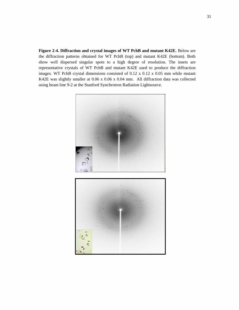
Figure 2-4. Diffraction and crystal images of WT PchB and mutant K42E. Below are
the diffraction patterns obtained for WT PchB (top) and mutant K42E (bottom). Both
show well dispersed singular spots to a high degree of resolution. The insets are
representative crystals of WT PchB and mutant K42E used to produce the diffraction
images. WT PchB crystal dimensions consisted of 0.12 x 0.12 x 0.05 mm while mutant
K42E was slightly smaller at 0.06 x 0.06 x 0.04 mm. All diffraction data was collected
using beam line 9-2 at the Stanford Synchrotron Radiation Lightsource.

32
and 6% glycerol. Parallelepiped crystals grew to ~ 0.12 mm x 0.12 mm x 0.05 mm within
twenty-four hours.
K42E PchB with Salicylate and Pyruvate.
Crystallization of the PchB mutant K42E was carried out using the hanging drop vapor
diffusion method at 25o C. Purified K42E (34 mg/ml) was mixed with a salicylate-puruvate
solution in a 1:20 ratio of K42E to Sal-Pyr (2.9 mM: 58.0 mM) and incubated on ice for 30 min.
Hanging drops consisted of 1.0 μL of the K42E-Sal-Pyr solution mixed with 1.0 μL of well
solution containing 0.004 M Gly-Gly, 0.100 M sodium acetate (pH 3.6) and 12% glycerol.
Parallelepiped crystals grew to ~ 0.06 mm x 0.06 mm x 0.04 mm in two days.
Collection of Crystallographic Data and Structure Determination for PchB
WT PchB with Salicylate and Pyruvate.
A WT PchB crystal was transferred to a drop containing 20% glycerol and flash cooled to
-180o C with liquid nitrogen. Diffraction data were collected remotely at beam line 9-2 at the
Stanford Synchrotron Radiation Lightsource. The collected diffraction images consisted of 1o
oscillations with an exposure time of 20 sec and a detector distance of 290 mm (Figure 2-4). The
diffraction data were indexed and scaled using the XDS program package.24
PchB K42E with Salicylate and Pyruvate.
A PchB K42E crystal was transferred to a drop containing 20% glycerol and flash cooled
to -180o C with liquid nitrogen. Diffraction data were collected remotely at beam line 9-2 at the
Stanford Synchrotron Radiation Lightsource. The collected diffraction images consisted of 1o
oscillations with an exposure time of 15 sec and a detector distance of 100 mm (Figure 2-4). The
diffraction data were indexed and scaled using the XDS program package.24
33
Structure Determination
Structure determination was performed by molecular replacement for both WT and K42E
PchB using Phaser25
from the CCP426
suite using the PchB pyruvate-bound structure (PDB
Code: 2H9D) as a model with the water and ligands omitted. The resultant models were refined
using Coot27
, Phenix28
and REFMAC with resultant statistics (Table 2-2).
34
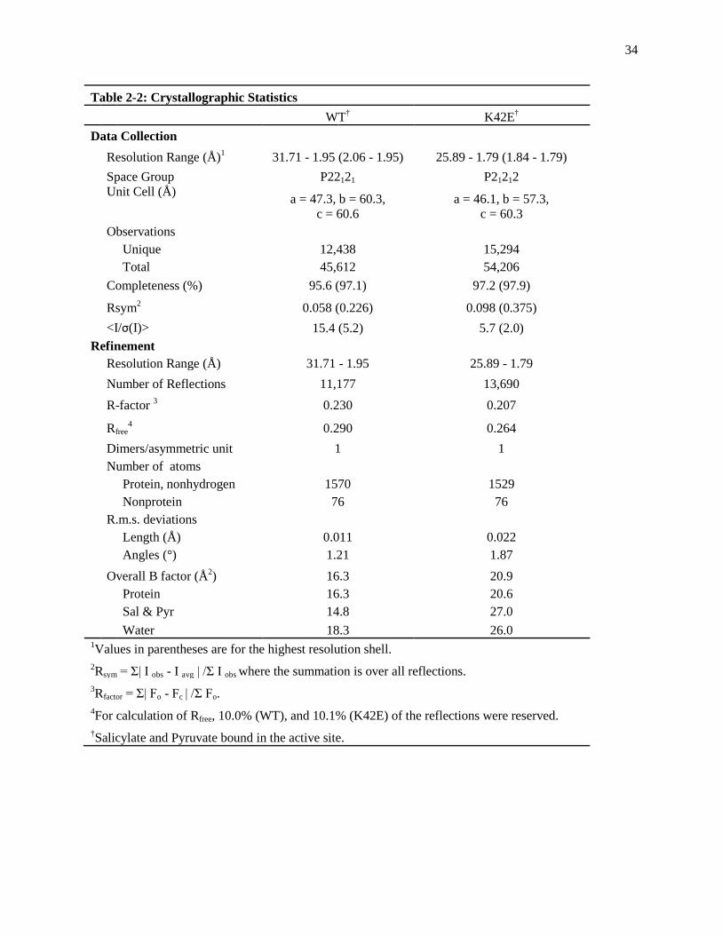
Table 2-2: Crystallographic Statistics
WT† K42E
†
Data Collection
Resolution Range (Å)1 31.71 - 1.95 (2.06 - 1.95) 25.89 - 1.79 (1.84 - 1.79)
Space Group P22121 P21212
Unit Cell (Å) a = 47.3, b = 60.3,
c = 60.6
a = 46.1, b = 57.3,
c = 60.3
Observations
Unique 12,438 15,294
Total 45,612 54,206
Completeness (%) 95.6 (97.1) 97.2 (97.9)
Rsym2 0.058 (0.226) 0.098 (0.375)
<I/σ(I)> 15.4 (5.2) 5.7 (2.0)
Refinement
Resolution Range (Å) 31.71 - 1.95 25.89 - 1.79
Number of Reflections 11,177 13,690
R-factor 3 0.230 0.207
Rfree4 0.290 0.264
Dimers/asymmetric unit 1 1
Number of atoms
Protein, nonhydrogen 1570 1529
Nonprotein 76 76
R.m.s. deviations
Length (Å) 0.011 0.022
Angles (°) 1.21 1.87
Overall B factor (Å2) 16.3 20.9
Protein 16.3 20.6
Sal & Pyr 14.8 27.0
Water 18.3 26.0 1Values in parentheses are for the highest resolution shell.
2Rsym = Σ| I obs - I avg | /Σ I obs where the summation is over all reflections.
3Rfactor = Σ| Fo - Fc | /Σ Fo.
4For calculation of Rfree, 10.0% (WT), and 10.1% (K42E) of the reflections were reserved.
†Salicylate and Pyruvate bound in the active site.
35
2.3 Results
Crystal Structure of WT PchB with Salicylate and Pyruvate Bound
The crystal structure of WT PchB with salicylate and pyruvate bound in the active site
was determined to a resolution of 1.95 Å. The crystals of WT PchB belong to the space group
P22121. This WT PchB structure crystallized in a similar space group to previous apo WT PchB,
PchB mutant K42A and I87T structures (P212121).5,6
The unit cell for WT PchB with salicylate
and pyruvate bound was also different than those previously reported. The WT PchB structure
consists of two chains (A and B) that form intertwined dimers that overall make a homodimer
with two equivalent active sites (Figure 2-5, A). The two equivalent active sites were closed with
one molecule of pyruvate and one molecule of salicylate bound in each. The WT PchB structure
is overall well-ordered throughout with disordered regions only consisting of the N-terminal
methionine and last three amino acids at the C-terminus (Table 2-3, G99, A100, A101). The
salicylate and pyruvate in the active site are oriented by the carboxylate group of arginine 31 and
arginine 14, respectively (Figure 2-5,C). The root mean squared deviation (RMSD) for the
homodimer of WT PchB with salicylate- and pyruvate-bound compared to a homodimer of WT
PchB with two pyruvate molecules bound in the active site is 0.27 Å for 196 α-carbons showing
a highly conserved overall structure between the two wild-type forms. Comparing the α-carbon
at residue 42 in both structures yields an RMSD of 0.53 Å29
. This is higher than the overall
structure but corresponds to previous RMSD values of residue 42 comparisons between the WT
PchB (two pyruvate) and PchB mutant K42A structures.5 The active site of WT PchB with
salicylate and pyruvate is shown in stereo in Figure 2-5 allowing a clear view of the products of
IPL activity and interacting amino acids.
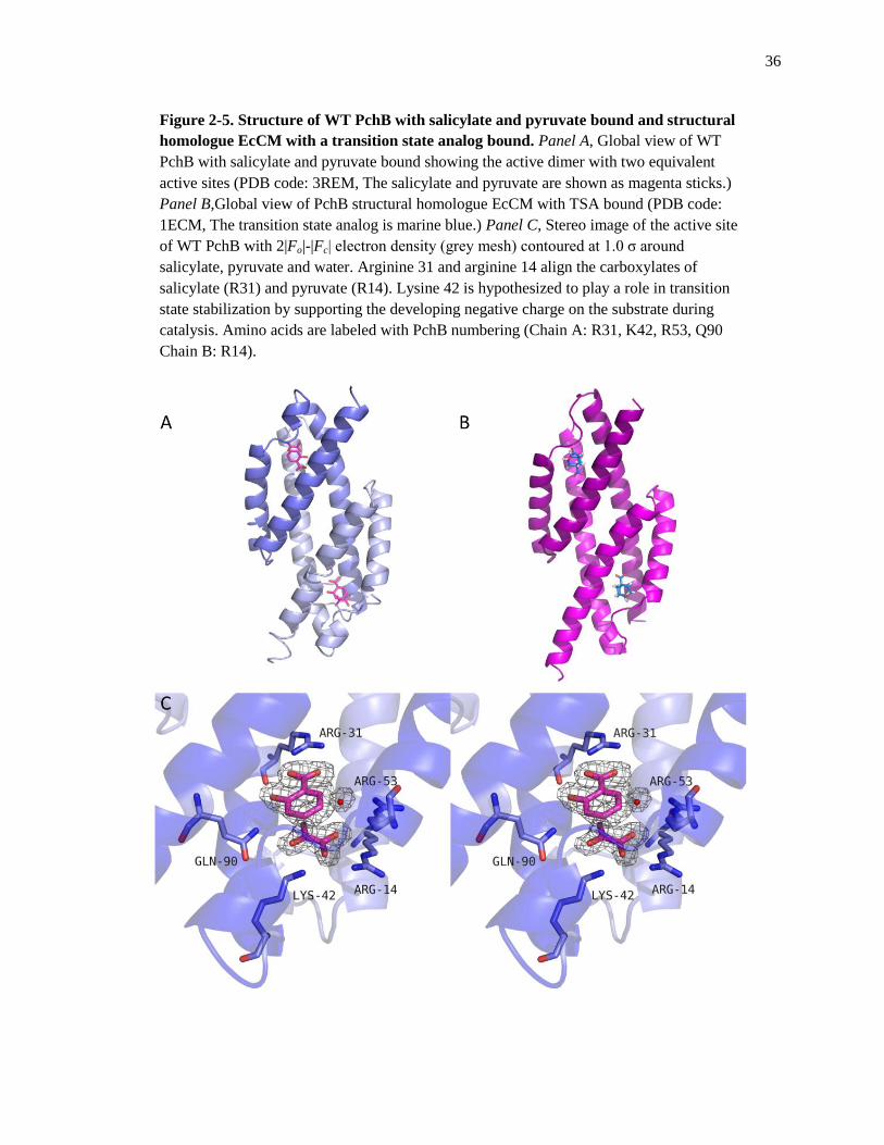
36
Figure 2-5. Structure of WT PchB with salicylate and pyruvate bound and structural
homologue EcCM with a transition state analog bound. Panel A, Global view of WT
PchB with salicylate and pyruvate bound showing the active dimer with two equivalent
active sites (PDB code: 3REM, The salicylate and pyruvate are shown as magenta sticks.)
Panel B,Global view of PchB structural homologue EcCM with TSA bound (PDB code:
1ECM, The transition state analog is marine blue.) Panel C, Stereo image of the active site
of WT PchB with 2|Fo|-|Fc| electron density (grey mesh) contoured at 1.0 σ around
salicylate, pyruvate and water. Arginine 31 and arginine 14 align the carboxylates of
salicylate (R31) and pyruvate (R14). Lysine 42 is hypothesized to play a role in transition
state stabilization by supporting the developing negative charge on the substrate during
catalysis. Amino acids are labeled with PchB numbering (Chain A: R31, K42, R53, Q90
Chain B: R14).

37
Table 2-3: Summary of WT and K42E Structures
Ordered Regions Ligands Bound in the Active Site
WT†
Chain A
2-98 Salicylate and Pyruvate
Chain B
2-98 Salicylate and Pyruvate
K42E†
Chain A
1-97 Salicylate and Pyruvate
Chain B 1-41, 48-97 Salicylate and Pyruvate †Full length PchB consists of 101 amino acids

38
Crystal Structure of PchB mutant K42E with Salicylate and Pyruvate Bound
The crystal structure of PchB mutant K42E with salicylate and pyruvate bound in the
active was determined to a resolution of 1.79 Å. The crystals of PchB mutant K42E belong to the
space group P21212. The space group of K42E is a similar space group to previous apo WT
PchB, PchB mutant K42A and I87T (P212121) structures.5,6
The PchB mutant K42E is also
slightly different than that of WT PchB with salicylate and pyruvate bound (P22121) differing by
one screw axis. The unit cell for K42E was different than those of previous PchB crystals but
very similar to WT PchB with salicylate and pyruvate bound (WT a=47.3 b=60.3 c=60.6, K42E
a=46.1 b=57.3 c=60.3). The K42E structure consists of chains A and B that form intertwined
dimers that overall make a homodimer with two equivalent active sites (Figure 2-6,A). The
active site is closed in both monomers with one molecule each of salicylate and pyruvate bound
in each active site. The K42E structure is overall well-ordered throughout chain A with
disordered regions consisting of only last 4 amino acids at the C-terminus (Table 2-3, R98, G99,
A100, A101) and a portion of the chain B loop region consisting of E42 - I47. This disorder in
the active site loop in chain B does not affect the binding of salicylate or pyruvate within the
active site. The salicylate and pyruvate in the active site are oriented by the carboxylate group of
arginine 31 and arginine 14 (Figure 2-6,C). The root mean squared deviation (RMSD) for the
homodimer of WT PchB with salicylate- and pyruvate-bound compared to K42E was 0.29 Å for
188 α-carbons of the dimer showing a conserved overall structure between the two wild-type
forms. Focusing on the α-carbon at residue 42 for chain A yields an RMSD of 0.34 Å.29
The
active site of PchB mutant K42E with salicylate and pyruvate is shown in stereo in Figure 2-6
allowing a clear view of the products of IPL activity and interacting amino acids.

39
Figure 2-6. Structure of PchB mutant K42E with salicylate and pyruvate bound with
comparative WT PchB. Panel A, Global view of PchB mutant K42E with salicylate and
pyruvate bound (cyan sticks) showing the active dimer with two equivalent active sites
(PDB code: 3RET). Panel B, Global view of WT PchB with salicylate and pyruvate bound
(magenta sticks) showing the active dimer with two equivalent active sites (PDB code:
3REM). Panel C, Stereo image of the active site of PchB mutant K42E with 2|Fo|-|Fc|
electron density (grey mesh) contoured at 1.0 σ around salicylate, pyruvate and water.
Arginine 31 and arginine 14 align the carboxylates of salicylate (R31) and pyruvate (R14).
The K42E mutation is hypothesized to disrupt transition state stabilization by repelling the
developing negative charge on the substrate during catalysis. Amino acids are labeled with
PchB numbering (Chain A: R31, K42, R53, Q90 Chain B: R14).

40
2.4 Discussion
Comparison of WT PchB Structures (2Pyr and Sal & Pyr)
The stereo overlay of the two WT PchB structures (PDB codes: 3REM, 2H9D) show a
good agreement of the overall active site architecture (Figure 2-7). One noticeable difference is
in the position of R53 in the WT PchB with 2 molecules of pyruvate bound (2H9D) structure
(green sticks) relative to that of R53 in the WT PchB with salicylate and pyruvate bound
structure (3REM). The side chain is flipped in towards the pyruvate molecules bound in the
active site (yellow sticks). In the WT PchB with salicylate and pyruvate structure the flipped R53
is replaced by a water molecule that appears to provide the same hydrogen bonding network in
place of R53. The carboxylic group of the two pyruvates are organized in the active site by R31
and R14 in the WT PchB structure with two molecules of pyruvate bound which is consistent
with the organization of salicylate (R31) and pyruvate (R14) in the WT PchB salicylate and
pyruvate bound structure. Another slight difference between the two structures is in the position
of glutamine 90. In the 2H9D structure the glutamine is position to interact specifically with only
one pyruvate molecule, which is in contrast to the 3RET structure where glutamine 90 is position
to interact with not only pyruvate but also salicylate. The relative position of K42 is shared
among the two crystal structures. The position of K42 within both structures places it in
proximity of stabilizing the developing negative charge on the ether oxygen during the transition
state of PchB’s IPL activity. Overall the active site architecture is conserved among both
structures. Even with the presence of salicylate, with its aromatic ring structure, there are no
drastic changes in the active site architecture. This new view of WT PchB with salicylate and
pyruvate bound in the active site shows the conserved interactions while at the same time shows

41
Figure 2-7. Stereo overlay comparing the active sites of two WT PchB crystal
structures. WT PchB with salicylate and pyruvate bound (blue and magenta sticks, PDB
Code: 3REM). WT PchB with two molecules of pyruvate bound (green and yellow sticks,
PDB Code: 2H9D). Arginine 31 and arginine 14 align the carboxylates of salicylate (R31)
and pyruvate (R14) in 3REM and also both pyruvate molecules in 2H9D. Lysine at
position 42 is hypothesized to aid in transition state stabilization by supporting the
developing negative charge on the substrate during catalysis. Differences in the active site
architecture include positioning of arginine 53 in 2H9D (green sticks) relative to the water
arginine 53 position in 3REM (red sphere, blue sticks). Amino acids are labeled with PchB
numbering (Chain A: R31, K42, R53, Q90, Chain B: R14).
42
the conserved interactions while at the same time showing the subtle differences between these
two structures.
Comparison of WT PchB (Sal & Pyr) & K42E Structures
Comparison of the active sites for the crystal structures for WT PchB (blue sticks) and
PchB mutant K42E (orange sticks) also shows a highly conserved active site architecture (Figure
2-8). This comes as a surprise because due to the recent mutational analysis study by Luo and
co-workers that reports no detectable IPL or CM activity for the PchB mutant K42E.5 Both
arginines at position 31 orient the carboxylic group of salicylate within the active site. The
pyruvate is also oriented in the active site by carboxylic interactions with arginine 14. The major
difference between these two structures is in the length of the variant side chain of glutamic acid
at position 42 relative to that of the native lysine in the WT structure and the charge carried by
both residues.
Comparison of K42E (Sal & Pyr) with K42A (Sal & Pyr)
Comparison of the PchB mutant structures K42E (PDB code: 3RET, orange sticks) and
K42A (Pdb code: 3HGX, pink sticks) also shows a conserved active site architecture (Figure 2-
9) between both structures. The salicylate and pyruvate molecules are orientated in the active site
by the same interactions of their carboxylic acid groups with arginines 31 and 14 in both
structures. The most noticeable difference is in the length of the side chain at position 42.
43
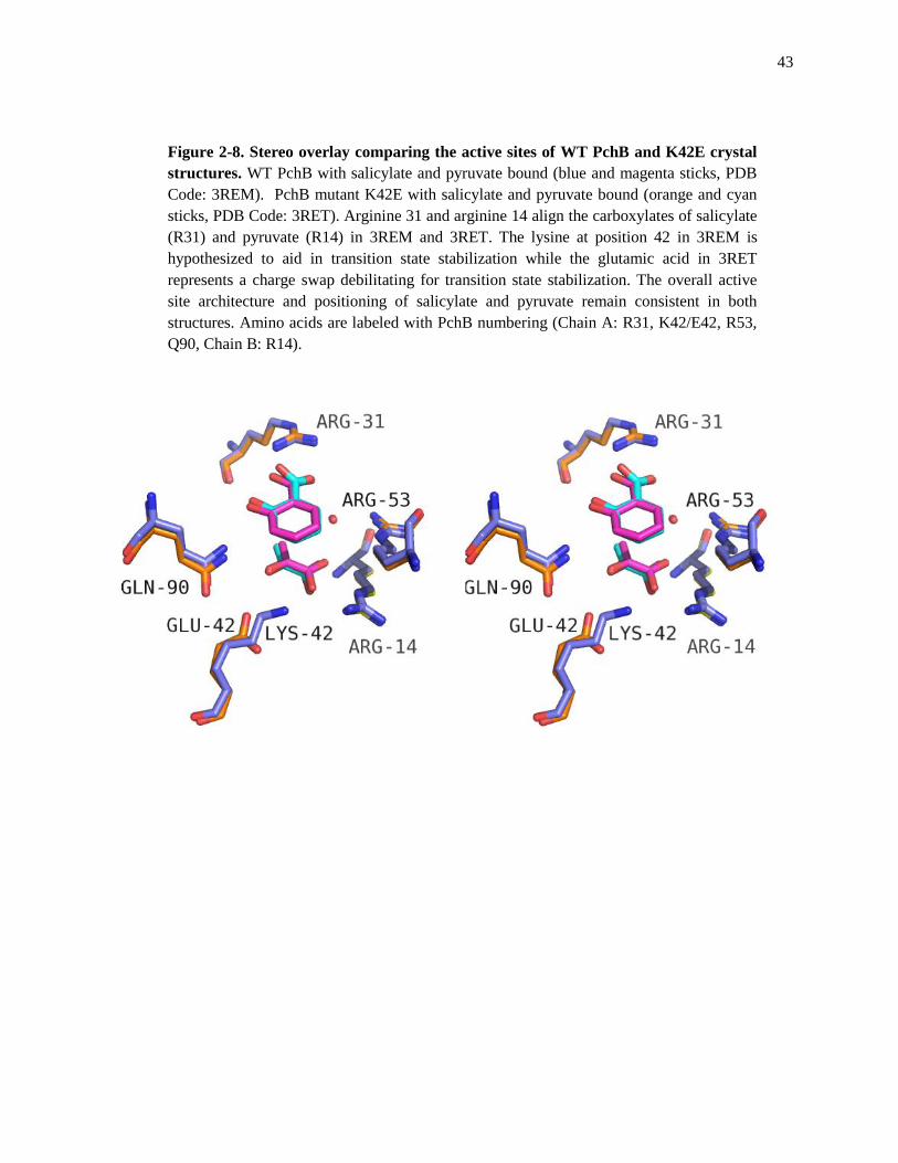
Figure 2-8. Stereo overlay comparing the active sites of WT PchB and K42E crystal
structures. WT PchB with salicylate and pyruvate bound (blue and magenta sticks, PDB
Code: 3REM). PchB mutant K42E with salicylate and pyruvate bound (orange and cyan
sticks, PDB Code: 3RET). Arginine 31 and arginine 14 align the carboxylates of salicylate
(R31) and pyruvate (R14) in 3REM and 3RET. The lysine at position 42 in 3REM is
hypothesized to aid in transition state stabilization while the glutamic acid in 3RET
represents a charge swap debilitating for transition state stabilization. The overall active
site architecture and positioning of salicylate and pyruvate remain consistent in both
structures. Amino acids are labeled with PchB numbering (Chain A: R31, K42/E42, R53,
Q90, Chain B: R14).

44
Figure 2-9. Stereo overlay comparing the active sites of PchB mutants K42E and
K42A crystal structures. PchB mutant K42E with salicylate and pyruvate bound (orange
and cyan sticks, PDB Code: 3RET). PchB mutant K42A with salicylate and pyruvate
bound (magenta and green sticks, PDB Code: 3HGX). Arginine 31 and arginine 14 are
shown to align the carboxylates of salicylate (R31) and pyruvate (R14) in 3RET and
3HGX. The glutamic acid at position 42 in 3RET represents a charge swap with
debilitating effects to transition state stabilization. The alanine at position 42 represents an
absence of supporting charge compared to WT PchB and mutant K42E. The overall active
site architecture and positioning of salicylate and pyruvate remain consistent in both K42E
and K42A mutant structures. Amino acids are labeled with PchB numbering (Chain A:
R31, E42/A42, R53, Q90, Chain B: R14).

45
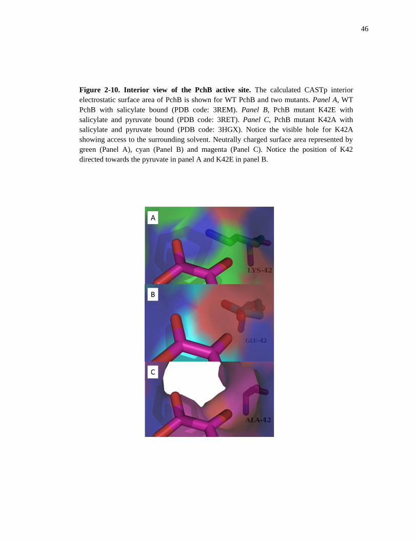
Comparison of WT PchB and Mutant Active Site Surface Area
Analysis of the electrostatic surface area has provided a detailed look at importance of
charged residues at position 42 in PchB. CASTp30
calculations performed on the active site
surface area of WT PchB and mutants K42E and K42A yield an electrostatic surface area image
of what can be imagined as a wine bottle with cork appearance (Figure 2-10). When the active
site loop is ordered as is the case of residues at position 42 (Figure 2-10, A-C). The side chains
directly position themselves into the active site of PchB. One major difference is the “un-corked”
appearance of the active site in the case of PchB mutant K42A (Figure 2-10, C). The lysine to
alanine mutation has physically shortened the side chain at position 42, resulting in an ordered
active site loop region upon ligand binding, but at the same time exposing the active site to the
surrounding solvent. This is in contrast to the K42E mutation which closes off the active site
from the surrounding solvent when the active site loop is ordered (Figure 2-10, B). The K42E
mutant however provides a strong negative charge as opposed to the positive charge found with
lysine at position 42 in WT PchB (Figure 2-10, A). It is important to note that the K42A
mutation decreases the IPL and CM activity 100-fold while the K42E mutant has no detectable
activity for either IPL or CM activity.5
46
Figure 2-10. Interior view of the PchB active site. The calculated CASTp interior
electrostatic surface area of PchB is shown for WT PchB and two mutants. Panel A, WT
PchB with salicylate bound (PDB code: 3REM). Panel B, PchB mutant K42E with
salicylate and pyruvate bound (PDB code: 3RET). Panel C, PchB mutant K42A with
salicylate and pyruvate bound (PDB code: 3HGX). Notice the visible hole for K42A
showing access to the surrounding solvent. Neutrally charged surface area represented by
green (Panel A), cyan (Panel B) and magenta (Panel C). Notice the position of K42
directed towards the pyruvate in panel A and K42E in panel B.

47
The Conserved Active Site Architecture of PchB
The WT PchB and the PchB mutants show a conserved active site architecture that does
not drastically change among the individual mutants. This is the result of the arginines 31 and 53
correlating the carboxylates of the salicylate and pyruvate in the active site. These two
interactions are fundamental in the stabilization of the products in the active site and therefore
can be viewed as important for the binding of the substrate in the active site. These two
interactions do not change among the mutants and so the major contributors orienting the
substrate in the active site do not change. Even when the side chain length is drastically reduced,
as in the case of the PchB mutant K42A, the active site loop is ordered and pyruvate and
salicylate are oriented in the same manner with very little difference in the α-carbon at position
42 (Figure 2-11). The various lengths of the side chains themselves also show no overall
apparent change in the active site architecture (Figure 2-11). The various lengths of WT, K42E
and K42A PchB side chains represent major differences in the IPL and CM activity of PchB but
are not by themselves structurally significant when stabilizing the products of IPL activity in the
active site (Figure 2-11).
If the active site architecture is the same among WT PchB and mutant structures when
stabilizing the products of IPL activity then the differences in activity are likely the result of the
charge at position 42 and the dynamics of the active site loop. The initial binding event between
the substrate and enzyme can be hypothesized to be mainly coordinated by the two arginines
(R31 and R53) within the active site. We know that the active site loop has been shown to be
ordered in the presence of substrate.6
This ordering of the active site loop consequently aligns the
charged residue at position 42 into the active site as shown with the previous crystal structures
above along with an interaction necessary for PchB activity5,6
. Once the substrate isochorismate

48
Figure 2-11. Overlay comparing the active sites of WT PchB and PchB mutants
K42E and K42A. WT PchB with salicylate and pyruvate bound (blue and magenta sitcks,
PDB code: 3REM). PchB mutant K42E with salicylate and pyruvate bound (orange and
cyan sticks, PDB Code: 3RET). PchB mutant K42A with salicylate and pyruvate bound
(magenta and green sticks, PDB Code: 3HGX). Arginine 31 and arginine 14 are shown to
align the carboxylates of salicylate (R31) and pyruvate (R14). The overall active site
architecture and positioning of salicylate and pyruvate remain consistent in both WT PchB
and PchB K42E and K42A mutant structures. Notice the difference in physical length
among the side chains at position 42. Amino acids are labeled with PchB numbering
(Chain A: R31, K42/E42/A42, R53, Q90, Chain B: R14).

49
has passed through the transition state, the stabilization of the products within the active site are
similar among WT PchB and PchB mutants. This highlights the importance of providing the
correct charge at position 42 prior to catalysis allowing for IPL and CM activity to proceed.
Whereas the placement of the positive charge in K42 is hypothesized to stabilize the developing
negative charge on the ether oxygen of isochorismate, the placement of a negative charge in
K42E can be hypothesized to destabilize the transition state through electrostatic interactions.5,18
While the positive charge at position 42 has been shown to be the most efficient, the PchB
mutant K42A still provides sufficient stabilization of the transition state allowing for IPL and
CM activity, albeit at a reduced rate.5
The evidence for the importance of the correct charge at position 42 allowing for efficient
catalysis is complimented by the ordered loop region upon ligand binding6 with alignment of the
charged residue at position 42 and subsequent release of the products once catalysis occurs. The
structural studies so far have provided evidence for the importance of residue 42 while also
raising the question of how loop dynamics are responsible for the positioning of residue 42 and
therefor contribute to catalysis.

50
References
1. Gaille, C., Kast, P. & Haas, D. Salicylate biosynthesis in Pseudomonas aeruginosa.
Purification and characterization of PchB, a novel bifunctional enzyme displaying
isochorismate pyruvate-lyase and chorismate mutase activities. J Biol Chem 277, 21768-
21775 (2002).
2. Gaille, C., Reimmann, C. & Haas, D. Isochorismate synthase (PchA), the first and rate-
limiting enzyme in salicylate biosynthesis of Pseudomonas aeruginosa. J Biol Chem 278,
16893-16898 (2003).
3. DeClue, M.S., Baldridge, K.K., Kunzler, D.E., Kast, P. & Hilvert, D. Isochorismate
pyruvate lyase: a pericyclic reaction mechanism? J Am Chem Soc 127, 15002-15003
(2005).
4. Rajagopalan, J.S., Taylor, K.M. & Jaffe, E.K. 13C NMR studies of the enzyme-product
complex of Bacillus subtilis chorismate mutase. Biochemistry 32, 3965-3972 (1993).
5. Luo, Q., Olucha, J. & Lamb, A.L. Structure-Function Analyses of Isochorismate-
Pyruvate Lyase from Pseudomonas aeruginosa Suggest Differeing Catalytic Mechanisms
for the Two Pericyclic Reactions of This Bifunctional Enzyme. Biochemistry 48, 5239-
5245 (2009).
6. Zaitseva, J., Lu, J., Olechoski, K.L. & Lamb, A.L. Two crystal structures of the
isochorismate pyruvate lyase from Pseudomonas aeruginosa. J Biol Chem 281, 33441-
33449 (2006).
7. Andrews, P.R., Smith, G.D. & Young, I.G. Transition-state stabilization and enzymic
catalysis. Kinetic and molecular orbital studies of the rearrangement of chorismate to
prephenate. Biochemistry 12, 3492-3498 (1973).
8. Hilvert, D., Carpenter, S.H., Nared, K.D. & Auditor, M.T. Catalysis of concerted
reactions by antibodies: the Claisen rearrangement. Proc Natl Acad Sci U S A 85, 4953-
4955 (1988).
9. Copley, S.D. & Knowles, J.R. The Uncatalyzed Claisen Rearrangement of Chorismate to
Prephenate Prefers a Transition-State of Chairlike Geometry. J Am Chem Soc 107, 5306-
5308 (1985).
10. Copley, S.D. & Knowles, J.R. The Conformational Equilibrium of Chorismate in
Solution - Implications for the Mechanism of the Nonenzymatic and the Enzyme-
Catalyzed Rearrangement of Chorismate to Prephenate. J Am Chem Soc 109, 5008-5013
(1987).
11. Warshel, A. Energetics of Enzyme Catalysis. Proc Natl Acad Sci U S A 75, 5250-5254
(1978).
12. Warshel, A. et al. Electrostatic basis for enzyme catalysis. Chemical Reviews 106, 3210-
3235 (2006).
13. Lee, A.Y., Karplus, P.A., Ganem, B. & Clardy, J. Atomic structrue of the buried catalytic
pocket of Escherichia coli chorismate mutase. J Am Chem Soc 117, 3627-3628 (1995).
14. Lee, A.Y., Stewart, J.D., Clardy, J. & Ganem, B. New insight into the catalytic
mechanism of chorismate mutases from structural studies. Chem Biol 2, 195-203 (1995).
15. Liu, D.R., Cload, S.T., Pastor, R.M. & Schultz, P.G. Analysis of active site residues in
Escherichia coli chorismate mutase by site-directed mutagenesis. J Am Chem Soc 118,
1789-1790 (1996).
16. Cload, S.T., Liu, D.R., Pastor, R.M. & Schultz, P.G. Mutagenesis study of

51
active site residues in chorismae mutase from Bacillus subtilis. J Am Chem Soc 118,
1787-1788 (1996).
17. Kast, P., Asif-Ullah, M., Jiang, N. & Hilvert, D. Exploring the active site of chorismate
mutase by combinatorial mutagenesis and selection: the importance of electrostatic
catalysis. Proc Natl Acad Sci U S A 93, 5043-5048 (1996).
18. Kast, P. et al. A strategically positioned cation is crucial for efficient catalysis by
chorismate mutase. J Biol Chem 275, 36832-36838 (2000).
19. Kienhofer, A., Kast, P. & Hilvert, D. Selective stabilization of the chorismate mutase
transition state by a positively charged hydrogen bond donor. J Am Chem Soc 125, 3206-
3207 (2003).
20. Hur, S. & Bruice, T.C. Comparison of formation of reactive conformers (NACs) for the
Claisen rearrangement of chorismate to prephenate in water and in the E. coli mutase: the
efficiency of the enzyme catalysis. J Am Chem Soc 125, 5964-5972 (2003).
21. Zhang, X. & Bruice, T.C. A definitive mechanism for chorismate mutase. Biochemistry
44, 10443-10448 (2005).
22. Hur, S. & Bruice, T.C. Just a near attack conformer for catalysis (chorismate to
prephenate rearrangements in water, antibody, enzymes, and their mutants). J Am Chem
Soc 125, 10540-10542 (2003).
23. Hur, S. & Bruice, T.C. The near attack conformation approach to the study of the
chorismate to prephenate reaction. Proc Natl Acad Sci U S A 100, 12015- 12020 (2003).
24. Kabsch, W. XDS. Acta Cryst. D 66, 125-132 (2010).
25. McCoy A.J., et al. Phaser crystallographic software. J Appl Cryst 40, 658-674 (2007).
26. "The CCP4 Suite: Programs for Protein Crystallography". Acta Cryst D 50, 760-763
(1994).
27. Emsley, P. and Cowtan, K. Acta Cryst D 60, 2126-2132 (2004).
28. Adams, P., et al. PHENIX: a comprehensive Python-based system for macromolecular
structure solution. Acta Cryst D 66, 213-221 (2010).
29. Kleywegt, G.J., & Jones, T.A. Detecting folding motifs and similarities in protein
structures. Methods in Enzymology 277, 525-545 (1997).
30. Dundas, J. et al. CASTp: computed atas of surface topography of proteins with structural
and topographical mapping of functionally annotated residues. Nucl Acids Res 34, 116-
118 (2006).

52
CHAPTER 3
Nuclear Magnetic Resonance Relaxation Studies of the PchB Mutant K42H
3.1 Introduction
Conformational changes and the macromolecular motions of an enzyme upon binding of
a substrate contribute to moving the substrate farther down the reaction coordinate path towards
the transition state.1,2
These macromolecular motions vary in length from tenths to tens of
angstroms and can vary in time from sub-pico seconds to over a second in length.1 How much
then do conformational changes and macromolecular motion contribute to the formation of the
transition state when the transition state itself is estimated to exist for a mere 100 femtoseconds?2
In chapter 1 we discussed how rate enhancement is proportional to how well an enzyme binds
the transition state for a given substrate.3 Applying this directly to the mutational studies
performed on PchB at residue K42 would lead one to believe that decreases in catalytic rate4 are
fundamentally related to how well PchB can bind the transition state for isochorismate pyruvate
lyase (IPL) and chorismate mutase (CM) activity. Positioning of residue K42 within the active
site loop then brings into question how the dynamics of the active site loop region effect the rate
of catalysis.

53
Dynamics of the Active Site Loop in PchB
The active site loop region of PchB (residues 39-51, monomers A &B) has been shown to
be quite flexible.5 The apo crystal structure of PchB (PDB code: 2H9C) shows a disordered
active site loop region for both monomers, with one of the two equivalent active sites shown in
Figure 3-1A. Well defined electron density around the active site exists for amino acids on both
sides of the active site loop. The region associated with the active site loop in Figure 3-1A shows
no electron density with magenta amino acids marking the beginning and end of the missing
electron density. The lack of electron density for the loop region in the apo structure is the result
of averaging the individual positions of each active site loop for all PchB molecules within the
given crystal used for structure determination through x-ray diffraction. This is to say that the
active site loop region adopts many distinct positions over time within any population of apo
PchB molecules, resulting in weak diffraction and thereby producing electron density that is a
blurred view of all super positions with no definable regions6. This is in direct contrast to the
electron density found in the WT PchB crystal structure with pyruvate bound in the active site as
shown in Figure 3-1, B (PDB code: 2H9D).5 In this case, the ordered active site loop region
shows well defined electron throughout the active site loop region. Also noticeable is the
appearance of two pyruvate molecules with in the active site. This ordering of the active site loop
was hypothesized to occur when a ligand is bound within the active site of PchB.5 This
hypothesis has been shown to be the case for previous K42A3 (salicylate and pyruvate bound)
and current WT PchB (salicylate and pyruvate bound) and K42E (salicylate and pyruvate bound)
crystal structures.

54
Figure 3-1. View of the active site loop region in crystal structures of apo WT PchB and WT
PchB with pyruvate bound. The pyruvate is green sticks. Panel A, Active site loop region of apo
WT PchB (orange sticks: monomer B, cyan sticks: monomer A) showing one of two equivalent
active sites with 2|Fo|-|Fc| electron density (grey mesh) contoured at 1.0 σ around the residues
(PDB code: 2H9C). Missing active site loop region is highlighted with magenta residues. Panel B,
Ordered active site loop region of WT PchB (yellow sticks: monomer A, blue sticks: monomer B)
with pyruvate bound showing one of two equivalent active sites with 2|Fo|-|Fc| electron density
(grey mesh) contoured at 1.0 σ around the residues and two pyruvate molecules (PDB code:
2H9D). Active site residue K42 is shown in green with two pyruvate molecules and magenta
residues are used as reference for Panel A.

55
Coordination of Salicylate and Pyruvate in the Active Site
The coordination of salicylate and pyruvate (IPL products) in the active site of PchB has
been shown to be facilitated by two arginines (R14 & R31) in the active site pocket of the crystal
structure of PchB mutant K42A.4 The K42A structure has completely negated the positive charge
at position 42 in WT PchB and consequentially exhibits a 100-fold decrease in activity for both
IPL and CM activity.4 The significance of activity without electrostatic interactions from residue
42 within the flexible active site loop support the hypothesis that the active site loop contributes
to catalysis through stabilizing the transition state. How much does the active site loop move in
the presence and absence of substrate is yet to be determined.
In this chapter Nuclear Magnetic Resonance (NMR) experiments were carried out to
provide insight into the relationship between dynamic active site loop movement and catalysis in
PchB. The backbone resonance assignments were obtained from collections of 2D 1H-
15N
heteronuclear single-quantum coherence (HSQC)8 and HNCA
9, CBCA(CO)NH
9, HNCACB
10
3D data sets in combination with 15
N alanine and15
N leucine labeled HSQC
11 experiments. These
results lay the ground for determining the relative contributions of WT and PchB mutants in
position 42 of the active site loop region towards stabilizing the transition or generation of a
NAC.
3.2 Materials and Methods
Expression and Purification of 13
C-,15
N- and 15
N-labeled recombinant and WT PchB
The sub-cloning of the PchB expression plasmid has previously been reported.4
Expression and purification of double labeled (13
C-, 15
N-) and single labeled (15
N-) recombinant

56
and WT PchB was performed as previously described5 with modifications. Uniformly
13C,
15N-
and 15
N-labeled PchB were obtained from bacterial cells (E. coli BL21(DE3), BL21 Star
(DE3)pLysS, Invitrogen) that were grown up in 1 liter non-baffled Fernbach flasks containing
minimal media supplemented with 2g/L of [13
C]-glucose for double labeled or 4g/L of glucose
for single labeled sample with 1g/L [15
N]-ammonium chloride at 37 °C with shaking at 250 rpm
until an optical density of 0.8 at 600 nm was reached (Cary 50 UV-Visible Spectrophotometer).
The bacterial cells were induced with 0.2 mM isopropyl-β-D-thiogalactopyranoside (IPTG) and
transferred to a 15 oC incubator with shaking at 250 rpm for overnight growth. Cells were
harvested by centrifugation at 4,000 x g for 10 min at 4 °C. The pelleted bacterial cells were
resuspended with ~15 mL buffer A (25 mM Tris-HCl (pH 8.0)) and lysed by sonication (Digital
Sonifier, Branson) on ice for a total of 3 min consisting of 3 second bursts with intervening 20
sec resting periods. The lysed bacterial cells were subjected to ultracentrifugation at 142,000 x g
for 45 min at 4 °C to remove cellular debris. The supernatant (~30 mL) was diluted to 150 mL
with buffer A before being applied to a 30Q Sepharose Fast Flow column (Amersham
Biosciences) equilibrated with buffer A for anion-exchange chromatography. A linear gradient
from 0 – 50% buffer B (25 mM Tris-HCl (pH 8.0), 500 mM NaCl) was applied to the 30Q
column and PchB eluted at ~18% B into 6 mL fractions. The fractions were analyzed using 15%
SDS-PAGE and those containing PchB were pooled and concentrated to ~4 mL using a stirred
cell apparatus (Amicon) with a YM-3 membrane (NMWL of 3 kDa) under nitrogen gas at 73 psi.
The concentrated pooled fractions were applied to a HiLoad 16/60 Superdex 75 gel filtration
column (GE Healthcare) equilibrated with 50 mM Tris-HCl (pH 8.0), 150 mM NaCl, and 10%
glycerol. The resulting fractions were analyzed using 15% SDS-PAGE and found to be of high
purity (≥99%). The protein concentration was determined by UV absorbance at 280 nm after

57
treatment with 6M guanidine-HCl. The purified protein was aliquoted into Eppendorf tubes and
stored at -80 oC for NMR studies.
Expression and Purification of Selectively 15
N-labeled Ala and Leu PchB
Expression and purification of selectively 15
N-labeled Ala and Leu PchB was performed
as previously described11
with modifications shown above. The minimal media was
supplemented with 250 mg of the labeled amino acid per one liter growth with an additional 900
mg of unlabeled Val, Leu, Gly and Ile for 15
N-labeled Ala growths with Val and Ile for 15
N-
labeled Leu growths.
NMR Spectroscopy
NMR data were acquired at room temperature (25 o
C) on a Bruker Avance 800 MHz
spectrometer and was processed and analyzed using NMRPipe12
and NMRView13
. A uniformly
13C-
15N labeled PchB mutant K42H protein sample at a 2.0 mM concentration was used to
collect the following 3D data sets: HNCA9, CBCA(CO)NH
9 HNCACB.
10 Resonance
assignments were determined by 2D 1H-
15N HSQC experiments at various concentrations
between 0.3 mM and 1.5 mM for 15
N Ala and 15
N Leu selectively11
labeled PchB mutant K42E
and uniformly labeled 15
N PchB mutant K42H along with previously mentioned 3D data sets.
Flip-back pulses and pulsed field gradients were used to achieve water suppression.

58
Optimization of NMR Buffers
Preparation of NMR Samples
The preparation of NMR samples consisted of protein concentration using ultra
centrifugal devices (Amicon, 3K NMWL) until ~two milliliters of concentrated PchB was
achieved. The buffer system was swapped with an NMR suitable buffer using a PD-10 desalting
column (GE Healthcare) using the spin protocol to prevent any substantial loss. The resultant
protein sample was concentrated again using ultra centrifugal devices (Amicon, 3K NMWL)
until a volume of 500 uL was achieved. The protein concentration was determined by UV
absorbance at 280 nm after treatment with 6M guanidine-HCl. The sample was transferred to an
eight inch NMR tube (Kontes) suitable for use in an 800 MHz spectrometer. Samples that
showed good HSQC results were hermetically sealed to prevent contamination and stored at 4 O
C
for use in future NMR studies.
Phosphate-NaCl Buffer Conditions
The original buffer conditions for PchB NMR samples consisted of 20 mM phosphate, 20
mM NaCl and 10% D2O (pH=7). The pH of the buffer system was determined by combining
equal molar concentrations of mono and di-basic phosphate solution until the desired pH was
reached. Samples were originally found to be stable after 24 hours. However, these same
samples began to show signs of precipitation (white & cloudy) at 36 hours with increasing
precipitation as at time went on. This resulted in a decreased NMR signal and poor HSQC
spectra. Optimization steps were taken to increase solubility of PchB with progressively high
NaCl concentrations (50 mM, 75mM). The changes in NaCl concentration yielded stabilization
in the short term (24-48 hours) but ultimately resulted in a delayed precipitation effect similar to
59
the original buffer conditions with precipitation persistently increasing over time and found to be
unsuitable for consistency among long term and future NMR studies.
Deuterated Tris Buffer Conditions
To increase the solubility of PchB in an NMR suitable buffer, deuterated Tris (50 mM)
and NaCl (75-100mM) were incorporated. The new NMR buffer consisted of deuterated 50 mM
Tris pH=7,100 mM NaCl and 10% D2O. The pH of the buffer system was determined by
titration with concentrated HCl (12 N) until the desired pH of 7.5 was reached. This new buffer
condition resulted in highly soluble PchB with no protein precipitation over time. It should be
noted that this buffer system is closely related to the buffer system used during the final stage of
non-labeled PchB purification (50 mM Tris- pH=8, 150 mM NaCl, 10% glycerol). The resultant
protein sample showed an improved HSQC over the original NMR buffer conditions and was
subsequently used for collection of 3D data sets.
3.3 Results
The PchB mutant K42H was used in this NMR analysis. Analysis of the CBCA(CO)NH
and HNCA data sets resulted in 32 out of the 101 amino acids assigned (Figure 3-2). The method
of analysis for resonance assignments was dependent on the HNCA and CBCA(CO)NH data
sets. The process of assignment in NMR analysis is dependent on correlating protons through
other spin systems (13
C, 15
N) or through other protons in what is known as through-bond
relationships.14
Protons produce what is referred to as a chemical shift position which can be
thought of as a peak dependent on the specific environment experienced within the protein. This
idea is illustrated in Figure 3-3 where each peak can be thought of as having a unique 1H and
15N
chemical shift resulting in a 2D map of each specific backbone proton seen through the bond of

60
Figure 3-2. Assigned 1H-
15N HSQC Spectra of
PchB K42H. Full view of the
1H-
15N HSQC
spectra for the PchB mutant K42H at pH=7. The assigned peaks below represent 32 out of the 101
amino acids of PchB.

61
Figure 3-3. 1H-
13C HNCA Spectra of PchB K42H. A set of ten assigned sequential amino acids
are shown below with specific designations shown at the top of the strips. Notice the G69 which
has a very characteristic α-carbon (13
C) chemical shift ~45 ppm which was used as a
starting point in assignment. The resultant strips show the matching of the α-carbon (13
C)
shifts and sequential “stair-stepping” used in backbone resonance assignment.
62
an attached 15
N in PchB.14
In essence the HSQC shown in Figure 3-2 is representative of each
amino acid within PchB. The challenge is assigning each specific peak in the HSQC to a specific
amino acid in the sequence of PchB, which in our case is aided by the use of another spin system
(13
C) and further experiments.
The HNCA experiment is used for assignments by its ability to correlate the chemical
shifts of the α-carbon (13
C) of the current amino acid (n, strong peak) and the α-carbon (13
C) of
the previous peak (n-1, usually weaker peak). Since the magnetism originates at the amide bond
of a specific amino acid and is then sequentially transferred through J coupling (through-bond) to
the α-carbons of the current and previous amino acid the two resultant peaks will share the same
1H and
15N chemical shifts but exhibit a different
13C chemical shift
N as seen in the individual
strips in Figure 3-3. Since all amides in the protein are excited at once, these two unique peak
signals are also produced for every single α-carbon (13
C) amino in the protein, resulting in a
strong and weak peak that share the same 13
C chemical shift but with different 1H chemical
shifts, which are the result of “seeing” the α-carbon through its amide bond (n, strong peak) and
the amide bond of the next amino acid (n+1, weak peak) as seen in the adjacent strips in Figure
3-3.14
This results in every amino acid being able to “see” the previous amino acid (n-1) while at
the same time being “seen” by the following amino acid (n+1). These chemical shifts peaks can
then be used to correlate the 13
C α-carbons and piece together the backbone resonances resulting
in assigning the individual peaks shown in the HSQC with a specific amino acid (Figure 3-2).14
Confirmation of the assignments made through HNCA data analysis was performed
through the CACB(CO)NH experiment. Analysis of this experiment yields two peaks
representative of the α and ß-carbons (13
C) of the preceding residue in the peptide sequence each
with a differing 13
C chemical shift as shown in the individual strips in Figure 3-4.14
The α and ß-

63
carbons (13
C) shifts are correlated to a range of expected values recorded for the individual
twenty amino acids thereby supporting or ruling out “matching” strips in the HNCA.
The process of assignment can also be aided in through selective labeling of individual
amino acids.11
As mentioned previously labeling results in HSQC peaks representative of the
specific labeled amino acids chosen. This can be very helpful in identifying the nature of peaks
that are difficult to assign otherwise but only serves to narrow down the possible peaks out of the
total number of peaks. Take for example the results for 15
N-labeled alanine (green) and 15
N-
labeled leucine (red) as shown in Figure 3-5. PchB contains a total of 18 alanine and six leucine
residues which closely match the number of peaks shown for each respective HSQC. While the
selectively labeled approach narrows down the field by identifying the alanine or leucine peaks
within a spectra it still does not result in specific assignments and as such must be used in
conjunction with data from other experiments such as the HNCA.

64
Figure 3-4. 1H-
13C CACB(CO)NH Spectra of PchB K42H. A set of nine assigned (R61-G69)
and one yet to be assigned (E60) sequential amino acids are shown below with specific
designations shown at the top of the strips. Each strip is representative of the (n-1) 13
C-α and ß
carbons. Notice the G69 which has a very characteristic α-carbon (13
C) chemical shift ~45
ppm and other strips which was used to check assignments made through the HNCA.

65
Figure 3-5. 1H-
15N HSQC Spectra of
PchB K42H with
15N-labeled Ala and Leu Overlays. Full
view of the 1H-
15N HSQC spectra for the PchB mutant K42H at pH=7. The green peaks are
representative of the 1H-
15N alanine HSQC of PchB K42E. Notice ~15 peaks are visible out of the
total 18 within the protein. The red peaks are representative of the 1H-
15N leucine HSQC of PchB
K42E. Notice that five peaks are visible out of a total of five leucine within PchB.

66
3.4 Discussion
Through the use of NMR spectroscopy, protein dynamics can be studied by looking at the
relaxation rates of individual nuclei within a given protein.14,15
These relaxation rates are used to
correlate the overall flexibility of the protein through the summation of the flexibility of
individual amino acids.14
This information can be used to correlate structural dynamics to aspects
of protein function such as contributions to catalytic activity.15
Before relaxation data can be utilized the backbone resonance assignments must first be
made for the regions of interest. While the PchB mutant K42H was chosen as the main
foundation for this thesis, initially selectively labeled 15
N-alanine and 15
N-leucine K42E were
generated to aid in the backbone resonance assignments of previously collected NMR data for
the PchB mutant K42E and were used to aid in the assignment of PchB mutant K42H (Figure 3-
5). The PchB mutant K42H was chosen due its unique titratable IPL and CM activity4 and also
for the potential of future structure determination. The majority of peaks shown in the PchB
mutant K42H HSQC in Figure 3-2 are associated with backbone resonances of the individual
amino acids in PchB. The peaks can then be assigned to a particular amino acid based on
chemical shift data (Figure 3-3, 3-4). Currently out of the 101 amino acids in PchB mutant K42H
32 backbone resonances have been assigned (Figure 3-2). All of the current assignments have
been made in areas away from the active site loop region (light blue, Figure 3-6). Future work is
needed to complete the backbone resonance assignments. Once all of the backbone resonance
assignments have been made they can be used to evaluate relaxation data that provides structural
dynamic information for individual amino acids and also the flexibility of the overall structure.
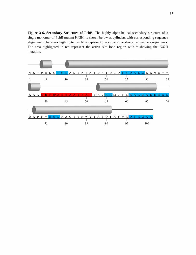
67
Figure 3-6. Secondary Structure of PchB. The highly alpha-helical secondary structure of a
single monomer of PchB mutant K42H is shown below as cylinders with corresponding sequence
alignment. The areas highlighted in blue represent the current backbone resonance assignments.
The area highlighted in red represent the active site loop region with * showing the K42H
mutation.

68
This information can then be used to correlate the active site loop dynamics and characterize its
contribution to catalytic activity in PchB.
In the case of PchB, the relaxation data (T1, T2, hetNOE) can be used to correlate the
active site loop dynamics and contribution to catalysis.15,16
For the active site loop dynamics to
play a role in the catalysis, the motions of the active site loop must first occur on a time scale
relevant to catalysis. The kcat for the IPL activity of the PchB mutant K42H is ~0.037 s-1
3. In
other words, one molecule of isochorismate is converted to salicylate and pyruvate every 27
seconds (WT PchB = 6 seconds). This is not to say that only one molecule of isochorismate
binds within the active site and then proceeds to take 27 seconds to surmount the transition state.
However only once every 27 seconds does the combined effort of initial binding, electrostatics,
loop dynamics and molecular breathing, result in a catalytic event. NMR relaxation
measurements will be able to probe the timescale of the active site loop dynamics while also
providing information on the flexibility of the macromolecule. Future NMR analysis will be able
to elucidate and assess the contribution of PchB’s active site loop dynamics towards IPL and CM
activity.

69
References
1. Karplus, M. Molecular dynamics simulations of biomolecules. Acc Chem Res 35, 321-
323 (2002).
2. Schramm, V.L. Enzymatic transition states and transition state analog design. Annu Rev
Biochem 67, 693-720 (1998).
3. Mader, M.M. & Bartlett, P.A. Binding Energy and Catalysis: The Implications for
Transition-State Analogs and Catalytic Antibodies. Chem Rev 97, 1281-1302 (1997).
4. Lou, Q., Oulcha, J. & Lamb, A.L. Structure-function analyses of isochorismate-pyruvate
lyase from Pseudomonas aeruginosa suggest differing catalytic mechanisms for the two
pericyclic reactions of this bifunctional enzyme. J Biol Chem 48, 5239-5245 (2009).
5. Zaitseva, J., Lu, J., Olechoski, K.L. & Lamb, A.L. Two crystal structures of the
isochorismate pyruvate lyase from Pseudomonas aeruginosa. J Biol Chem 281, 33441-
33449 (2006).
6. Dernth, J. X-ray Diffraction: Princples. eLS. 2003.
7. Dundas, J., et al. CASTp: computed atas of surface topography of proteins with structural
and topographical mapping of functionally annotated residues. Nucl Acids Res 34, 116-
118 (2006).
8. Grzesiek, S. & Bax, A. The importance of not saturating H2O in protein NMR.
Application to sensitivity enhancement and NOE measurements. J Am Chem Soc 115,
12593–12594 (1993).
9. Grzesiek, S., Dobeli, H., Gentz, R., Garotta, G., Labhardt, A. M. & Bax, A. 1H, 13C, and
15N NMR backbone assignments and secondary structure of human interferon-gamma.
Biochemistry 31, 8180–8190 (1992).
10. Wittekind, M. & Mueller, L. (1993). HNCACB, a high sensitivity 3D NMR experiment
to correlate amide proton and nitrogen resonances with the α-carbon and β-carbon
resonances in proteins. J Magn Reson 101, 201–205 (1993).
11. Dahlquist, W., et al. Proton NMR Measurements of Bacteriophage T4 Lysozyme Aided
by 15N Isotopic Labeling: Structural and Dynamic Studies of Larger Proteins. PNAS 84,
1244-1248 (1987).
12. Delaglio F., et al. NMRPipe: a multidimensional spectral processing system based on
UNIX pipes. J Biomol NMR 6, 277–293 (1995).
13. Johnson BA., Using NMRView to visualize and analyze the NMR spectra of
macromolecules. Methods Mol Biol 278, 313–352 (2004).
14. Jacobsen, N., NMR Spectroscopy Explained (Wiley, 2007).
15. Kay, L., Torchia, D., & Bax, A. Backbone Dynamics of Proteins As Studied by 15
N
Inverse Detected Heteronuclear NMR Spectroscopy: Application to Staphylococcal
Nuclease. Biochemistry 28, 8972-8979 (1989).
16. Flemming, H., &Vallurupalli, P. Using relaxation dispersion NMR spectroscopy to
determine structures of excited, invisible protein states. J Biomol NMR 41, 113-120
(2008).

70
CHAPTER 4
Conclusion
Enzymes act as catalysts for chemical reactions in all biological systems. Some enzymes
exhibit enhancement rates of 1020
that produce products with every encounter within solution.1,2,3
The catalytic mechanisms that drive these reactions can be as fundamental as the initial binding
interactions between enzyme and substrate or can include a consortium of specialized catalytic
mechanisms such as general acid-base, covalent intermediates, metal ions and proximity effects
to lower the activation energy of a reaction and stabilize the transition state.4,5
The major goal of
this thesis is elucidating the fundamental forces that drive catalysis in PchB. Implications for this
work include advancing fundamental theories of enzyme catalysis and also the rational drug
design of novel antibiotics based on our understanding of these fundamental forces. These are
age old pursuits in the field of enzymology answered through the ideas of transition state
theory6,7
and reactive substrate destabilization8 each striving to explain the forces behind enzyme
catalysis from different viewpoints.
This thesis focuses on answering these questions through structural and dynamic studies
of PchB , an isochorismate pyruvate-lyase (IPL) from Pseudomonas aeruginosa that shows
adventitious activity as a chorismate mutase (CM).9 There has been significant work done
involving chorismate mutases, of which PchB is a structural homologue, in understanding the
fundamental forces that drive catalysis and the pericyclic reactions they catalize.7,8,10-12
PchB is
unique in biology in its ability to catalyze two pericyclic reactions within one active site.9,13
71
Pericyclic reactions are single substrate reactions that proceed through none of the classical
mechanisms of general acid-base, covalent or metal ion catalysis and thus make an ideal system
from which to elucidate the fundamental forces that drive enzyme catalysis.
Researchers that propose that transition state stabilization is responsible for activity
within chorismate mutases argue that electrostatic stabilization of developing negative charge
within the transition state intermediate is essential for activity.7 This is in direct contrast to those
that support a NAC8 idea of catalysis where the enzyme constrains the substrate into what can be
thought of as a reactive substrate conformation that can then proceed to the transition state
without any further input from the enzyme. The structural and mechanistic studies within this
thesis support a view that both of these theories contribute to the activity of PchB.
The structural and mutational mechanistic studies on residue 42 show that when a
positive charge is present, such as in WT PchB with lysine 42, catalysis is most efficient.13
The
structural studies of WT PchB support the idea of transition state theory through placement of
residue 42 in which the lysine supports the developing negative charge in the transition state
intermediate. This idea of charge stabilization is also supported in the structural and mechanism
studies of PchB mutant K42E.13
When the positive charge of lysine is replaced with a negative
charge in glutamic acid, no detectable IPL or CM activity is exhibited. The structural studies of
K42E show that the active site architecture is conserved among both WT and PchB mutant
K42E. This means that the placement of a negative charge in position 42 would not support a
developing negative charge in the transition state intermediate, hindering passage to the
transition state.
While there is structural evidence to support a positive charge for efficient catalysis, this
does not mean that a positive charge is required for catalysis. The mutational and structural

72
studies of PchB mutant K42A support the idea of a NAC for PchB catalysis through the removal
of any charge at position 42.13
While a positive charge exhibits the most efficient catalysis, the
removal of charge is still sufficient to promote catalysis. The active site architecture is conserved
among the WT and PchB mutant K42A with the only major difference is that of the length of the
side chain. While the K42A mutant is sufficient for catalysis the catalytic efficiency (kcat /Km) is
102
compared to 104 (M
-1s
-1) for WT PchB.
13 The activity of K42A is significantly less than that
of WT PchB but K42A is still able to catalyze the reaction with a rate enhancement over that of
the uncatalyzed reaction.
While our findings here support both the ideas of transition state theory and that of the
NAC more questions arise in regards to the active site loop dynamics of PchB. If all of the active
sites of the WT and PchB mutants have similar active site architectures and exhibit multiple
orders of magnitude differences in activity what role does the active site loop dynamics play in
catalysis? If the ability to bind the transition state is impaired relative to each PchB mutant but
the active site architecture is maintained when the products of catalysis are bound in the active
site, then there must be an event prior to product formation that differs among WT and PchB
mutants. The answer likely has to do with the highly flexible active site loop region of PchB.14
In
this thesis the foundation of the Nuclear Magnetic Resonance (NMR) studies is laid out for
further investigation. Further investigation into NMR relaxation rates of the active site loop is
needed to fully elucidate the dynamics and the role of the active site loop in PchB catalysis.
The enormous rate enhancements that enzymes produce for chemical reactions are truly a
summation of the fundamental forces that drive catalysis and a testament of the evolutionary
processes that hone these forces. By breaking these forces down into their component vectors
through targeted mutagenesis, selective co-crystallization and relaxation NMR studies we are

73
able to elucidate the fundamental forces that drive enzyme catalysis and make significant
contributions to the field of enzymology as a whole.

74
References
1. Borman, S. Much ado about enzyme mechanisms. C&E News 88, 35-39 (2004).
2. Wolfenden, R. & Snider, M.J. The depth of chemical time and the power of enzymes as
catalysts. Acc Chem Res 34, 938-945 (2001).
3. Alberty, R., Hammes, G., Application of the Theory of Diffusion-Controlled Reactions to
Enzyme Kinetics. J Phys Chem 62, 154-162 (1958).
4. Nelson, D.L. & Cox, M.M. Lehninger Principles of Biochemistry (2005).
5. Voet, D. & Voet, J.G. Biochemistry (Wiley, 2004).
6. Pauling, L. Nature of forces between large molecules of biological interest. Nature 161,
707-709 (1948).
7. Warshel, A. et al. Electrostatic basis for enzyme catalysis. Chem Rev 106, 3210-3235
(2006).
8. Bruice, T.C. & Lightstone, F.C. Ground state and transition state contributions to the
rates of intramolecular and enzymatic reactions. Acc Chem Res 32, 127-136 (1999).
9. Gaille, C., Kast, P. & Haas, D. Salicylate biosynthesis in Pseudomonas aeruginosa.
Purification and characterization of PchB, a novel bifunctional enzyme displaying
isochorismate pyruvate-lyase and chorismate mutase activities. J Biol Chem 277, 21768-
21775 (2002).
10. Liu, D.R., Cload, S.T., Pastor, R.M. & Schultz, P.G. Analysis of active site residues in
Escherichia coli chorismate mutase by site-directed mutagenesis. J Am Chem Soc 118,
1789-1790 (1996).
11. Hur, S. and Bruice, T. C. The mechanism of catalysis of the chorismate to prephenate
reaction by the Escherichia coli mutase enzyme Proc Natl Acad Sci U S A 99, 1176-
1181 (2002).
12. Hur, S. and Bruice, T. C. The near attack conformation approach to the study of the
chorismate to prephenate reaction Proc Natl Acad Sci U S A 100, 12015- 12020 (2003).
13. Luo, Q., Olucha, J. & Lamb, A.L. Structure-Function Analyses of Isochorismate-
Pyruvate Lyase from Pseudomonas aeruginosa Suggest Differeing Catalytic Mechanisms
for the Two Pericyclic Reactions of This Bifunctional Enzyme. Biochemistry 48, 5239-
5245 (2009).
14. Zaitseva, J., Lu, J., Olechoski, K.L. & Lamb, A.L. Two crystal structures of the
isochorismate pyruvate lyase from Pseudomonas aeruginosa. J Biol Chem 281, 33441-
33449 (2006).