Embed Size (px)
Citation preview

36
Acta Cryst. (1990). C46, 36-38
Structure of Bis(aniline)bis(hexylmethylglyoximato)eobalt(l~ Chloride and Bis(aniline)bis(methyloetylglyoximato)eobaltOll) Chloride
BY Y. YArqASE, H. YOSmmYRA, S. KrNOSI-nTA, T. YAMAGUCrn* AND H. WAraTA
Department of Chemistry, Faculty of Science, Fukuoka University, Nanakuma, Jonan-ku, Fukuoka 814-01, Japan
(Received 16 February 1989; accepted 26 April 1989)
Abstract. (I): [Co(C9H17N202)2(C6H7N)2]C1 , Mr = 651"1, triclinic, P1, a = 8.972 (2), b = 15.097 (4), c = 6-390 (4)/~, a = 81.62 (3), fl = 99.12 (3), y = 103-18 (2) °, V= 826.7 (6) A 3, Z = 1, D m = 1"31 , O x =
1"31 gcm -3, ,~(Mo Ka) = 0-71069/~,/~ = 6"39 cm -1, F(000) = 172, T = 2 9 4 ( 1 ) K , RF=0.051 for 3473 observed unique reflections. (II): [Co(C11H21N202)2- (C6HTN)2]CI, Mr = 707.2, triclinic, PT, a = 8.995 (2), b = 17.291 (3), c = 6-346 (1)A, cr = 100.01 (1), fl = 98-49 (1), y = 95-98 (2) °, V = 952-7 (3) A 3, Z = 1, O m = 1.21, Dx = 1.23 g cm -3, ,~(Mo Kcr) = 0.71069 A,/z = 5-59 cm -~, F(000) = 188, T= 294 (1) K, Re = 0.050 for 2908 observed unique reflections. In both (I) and (II), a cobalt(II) ion is octahedrally coordi- nated by four N atoms from two glyoximato ligands [Co--N = 1-894 (3)( x 2) and 1.904 (3)(x 2) A for (I), and 1.885 (3)( x 2) and 1-913 (3)( x 2) A for (II)], and by two N atoms from two aniline molecules [Co--N =2.011 (3)(×2)A for (I) and 2.016 (3)( x 2) A for (II)]. A possible hydrogen bond is found between the chloride ion and an aniline N atom with a C1...N distance of 3.172 (3)A for (I) and 3.176 (3)/~ for (ii).
Introduction. Bis(dioximato)cobalt(III) complexes have been investigated extensively in the last decade, since they are regarded as models for biochemically important complexes and are useful in catalytic re- actions and template syntheses (Bresciani-Pahor, Forcolin, Marzilli, Randaccio, Summers & Toscano, 1985). The dynamic structural analysis of crystalline- state racemization has also been reported (Ohashi, 1988). We have studied the molecular structure and dynamic properties of several bis(dioximato)- cobalt(III) complexes in the solid state and in solu- tion with 1H and ~3C NMR spectroscopy (Kinoshita & Masuda, 1985; Kinoshita, Wakita & Masuda, 1986). As part of these investigations, we have syn- thesized the title compounds (I) and (II). In the series of bis(aniline)bis(2,3-R-methylglyoximato)cobalt(III) (R=n-CnH2~ + 1) complexes, only [Co(dmgH)z- (C6I-I5NH2)2]C1 (dmgH is the dimethyglyoximato
* Author to whom correspondence should be addressed.
0108-2701/90/010036-03 $03.00
monoanion) has been determined (Battaglia, Cor- radi, Palmieri, Nardelli & Tani, 1974). In the present paper the structures of the title compounds [(I) and (II)] are reported and compared with that of the previously reported [Co(dmgH)2(C6HsNH2)2]C1.
Experimental. Title compounds (I) and (II) were synthesized in the following manner. A hot ethanol solution (95%, 100m 1), in which COC12.6H20 (2.38 g, 0-01 mol) had been dissolved, was added to aniline (3.73 g, 0-04 mol). A hot ethanol solution (95%, 300 ml) containing hexylmethylglyoxime (3-72 g, 0.02 mol) or methyloctylglyoxime (4-28 g, 0.02 mol) was then added to the solution. The com- pound formed was oxidized by bubbling air for several hours. The precipitated yellow crystals were filtered and then washed with acetone. Single crystals were obtained by slow evaporation of a solution of the complex in a mixture of methanol and ethanol. D m was measured by flotation in chloroform/ethanol.
Experimental details are as follows. Values for (II) are the same as those for (I) unless given in paren- theses. Yellow rectangular crystals; 0.10 × 0.15 x 0.20 mm (0.10 × 0.20 x 0.25 mm); Rigaku AFC-5R diffractometer; graphite-monochromatized M o K a radiation; at--20 scan. No systematic absences for (I) and (II) indicated the Laue symmetry 1 and the possible space group P1 or PT. The correct space group P1 was confirmed from subsequent calcula- tions. Cell parameters from setting angles of 25 reflections with 8 <_ 20 _< 30°; data corrected for Lor- entz and polarization effects, but no absorption cor- rection was made. The intensities of three standard reflections did not show any systematic variation during the data collection; 9009 (4992) unique reflections were measured with 2 _< 20 __< 70 ° (2 ___ 20 _< 50°); 3473 (2908) with IFol/tr(IbT)>3"0 used in refinement. Data collected: _+ h, ___ k, l; h 0-14 (0-10), k 0-27 (0-20), l 0-10 (0-7). Structure solved by direct methods (MULTAN80; Main, Fiske, Hull, Lessinger, Germain, Declercq & Woolfson, 1980). Non-H atoms refined with anisotropic thermal parameters and all H atoms, located from sub- sequent difference syntheses, refined isotropically
© 1990 International Union of Crystallography
YANASE, YOSHIMURA, KINOSHITA, YAMAGUCHI A N D WAKITA 37
Table 1. Fractional atomic coordinates and isotropic thermal parameters (/~2) with e.s.d.'s in parentheses,
for (I) ( U 2) = 13( l / 2 ~.2) Y~,~jfl o(a,.a/).
x y z (U 2) x 102 Co 0"0000 0"0000 0"0000 2"23 (3) El 0"5000 0"0000 0"0000 4"23 (6) O(1) 0"1045 (3) 0"9410 (2) 0"4301 (3) 3"6 (1) 0(2) 0"1341 (3) 0"1574 (2) 0"7312 (3) 4"0 (1) N(I) 0"1375 (3) 0"9324 (2) 0"8955 (4) 2"8 (1) N(2) 0.1315 (3) 0-0047 (2) 0-2649 (4) 2-9 (1) N(3) 0-1450 (3) 0.1086 (2) 0.9281 (4) 2.9 (1) C(1) 0.1263 (4) 0.8359 (2) 0.9634 (5) 3.3 (1) C(2) 0-2076 (5) 0-8090 (3) 0-1564 (6) 4-7 (1) C(3) 0-1944 (6) 0.7161 (3) 0.2235 (8) 6.6 (3) C(4) 0-1029 (6) 0.6515 (3) 0.I013 (10) 7.1 (3) C(5) 0.0233 (6) 0-6791 (3) 0-9111 (10) 6.8 (3) C(6) 0-0335 (4) 0.7714 (3) 0.8371 (7) 4-9 (2) C(7) 0-2526 (3) 0-0712 (2) 0.2713 (5) 3.2 (1) C(8) 0-3712 (4) 0.0818 (3) 0-4620 (6) 4.4 (2) C(9) 0-2615 (4) 0-1324 (2) 0-0710 (5) 3-2 (2) C(10) 0.3952 (4) 0.2105 (3) 0.0362 (6) 4.2 (2) C(I I) 0-4048 (5) 0.2906 (3) 0.1618 (8) 5.0 (2) C(12) 0.5607 (5) 0-3574 (3) 0.1527 (9) 5.8 (2) C(13) 0-5832 (5) 0.4354 (3) 0.2857 (9) 5.9 (3) C(14) 0-7397 (7) 0-5005 (4) 0.2749 (12) 7.7 (3) C(15) 0-7641 (10) 0.5786 (6) 0.4088 (15) 10.4 (5)
Table 2. Fractional atomic coordinates and isotropic thermal parameters (,~2) with e.s.d.'s in parentheses,
for (II) x y z (U 2) x 102
Co 0"0000 0"0000 0"0000 2"71 (4) CI 0-5000 0-0000 0.0000 4.78 (8) O(I) 0.1139 (3) -0.0502 (2) 0.3878 (4) 4.2 (1) 0(2) 0.1104 (3) 0.1363 (2) -0.1599 (4) 4.9 (2) N(I) -0.1478 (3) 0.0583 (2) 0.1544 (4) 3.1 (2) N(2) 0.1324 (3) 0-0058 (2) 0-2691 (4) 3.3 (2) N(3) 0.1286 (3) 0.0937 (2) 0.0033 (4) 3.4 (2) C(1) -0.1477 (4) 0-1416 (2) 0.1582 (6) 3.9 (2) C(2) -0.2311 (6) 0-1663 (3) -0-0150 (8) 5.5 (3) C(3) -0-2298 (8) 0-2462 (4) -0-0110 (10) 8.1 (4) C(4) -0.1490 (8) 0-3022 (3) 0-1610 (12) 8.5 (4) C(5) -0.0687 (8) 0-2761 (4) 0-3299 (12) 7.8 (4) C(6) -0-0670 (6) 0.1971 (3) 0.3309 (8) 5.2 (3) C(7) 0.2421 (4) 0-0620 (2) 0.3192 (5) 3.4 (2) C(8) 0.3619 (5) 0.0716 (3) 0.5164 (7) 5.1 (3) C(9) 0.2428 (4) 0.1161 (2) 0.1618 (6) 3.7 (2) C(10) 0-3624 (6) 0.1837 (3) 0.1820 (8) 5.0 (3) C(11) 0.3603 (6) 0.2525 (3) 0.3680 (10) 6.5 (3) C(12) 0-5056 (7) 0.3105 (4) 0.4089 (11) 7.4 (4) C(13) 0.5179 (7) 0-3765 (4) 0.6041 (11) 7.6 (4) C(14) 0.6633 (8) 0.4334 (4) 0-6444 (12) 8.9 (4) C(15) 0.6793 (9) 0-4983 (4) 0.8458 (12) 9-5 (5) C(16) 0.8173 (10) 0.5565 (5) 0-8810 (14) 11.2 (6) C(17) 0-8309 (14) 0-6205 (7) 1.0850 (16) 16.2 (8)
Final atomic parameters for non-H atoms are given in Tables 1 and 2*,for compounds (I) and (II), respectively.
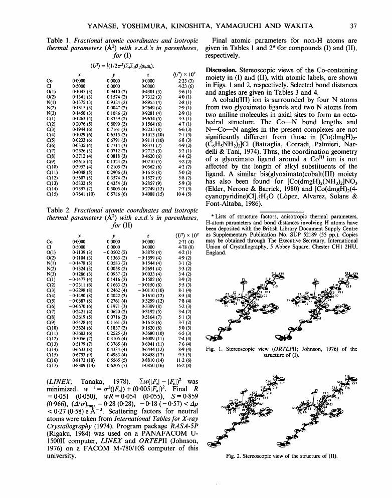
Discussion. Stereoscopic views of the Co-containing moiety in (I) and (II), with atomic labels, are shown in Figs. 1 and 2, respectively. Selected bond distances and angles are given in Tables 3 and 4.
A cobalt(Ill) ion is surrounded by four N atoms from two glyoximato ligands and two N atoms from two aniline molecules in axial sites to form an octa- hedral structure. The Co---N bond lengths and N---Co---N angles in the present complexes are not significantly different from those in [Co(dmgH)2- (C6HsNH2)2]C1 (Battaglia, Corradi, Palmieri, Nar- delli & Tani, 1974). Thus, the coordination geometry of a glyoximato ligand around a Co m ion is not affected by the length of alkyl substituents of the ligand. A similar bis(glyoximato)cobalt(III) moiety has also been found for [Co(dmgH)2(NH3)2]NO3 (Elder, Nerone & Barrick, 1980) and [Co(dmgH)2(4- cyanopyridine)C1].]HzO (L6pez, Alvarez, Solans & Font-Altaba, 1986).
* Lists of structure factors, anisotropic thermal parameters, H-atom parameters and bond distances involving H atoms have been deposited with the British Library Document Supply Centre as Supplementary Publication No. SUP 52189 (55 pp.). Copies may be obtained through The Executive Secretary, International Union of Crystallography, 5 Abbey Square, Chester CH1 2HU, England.
c41
Cl2 )
Cl3
¢11
CL C!2
~ 0~ ctt N3
Fig. 1. Stereoscopic view (ORTEPII; Johnson, 1976) of the structure of (I).
(LINEX; Tanaka, 1978). ~w(IFol- IFcl) 2 was minimized, w-1 = o~(iFol) + (0.0051Fol)2. Final R = 0.051 (0.050), wR = 0.054 (0.055), S = 0.859 (0.966), (A/tr)max = 0"28 (0"28), -0"18 (-0"57) < Ap < 0"27 (0"58) e A -3. Scattering factors for neutral atoms were taken from International Tables for X-ray Crystallography (1974). Program package RASA-5P (Rigaku, 1984) was used on a PANAFACOM U- 150011 computer, LINEX and ORTEPII (Johnson, 1976) on a FACOM M-780/10S computer of this university.
C16
CL C12~-~S q- . ; ~ c,, :3 c2 . . j cT, n~a,,__:-
CI3
I1
~0~
Fig. 2. Stereoscopic view of the structure of (II).
Ct6
CIS

38 [Co(CgH17N202)2(C6HTN)2]CI AND [Co(CllH21N202)2(C6H7b02]CI
Table 3. Selected bond lengths (A) and angles (o) for (I)
(a) Coordination polyhedron Co===N(1) 2.011 (3) N(1)--Co---N(2) 89.4 (1) Co---N(2) 1.904 (3) N(1)--Co---N(3) 86-8 (1) Co--N(3) 1-894 (3) N(2)--Co---N(3) 81.3 (1)
(b) Aniline molecule N(1)--C(1) 1-444 (4) Cty--N(1)--C(1) 119.8 (2) C(1)----C(2) 1.386 (5) N(I)---C(1)---C(2) 119.0 (3) C(2)---C(3) 1-388 (6) N(1)--C(1)--C(6) 120.0 (3) C(3)---C(4) 1.375 (7) C(2)----C(1)---C(6) 121.0 (3) C(4)----C(5) 1.368 (8) C(I)--C(2)--C(3) 118.9 (4) C(5)--C(6) 1.392 (6) C(2)--C(3)--C(4) 120.9 (5) C(6)--C(1) 1.392 (5) C(3)--C(4)---C(5) 119.5 (5)
C(4)--C(5)---42(6) 121.6 (5) C(1)--C(6)--C(5) 118.1 (4)
(c) Hexylmethylglyoxime molecule N(2)----O(1) 1.333 (3) Co---N(2)---O(1) 121.8 (2)
(4) Co--N(2)--C(7) 115.9 (2) (5) O(1)--N(2)--C(7) 122.2 (3) (4) N(2)---C(7)--C(8) 122.7 (3) (4) N(2)--C(7)--C(9) 113.3 (3) (3) C(8)--C(7)--C(9) 124.0 (3) (5) C(7)--C(9)--C(10) 122.9 (3) (6) C(7)--C(9)--N(3) 112.4 (3) (6) C(10)--C(9)--N(3) 124.7 (3) (7) C(9)--N(3)---O(2) 120-6 (3) (8) O(2)--N(3)--Co 122-3 (2) (11) C(9)--N(3)--Co 117.0 (2)
C(9)----C(10)--C(I l) 114.5 (3) C(10)--C(I1)--C(12) 110.5 (4) C(11)--C(12)--C(13) 113.7 (4) C(12)--C(13)--C(14) I12.6 (4) C(13)--C(14)--C(15) 113-0 (6)
N(2)---C(7) 1.300 C(7)---C(8) 1.488 C(7)---C(9) 1.467 C(9)---N(3) 1.296 N(3)--O(2) 1.364 C(9)--C(10) 1-497 C(10)--C(I 1) 1.526 C(1 I)---C(12) 1.530 C(12)--C(13) 1.510 C(13)----C(14) 1.525 C(14)--C(15) 1.513
Table 4. Selected bond lengths (A) and angles (o) for (II)
(a) Coordination polyhedron Co--N0) 2.016 (3) C(y--N(2) 1.913 (3) Cry--N(3) 1.885 (3)
(b) Aniline molecule N(1)----C(1) 1.437 (5) C(1)--C(2) 1.393 (6) C(2)--C(3) 1.375 (8) C(3)--C(4) 1.382 (9) C(4)--C(5) 1.371 (10) C(5)--C(6) 1.369 (8) C(6)--C(1) 1.380 (6)
(c) Methyloctylglyoxime molecule N(2)---<)(1) 1.339 (4) N(2)--C(7) 1.272 (5) C(7)---C(8) 1.499 (6) C(7)--C(9) 1.482 (5) C(9)--N(3) 1.302 (5) N(3)--O(2) 1.372 (4) C(9)--C(I0) 1.479 (6) CO0)--CO 1) 1.527 (8) C(11)--C(12) 1.522 (8) C(12)--C(13) 1.515 (9) C(13)--C(14) 1.513 (10) C(14)----C(15) 1.525 (10)
N(1)--Co---N(2) 88.9 (1) N(1)--Co--N(3) 86.6 (1) N(2)--Co---N(3) 80.6 (1)
Co---N(1)--C(1) 119.5 (2) N(1)--C(1)--C(2) 119.2 (4) N(1)--C(1)--C(6) 121-0 (4) C(2)--C(1)--C(6) 119.8 (4) C(1)--C(2)--C(3) 119.0 (5) C(2)--C(3)--C(4) 121.6 (6) C(3)--C(4)--C(5) 118.2 (6) C(4)--C(5)----C(6) 121-8 (6) C(1)--C(6)---C(5) 119.7 (5)
Co---N(2)---O(1) 120.4 (2) Co--N(2)--C(7) 116.5 (3) O(1)--N(2)--C(7) 122.9 (3) N(2)--C(7)--C(8) 123-2 (4) N(2)---C(7)---C(9) 113.8 (3) C(8)--C(7)--C(9) 123.0 (4) C(7)--C(9)--C(10) 123.4 (3) C(7)--C(9)--N(3) 110-7 (3) C(I 0)--C(9)~N(3) 125.8 (4) C(9)----N(3)---O(2) 119-3 (3) O(2)--N(3)--Co 122.4 (2) C(9)--N(3)--Co 118.2 (3) C(9)--C(10)--C(11) 114.5 (4) C(10)--C(ll)--C(12) 110.8 (5) C(11)--C(12)--C(13) 113.9 (5) C(12)--C(13)--C(14) 113.5 (6) C(13)--C(14)--C(15) 113.8 (6)
The Co---N(aniline) distances (2.011, 2.016A) found in (I) and (II) are considerably longer than the Co--N(NH3) length (1.951 A) in [Co(dmgH)2- (NH3)2]NO3 and the" Co~N(4-cyanopyridine) distance (1.95-1.97 A) in [Co(dmgH)2(4-cyano- pyridine)C1].~H20. This lengthening of the Co---N distance may reflect the basicity of the N atom of the ligands in the axial positions (the acid dis- sociation constant, pKa, is 9"24 for NH2- and 4.65 for aniline); the small electron donating power of the N atom of the axial ligand leads to the weak Co--N bond, resulting in the long Co---N bond length. The N(1)---C(1) distance within the coordinated aniline molecules len[gthens by 0-06/~ from the mean N---C value (1-382 A) in a free aniline molecule (Fukuyo, Hirotsu & Higuchi, 1982).
As seen in Tables 1 and 2, the isotropic thermal parameters of the C atoms in the hexyl and octyl groups of the glyoximato ligand increased with increasing distance of each C atom from the C(9) atom of the glyoximato ligand. This suggests a seg- mental motion of the alkyl substituent around the C(9) atom.
A hydrogen bond possibly forms between a CI atom and the N(1) atom of an aniline molecule with the C1--N(1) distance 3.172(3)/~ for (I) and 3.176 (3)A for (II) (see Figs. 1 and 2). Such a hydrogen bond has been found in [Co(dmgH)2- (C6HsNH2)2]C1 (3.19 A).
References
BATI'AGLIA, L., CORRADI, A. B., PALMIERI, C. G., NARDELLI, M. & TANL M. E. V. (1974). Acta Cryst. B30, 1114-1116.
BRESCIA~-PAHoa, N., FORCOLIN, M., MARZILLI, L. G., RANDAC- CIO, L., SUMMERS, M. F. & TOSCANO, P. J. (1985). Coord. Chem. Rev. 63, 1-125.
ELDER, R. C., NERONE, A. & BARRICK, J. C. (1980). Acta Cryst. B36, 2428-2431.
Ft~uYo, M., HmOTSO, K. & HIGUCm, T. (1982). Acta Cryst. B38, 640-643.
International Tables for X-ray Crystallography (1974). Vol. IV. Birmingham: Kynoch Press. (Present distributor Kluwer Academic Publishers, Dordrecht.)
JOHNSON, C. K. (1976). ORTEPII. Report ORNL-5138. Oak Ridge National Laboratory, Oak Ridge, Tennessee, USA.
KINOSHITA, S. & MASUDA, I. (1985). Polyhedron, 4, 1245--1251. KINOSHrrA, S., WAr, IrA, H. & MASUDA, I. (1986). Bull. Chem. Soc.
Jpn, 59, 651-652. L6PEZ, C., ALVAREZ, S., SOLANS, X. & FONT-ALTABA, M. (1986).
Inorg. Chem. 25, 2962-2969. MAIN, P., FtSKE, S. J., HULL, S. E., LESSINGER, L., GERMArN, G.,
DECLERC'Q, J.-P. & WOOLFSON, M. M. (1980). MULTANSO. A System of Computer Programs for the Automatic Solution of Crystal Structures from X-ray Diffraction Data. Univs. of York, England, and Louvain, Belgium.
Oagsm, Y. (1988). Ace. Chem. Res. 21. 268-274. Rigaku (1984). RASA-5P. Rigaku Ltd, Matsushima, Akishima,
Tokyo 196, Japan. TANAKA, K. (1978). LINEX. Tokyo Institute of Technology,
Nagatsuta, Midori-ku, Yokohama, Japan.