Embed Size (px)
Citation preview
electronic reprint
ISSN: 1399-0047
journals.iucr.org/d
Structure of the adenylation–peptidyl carrier proteindidomain of the Microcystis aeruginosa microcystinsynthetase McyG
Xiao-Feng Tan, Ya-Nan Dai, Kang Zhou, Yong-Liang Jiang, Yan-Min Ren,Yuxing Chen and Cong-Zhao Zhou
Acta Cryst. (2015). D71, 873–881
IUCr JournalsCRYSTALLOGRAPHY JOURNALS ONLINE
Copyright c© International Union of Crystallography
Author(s) of this paper may load this reprint on their own web site or institutional repository provided thatthis cover page is retained. Republication of this article or its storage in electronic databases other than asspecified above is not permitted without prior permission in writing from the IUCr.
For further information see http://journals.iucr.org/services/authorrights.html
Acta Cryst. (2015). D71, 873–881 Tan et al. · McyG adenylation–peptidyl carrier didomain
research papers
Acta Cryst. (2015). D71, 873–881 http://dx.doi.org/10.1107/S1399004715001716 873
Received 11 September 2014
Accepted 27 January 2015
Edited by Q. Hao, University of Hong Kong
Keywords: microcystin; NRPS/PKS; McyG;
adenylation–peptidyl carrier protein didomain;
reaction cycle.
PDB reference: McyG A–PCP didomain
complexed with L-Phe-AMP, 4r0m
Supporting information: this article has
supporting information at journals.iucr.org/d
Structure of the adenylation–peptidyl carrierprotein didomain of the Microcystis aeruginosamicrocystin synthetase McyG
Xiao-Feng Tan, Ya-Nan Dai,* Kang Zhou, Yong-Liang Jiang, Yan-Min Ren,
Yuxing Chen and Cong-Zhao Zhou*
Hefei National Laboratory for Physical Sciences at the Microscale and School of Life Sciences, University of Science and
Technology of China, Hefei, Anhui 230027, People’s Republic of China. *Correspondence e-mail:
[email protected], [email protected]
Microcystins, which are the most common cause of hepatotoxicity associated
with cyanobacterial water blooms, are assembled in vivo on a large multienzyme
complex via a mixed nonribosomal peptide synthetase/polyketide synthetase
(NRPS/PKS). The biosynthesis of microcystin in Microcystis aeruginosa PCC
7806 starts with the enzyme McyG, which contains an adenylation–peptidyl
carrier protein (A–PCP) didomain for loading the starter unit to assemble the
side chain of an Adda residue. However, the catalytic mechanism remains
unclear. Here, the 2.45 A resolution crystal structure of the McyG A–PCP
didomain complexed with the catalytic intermediate l-phenylalanyl-adenylate
(l-Phe-AMP) is reported. Each asymmetric unit contains two protein molecules,
one of which consists of the A–PCP didomain and the other of which comprises
only the A domain. Structural analyses suggest that Val227 is likely to be critical
for the selection of hydrophobic substrates. Moreover, two distinct interfaces
demonstrating variable crosstalk between the PCP domain and the A domain
were observed. A catalytic cycle for the adenylation and peptide transfer of the
A–PCP didomain is proposed.
1. Introduction
Cyanobacterial water blooms are of worldwide concern owing
to their production of a wide range of hepatotoxins and
neurotoxins. Of the known toxins, the hepatotoxic micro-
cystins are produced by a diverse range of cyanobacteria,
including species of the genera Microcystis, Anabaena, Nostoc
and Oscillatoria (Rinehart et al., 1994). It is known that acute
poisoning by microcystins leads to death owing to hepatic
haemorrhage (Jochimsen et al., 1998; Pouria et al., 1998),
whereas the ingestion of sublethal amounts of microcystins
increases the incidence rate of liver cancer (Nishiwaki-
Matsushima et al., 1992). Microcystins, which contain the
novel aromatic �-amino-acid residue [(2S,3S,8S,9S)-3-amino-
9-methoxy-2,6,8-trimethyl-10-phenyl-4,6-decadienoic acid;
Adda], can inactivate serine/threonine protein phosphatase
1/2A (PP1/2A) in eukaryotes (Honkanen et al., 1990) by
specifically identifying the hydrophobic groove of PP1/2A
(Goldberg et al., 1995).
Previous biochemical and genetic studies have shown that
microcystins are assembled by a mixed polyketide synthetase/
nonribosomal peptide synthetase (PKS/NRPS; Moore et al.,
1991; Dittmann et al., 1997) encoded by the microcystin
synthetase (mcy) gene cluster (Tillett et al., 2000). The PKS/
NRPS belongs to a family of large multi-domain enzymes that
ISSN 1399-0047
# 2015 International Union of Crystallography
electronic reprint
use a modular architecture to produce important biological
polyketides/nonribosomal peptides such as antibiotics. The
NRPS responsible for peptide elongation is composed of at
least three domains: an adenylation domain (A domain) which
is responsible for substrate activation, a peptidyl carrier
domain (PCP domain) that transfers the intermediate between
domains and a condensation domain (C domain) which carries
out peptide formation in the order C–A–PCP. PKS is
composed of three domains as a basic unit: an acyltransferase
domain (AT domain) responsible for substrate activation, an
acyl carrier domain (ACP domain) homologous to the PCP
domain and a �-ketoacyl synthase domain (KS domain) which
catalyzes polyketide elongation assembled as KS–AT–ACP.
In addition, other domains such as a carbon-methyltransferase
domain (CM domain), a ketoacyl reductase domain (KR
domain) and a thioesterase domain (TE domain) are required
for further modification.
Comparing the modular organization of the mcy gene
cluster with those of other PKS/NPRSs, a biosynthetic scheme
for microcystin has been proposed (Tillett et al., 2000).
Biosynthesis of microcystin starts with McyG to assemble the
side chain of the Adda residue, which is then synthesized
by McyD and McyE. McyG consists of an A–PCP didomain
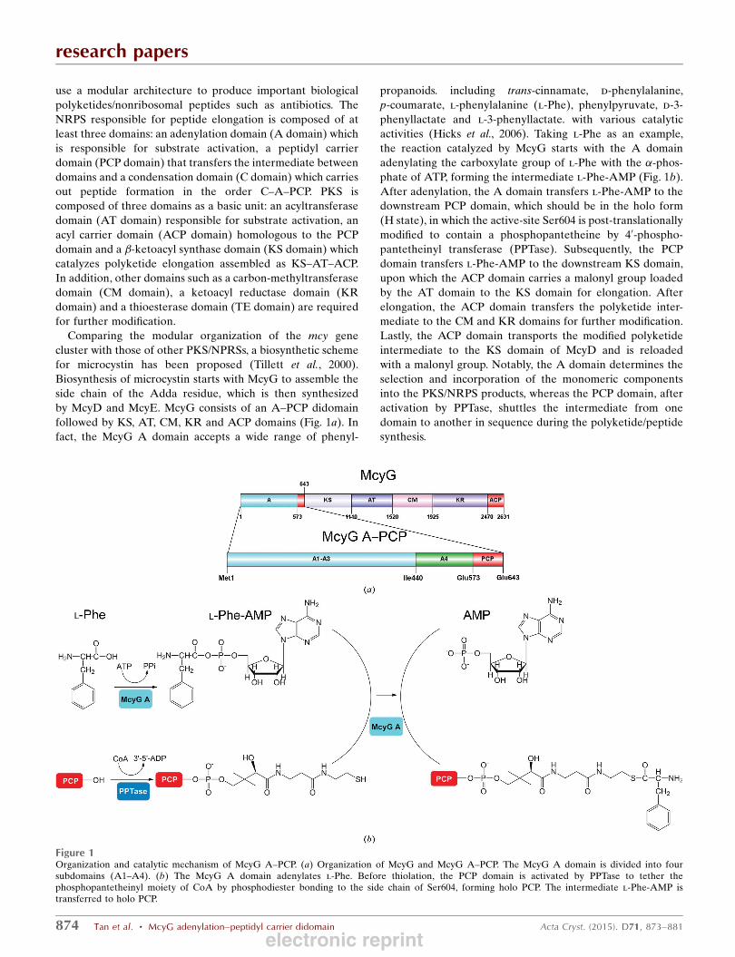
followed by KS, AT, CM, KR and ACP domains (Fig. 1a). In
fact, the McyG A domain accepts a wide range of phenyl-
propanoids. including trans-cinnamate, d-phenylalanine,
p-coumarate, l-phenylalanine (l-Phe), phenylpyruvate, d-3-
phenyllactate and l-3-phenyllactate. with various catalytic
activities (Hicks et al., 2006). Taking l-Phe as an example,
the reaction catalyzed by McyG starts with the A domain
adenylating the carboxylate group of l-Phe with the �-phos-phate of ATP, forming the intermediate l-Phe-AMP (Fig. 1b).
After adenylation, the A domain transfers l-Phe-AMP to the
downstream PCP domain, which should be in the holo form
(H state), in which the active-site Ser604 is post-translationally
modified to contain a phosphopantetheine by 40-phospho-pantetheinyl transferase (PPTase). Subsequently, the PCP
domain transfers l-Phe-AMP to the downstream KS domain,
upon which the ACP domain carries a malonyl group loaded
by the AT domain to the KS domain for elongation. After
elongation, the ACP domain transfers the polyketide inter-
mediate to the CM and KR domains for further modification.
Lastly, the ACP domain transports the modified polyketide
intermediate to the KS domain of McyD and is reloaded
with a malonyl group. Notably, the A domain determines the
selection and incorporation of the monomeric components
into the PKS/NRPS products, whereas the PCP domain, after
activation by PPTase, shuttles the intermediate from one
domain to another in sequence during the polyketide/peptide
synthesis.
research papers
874 Tan et al. � McyG adenylation–peptidyl carrier didomain Acta Cryst. (2015). D71, 873–881
Figure 1Organization and catalytic mechanism of McyG A–PCP. (a) Organization of McyG and McyG A–PCP. The McyG A domain is divided into foursubdomains (A1–A4). (b) The McyG A domain adenylates l-Phe. Before thiolation, the PCP domain is activated by PPTase to tether thephosphopantetheinyl moiety of CoA by phosphodiester bonding to the side chain of Ser604, forming holo PCP. The intermediate l-Phe-AMP istransferred to holo PCP.
electronic reprint

To date, two structures containing an A–PCP didomain
have been deposited in the Protein Data Bank (PDB): SrfA-C
(the termination module of surfactin synthetase assembled
as C–A–PCP–TE; PDB entry 2vsq; Tanovic et al., 2008) and
PA1221 (an A–PCP didomain from Pseudomonas aeruginosa;
PDB entry 4dg9; Mitchell et al., 2012), demonstrating two
different conformations of the A–PCP didomain during the
catalytic cycle. In detail, the A domain of SrfA-C displays
an open conformation for accepting ATP, showing the inter-
mediate to be transferred from the downstream PCP domain
to the upstream C domain (Tanovic et al., 2008). In PA1221,
the A domain is in a thiolated conformation guiding the holo
PCP domain into the active site for thioester formation
(Mitchell et al., 2012). However, the mode of communication
between the PCP domain and the adenylated A domain is still
unknown.
Here, we have determined the crystal structure of McyG A–
PCP complexed with the catalytic intermediate l-Phe-AMP.
There are two molecules in one asymmetric unit, with one
molecule consisting of the A–PCP didomain and the other
comprising only the A domain. Detailed examination of the
l-Phe-AMP binding pocket demonstrated Val227 to be a
crucial residue in the selection of hydrophobic substrates.
Further structural analyses identified two different interfaces
displaying varied communications between the PCP domain
and the A domain. We propose a catalytic cycle for adenyla-
tion and peptide transfer of the A–PCP didomain.
2. Materials and methods
2.1. Cloning, expression and purification of McyG A–PCP
The gene encoding McyG A–PCP was amplified by PCR
from genomic DNA of M. aeruginosa PCC 7806 using primers
McyG-NdeI (50-GCACATATGATGTCTAAGCATTCCATC-
AGTCT-30) and McyG-XhoI (50-TGCCTCGAGTTATTCT-
TCGCTTAAGAAACG-30). The amplified fragment was
cloned into pET-28b with an N-terminal hexahistidine tag.
Escherichia coli BL21 (DE3) cells (Novagen) were trans-
formed with the recombinant plasmid and grown in LB
medium (10 g l�1 NaCl, 10 g l�1 Bacto tryptone, 5 g l�1 yeast
extract) containing 30 mg ml�1 kanamycin at 37�C until an
OD600 nm of 0.6 was reached. The temperature was shifted to
16�C and the cultures were induced with 0.2 mM isopropyl
�-d-1-thiogalactopyranoside. After 20 h of growth, the cells
were harvested and resuspended in lysis buffer (100 mM
NaCl, 20 mM Tris–HCl pH 8.0). To purify the McyG A–PCP
protein, resuspended cells were lysed by sonication for 30 min.
After centrifugation at 12 000g for 30 min, the supernatant
containing the soluble target protein was collected and loaded
onto a nickel-chelating column (GE Healthcare). The binding
buffer (100 mM NaCl, 20 mM Tris–HCl pH 8.0) contained
20 mM imidazole to reduce nonspecific binding of untagged
proteins. The target protein was eluted with 300 mM imidazole
in 6 ml binding buffer and further purified using a HiLoad
16/60 Superdex 200 column (GE Healthcare) pre-equilibrated
with 100 mM NaCl, 20 mM Tris–HCl pH 8.0.
2.2. Preparation of selenomethionine-labelled protein
Selenomethionine (SeMet)-labelled McyG A–PCP (Se-
McyG A–PCP) was prepared for phase determination. Se-
McyG A–PCP was obtained by expressing McyG A–PCP in
E. coli strain B834 (DE3) (Novagen). The transformed cells
were inoculated into LB medium and grown at 37�C until an
OD600 nm of 0.2 was reached. The cells were harvested and
then washed twice with M9 medium. The cells were then
cultured in SeMet medium (M9 medium with 25 mg l�1
l-SeMet and the other essential amino acids at 50 mg l�1) to
an OD600 nm of 0.6 and induced using 0.2 mM isopropyl �-d-1-thiogalactopyranoside. The remaining steps of protein
expression and purification were identical to those used for
the native McyG A–PCP except that all buffers contained
5%(v/v) glycerol and 14 mM �-mercaptoethanol. Se-McyG
A–PCP was concentrated to a final concentration of
58 mg ml�1 for crystallization. The purity was estimated using
10% SDS–PAGE. The protein concentration was determined
by A280 nm using an extinction coefficient of 75 860 M�1 cm�1.
2.3. Crystallization, data collection and processing
Before crystallization, Se-McyGA–PCP was incubated with
5 mM l-Phe, 5 mM ATP, 1 mM MgCl2 for 30 min at 25�C.Initial crystallization screening for Se-McyG A–PCP was
performed using the hanging-drop vapour-diffusion method at
research papers
Acta Cryst. (2015). D71, 873–881 Tan et al. � McyG adenylation–peptidyl carrier didomain 875
Table 1Crystal parameters and data-collection and structure-refinement statisticsfor Se-McyG A–PCP.
Values in parentheses are for the highest resolution bin.
Data collectionSpace group P3221Unit-cell parameters (A, �) a = b = 120.709, c = 262.780,
� = � = 90.00, � = 120.00Resolution range (A) 54.84–2.45 (2.58–2.45)Unique reflections 81008 (11705)Completeness (%) 98.8 (98.7)hI/�(I)i 9.6 (3.0)Rmerge† (%) 11.8 (59.9)Average multiplicity 5.5 (5.6)
Structure refinementResolution range (A) 54.84–2.45R factor‡/Rfree§ (%) 24.8/27.3No. of protein atoms 9475No. of ligand atoms 68No. of water atoms 26R.m.s.d.}, bond lengths (A) 0.005R.m.s.d., bond angles (�) 0.956Mean B factors (A2)Overall 43.4Protein 43.5Ligand 31.7Water 32.4
Ramachandran plot††Most favoured (%) 97.22Additionally allowed (%) 2.78Outliers (%) 0
PDB entry 4r0m
† Rmerge =P
hkl
Pi jIiðhklÞ � hIðhklÞij=Phkl
Pi IiðhklÞ, where Ii(hkl) is the intensity of
an observation and hI(hkl)i is the mean value for its unique reflection; summations areover all reflections. ‡ R factor =
Phkl
��jFobsj � jFcalcj
��=P
hkl jFobsj, where Fobs and Fcalc
are the observed and calculated structure-factor amplitudes, respectively. § Rfree wascalculated using 5% of the data, which were excluded from the refinement. } Root-mean square-deviation from ideal values. †† Categories were defined by MolProbity.
electronic reprint
16�C in 96-well plates. The crystallization screening kits
comprised Crystal Screen, Crystal Screen 2, Index, Grid
Screen, SaltRx (Hampton Research) and Buffer and pH
screen (Radaev et al., 2006). Se-McyG A–PCP crystals were
obtained in a condition consisting of 1.8 M ammonium sulfate,
0.1 M NaKHPO4 pH 6.5. During optimization in 24-well
plates, the hanging drop consisted of 1 ml protein sample and
1 ml reservoir solution consisting of 1.2–2.2 M ammonium
sulfate, 0.1 M NaKHPO4 pH 6.5. Crystals appeared in one
week and grew to maximum size over three weeks. Se-McyG
A–PCP crystals were briefly soaked in reservoir solution
supplemented with 30% glycerol as a cryoprotectant, mounted
on loops and flash-cooled in liquid nitrogen. SeMet-derivative
data were collected from single crystals at 100 K in a liquid-
nitrogen stream on beamline 17U at the Shanghai Synchro-
tron Radiation Facility (SSRF) using an ADSC Q315r CCD
detector (MAR Research, Germany). All diffraction data
were indexed, integrated and scaled with iMosflm (Battye et
al., 2011).
2.4. Structure determination and refinement
The crystal structure of McyG A–PCP was determined
using the single-wavelength anomalous dispersion phasing
method from a single SeMet-substituted protein crystal to a
maximum resolution of 2.45 A. The AutoSol program from
PHENIX (Adams et al., 2002) was used to locate the heavy
atoms, and the phases were calculated and further improved
using partial model extension with OASIS (Hao et al., 2000).
Automatic model building was carried out using AutoBuild in
PHENIX. Refinement was carried out using the maximum-
likelihood method implemented in REFMAC5 (Murshudov et
research papers
876 Tan et al. � McyG adenylation–peptidyl carrier didomain Acta Cryst. (2015). D71, 873–881
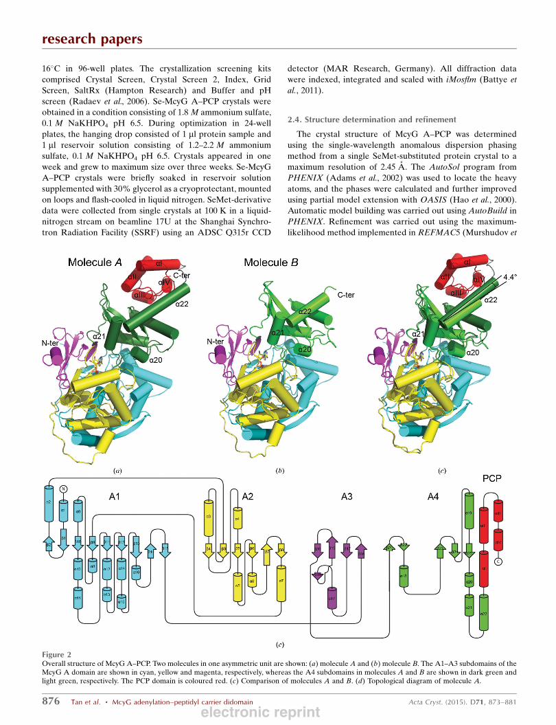
Figure 2Overall structure of McyG A–PCP. Two molecules in one asymmetric unit are shown: (a) molecule A and (b) molecule B. The A1–A3 subdomains of theMcyG A domain are shown in cyan, yellow and magenta, respectively, whereas the A4 subdomains in molecules A and B are shown in dark green andlight green, respectively. The PCP domain is coloured red. (c) Comparison of molecules A and B. (d) Topological diagram of molecule A.
electronic reprint
al., 2011) as part of the CCP4 program suite (Winn et al., 2011)
and rebuilt interactively using Coot (Emsley & Cowtan, 2004).
The final model was evaluated with MolProbity (Chen et al.,
2010) and PROCHECK (Laskowski et al., 1993). Crystallo-
graphic parameters and data-collection statistics are listed in
Table 1. All figures showing the structure were prepared with
PyMOL (DeLano, 2002).
3. Results and discussion
3.1. Overall structure of McyG A–PCP
The structure of McyG A–PCP was determined at 2.45 A
resolution in space group P3221, with two monomers (namely
molecules A and B) in one asymmetric unit. Molecule A
consists of an A domain (Ser5–Ser572) and a PCP domain
(Glu573–Ser641) (Fig. 2a). However, molecule B comprises
only an A domain corresponding to residues Ser7–Lys561
(Fig. 2b) owing to the electron density of residues Glu562–
Ser643 being disordered. The A domain of both molecules is
bound to one molecule of l-Phe-AMP.
The McyG A domain is composed of two
domains: the large domain (A1–A3 subdo-
mains; Ser5–Gly434) and the small domain
(A4 subdomain; Ile440–Lys566) connected
by a flexible loop (Arg435–Glu439) which
permits the A4 subdomain to adopt
different orientations with respect to the
A1–A3 subdomains. The A1 subdomain
consists of a nine-stranded �-sheet (�1, �2,�9–�15) surrounded by 11 �-helices (�1, �2,�8–�16), forming a distorted �-barrel. TheA2 subdomain is composed of a six-stranded
�-sheet (�3–�8) flanked by five �-helices(�3–�7), whereas the A3 subdomain
contains a distorted five-stranded �-sheet(�16–�20) in addition to one �-helix (�17).Collectively, the A1–A3 subdomains display
a distorted �-barrel and two �-sheets whichpack against each other to form a five-
layered ����� domain structure. The A4
subdomain begins with an antiparallel �-sheet containing two small �-strands (�21and �22) followed by a central three-
stranded �-sheet (�23–�25) which is
surrounded by five �-helices (�18–�22).Notably, the structure of the McyG A
domain displays an adenylated conforma-
tion, binding to one molecule of l-Phe-
AMP.
As is well known, three states of the PCP
domain have been observed according to
the variation of four �-helices (�I–�IV): anunmodified state (A state), a modified state
(H state) and an intermediary state between
the A state and the H state (the A/H state)
(Koglin et al., 2006). Specifically, in the A
state �III changes to a loop and the other three helices are
shorter, in the H state �III is also disordered and helix �IVmoves parallel to �II, while the A/H state is composed of four
�-helices named �I–�IV. In McyG A–PCP, the PCP domain of
molecule A consists of four �-helices (�I–�IV) which were
well defined in the electron-density map in the conformation
of the A/H state with the active-site Ser604 unmodified.
3.2. Comparison of the two A–PCP didomains in eachasymmetric unit
Superposition of the two A–PCP molecules (molecules A
and B) in each asymmetric unit yielded root-mean-square
deviations (r.m.s.d.s) of 0.466 and 0.901 A over 427 C� atoms
of the A1–A3 subdomains and 114 C� atoms of the A4
subdomain, respectively. It is notable that three �-helices(�20–�22) of the A4 subdomain slant, with helix �22 flipped
by 4.4� (Fig. 2c). In the structure of SrfA-C, the segment
corresponding to helix �22 adopts a loop conformation to
enable the PCP domain to approach the upstream C domain,
research papers
Acta Cryst. (2015). D71, 873–881 Tan et al. � McyG adenylation–peptidyl carrier didomain 877
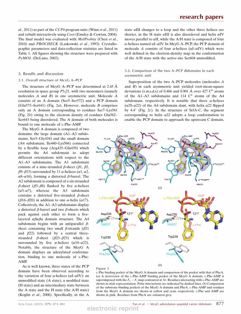
Figure 3l-Phe binding pocket of the McyG A domain and comparison of the pocket with that of PheA.(a) A stereoview of the l-Phe-AMP binding pocket of the McyG A domain. l-Phe-AMP issuperimposed with the Fo� Fc map contoured at 3�. Residues interacting with l-Phe-AMP areshown in stick representation. Polar interactions are indicated by dashed lines. (b) Comparisonof the substrate-binding pockets of the McyG A domain and PheA. l-Phe-AMP and residuesfrom the McyG A domain are shown in yellow and cyan, respectively. l-Phe and AMP areshown in pink. Residues from PheA are coloured grey.
electronic reprint
whereas the counterpart in PA1221 is more flexible, which
facilitates interaction between the PCP domain and the A
domain (Tanovic et al., 2008; Mitchell et al., 2012). Therefore,
we speculated that the slanting of the �20–�22 helices in
addition to the high flexibility of the segment corresponding to
helix �22 is involved in the shuttling and/or conformational
changes of the PCP domain during the catalytic cycle.
3.3. The L-Phe-AMP binding site
Taking molecule B as an example, the l-Phe-AMP binding
pocket is positioned at the interface between the large and
small domains. From a closer
viewpoint, the l-Phe-AMP is
mainly surrounded by residues
from the A1 subdomain and is
covered by the A4 subdomain
like a lid. In detail, l-Phe-AMP
lies in a slot sandwiched between
two loops consisting of
A304GEQV308 and A333FGMT337,
respectively (Fig. 3a). With
respect to the AMP moiety of l-
Phe-AMP, the adenine base is
fixed by two hydrogen bonds, one
of which is formed between the
N6 amino group of the adenine
base and the �-carbonyl of
Ala333 and the other of which is
formed between the N7 amino
group and the �-carbonyl of
Gly305. The ribose 20 and 30
hydroxyls interact with Asp421
and Arg435 from the A3 subdo-
main and the ribose 40 and 50 Oatoms bind to the side chain of
residue Lys533. The �-phosphateinteracts with the side chain of
Thr337. In addition, His226 forms
a hydrogen bond to the bridging
O atom between the l-Phe group
and the AMP part. Moreover, the
�-amino group of the l-Phe
group is fixed by the formation of
one hydrogen bond to the
�-carbonyl of Gly335. The
benzene ring of the l-Phe group
is stabilized by hydrophobic
interactions via being sandwiched
between the side chain of Trp272
and the main chains of Ala333,
Gly335 and Ser341.
To reveal further character-
istics of the substrate-binding
pocket of McyG A–PCP, super-
position with PheA, which is
a classic A domain activating
l-Phe (Conti et al., 1997), was
performed. Generally, residues that participate in fixing the
AMP part of McyG A–PCP and PheA are highly conserved
(data not shown), whereas two notable features among the
l-Phe group binding residues were observed in McyG A–PCP.
Val227 in the McyG A domain is positioned at the same
position as the corresponding residue Asp235 of PheA
(Fig. 3b). It is known that Asp235 is conserved in all amino-
acid adenylation domains, participating in the formation of
hydrogen bonds to the �-amino group of the amino-acid
substrate (Stachelhaus et al., 1999). Remarkably, the side chain
of Val227 provides a hydrophobic environment which rotates
research papers
878 Tan et al. � McyG adenylation–peptidyl carrier didomain Acta Cryst. (2015). D71, 873–881
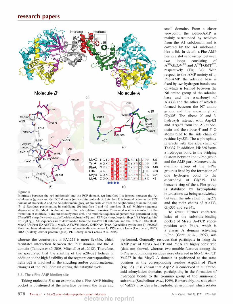
Figure 4Interfaces between the A4 subdomain and the PCP domain. (a) Interface I is formed between the A4subdomain (green) and the PCP domain (red) within molecule A. Interface II is formed between the PCPdomain of molecule A and the A4 subdomain (grey) of molecule B0 from the neighbouring asymmetric unit.(b, c) Residues participating in stabilizing (b) interface I and (c) interface II. (d) Multiple sequencealignment of the McyG A domain and other adenylation domains. Conserved residues involved in theformation of interface II are indicated by blue dots. The multiple sequence alignment was performed usingClustalW2 (http://www.ebi.ac.uk/Tools/msa/clustalw2/) and ESPript (http://espript.ibcp.fr/ESPript/cgi-bin/ESPript.cgi). All sequences were downloaded from the UniProtKB database and the Protein Data Bank.McyG, UniProt ID A8YJW1; McyB, A8YJV6; McyC, Q9RNA9; TycA (tyrocidine synthetase 1), P09095;Phe (the phenylalanine-activating subunit of gramicidin synthetase 1), PDB entry 1amu (Conti et al., 1997);DltA (d-alanyl carrier protein ligase), PDB entry 3e7w (Yonus et al., 2008).
electronic reprint
the �-amino group of l-Phe-AMP in the McyG A domain
through 121� away from the counterpart �-amino group of
l-Phe in PheA. Moreover, an ATP–PPi exchange assay of
McyG A–PCP towards various substrates identified two
hydrophobic substrates lacking �-amino groups as displaying
the highest activities: trans-cinnamate has the highest activity,
while that of hydrocinnamate was lower (Hicks et al., 2006).
In fact, compared with wild-type McyG A–PCP, V227T and
V227D mutations resulted in an increased Km value towards
trans-cinnamate by approximately threefold and 23-fold and a
kcat/Km value that was decreased by 43 and 96.6%, respec-
tively (Supplementary Table S1). Therefore, we speculated
that Val227 in McyG is most likely to be critical for the
selection of hydrophobic substrates. In PheA, the side chain of
Trp239 is positioned at the bottom as a lid, covering the pocket
with an average distance of 3.6 A from the benzene ring of l-
Phe. However, the corresponding distance from the benzene
ring of l-Phe to the side chain of Phe232 in McyG A is as long
as 5 A, leading to a deeper substrate-binding pocket which
may accommodate substrates that are longer than l-Phe to
some degree. Indeed, the longer substrate hydroxycinnamate
can be recognized by the McyG A domain (Hicks et al., 2006).
3.4. Interfaces between the A domain and the PCP domain
We observed two different interfaces between the A
domain and the PCP domain: interface I of 641 A2 buried area
is formed between the A domain and the PCP domain within
molecule A, while interface II of 952 A2 is formed between the
PCP domain of molecule A and the A domain of molecule B0
from the neighbouring asymmetric unit (Fig. 4a). In interface
I, hydrophobic interactions are formed between Gly512 (�19),Thr508 (�19) and Phe628 (�III). In addition, Glu599 from
loop I (LI) is stabilized by forming hydrogen bonds to Arg501
(�19), Arg504 (�19) and Arg563
(�22) (Fig. 4b). It is known that in
the structure of the TycC_PCP
(S45A)–Sfp complex (the PCP
domain of tyrocidine synthetase 3
complexed with the PPTase Sfp),
Gln40, which corresponds to
Glu599 of McyG A–PCP, is
exposed to participate in the
interaction between the PCP
domain and the PPTase Sfp
(Tufar et al., 2014). Additionally,
in EntB_PCP (the PCP domain
of enterobactin synthetase B),
Ala268, which corresponds to
Phe628 in McyG A–PCP, is
crucial for interaction with the
downstream C domain (Lai et al.,
2006). Clearly, both Glu599 and
Phe628 are hindered from parti-
cipating in forming an interface
between the PCP domain and the
downstream domains or PPTase
in interface I. Moreover, the
active-site Ser604 orients towards
the A domain and is buried in
interface I; thus, the PCP domain
in molecule A adopts a blocked
conformation which is prevented
from interacting with down-
stream domains and PPTase.
Interface II is stabilized by a
network of hydrogen bonds and
salt bridges (Fig. 4c). The
hydrogen bonds are formed as
follows: from the main chain of
Glu573 (�I) to the side chain of
Gln5090 (�190), from the side
chain of Arg576 (�I) to the
�-carbonyl of Thr5080 (�190) and
research papers
Acta Cryst. (2015). D71, 873–881 Tan et al. � McyG adenylation–peptidyl carrier didomain 879
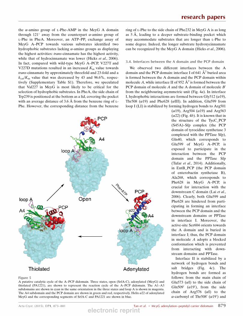
Figure 5A putative catalytic cycle of the A–PCP didomain. Three states, open (SrfA-C), adenylated (McyG) andthiolated (PA1221), are shown to represent the reaction cycle of the A–PCP didomain. The A1–A3subdomains are shown in cyan in the same orientation in the three states and loop A is shown in magenta.The A4 subdomain and the PCP domain are shown in green and red, respectively. Helix �22 of adenylatedMcyG and the corresponding segments of SrfA-C and PA1221 are shown in blue.
electronic reprint
Gln5090 (�190), from the side chain of Ser583 (�I) to the side
chain of Arg5040 (�190) and the �-carbonyl of Pro5150
(loop190; L190), from Gln587 (LI) to Tyr5170 (L190) and from
the side chain of Thr589 (LI) to the �-carbonyl of Ala5140
(L190) and Ala5160 (L190). In addition, the NH1 and NH2
amino groups of Arg609 (�II) form two salt bridges to the side
chain of Asp5560 (�220). The side chains of Glu584 (�I) andGlu613 (�II) form four salt bridges to Arg5010. Biochemical
analyses of the adenylation reaction of TycA-A (the A domain
of tyrocidine synthetase 1) demonstrated that residues Pro485
and Tyr487, which correspond to Pro5150 and Tyr5170 fromL190 of McyG A–PCP, are crucial in transferring the inter-
mediate to the PCP domain (Bucevic-Popovic et al., 2012).
Multiple sequence alignment showed that Pro515 and Tyr517
of McyG A–PCP are conserved among the adenylation
domains, suggesting that the interaction between L190 and the
PCP domain may be related to intermolecular recognition
and intermediate transfer between the A domain and the PCP
domain (Fig. 4d).
3.5. A putative catalytic cycle of the A–PCP didomain
We observed the intact A–PCP didomain of McyG in the
adenylated conformation in molecule A. Structural compar-
ison of McyG A–PCP with SrfA-C and PA1221 showed that
the conformation of the A1–A3 subdomains varies little,
whereas both the A4 subdomain and the PCP domain adopt
changing conformations and move in relation to the A1–A3
subdomains during the catalytic reaction. Specifically, in
adenylated McyG A–PCP loop A (Pro527–Gln535) moves to
cover the substrate-binding pocket, with Lys533 hydrogen-
bonding to the intermediate l-Phe-AMP, whereas the corre-
sponding loop in SrfA-C A–PCP is located far away, resulting
in a more open pocket. Moreover, in adenylated McyG helix
�22 is well ordered and the PCP domain is more close to the
A4 subdomain, with the active-site Ser604 blocked to hinder
the PCP domain from interacting with the downstream
KS domain or A domain. However, in SrfA-C A–PCP the
segment corresponding to helix �22 of McyG transforms into a
flexible loop and the PCP domain is positioned far away from
the A4 subdomain, with the active-site serine approaching the
upstream C domain. Further structural analyses of McyG A–
PCP and PA1221 revealed that the A4 subdomain of McyG
A–PCP needs to rotate 140� to enable the holo PCP to interact
with the intermediate, along with the rearrangement of helix
�22 and the movement of the PCP domain to form the thio-
lated conformation.
It is known that in three-dimensional cryo-microscopy
structures of holo PikAIII (module 5 of pikromycin PKS
with a modified ACP domain organized as KS–AT–KR–holo
ACP5), pentaketide–KS5–PikAIII and �-ketohexaketide–PikAIII, the ACP domain, AT domain and KS domain
undergo conformational rearrangements during the enzymatic
cycle. In detail, once the ACP5 domain has transported the
�-ketohexaketide to the KR domain for modification, the AT
domain moves to cover the side entrance of the KS domain in
order to prevent the KS domain from accepting the inter-
mediate transported by the upstream ACP4 domain and to
keep the catalytic cycle in sequence (Whicher et al., 2014;
Dutta et al., 2014). The adenylation of the A–PCP didomain in
our structure prevents the PCP domain from interacting with
the downstream KS domain and PPTase until the downstream
ACP domain has finished transporting the intermediate to the
KS domain of McyD and has reloaded the malonyl group.
Thus, we captured a transient conformation of the A–PCP
didomain that induces the downstream domains to react in
sequence.
Based on these findings, we propose a putative catalytic
cycle for McyG A–PCP. Upon adenylation, the A–PCP di-
domain is blocked in a conformation in which the PCP domain
is prevented from interacting with the downstream domains
or PPTase. After reloading of the malonyl group onto the
downstream ACP domain by the AT domain, the A4 sub-
domain rotates by 140� and helix �22 rearranges to help the
PCP domain attach to the intermediate, forming the thiolated
conformation. Finally, the A–PCP didomain converts into an
open conformation to allow the PCP domain to more easily
transport the intermediate to the downstream KS domain for
elongation and to accept substrates for next reaction cycle
(Fig. 5).
Acknowledgements
We thank the beamline staff of the Shanghai Synchrotron
Radiation Facility (SSRF) for technical help during X-ray data
collection. This work was supported by the Chinese National
Natural Science Foundation (Grant Nos. 31370757 and
31070652), a Scientific Research Grant from Hefei Science
Center of CAS and the Ministry of Education of China (Grant
No. 20133402110023) and the Program for Changjiang Scho-
lars and Innovative Research Team in University.
References
Adams, P. D., Grosse-Kunstleve, R. W., Hung, L.-W., Ioerger, T. R.,McCoy, A. J., Moriarty, N. W., Read, R. J., Sacchettini, J. C., Sauter,N. K. & Terwilliger, T. C. (2002). Acta Cryst. D58, 1948–1954.
Battye, T. G. G., Kontogiannis, L., Johnson, O., Powell, H. R. & Leslie,A. G. W. (2011). Acta Cryst. D67, 271–281.
Bucevic-Popovic, V., Sprung, M., Soldo, B. & Pavela-Vrancic, M.(2012). Chembiochem, 13, 1913–1920.
Chen, V. B., Arendall, W. B., Headd, J. J., Keedy, D. A., Immormino,R. M., Kapral, G. J., Murray, L. W., Richardson, J. S. & Richardson,D. C. (2010). Acta Cryst. D66, 12–21.
Conti, E., Stachelhaus, T., Marahiel, M. A. & Brick, P. (1997). EMBOJ. 16, 4174–4183.
DeLano, W. L. (2002). PyMOL. http://www.pymol.org.Dittmann, E., Neilan, B. A., Erhard, M., von Dohren, H. & Borner, T.(1997). Mol. Microbiol. 26, 779–787.
Dutta, S., Whicher, J. R., Hansen, D. A., Hale, W. A., Chemler, J. A.,Congdon, G. R., Narayan, A. R., Hakansson, K., Sherman, D. H.,Smith, J. L. & Skiniotis, G. (2014). Nature (London), 510, 512–517.
Emsley, P. & Cowtan, K. (2004). Acta Cryst. D60, 2126–2132.Goldberg, J., Huang, H.-B., Kwon, Y.-G., Greengard, P., Nairn, A. C.& Kuriyan, J. (1995). Nature (London), 376, 745–753.
Hao, Q., Gu, Y. X., Zheng, C. D. & Fan, H. F. (2000). J. Appl. Cryst.33, 980–981.
Hicks, L. M., Moffitt, M. C., Beer, L. L., Moore, B. S. & Kelleher, N. L.(2006). ACS Chem. Biol. 1, 93–102.
research papers
880 Tan et al. � McyG adenylation–peptidyl carrier didomain Acta Cryst. (2015). D71, 873–881
electronic reprint

Honkanen, R. E., Zwiller, J., Moore, R. E., Daily, S. L., Khatra, B. S.,Dukelow, M. & Boynton, A. L. (1990). J. Biol. Chem. 265, 19401–19404.
Jochimsen, E. M., Carmichael, W. W., An, J. S., Cardo, D. M.,Cookson, S. T., Holmes, C. E. M., Antunes, M. B., de Melo Filho,D. A., Lyra, T. M., Barreto, V. S. T., Azevedo, S. M. F. O. & Jarvis,W. R. (1998). N. Engl. J. Med. 338, 873–878.
Koglin, A., Mofid, M. R., Lohr, F., Schafer, B., Rogov, V. V., Blum,M. M., Mittag, T., Marahiel, M. A., Bernhard, F. & Dotsch, V.(2006). Science, 312, 273–276.
Lai, J. R., Koglin, A. & Walsh, C. T. (2006). Biochemistry, 45, 14869–14879.
Laskowski, R. A., MacArthur, M. W., Moss, D. S. & Thornton, J. M.(1993). J. Appl. Cryst. 26, 283–291.
Mitchell, C. A., Shi, C., Aldrich, C. C. & Gulick, A. M. (2012).Biochemistry, 51, 3252–3263.
Moore, R. E., Chen, J. L., Moore, B. S., Patterson, G. M. L. &Carmichael, W. W. (1991). J. Am. Chem. Soc. 113, 5083–5084.
Murshudov, G. N., Skubak, P., Lebedev, A. A., Pannu, N. S., Steiner,R. A., Nicholls, R. A., Winn, M. D., Long, F. & Vagin, A. A. (2011).Acta Cryst. D67, 355–367.
Nishiwaki-Matsushima, R., Ohta, T., Nishiwaki, S., Suganuma, M.,Kohyama, K., Ishikawa, T., Carmichael, W. W. & Fujiki, H. (1992).J. Cancer Res. Clin. Oncol. 118, 420–424.
Pouria, S., de Andrade, A., Barbosa, J., Cavalcanti, R. L., Barreto,V. T., Ward, C. J., Preiser, W., Poon, G. K., Neild, G. H. & Codd,G. A. (1998). Lancet, 352, 21–26.
Radaev, S., Li, S. & Sun, P. D. (2006). Acta Cryst. D62, 605–612.
Rinehart, K. L., Namikoshi, M. & Choi, B. W. (1994). J. Appl. Phycol.6, 159–176.
Stachelhaus, T., Mootz, H. D. & Marahiel, M. A. (1999). Chem. Biol.6, 493–505.
Tanovic, A., Samel, S. A., Essen, L.-O. & Marahiel, M. A. (2008).Science, 321, 659–663.
Tillett, D., Dittmann, E., Erhard, M., von Dohren, H., Borner, T. &Neilan, B. A. (2000). Chem. Biol. 7, 753–764.
Tufar, P., Rahighi, S., Kraas, F. I., Kirchner, D. K., Lohr, F., Henrich,E., Kopke, J., Dikic, I., Guntert, P., Marahiel, M. A. & Dotsch, V.(2014). Chem. Biol. 21, 552–562.
Whicher, J. R., Dutta, S., Hansen, D. A., Hale, W. A., Chemler, J. A.,Dosey, A. M., Narayan, A. R., Hakansson, K., Sherman, D. H.,Smith, J. L. & Skiniotis, G. (2014). Nature (London), 510, 560–564.
Winn, M. D. et al. (2011). Acta Cryst. D67, 235–242.Yonus, H., Neumann, P., Zimmermann, S., May, J. J., Marahiel, M. A.& Stubbs, M. T. (2008). J. Biol. Chem. 283, 32484–32491.
research papers
Acta Cryst. (2015). D71, 873–881 Tan et al. � McyG adenylation–peptidyl carrier didomain 881electronic reprint