Embed Size (px)
Citation preview
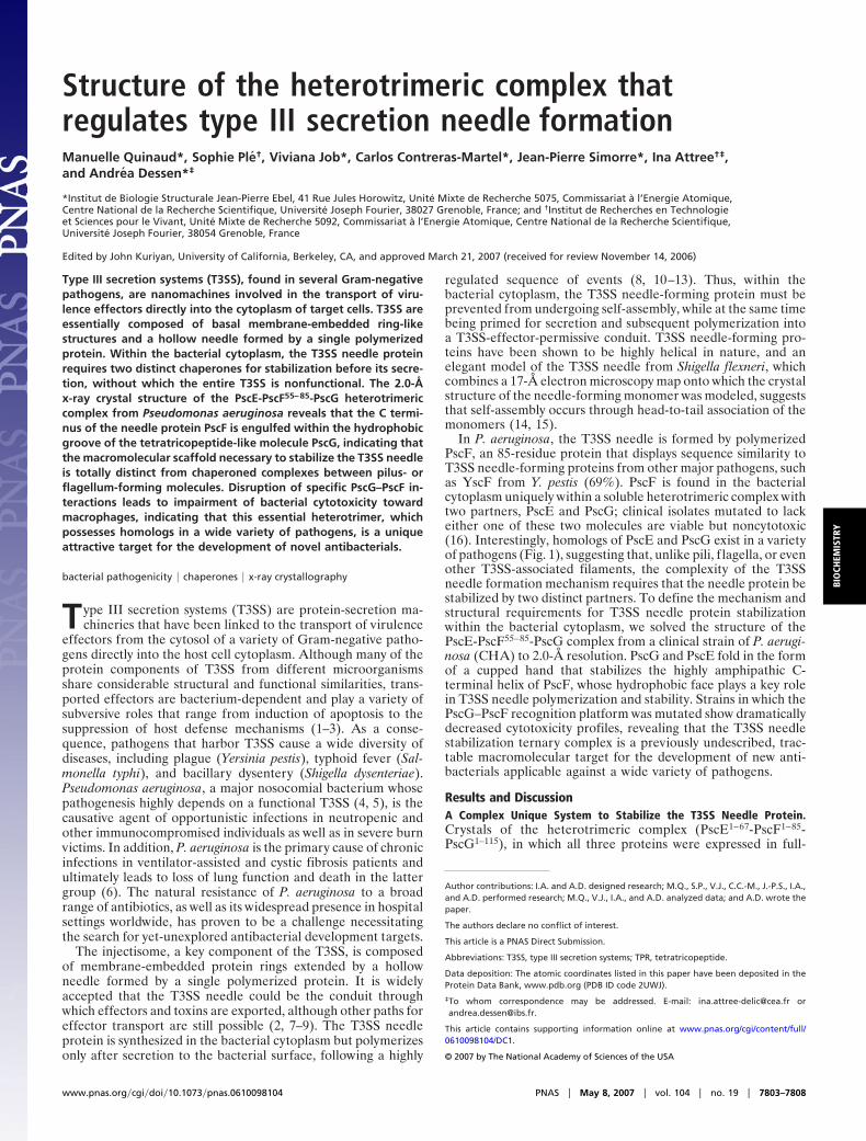
Structure of the heterotrimeric complex thatregulates type III secretion needle formationManuelle Quinaud*, Sophie Ple†, Viviana Job*, Carlos Contreras-Martel*, Jean-Pierre Simorre*, Ina Attree†‡,and Andrea Dessen*‡
*Institut de Biologie Structurale Jean-Pierre Ebel, 41 Rue Jules Horowitz, Unite Mixte de Recherche 5075, Commissariat a l’Energie Atomique,Centre National de la Recherche Scientifique, Universite Joseph Fourier, 38027 Grenoble, France; and †Institut de Recherches en Technologieet Sciences pour le Vivant, Unite Mixte de Recherche 5092, Commissariat a l’Energie Atomique, Centre National de la Recherche Scientifique,Universite Joseph Fourier, 38054 Grenoble, France
Edited by John Kuriyan, University of California, Berkeley, CA, and approved March 21, 2007 (received for review November 14, 2006)
Type III secretion systems (T3SS), found in several Gram-negativepathogens, are nanomachines involved in the transport of viru-lence effectors directly into the cytoplasm of target cells. T3SS areessentially composed of basal membrane-embedded ring-likestructures and a hollow needle formed by a single polymerizedprotein. Within the bacterial cytoplasm, the T3SS needle proteinrequires two distinct chaperones for stabilization before its secre-tion, without which the entire T3SS is nonfunctional. The 2.0-Åx-ray crystal structure of the PscE-PscF55–85-PscG heterotrimericcomplex from Pseudomonas aeruginosa reveals that the C termi-nus of the needle protein PscF is engulfed within the hydrophobicgroove of the tetratricopeptide-like molecule PscG, indicating thatthe macromolecular scaffold necessary to stabilize the T3SS needleis totally distinct from chaperoned complexes between pilus- orflagellum-forming molecules. Disruption of specific PscG–PscF in-teractions leads to impairment of bacterial cytotoxicity towardmacrophages, indicating that this essential heterotrimer, whichpossesses homologs in a wide variety of pathogens, is a uniqueattractive target for the development of novel antibacterials.
bacterial pathogenicity � chaperones � x-ray crystallography
Type III secretion systems (T3SS) are protein-secretion ma-chineries that have been linked to the transport of virulence
effectors from the cytosol of a variety of Gram-negative patho-gens directly into the host cell cytoplasm. Although many of theprotein components of T3SS from different microorganismsshare considerable structural and functional similarities, trans-ported effectors are bacterium-dependent and play a variety ofsubversive roles that range from induction of apoptosis to thesuppression of host defense mechanisms (1–3). As a conse-quence, pathogens that harbor T3SS cause a wide diversity ofdiseases, including plague (Yersinia pestis), typhoid fever (Sal-monella typhi), and bacillary dysentery (Shigella dysenteriae).Pseudomonas aeruginosa, a major nosocomial bacterium whosepathogenesis highly depends on a functional T3SS (4, 5), is thecausative agent of opportunistic infections in neutropenic andother immunocompromised individuals as well as in severe burnvictims. In addition, P. aeruginosa is the primary cause of chronicinfections in ventilator-assisted and cystic fibrosis patients andultimately leads to loss of lung function and death in the lattergroup (6). The natural resistance of P. aeruginosa to a broadrange of antibiotics, as well as its widespread presence in hospitalsettings worldwide, has proven to be a challenge necessitatingthe search for yet-unexplored antibacterial development targets.
The injectisome, a key component of the T3SS, is composedof membrane-embedded protein rings extended by a hollowneedle formed by a single polymerized protein. It is widelyaccepted that the T3SS needle could be the conduit throughwhich effectors and toxins are exported, although other paths foreffector transport are still possible (2, 7–9). The T3SS needleprotein is synthesized in the bacterial cytoplasm but polymerizesonly after secretion to the bacterial surface, following a highly
regulated sequence of events (8, 10–13). Thus, within thebacterial cytoplasm, the T3SS needle-forming protein must beprevented from undergoing self-assembly, while at the same timebeing primed for secretion and subsequent polymerization intoa T3SS-effector-permissive conduit. T3SS needle-forming pro-teins have been shown to be highly helical in nature, and anelegant model of the T3SS needle from Shigella flexneri, whichcombines a 17-Å electron microscopy map onto which the crystalstructure of the needle-forming monomer was modeled, suggeststhat self-assembly occurs through head-to-tail association of themonomers (14, 15).
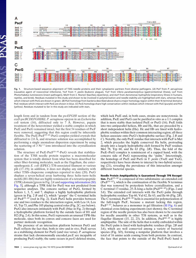
In P. aeruginosa, the T3SS needle is formed by polymerizedPscF, an 85-residue protein that displays sequence similarity toT3SS needle-forming proteins from other major pathogens, suchas YscF from Y. pestis (69%). PscF is found in the bacterialcytoplasm uniquely within a soluble heterotrimeric complex withtwo partners, PscE and PscG; clinical isolates mutated to lackeither one of these two molecules are viable but noncytotoxic(16). Interestingly, homologs of PscE and PscG exist in a varietyof pathogens (Fig. 1), suggesting that, unlike pili, f lagella, or evenother T3SS-associated filaments, the complexity of the T3SSneedle formation mechanism requires that the needle protein bestabilized by two distinct partners. To define the mechanism andstructural requirements for T3SS needle protein stabilizationwithin the bacterial cytoplasm, we solved the structure of thePscE-PscF55–85-PscG complex from a clinical strain of P. aerugi-nosa (CHA) to 2.0-Å resolution. PscG and PscE fold in the formof a cupped hand that stabilizes the highly amphipathic C-terminal helix of PscF, whose hydrophobic face plays a key rolein T3SS needle polymerization and stability. Strains in which thePscG–PscF recognition platform was mutated show dramaticallydecreased cytotoxicity profiles, revealing that the T3SS needlestabilization ternary complex is a previously undescribed, trac-table macromolecular target for the development of new anti-bacterials applicable against a wide variety of pathogens.
Results and DiscussionA Complex Unique System to Stabilize the T3SS Needle Protein.Crystals of the heterotrimeric complex (PscE1–67-PscF1–85-PscG1–115), in which all three proteins were expressed in full-
Author contributions: I.A. and A.D. designed research; M.Q., S.P., V.J., C.C.-M., J.-P.S., I.A.,and A.D. performed research; M.Q., V.J., I.A., and A.D. analyzed data; and A.D. wrote thepaper.
The authors declare no conflict of interest.
This article is a PNAS Direct Submission.
Abbreviations: T3SS, type III secretion systems; TPR, tetratricopeptide.
Data deposition: The atomic coordinates listed in this paper have been deposited in theProtein Data Bank, www.pdb.org (PDB ID code 2UWJ).
‡To whom correspondence may be addressed. E-mail: [email protected] [email protected].
This article contains supporting information online at www.pnas.org/cgi/content/full/0610098104/DC1.
© 2007 by The National Academy of Sciences of the USA
www.pnas.org�cgi�doi�10.1073�pnas.0610098104 PNAS � May 8, 2007 � vol. 104 � no. 19 � 7803–7808
BIO
CHEM
ISTR
Y
length form and in tandem from the pscEFGHI section of theexsD-pscBCDEFGHIJKL P. aeruginosa operon in an Escherichiacoli system (16), diffracted only to 7 Å. However, papaintreatment of the heterotrimer yielded a stable complex in whichPscE and PscG remained intact, but the first 54 residues of PscFwere removed, suggesting that this region could be inherentlyflexible. The PscE-PscF55–85-PscG complex yielded crystals thatdiffracted to 2.0 Å, and structure solution was accomplished byperforming a single anomalous dispersion experiment by usingthe scattering of 3 Ni�2 ions introduced into the crystallizationsolution.
The structure of PscE-PscF55–85-PscG reveals that stabiliza-tion of the T3SS needle protein requires a macromolecularsystem that is totally distinct from what has been described forother fiber-forming molecules, such as the flagellum, the enter-opathogenic E. coli (EPEC) T3S-associated filament or variouspili (17–19); in addition, it does not display any similarity withother T3SS–chaperone complexes reported to date (20). PscGdisplays a seven-helical array harboring three helix–turn–helixmotifs (H1-H6) that are highly reminiscent of a tetratricopeptide(TPR) domain [green in Fig. 2A and supporting information (SI)Fig. 5], although a TPR fold for PscG was not predicted fromsequence analyses. The concave surface of PscG, formed byhelices 1, 3, 5, and 7, displays a highly hydrophobic platform(green in Fig. 2 B and C), which ‘‘grasps’’ the C-terminal helixof PscF55–85 (red in Fig. 2). Each PscG helix provides betweenone and two residues to the interaction region, with Leu 14, Leu43, Trp 73, and Phe 105 playing central roles (Fig. 2C; see below).PscG also interacts intimately with PscE (blue in Fig. 2) througha hydrophobic platform formed by the outside faces of H1 andH2 (Fig. 2 A). In this sense, PscG represents an unusual TPR-likemolecule, since both its convex and concave faces are used forpartner molecule recognition.
The presence of an interaction platform between PscG andPscE reflects the fact that, both in vitro and in vivo, PscE servesas a stabilizing element for PscG (and vice versa). P. aeruginosastrains that lack chromosomally encoded pscE are incapable ofproducing PscG stably; the same occurs in pscG-deleted strains,
which lack PscE and, in both cases, strains are noncytotoxic. Inaddition, PscE and PscG can be purified in vitro as a 1:1 complexthat is more stable than isolated PscE or PscG (16). PscE foldsinto two antiparallel helices, Hb and Hc, that are preceded by ashort independent helix (Ha). Hc and Hb are lined with hydro-phobic residues within their common interacting region; all threehelices associate onto PscG’s hydrophobic surface (Fig. 2 B andC). Notably, the only PscE residue that interacts with PscF is Met2, which precedes the highly conserved Ha helix, protrudingdeeply into a largely hydrophobic cleft formed by PscF residuesMet 78, Trp 60, and Ile 63 (Fig. 2B). Thus, the fold of thePscE–PscG complex is reminiscent of a cupped hand, with theconcave side of PscG representing the ‘‘palm.’’ Interestingly,the homologs of PscE and PscG in Y. pestis (YscE and YscG,respectively) have been shown to interact by two-hybrid screen-ing (21), revealing the prevalence of this interaction amongstdifferent bacterial species.
Needle Protein Amphipathicity Is Guaranteed Through TPR Recogni-tion. PscF55–85 is composed of two subdomains: an extended coil(PscF55–67), which is the continuation of the flexible N terminusthat was removed by proteolysis before crystallization, and aC-terminal 17-residue, 25-Å-long �-helix (PscF68–85) (Figs. 2 and3A). The extended coil interacts with the PscG palm throughhydrophobic residues Trp 60, Val 62, Ile 63, and Ile 66 (Fig. 2B).The C-terminal, PscF68–85 helix is essential for polymerization ofthe full-length PscF, because a mutant lacking this region,PscF1–67, behaves as a monomer in gel filtration (SI Fig. 6) andchemical cross-linking experiments (not shown); the C-terminalregion of the needle protein has also been shown to be essentialfor needle assembly in other T3S systems, as well as in theflagellar filament (15, 22, 23). In addition, PscF68–85 is highlyamphipathic. The side of the helix that is stably embedded withinthe PscG palm is lined with hydrophobic residues (Figs. 2B and3A), which are well conserved among a variety of bacterialspecies (Fig. 3D), forming a nonpolar platform that involves atotal of 16 amino acids coming from both proteins. In contrast,the face that points to the outside of the PscE-PscG hand is
Fig. 1. Structure-based sequence alignment of T3SS needle proteins and their cytoplasmic partners from diverse pathogens. (A) PscF from P. aeruginosa(causative agent of nosocomial infections), YscF from Y. pestis (bubonic plague), YscF from Vibrio parahaemolyticus (gastrointestinal illness), LscF fromPhotorhabdus luminescens (insect pathogen), MxiH from S. flexneri (bacillary dysentery), and AscF from Aeromonas hydrophila (respiratory illness in humans,reptiles, and birds). Residues mutated in this study and shown to be involved in polymerization and needle stability are highlighted with stars, whereas thosewhich interact with PscG are shown in green. (B) PscE homologs from bacteria described above share a major homology region within their N-terminal domains.PscE residues which interact with PscG are shown in blue. (C) PscG homologs share high conservation within residues which interact with PscE (purple) and PscF(yellow). Residues mutated to Ser in this study are indicated with stars.
7804 � www.pnas.org�cgi�doi�10.1073�pnas.0610098104 Quinaud et al.
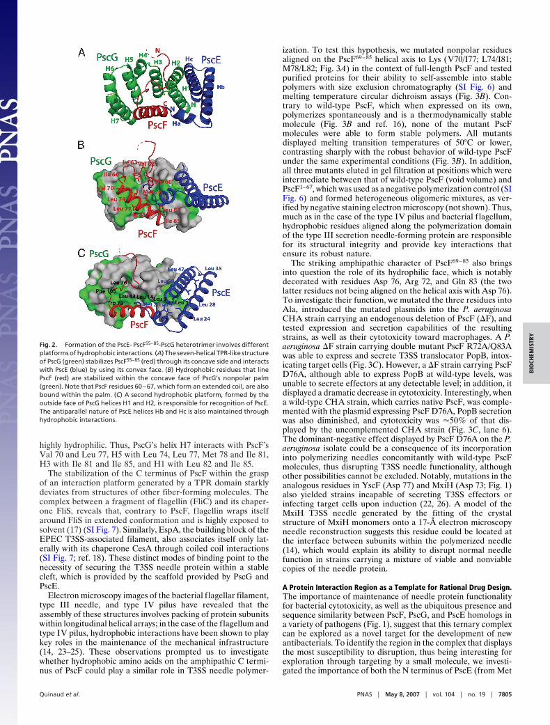
highly hydrophilic. Thus, PscG’s helix H7 interacts with PscF’sVal 70 and Leu 77, H5 with Leu 74, Leu 77, Met 78 and Ile 81,H3 with Ile 81 and Ile 85, and H1 with Leu 82 and Ile 85.
The stabilization of the C terminus of PscF within the graspof an interaction platform generated by a TPR domain starklydeviates from structures of other fiber-forming molecules. Thecomplex between a fragment of flagellin (FliC) and its chaper-one FliS, reveals that, contrary to PscF, f lagellin wraps itselfaround FliS in extended conformation and is highly exposed tosolvent (17) (SI Fig. 7). Similarly, EspA, the building block of theEPEC T3SS-associated filament, also associates itself only lat-erally with its chaperone CesA through coiled coil interactions(SI Fig. 7; ref. 18). These distinct modes of binding point to thenecessity of securing the T3SS needle protein within a stablecleft, which is provided by the scaffold provided by PscG andPscE.
Electron microscopy images of the bacterial f lagellar filament,type III needle, and type IV pilus have revealed that theassembly of these structures involves packing of protein subunitswithin longitudinal helical arrays; in the case of the flagellum andtype IV pilus, hydrophobic interactions have been shown to playkey roles in the maintenance of the mechanical infrastructure(14, 23–25). These observations prompted us to investigatewhether hydrophobic amino acids on the amphipathic C termi-nus of PscF could play a similar role in T3SS needle polymer-
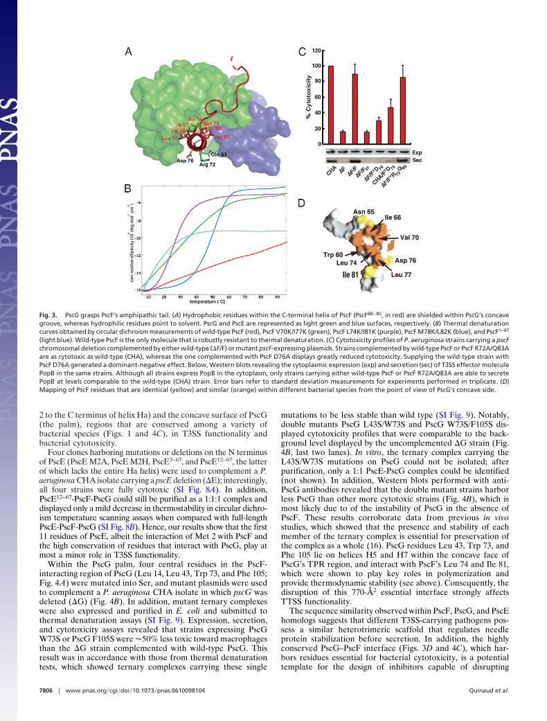
ization. To test this hypothesis, we mutated nonpolar residuesaligned on the PscF69–85 helical axis to Lys (V70/I77; L74/I81;M78/L82; Fig. 3A) in the context of full-length PscF and testedpurified proteins for their ability to self-assemble into stablepolymers with size exclusion chromatography (SI Fig. 6) andmelting temperature circular dichroism assays (Fig. 3B). Con-trary to wild-type PscF, which when expressed on its own,polymerizes spontaneously and is a thermodynamically stablemolecule (Fig. 3B and ref. 16), none of the mutant PscFmolecules were able to form stable polymers. All mutantsdisplayed melting transition temperatures of 50°C or lower,contrasting sharply with the robust behavior of wild-type PscFunder the same experimental conditions (Fig. 3B). In addition,all three mutants eluted in gel filtration at positions which wereintermediate between that of wild-type PscF (void volume) andPscF1–67, which was used as a negative polymerization control (SIFig. 6) and formed heterogeneous oligomeric mixtures, as ver-ified by negative staining electron microscopy (not shown). Thus,much as in the case of the type IV pilus and bacterial f lagellum,hydrophobic residues aligned along the polymerization domainof the type III secretion needle-forming protein are responsiblefor its structural integrity and provide key interactions thatensure its robust nature.
The striking amphipathic character of PscF69–85 also bringsinto question the role of its hydrophilic face, which is notablydecorated with residues Asp 76, Arg 72, and Gln 83 (the twolatter residues not being aligned on the helical axis with Asp 76).To investigate their function, we mutated the three residues intoAla, introduced the mutated plasmids into the P. aeruginosaCHA strain carrying an endogenous deletion of PscF (�F), andtested expression and secretion capabilities of the resultingstrains, as well as their cytotoxicity toward macrophages. A P.aeruginosa �F strain carrying double mutant PscF R72A/Q83Awas able to express and secrete T3SS translocator PopB, intox-icating target cells (Fig. 3C). However, a �F strain carrying PscFD76A, although able to express PopB at wild-type levels, wasunable to secrete effectors at any detectable level; in addition, itdisplayed a dramatic decrease in cytotoxicity. Interestingly, whena wild-type CHA strain, which carries native PscF, was comple-mented with the plasmid expressing PscF D76A, PopB secretionwas also diminished, and cytotoxicity was �50% of that dis-played by the uncomplemented CHA strain (Fig. 3C, lane 6).The dominant-negative effect displayed by PscF D76A on the P.aeruginosa isolate could be a consequence of its incorporationinto polymerizing needles concomitantly with wild-type PscFmolecules, thus disrupting T3SS needle functionality, althoughother possibilities cannot be excluded. Notably, mutations in theanalogous residues in YscF (Asp 77) and MxiH (Asp 73; Fig. 1)also yielded strains incapable of secreting T3SS effectors orinfecting target cells upon induction (22, 26). A model of theMxiH T3SS needle generated by the fitting of the crystalstructure of MxiH monomers onto a 17-Å electron microscopyneedle reconstruction suggests this residue could be located atthe interface between subunits within the polymerized needle(14), which would explain its ability to disrupt normal needlefunction in strains carrying a mixture of viable and nonviablecopies of the needle protein.
A Protein Interaction Region as a Template for Rational Drug Design.The importance of maintenance of needle protein functionalityfor bacterial cytotoxicity, as well as the ubiquitous presence andsequence similarity between PscF, PscG, and PscE homologs ina variety of pathogens (Fig. 1), suggest that this ternary complexcan be explored as a novel target for the development of newantibacterials. To identify the region in the complex that displaysthe most susceptibility to disruption, thus being interesting forexploration through targeting by a small molecule, we investi-gated the importance of both the N terminus of PscE (from Met
Fig. 2. Formation of the PscE- PscF55–85-PscG heterotrimer involves differentplatforms of hydrophobic interactions. (A) The seven-helical TPR-like structureof PscG (green) stabilizes PscF55–85 (red) through its concave side and interactswith PscE (blue) by using its convex face. (B) Hydrophobic residues that linePscF (red) are stabilized within the concave face of PscG’s nonpolar palm(green). Note that PscF residues 60–67, which form an extended coil, are alsobound within the palm. (C) A second hydrophobic platform, formed by theoutside face of PscG helices H1 and H2, is responsible for recognition of PscE.The antiparallel nature of PscE helices Hb and Hc is also maintained throughhydrophobic interactions.
Quinaud et al. PNAS � May 8, 2007 � vol. 104 � no. 19 � 7805
BIO
CHEM
ISTR
Y
2 to the C terminus of helix Ha) and the concave surface of PscG(the palm), regions that are conserved among a variety ofbacterial species (Figs. 1 and 4C), in T3SS functionality andbacterial cytotoxicity.
Four clones harboring mutations or deletions on the N terminusof PscE (PscE M2A, PscE M2H, PscE3–67, and PscE12–67, the latterof which lacks the entire Ha helix) were used to complement a P.aeruginosa CHA isolate carrying a pscE deletion (�E); interestingly,all four strains were fully cytotoxic (SI Fig. 8A). In addition,PscE12–67-PscF-PscG could still be purified as a 1:1:1 complex anddisplayed only a mild decrease in thermostability in circular dichro-ism temperature scanning assays when compared with full-lengthPscE-PscF-PscG (SI Fig. 8B). Hence, our results show that the first11 residues of PscE, albeit the interaction of Met 2 with PscF andthe high conservation of residues that interact with PscG, play atmost a minor role in T3SS functionality.
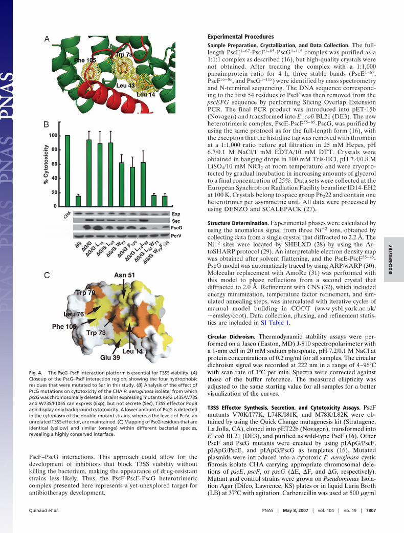
Within the PscG palm, four central residues in the PscF-interacting region of PscG (Leu 14, Leu 43, Trp 73, and Phe 105;Fig. 4A) were mutated into Ser, and mutant plasmids were usedto complement a P. aeruginosa CHA isolate in which pscG wasdeleted (�G) (Fig. 4B). In addition, mutant ternary complexeswere also expressed and purified in E. coli and submitted tothermal denaturation assays (SI Fig. 9). Expression, secretion,and cytotoxicity assays revealed that strains expressing PscGW73S or PscG F105S were �50% less toxic toward macrophagesthan the �G strain complemented with wild-type PscG. Thisresult was in accordance with those from thermal denaturationtests, which showed ternary complexes carrying these single
mutations to be less stable than wild type (SI Fig. 9). Notably,double mutants PscG L43S/W73S and PscG W73S/F105S dis-played cytotoxicity profiles that were comparable to the back-ground level displayed by the uncomplemented �G strain (Fig.4B, last two lanes). In vitro, the ternary complex carrying theL43S/W73S mutations on PscG could not be isolated; afterpurification, only a 1:1 PscE-PscG complex could be identified(not shown). In addition, Western blots performed with anti-PscG antibodies revealed that the double mutant strains harborless PscG than other more cytotoxic strains (Fig. 4B), which ismost likely due to of the instability of PscG in the absence ofPscF. These results corroborate data from previous in vivostudies, which showed that the presence and stability of eachmember of the ternary complex is essential for preservation ofthe complex as a whole (16). PscG residues Leu 43, Trp 73, andPhe 105 lie on helices H5 and H7 within the concave face ofPscG’s TPR region, and interact with PscF’s Leu 74 and Ile 81,which were shown to play key roles in polymerization andprovide thermodynamic stability (see above). Consequently, thedisruption of this 770-Å2 essential interface strongly affectsTTSS functionality.
The sequence similarity observed within PscF, PscG, and PscEhomologs suggests that different T3SS-carrying pathogens pos-sess a similar heterotrimeric scaffold that regulates needleprotein stabilization before secretion. In addition, the highlyconserved PscG–PscF interface (Figs. 3D and 4C), which har-bors residues essential for bacterial cytotoxicity, is a potentialtemplate for the design of inhibitors capable of disrupting
Fig. 3. PscG grasps PscF’s amphipathic tail. (A) Hydrophobic residues within the C-terminal helix of PscF (PscF68–85, in red) are shielded within PscG’s concavegroove, whereas hydrophilic residues point to solvent. PscG and PscE are represented as light green and blue surfaces, respectively. (B) Thermal denaturationcurves obtained by circular dichroism measurements of wild-type PscF (red), PscF V70K/I77K (green), PscF L74K/I81K (purple), PscF M78K/L82K (blue), and PscF1–67
(light blue). Wild-type PscF is the only molecule that is robustly resistant to thermal denaturation. (C) Cytotoxicity profiles of P. aeruginosa strains carrying a pscFchromosomal deletion complemented by either wild-type (�F/F) or mutant pscF-expressing plasmids. Strains complemented by wild-type PscF or PscF R72A/Q83Aare as cytotoxic as wild-type (CHA), whereas the one complemented with PscF D76A displays greatly reduced cytotoxicity. Supplying the wild-type strain withPscF D76A generated a dominant-negative effect. Below, Western blots revealing the cytoplasmic expression (exp) and secretion (sec) of T3SS effector moleculePopB in the same strains. Although all strains express PopB in the cytoplasm, only strains carrying either wild-type PscF or PscF R72A/Q83A are able to secretePopB at levels comparable to the wild-type (CHA) strain. Error bars refer to standard deviation measurements for experiments performed in triplicate. (D)Mapping of PscF residues that are identical (yellow) and similar (orange) within different bacterial species from the point of view of PscG’s concave side.
7806 � www.pnas.org�cgi�doi�10.1073�pnas.0610098104 Quinaud et al.
PscF–PscG interactions. This approach could allow for thedevelopment of inhibitors that block T3SS viability withoutkilling the bacterium, making the appearance of drug-resistantstrains less likely. Thus, the PscF-PscE-PscG heterotrimericcomplex presented here represents a yet-unexplored target forantibiotherapy development.
Experimental ProceduresSample Preparation, Crystallization, and Data Collection. The full-length PscE1–67-PscF1–85-PscG1–115 complex was purified as a1:1:1 complex as described (16), but high-quality crystals werenot obtained. After treating the complex with a 1:1,000papain:protein ratio for 4 h, three stable bands (PscE1–67,PscF55–85, and PscG1–115) were identified by mass spectrometryand N-terminal sequencing. The DNA sequence correspond-ing to the first 54 residues of PscF was then removed from thepscEFG sequence by performing Slicing Overlap ExtensionPCR. The final PCR product was introduced into pET-15b(Novagen) and transformed into E. coli BL21 (DE3). The newheterotrimeric complex, PscE-PscF55–85-PscG, was purified byusing the same protocol as for the full-length form (16), withthe exception that the histidine tag was removed with thrombinat a 1:1,000 ratio before gel filtration in 25 mM Hepes, pH6.7/0.1 M NaCl/1 mM EDTA/10 mM DTT. Crystals wereobtained in hanging drops in 100 mM Tris�HCl, pH 7.4/0.8 MLiSO4/10 mM NiCl2 at room temperature and were cryopro-tected by gradual incubation in increasing amounts of glycerolto a final concentration of 25%. Data sets were collected at theEuropean Synchrotron Radiation Facility beamline ID14-EH2at 100 K. Crystals belong to space group P6222 and contain oneheterotrimer per asymmetric unit. All data were processed byusing DENZO and SCALEPACK (27).
Structure Determination. Experimental phases were calculated byusing the anomalous signal from three Ni�2 ions, obtained bycollecting data from a single crystal that diffracted to 2.2 Å. TheNi�2 sites were located by SHELXD (28) by using the Au-toSHARP protocol (29). An interpretable electron density mapwas obtained after solvent flattening, and the PscE-PscF55–85-PscG model was automatically traced by using ARP/wARP (30).Molecular replacement with AmoRe (31) was performed withthis model to phase reflections from a second crystal thatdiffracted to 2.0 Å. Refinement with CNS (32), which includedenergy minimization, temperature factor refinement, and sim-ulated annealing steps, was intercalated with iterative cycles ofmanual model building in COOT (www.ysbl.york.ac.uk/�emsley/coot). Data collection, phasing, and refinement statis-tics are included in SI Table 1.
Circular Dichroism. Thermodynamic stability assays were per-formed on a Jasco (Easton, MD) J-810 spectropolarimeter witha 1-mm cell in 20 mM sodium phosphate, pH 7.2/0.1 M NaCl atprotein concentrations of 0.2 mg/ml for all samples. The circulardichroism signal was recorded at 222 nm in a range of 4–96°Cwith scan rate of 1°C per min. Spectra were corrected againstthose of the buffer reference. The measured ellipticity wasadjusted to the same starting value for all samples for a bettervisualization of the curves.
T3SS Effector Synthesis, Secretion, and Cytotoxicity Assays. PscFmutants V70K/I77K, L74K/I81K, and M78K/L82K were ob-tained by using the Quick Change mutagenesis kit (Stratagene,La Jolla, CA), cloned into pET22b (Novagen), transformed intoE. coli BL21 (DE3), and purified as wild-type PscF (16). OtherPscF and PscG mutants were created by using pIApG/PscF,pIApG/PscE, and pIApG/PscG as templates (16). Mutatedplasmids were introduced into a cytotoxic P. aeruginosa cysticfibrosis isolate CHA carrying appropriate chromosomal dele-tions of pscE, pscF, or pscG (�E, �F, and �G, respectively).Mutant and control strains were grown on Pseudomonas Isola-tion Agar (Difco, Lawrence, KS) plates or in liquid Luria Broth(LB) at 37°C with agitation. Carbenicillin was used at 500 �g/ml
Fig. 4. The PscG–PscF interaction platform is essential for T3SS viability. (A)Closeup of the PscG–PscF interaction region, showing the four hydrophobicresidues that were mutated to Ser in this study. (B) Analysis of the effect ofPscG mutations on cytotoxicity of the CHA P. aeruginosa isolate, from whichpscG was chromosomally deleted. Strains expressing mutants PscG L43S/W73Sand W73S/F105S can express (Exp), but not secrete (Sec), T3SS effector PopBand display only background cytotoxicity. A lower amount of PscG is detectedin the cytoplasm of the double-mutant strains, whereas the levels of PcrV, anunrelated T3SS effector, are maintained. (C) Mapping of PscG residues that areidentical (yellow) and similar (orange) within different bacterial species,revealing a highly conserved interface.
Quinaud et al. PNAS � May 8, 2007 � vol. 104 � no. 19 � 7807
BIO
CHEM
ISTR
Y
for Pseudomonas Isolation Agar plates and 300 �g/ml in LB. Forinduction of type III secretion in vitro, P. aeruginosa overnightcultures were diluted to an optical density at 600 nm (A600) of 0.2in LB containing 5 mM EGTA and 20 mM MgCl2. Incubationwas prolonged for additional 3 h until the cultures reached A600
values of 1.0. The samples containing 30 �l of culture superna-tants or total cells were directly analyzed by Western blotting byusing anti-PopB, anti-PcrV, or anti-PscG antibodies. For cyto-toxicity assays, the bacteria were cultivated to an OD600 of 1 andadded to macrophage cell line J774 at a Multiplicity of Infectionof 5. Cell death was assessed at 3 h postinfection by using a
Cytotoxicity Detection Kit, LDH (Roche, Indianapolis, IN). Alltests were performed in triplicate.
We thank Otto Dideberg for continuous support, Guy Schoehn and theCIBB electron microscopy platform for help with negative staininganalyses, and laboratory members for helpful discussions. This work wassupported by grants from the French Cystic Fibrosis Foundation (Vain-cre la Mucoviscidose) and the Direction des Sciences du Vivant (DSV/Commissariat a l’Energie Atomique). M.Q. is the recipient of a CFRfellowship (Commissariat a l’Energie Atomique), and S.P. is the recip-ient of a postdoctoral fellowship from the DSV/Commissariat a l’EnergieAtomique. We thank D. Lemaire and B. Dublet for mass spectrometryanalyses, as well as the ESRF ID14 staff for access to beamlines.
1. Galan JE, Collmer A (1999) Science 284:1322–1328.2. Ghosh P (2004)) Mol Biol Rev 68:771–795.3. Cornelis G (2006) Nat Rev Microbiol 4:811–825.4. Vance RE, Retsch A, Mekalanos JJ (2005) Infect Immun 73:1706–1713.5. Roy-Burman A, Savel RH, Racine S, Swanson BL, Revadigar NS, Fujimoto J,
Sawa T, Frank DW, Wiener-Kornish JP (2001) J Infect Dis 183:1767–1774.6. Lyczak JB, Cannon CL, Pier GB (2000) Microbes Infect 2:1051–1060.7. Kubori T, Matsushima Y, Nakamura D, Uralil J, Lara-Tejero M, Sukhan A,
Galan J, Aizawa S-I (1998) Science 280:602–605.8. Marlovits TC, Kubori T, Sukhan A, Thomas DR, Galan JE, Unger VM (2004)
Science 306:1040–1042.9. Blocker A, Jouihri N, Larquet E, Gounon P, Ebel F, Parsot C, Sansonetti P,
Allaoui A (2001) Mol Microbiol 39:652–663.10. Kubori T, Sukhan A, Aizawa S-I, Galan JE (2000) Proc Natl Acad Sci USA
97:10225–10230.11. Marlovits TC, Kubori T, Lara-Tejero M, Thomas D, Unger VM, Galan JE
(2006) Nature 441:637–640.12. Kimbrough TG, Miller SI (2000) Proc Natl Acad Sci USA 97:11008–11013.13. Journet L, Agrain C, Broz P, Cornelis GR (2003) Science 302:1757–1760.14. Deane JE, Roversi P, Cordes FS, Johnson S, Kenjale R, Daniell S, Booy F,
Picking WD, Picking WL, Blocker AJ, Lea SM (2006) Proc Natl Acad Sci USA103:12529–12533.
15. Zhang L, Wang Y, Picking WL, Picking WD, De Guzman RN (2006) J Mol Biol359:322–330.
16. Quinaud M, Chabert J, Faudry E, Neumann E, Lemaire D, Pastor A, Elsen S,Dessen A, Attree I (2005) J Biol Chem 280:36293–36300.
17. Evdokimov AG, Phan J, Tropea JE, Routzahn KM, Peters III, H.K., PokrossM, Waugh DS (2003) Nat Struct Biol 10:789–793.
18. Yip CK, Finlay BB, Strynadka NC (2005) Nat Struct Mol Biol 12:75–81.19. Sauer FG, Barnhart M, Choudhury D, Knight SD, Waksman G, Hultgren SJ
(2000) Curr Opin Struct Biol 10:548–556.20. Yip CK, Strynadka NC (2006) Trends Biochem Sci 31:223–230.21. Day JB, Guller J, Plano GV (2000) Infect Immun 68:6466–6471.22. Kenjale R, Wilson J, Zenk SF, Saurya S, Picking WL, Picking WD, Blocker A
(2005)) J Biol Chem 280:42929–42937.23. Samatey FA, Imada K, Nagashima S, Vonderviszt F, Kumasaka T, Yamamoto
M, Namba K (2001) Nature 410:331–337.24. Craig L, Volkmann N, Arvai AS, Pique ME, Yeager M, Egelman EH, Tainer
JA (2006) Mol Cell 23:651–652.25. Yonekura K, Maki-Yonekura S, Namba K (2003) Nature 424:643–650.26. Torruellas J, Jackson MW, Pennock JW, Plano GV (2005) Mol Microbiol
57:1719–1733.27. Otwinowski Z, Minor W (1997) Methods Enzymol 276:307–326.28. Schneider TR, Sheldrick GM (2002) Acta Crystallogr D 58:1772–1779.29. de la Fortelle E, Bricogne G (1997) Methods Enzymol 276:472–494.30. Morris RJ, Perrakis A, Lamzin VS (2003) Methods Enzymol 374:229–
244.31. Navaza J (2001) Acta Crystallogr D 57:1367–1372.32. Brunger AT, Adams PD, Clore GM, DeLano WL, Gros P, Grosse-Kunstleve
RW, Jiang JS, Kuszewski J, Nilges M, Pannu NS, et al. (1998) Acta CrystallogrD 54:905–921.
7808 � www.pnas.org�cgi�doi�10.1073�pnas.0610098104 Quinaud et al.