Embed Size (px)
Citation preview

One of the main applications of gene tar-geting is to analyze protein function in vivo. When assessing overall proteinfunction via gene inactivation, conclusionsare usually unambiguous, particularlywhen the defective phenotype of aknockout line can be reverted to thewild-type phenotype by reintroducing anintact copy of the gene (complemen-tation). However, when assessing the con-tribution of certain residues or domainsin protein function (structure–functionanalysis), appropriate controls are essen-tial. Recently, two papers attempted astructure–function analysis of the throm-bospondin-related anonymous protein(TRAP) of malaria parasites1,2.
TRAP is a type 1 transmembrane pro-tein synthesized by the malaria sporo-zoite3, the stage of the parasite that infectsthe salivary glands of the mosquito vector
and the liver of the mammalian host.Previous use of the transformation systemin Plasmodium berghei, a malaria speciesthat infects rodents, has established thatTRAP is important for sporozoite glidingmotility (a form of substrate-dependentlocomotion characteristic of invasive stagesof apicomplexan parasites), infection ofthe mosquito salivary glands and infectionof the mammalian liver4. Gliding motilityand cell invasion by Apicomplexa wereproposed 20 years ago to result from theanterior-to-posterior redistribution, viathe actin cytoskeleton, of surface ligandsthat interact with the substrate (thus mov-ing the parasite forward) or the cell sur-face (leading to penetration into the cell)5,6.Recent experimental evidence suggeststhat TRAP may constitute the essentialtransmembrane link of the capping ma-chinery that drives gliding motility and cell
invasion by the malaria sporozoite2,4. TRAPmay thus link the parasite actin cytoskel-eton, probably via motor proteins, andthe extracellular receptors that are usedto exert force and allow traction.
Several methodological points and thecrucial importance of controls in validatingthe results of a structure–function analysisof TRAP1,2 merit further exploration.Wengelnik et al.1 addressed the functionof residues in the extracellular domain ofTRAP, whereas Kappe et al.2 tested thefunction of the cytoplasmic tail of TRAP.
Structure–Function Analysis
There are at least three ways to con-duct a structure–function analysis in vivo:the gene encoding the protein of interestcan be modified by a single recombinationevent, using: (1) an insertion; or (2) a re-placement targeting plasmid (Fig. 1); or (3)the endogenous gene can be inactivatedusing a first selectable marker and a modi-fied version of the gene introduced intothe knockout line using a second select-able marker. Until recently, only one se-lectable marker was available for P. bergheitransformation. Therefore, only the firsttwo approaches were feasible. Wengelniket al. used a replacement technique to in-troduce the TRAP modifications, whereasKappe et al. used an insertion strategy.
The advantages and potential draw-backs of each of these methods havebeen discussed elsewhere7,8. We want totake issue with two statements made inRef. 1 regarding the strategies for genemodification.
(1) ‘Genetic reversion at a locus cre-ated by integration of an insertion plasmidis a limitation’. Reversion to the WT gene,which we described at the TRAP locus in-activated by an insertion strategy2,4, con-sists of recombination between the dupli-cated sequences at the modified locus and leads to plasmid excision. When re-version occurs, the frequency is very low(,1/100), and the presence of revertantsdoes not complicate phenotypical analysisof the recombinant population. Not only isreversion not a limitation, but it can actu-ally be very advantageous. Indeed, thepresence of revertants that have regaineda wild-type cycle in a population of recom-binants that are arrested in their develop-ment demonstrates that the defect of therecombinants is because of the mutation.
Comment
222 Parasitology Today, vol. 16, no. 6, 20000169-4758/00/$ – see front matter © 2000 Elsevier Science Ltd. All rights reserved. PII: S0169-4758(00)01679-3
Structure–Function Analysis of MalariaProteins by Gene Targeting
R. Ménard and V. Nussenzweig
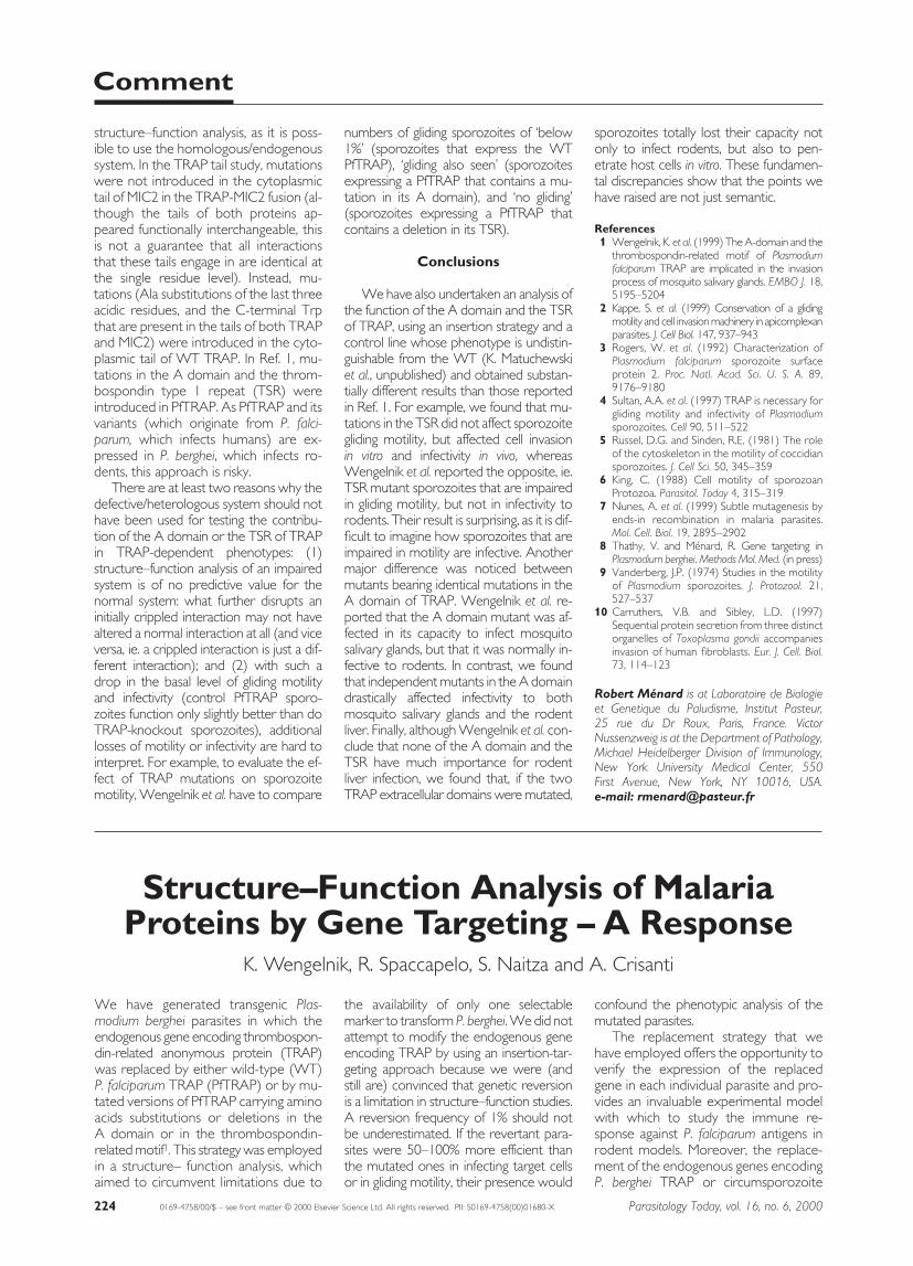
Fig. 1. Strategies for gene modification based on a single recombination event. The singlerecombination event can be promoted by a targeting plasmid of the replacement or ofthe insertion type. Using a replacement plasmid (a), the selectable marker should be in-serted upstream from the promoter region (or downstream from the 39 sequences nec-essary for gene expression) of the target gene. The transformed linear fragment containsan upstream region of homology consisting of 59 untranslated region (UTR) of the gene,and a downstream region of homology that starts upstream from the gene promoter andends after the mutation. The recombinant locus generated by the expected doublecrossover should therefore contain the modified gene flanked by the UTRs necessary forexpression. However, the selectable marker ends up inserted upstream (or downstreamif the construct is inverted) from the regulatory sequences of the modified gene, whichmay disrupt a closely linked locus or affect its expression. Using an insertion plasmid (b),the targeting sequence must lack the 59 end of the gene (to generate a truncated secondcopy) and end after its 39 regulatory elements (to allow expression of the first copy).When the plasmid is linearized within the region of homology upstream from the mu-tation, the recombinant locus generated by the crossover should contain a first copy ofthe gene that is full-length, modified and expressed, and a second copy that is truncatedand not expressed. The final locus contains uninterrupted sequences located upstreamfrom the target gene (up to the first duplicate) as well as downstream (starting from thesecond duplicate), minimizing the risk that plasmid integration may affect neighboringgenes. Mutations can be corrected during recombination in either case. Symbols: openbox, coding sequence of the target gene; thin lines, untranslated regions; arrow, gene pro-moter; open circle, 39 sequences necessary for gene expression; shaded box, resistancecassette; thick lines, bacterial plasmid sequences. E to E denotes the subtle modification.
Parasitology Today
Replacement
TARGETRGETA
TARGET
Insertion
TARGET
TARGET ARGETTARGET
(a) (b)

(2) ‘The replacement strategy over-comes the problems of mutation repairduring recombination’. The repair mecha-nisms that may correct mutations are op-erational when using either replacementor insertion plasmids, and we have verifiedthis in P. berghei (A. Nunes et al., unpub-lished). Mutations can be corrected duringDNA integration even if located hundredsof base pairs from the linearization sites(one edge of the construct in the case ofthe replacement construct), and strangephenomena, including discontinuous geneconversion events, can occur. This miscon-ception may have led the authors to re-frain from searching for the presence ofthe mutations in the recombinant loci.Thus, the question remains as to whetherthe mutations introduced by Wengelnik et al.1 are indeed present in the final loci.
Validation of the System
The control parasite line must have awild-type phenotype. Whatever the systemchosen to introduce the mutations, thefirst step is to construct a control line thatcontains the recombinant locus bearing awild-type gene, and to verify that the con-trol line has a wild-type phenotype. InKappe’s study2, the control line (namedINCO, for integration control) was foundto behave indistinguishably from the wild-type (WT) P. berghei in all TRAP-depen-dent phenotypes: (1) similar proportionsof WT and INCO sporozoites glidedwith a similar pattern; (2) WT and INCOsporozoites were present in similar num-bers in mosquito salivary glands; (3) WTand INCO sporozoites induced infectionin rats with similar prepatent periods; and(4) WT and INCO sporozoites gener-ated similar numbers of exo-erythrocyticforms in HepG2 cells in vitro. In Ref. 1, thecontrol line (referred to here as PfTRAP,in which the endogenous P. bergheiTRAP had been exchanged with P. falci-parum TRAP via the replacement strat-egy) displayed drastically impaired TRAP-dependent phenotypes: (1) whereas upto 80% of the WT P. berghei sporozoitesisolated from mosquito salivary glandsglide on solid substrates, as few as 2% of the PfTRAP sporozoites glided, andeven these produced shorter trails; (2)only 3700 PfTRAP sporozoites werefound associated with the mosquito sa-livary glands, when 5–10 times as manycan be routinely found in mosquitoes infected with WT P. berghei4,9; and (3) PfTRAP sporozoites collected fromeither midguts or salivary glands of mos-quitoes and injected intravenously intomice were approximately 2% as infectiveas the corresponding WT P. bergheisporozoites.
Regarding this last point, we were sur-prised to read the conclusions on infectiv-ity to rodents of PfTRAP sporozoites. Thiscan be quantified by measuring parasiterRNA in the liver, but it can be evaluatedby measuring pre-patent periods of infec-tion, ie. the number of days between in-travenous injection of sporozoites and de-tection of erythrocytic stages in animals. Inthis case, the goal should be to comparesporozoite infectivities under conditionsthat yield reproducible results, rather thanto utilize criteria that tend to minimize thedifferences. It can be misleading to use, asWengelnik et al. did, non-quantitative cri-teria in non-linear systems, such as thepercentage of animals that become in-fected (when these can become infectedwith very different pre-patent periods).The most reliable parameter is the aver-age pre-patent period induced by theminimum number of sporozoites that al-ways induce infection (in all experiments).Referral to the data in the tables from Ref. 1, rather than to the conclusions inthe text, will highlight that both midgutand salivary gland PfTRAP sporozoites (in-jection of 100 000 and 1000 sporozoitesrespectively) induce infections, with an ad-ditional 1.5 days in the pre-patent period,compared with infection induced by WTsporozoites. As a delay in patency of 24 hcorresponds to a <tenfold reduction inthe original inoculum (parasitemia in ro-dents increases ~tenfold per day, and in-jection of tenfold fewer sporozoites ex-tend the infection pre-patent period by 1day4,9)* , these results consistently show a~50-fold decrease in infectivity of PfTRAPsporozoites. Furthermore, the authorsclaim that the infectivity of PfTRAP sporo-zoites to rodents is similar to that of WTsporozoites, although PfTRAP sporo-zoites are severely impaired in gliding motility and in their capacity to infect themosquito salivary glands. This is in directcontradiction with the model that linksthese events via TRAP. A more plausibleconclusion is that PfTRAP sporozoites areseverely deficient in all TRAP-dependentactivities, and, therefore, that the PfTRAPsporozoites are not a valid system for further structure–function analysis.
The control line should produce wild-typeamounts of the protein under study. It is alsoimportant to verify that the protein underinvestigation is produced in normalamounts by the control line. In Kappe’sstudy2, western blot analysis showed that
the amounts of TRAP produced byINCO sporozoites were similar to thoseproduced by WT sporozoites. Importantly,deletions and amino acid substitutions inthe TRAP cytoplasmic tail, which pro-foundly affected sporozoite gliding and in-vasion, did not impair the production ofthe TRAP truncates/variants. These con-trols were crucial to the conclusion thatthe modifications of the cytoplasmic tail ofTRAP were responsible for the alteredphenotypes. In the study by Wengelnik etal.1, the issue of TRAP production is ofparticular importance, in view of the im-paired phenotype of PfTRAP sporozoites.Unfortunately, only immunofluorescencepictures are shown, but not western blotor other semi-quantitative methods tomeasure TRAP production.
A Homologous System
Heterelogous systems are valuable fortesting overall protein function in vivo …Heterologous systems have been widelyused for addressing protein function in vivo.There are many examples, in a variety oforganisms, of complementation of the de-fective phenotype of a knockout clonewith a heterologous product, demonstrat-ing that the transformed gene and the en-dogenous deleted gene have the same (oroverlapping) function. In Kappe’s study2, arelated approach was taken by testingwhether the cytoplasmic tail of the MIC2protein of Toxoplasma gondii, which hasbeen shown to undergo anterior to pos-terior translocation on the parasite surfaceduring cell invasion10, could function inplace of the cytoplasmic tail of TRAP. Thiswas not obvious, because the primary se-quences of the cytoplasmic tails of theseproteins are not conserved. The hybridmolecule made up of the extracellular do-main of TRAP and the cytoplasmic tail ofMIC2 efficiently mediated P. berghei sporo-zoite invasion into cultured cells, glidingmotility and infectivity to mosquito salivaryglands and the rodent liver. This indicatesthat the two proteins (and possibly otherstructurally related factors in Apicom-plexa) have similar functions and interactwith related partners in the parasite cyto-plasm. In Ref. 1, a heterologous comple-mentation strategy was used to testwhether PfTRAP could function in place ofPbTRAP. The answer was that it could, al-beit poorly. This confirmed what mighthave been predicted, ie. that TRAP pro-teins in different malaria species have thesame function.
… but not for protein structure–functionanalysis. While testing protein functionusing heterologous complementationmay be very informative, heterologoussystems should not be used for protein
Comment
Parasitology Today, vol. 16, no. 6, 2000 223
*One additional day in patency corresponds to atenfold decrease in infectivity; two days correspondsto a 100-fold difference and 1.5 days to a 50-fold difference in infectivity. [Editor’s note: a tenfold decrease is equivalent to a 90% reduction in theoriginal number, a 50-fold decrease is equivalent toa 98% reduction, and so on.]
We have generated transgenic Plas-modium berghei parasites in which theendogenous gene encoding thrombospon-din-related anonymous protein (TRAP)was replaced by either wild-type (WT)P. falciparum TRAP (PfTRAP) or by mu-tated versions of PfTRAP carrying aminoacids substitutions or deletions in the A domain or in the thrombospondin-related motif1. This strategy was employedin a structure– function analysis, whichaimed to circumvent limitations due to
the availability of only one selectablemarker to transform P. berghei. We did notattempt to modify the endogenous geneencoding TRAP by using an insertion-tar-geting approach because we were (andstill are) convinced that genetic reversionis a limitation in structure–function studies.A reversion frequency of 1% should notbe underestimated. If the revertant para-sites were 50–100% more efficient thanthe mutated ones in infecting target cellsor in gliding motility, their presence would
confound the phenotypic analysis of themutated parasites.
The replacement strategy that wehave employed offers the opportunity toverify the expression of the replacedgene in each individual parasite and pro-vides an invaluable experimental modelwith which to study the immune re-sponse against P. falciparum antigens inrodent models. Moreover, the replace-ment of the endogenous genes encodingP. berghei TRAP or circumsporozoite
structure–function analysis, as it is poss-ible to use the homologous/endogenoussystem. In the TRAP tail study, mutationswere not introduced in the cytoplasmictail of MIC2 in the TRAP-MIC2 fusion (al-though the tails of both proteins ap-peared functionally interchangeable, thisis not a guarantee that all interactionsthat these tails engage in are identical atthe single residue level). Instead, mu-tations (Ala substitutions of the last threeacidic residues, and the C-terminal Trpthat are present in the tails of both TRAPand MIC2) were introduced in the cyto-plasmic tail of WT TRAP. In Ref. 1, mu-tations in the A domain and the throm-bospondin type 1 repeat (TSR) wereintroduced in PfTRAP. As PfTRAP and itsvariants (which originate from P. falci-parum, which infects humans) are ex-pressed in P. berghei, which infects ro-dents, this approach is risky.
There are at least two reasons why thedefective/heterologous system should nothave been used for testing the contribu-tion of the A domain or the TSR of TRAPin TRAP-dependent phenotypes: (1)structure–function analysis of an impairedsystem is of no predictive value for thenormal system: what further disrupts aninitially crippled interaction may not havealtered a normal interaction at all (and viceversa, ie. a crippled interaction is just a dif-ferent interaction); and (2) with such adrop in the basal level of gliding motilityand infectivity (control PfTRAP sporo-zoites function only slightly better than doTRAP-knockout sporozoites), additionallosses of motility or infectivity are hard tointerpret. For example, to evaluate the ef-fect of TRAP mutations on sporozoitemotility, Wengelnik et al. have to compare
numbers of gliding sporozoites of ‘below1%’ (sporozoites that express the WTPfTRAP), ‘gliding also seen’ (sporozoitesexpressing a PfTRAP that contains a mu-tation in its A domain), and ‘no gliding’(sporozoites expressing a PfTRAP thatcontains a deletion in its TSR).
Conclusions
We have also undertaken an analysis ofthe function of the A domain and the TSRof TRAP, using an insertion strategy and acontrol line whose phenotype is undistin-guishable from the WT (K. Matuchewski et al., unpublished) and obtained substan-tially different results than those reportedin Ref. 1. For example, we found that mu-tations in the TSR did not affect sporozoitegliding motility, but affected cell invasion in vitro and infectivity in vivo, whereasWengelnik et al. reported the opposite, ie.TSR mutant sporozoites that are impairedin gliding motility, but not in infectivity torodents. Their result is surprising, as it is dif-ficult to imagine how sporozoites that areimpaired in motility are infective. Anothermajor difference was noticed betweenmutants bearing identical mutations in theA domain of TRAP. Wengelnik et al. re-ported that the A domain mutant was af-fected in its capacity to infect mosquitosalivary glands, but that it was normally in-fective to rodents. In contrast, we foundthat independent mutants in the A domaindrastically affected infectivity to both mosquito salivary glands and the rodentliver. Finally, although Wengelnik et al. con-clude that none of the A domain and theTSR have much importance for rodentliver infection, we found that, if the twoTRAP extracellular domains were mutated,
sporozoites totally lost their capacity notonly to infect rodents, but also to pen-etrate host cells in vitro. These fundamen-tal discrepancies show that the points wehave raised are not just semantic.
References1 Wengelnik, K. et al. (1999) The A-domain and the
thrombospondin-related motif of Plasmodiumfalciparum TRAP are implicated in the invasionprocess of mosquito salivary glands. EMBO J. 18,5195–5204
2 Kappe, S. et al. (1999) Conservation of a glidingmotility and cell invasion machinery in apicomplexanparasites. J. Cell Biol. 147, 937–943
3 Rogers, W. et al. (1992) Characterization ofPlasmodium falciparum sporozoite surfaceprotein 2. Proc. Natl. Acad. Sci. U. S. A. 89,9176–9180
4 Sultan, A.A. et al. (1997) TRAP is necessary forgliding motility and infectivity of Plasmodiumsporozoites. Cell 90, 511–522
5 Russel, D.G. and Sinden, R.E. (1981) The roleof the cytoskeleton in the motility of coccidiansporozoites. J. Cell Sci. 50, 345–359
6 King, C. (1988) Cell motility of sporozoanProtozoa. Parasitol. Today 4, 315–319
7 Nunes, A. et al. (1999) Subtle mutagenesis byends-in recombination in malaria parasites.Mol. Cell. Biol. 19, 2895–2902
8 Thathy, V. and Ménard, R. Gene targeting inPlasmodium berghei. Methods Mol. Med. (in press)
9 Vanderberg, J.P. (1974) Studies in the motilityof Plasmodium sporozoites. J. Protozool. 21,527–537
10 Carruthers, V.B. and Sibley, L.D. (1997)Sequential protein secretion from three distinctorganelles of Toxoplasma gondii accompaniesinvasion of human fibroblasts. Eur. J. Cell. Biol.73, 114–123
Robert Ménard is at Laboratoire de Biologieet Genetique du Paludisme, Institut Pasteur, 25 rue du Dr Roux, Paris, France. VictorNussenzweig is at the Department of Pathology,Michael Heidelberger Division of Immunology,New York University Medical Center, 550 First Avenue, New York, NY 10016, USA. e-mail: [email protected]
Comment
224 Parasitology Today, vol. 16, no. 6, 20000169-4758/00/$ – see front matter © 2000 Elsevier Science Ltd. All rights reserved. PII: S0169-4758(00)01680-X
Structure–Function Analysis of MalariaProteins by Gene Targeting – A Response
K. Wengelnik, R. Spaccapelo, S. Naitza and A. Crisanti