Embed Size (px)
Citation preview
Structures of protective antibodies reveal sites ofvulnerability on Ebola virusCharles D. Murina,b, Marnie L. Fuscob, Zachary A. Bornholdtb, Xiangguo Qiuc, Gene G. Olingerd, Larry Zeitline,Gary P. Kobingerc,f,g, Andrew B. Warda,1, and Erica Ollmann Saphireb,h,1
aDepartment of Integrative Structural and Computational Biology, bDepartment of Immunology and Microbial Science, and hThe Skaggs Institute forChemical Biology, The Scripps Research Institute, La Jolla, CA 92037; cNational Microbiology Laboratory, Public Health Agency of Canada, Winnipeg,MB, Canada R3E 3R2; dNational Institute of Allergy and Infectious Diseases/Integrated Research Facility, National Institutes of Health, Frederick, MD 21702;eMapp Biopharmaceutical, San Diego, CA 92121; fDepartment of Medical Microbiology, University ofManitoba,Winnipeg, MB, Canada R3E 0J9; and gDepartmentof Immunology, University of Manitoba, Winnipeg, MB, Canada R3E 0T5
Edited by Peter Palese, Icahn School of Medicine at Mount Sinai, New York, NY, and approved October 22, 2014 (received for review July 31, 2014)
Ebola virus (EBOV) and related filoviruses cause severe hemor-rhagic fever, with up to 90% lethality, and no treatments areapproved for human use. Multiple recent outbreaks of EBOV andthe likelihood of future human exposure highlight the need forpre- and postexposure treatments. Monoclonal antibody (mAb)cocktails are particularly attractive candidates due to their provenpostexposure efficacy in nonhuman primate models of EBOVinfection. Two candidate cocktails, MB-003 and ZMAb, have beenextensively evaluated in both in vitro and in vivo studies. Recently,these two therapeutics have been combined into a new cocktailnamed ZMapp, which showed increased efficacy and has beengiven compassionately to some human patients. Epitope informa-tion and mechanism of action are currently unknown for most ofthe component mAbs. Here we provide single-particle EM recon-structions of every mAb in the ZMapp cocktail, as well asadditional antibodies from MB-003 and ZMAb. Our results illumi-nate key and recurring sites of vulnerability on the EBOVglycoprotein and provide a structural rationale for the efficacy ofZMapp. Interestingly, two of its components recognize overlap-ping epitopes and compete with each other for binding. Goingforward, this work now provides a basis for strategic selection ofnext-generation antibody cocktails against Ebola and relatedviruses and a model for predicting the impact of ZMapp onpotential escape mutations in ongoing or future Ebola outbreaks.
Ebola | ZMapp | EM | antibodies
Ebolaviruses cause extremely lethal hemorrhagic fever. Sincefirst identified in 1976 (1), there have been at least 20 major
human outbreaks in Africa, the most recent of which has causedmore than 8,000 cases and more than 4,000 deaths (as of Oc-tober 10, 2014, World Health Organization). Further, the Restonspecies of ebolavirus, lethal to nonhuman primates (NHPs) (2)and other animals, is prevalent in Asia and has resulted in large-scale culling of swine farms (3, 4).Several candidate therapeutics against Ebola virus (EBOV)
are currently being evaluated, including postexposure vaccines(5–10), small molecule inhibitors (11–13), siRNA-based thera-peutics (14, 15), and mAbs (16–18). Passive administration ofmAbs offers an extended treatment window and has provenhighly efficacious in NHPs (19–24). Such mAbs could serve asa therapeutic for occupational or natural infection, either pro-phylactically or after exposure or infection. Initial studies ofprotective mAbs in rodent models showed that there was a syn-ergistic effect when antibodies are combined, increasing thepotency of protection. There were no major differences in pro-tection whether three or five antibodies are combined (25, 26),and antibody cocktails have since generally consisted of no morethan three antibodies. Key components of two of the most effi-cacious mAbs cocktails, titled MB-003 (MappBio) includingantibodies c13C6, h13F6, and c6D8 (27) and ZMAb (Defyrus)including antibodies c1H3, c2G4, and c4G7 (22), have been re-cently combined and are being developed for human use as
a cocktail named ZMapp (24). The mAb components of ZMappinclude c13C6, c2G4, and c4G7. Each of these antibodies wasraised in vaccinated mice (28, 29), chimerized into human IgG1scaffolds (21, 27, 30, 31), and are currently being mass producedin tobacco plants (31).A major knowledge gap is the current lack of information
regarding the epitopes of these and other mAbs and theirmechanism of action. In 1999, a neutralizing mAb KZ52 wasisolated from a human survivor (32). KZ52 protected mice andguinea pigs from lethal infection (33), but failed to protect NHPswhen delivered as a single mAb, in two dosages at 1 day beforeand 4 days after infection (34). The failure of this single mAbdelivered alone made it unclear, at the time, if neutralizingantibodies could confer protection against EBOV infection.Results have since shown that the presence of antibody doescorrelate with protection (35) and that several combinations ofdifferent mAbs do confer postexposure protection of NHPs (19–24). Therefore, it remains an open question if the failure ofKZ52 was because of some inadequacy of its epitope or functionor because it was delivered as a single mAb.A combination of two or more antibodies may confer pro-
tection via complementary mechanisms, involving neutralizationand neutralization-independent mechanisms and may reduce the
Significance
Ebola virus causes lethal hemorrhagic fever, and the current2014 outbreak in western Africa is the largest on record todate. No vaccines or therapeutics are yet approved for humanuse. Therapeutic antibody cocktails, however, have shown ef-ficacy against otherwise lethal Ebola virus infection and showsignificant promise for eventual human use. Here we providestructures of every mAb in the ZMapp cocktail, as well as ad-ditional antibodies from the MB-003 and ZMAb cocktails fromwhich ZMapp was derived, each in complex with the Ebolaglycoprotein. The set of structures illustrates sites of vulnera-bility of Ebola virus, and importantly, provides a roadmap todetermine their mechanism of protection and for ongoing se-lection and improvement of immunotherapeutic cocktailsagainst the filoviruses.
Author contributions: C.D.M., M.L.F., Z.A.B., A.B.W., and E.O.S. designed research; C.D.M.and M.L.F. performed research; X.Q., G.G.O., L.Z., and G.P.K. contributed new reagents/analytic tools; C.D.M., M.L.F., Z.A.B., X.Q., G.G.O., L.Z., G.P.K., A.B.W., and E.O.S. analyzeddata; and C.D.M., A.B.W., and E.O.S. wrote the paper.
The authors declare no conflict of interest.
This article is a PNAS Direct Submission.
Data deposition: EM reconstructions have been deposited in the Electron MicroscopyData Bank, www.emdatabank.org (accession nos. EMDB 6150–6153).1To whom correspondence may be addressed. Email: [email protected] or [email protected].
This article contains supporting information online at www.pnas.org/lookup/suppl/doi:10.1073/pnas.1414164111/-/DCSupplemental.
17182–17187 | PNAS | December 2, 2014 | vol. 111 | no. 48 www.pnas.org/cgi/doi/10.1073/pnas.1414164111
opportunity for selection of escape mutants. Two of the anti-bodies in the ZMAb cocktail (c4G7 and c2G4) are neutralizing,whereas the third (c1H3) is nonneutralizing (22, 26, 36). None ofthe MB-003 mAbs are neutralizing in the absence of comple-ment (29), but both ZMAb and MB-003 mixtures are protective(22, 27). Nonneutralizing antibodies could confer in vivo pro-tection by preventing budding of nascent virions, as has beenproposed for Marburg virus (37), or by conferring antibody-dependent cellular cytotoxicity or another immune mechanism.For EBOV, antibodies against the mucin-like domains of theglycoprotein (GP) are generally nonneutralizing because thesedomains, as well as any antibodies bound to them, are strippedfrom the viral surface by host cathepsins in the endosome,leaving behind an antibody-free, functional receptor-bindingcore of GP (16, 38).By contrast, the epitopes of antibodies against the base of GP,
including KZ52 (39) and the anti-Sudan virus (SUDV) antibody16F6 (38), do neutralize infection in vitro, because they areunaffected by cathepsin cleavage or the low pH of the endosome.KZ52 and 16F6 simultaneously bind both the GP1 and GP2subunits of GP in their prefusion complex and may neutralizeEbola virus by preventing the conformational rearrangements ofGP that drive membrane fusion (38, 39).We sought to identify the binding sites of each conformational
antibody contained in the highly efficacious MB-003 and ZMAbcocktails and subsequently a structure of the reformulatedZMapp cocktail as well. MB-003 contains antibodies c13C6,which binds the GP core, as well as c13F6 and c6D8, which bothbind to previously characterized linear epitopes in the mucin-likedomain (29, 40). ZMAb contains antibodies c1H3, c2G4, andc4G7, all of which bind the GP core. ZMapp contains c13C6,c2G4, and c4G7 (24). Here, we present single-particle EMreconstructions of recombinantly expressed, soluble, and fullyglycosylated Ebola GP ectodomain (GPΔTM) in complex withthe fragment antigen binding (Fabs) of the four antibodies thatbind to regions outside the mucin-like domain: c13C6 from MB-003 and c1H3, c2G4, and c4G7 from ZMAb/ZMapp. Thesestructures and additional competition data indicate that c13C6and c1H3 bind overlapping epitopes in the glycan cap andcompete and that c2G4 and c4G7 bind overlapping epitopes inthe base and also compete. The c2G4/c4G7 site further overlapswith that of human KZ52 and murine 16F6. The resulting bodyof data demonstrates that neutralizing antibodies (38) andnonneutralizing but protective antibodies collectively targetparticular regions on GP, irrespective of their source or methodof elicitation. These data suggest the mechanisms of protectionimparted by these mAbs and provide a roadmap for functionalanalysis. Further, our work builds a framework by which otherprotective mAbs can be mapped, and offers direction for de-velopment of a next-generation monoclonal antibody therapyagainst Ebola and similar viruses.
ResultsBinding Competition Analysis by Biolayer Interferometry. The GP1or GP2 subunit bound by each antibody of the MB-003 andZMAb cocktails has previously been reported: (i) h13F6 andc6D8 both bind linear epitopes within the GP1 mucin-likedomains (29), (ii) c1H3 and c13C6 bind to quaternary epitopeswithin a region in GP1 that is shared between GP and sGP (28,29), and (iii) c2G4 and c4G7 bind quaternary epitopes on the GPtrimer, with c2G4 mostly recognizing GP2 and c4G7 recognizinga portion of GP1 (28). Further, point mutants that lead to escapefrom c1H3 lie at the top of GP1 in the vicinity of the glycan cap,whereas those for c2G4 and c4G7 lie near the GP1–GP2 in-terface (22). Here we used biolayer interferometry (BLI) tocompare the binding of each of these antibodies side-by-side ina single assay (Fig. 1). A GP ectodomain, which is fully glyco-sylated and contains the mucin-like domains, but lacks the
transmembrane (TM) region (GPΔTM), was engineered witha double strep-tag at the C terminus, near the site of the entryinto the viral membrane. The position of this tag likely mimicsthe orientation of GP on the viral surface to allow proper ex-posure of relevant epitopes while bound to the sensor. Bio-sensors coated with GPΔTM were first saturated with 1 μM ofantibody 1 before saturation with the same concentration ofantibody 2. mAbs were considered to be competing for the samesite if maximum binding of antibody 2 was reduced to <10% ofits noncompeted binding (black boxes with white numbers).mAbs were considered noncompetitive if maximum binding ofantibody 2 was >30% of its binding to GP alone (white boxeswith red numbers). Gray boxes with black numbers indicate anintermediate phenotype (between 10% and 30% of uncompetedbinding) (Fig. 1).Analysis of the competition data revealed that the antibodies
bind to three general areas on GP, which we divide into threegroups (Fig. 1 and Fig. S1). Group 1 contains c4G7 and c2G4,which strongly compete, suggesting that these two antibodies’epitopes significantly overlap. Group 2 contains c13C6 andc1H3. Binding of c13C6 strongly blocks binding of c1H3, sug-gesting that these antibodies’ epitopes overlap as well. Bindingof c1H3 diminishes but does not completely block binding ofc13C6. Retention of some c13C6 binding even after binding ofc1H3 is likely due to incomplete saturation of GP by c1H3.Group 3, containing h13F6 and c6D8, corresponds to themucin-like domain. Both antibodies bind this domain, but thetwo antibodies do not compete with each other. The linearepitopes of these antibodies have previously been determined(residues 405–413 for 13F6 and residues 389–405 for 6D8)(29), and a crystal structure of 13F6 in complex with itspeptide epitope exists (40). These linear epitopes are either
Fig. 1. Competition binding assays. Antibodies from the anti-Ebola cocktailsMB-003 and ZMAb were compared for their binding to Ebola GPΔTM todetermine if there were any overlapping binding sites within or betweenmAb cocktails. The percent binding of the competing mAb in the presenceof the first mAb was determined by comparing the maximal signal ofcompeting mAb applied after the first mAb complex to the maximal signalof competing mAb alone. MAbs were considered competing for the samesite if maximum binding of antibody 2 was reduced to <10% of its bindingto GP alone (black boxes with white numbers). mAbs were considerednoncompetitive if maximum binding of antibody 2 was >30% of its bindingto GP alone (white boxes with black numbers). Gray boxes with red numbersindicate an intermediate phenotype (between 10% and 30% of its bindingto GP alone).
Murin et al. PNAS | December 2, 2014 | vol. 111 | no. 48 | 17183
BIOPH
YSICSAND
COMPU
TATIONALBIOLO
GY
physically separate in 3D space, or the mucin domain is flexibleenough to prevent significant overlap in the context of thetertiary complex. These data also support the current model ofthe mucin-like domain as a physically separate portion of GP1,likely extending outward from the top of GP (39, 41), because nomucin-like domain antibody in this study inhibits binding of anyantibody against the remaining core of GP.
Single-Particle EM Structures of c13C6, c2G4, c4G7, and c1H3 FabsBound to Mucin-Containing Ebola GP. To determine the epitopesand locate each competition group in the context of GP, single-particle EM was used to generate reconstructions of the con-formational antibody Fabs in the MB-003 and ZMAb cocktails incomplex with GPΔTM, (Fig. 2 A–D, Figs. S2 and S3 A and B, andTable S1). Fabs were added in 10-molar excess to GP and pu-rified by size-exclusion chromatography (SEC) before sub-sequent staining and reconstruction by single-particle EM. Thesestructures allowed us to make a hybrid map of the ZMappcocktail (24), which is composed of c13C6 from MB-003 andc2G4 and c4G7 from ZMAb (Fig. 2E and Fig. S3C).The structure of the c13C6-GP complex shows that the anti-
body binds perpendicularly to the expected plane of the mem-brane, straight down onto the surface of the GP, in the region ofthe glycan cap (Fig. 2A and Fig. S2A). To verify the binding siteof c13C6 relative to the GP, we also added antibody KZ52, forwhich the crystal structure bound to mucin-deleted GP has al-ready been determined (39). KZ52 binds the base of GP, not the
glycan cap, and serves as an internal validation in our structurefor the binding site of c13C6 (Fig. S2A).The mucin-like domains comprise nearly half the mass of GP
and are thought to be heavily glycosylated and disordered (39, 41–44). Notably, these domains are not visible in our class averages(Fig. S2), due to their flexibility and poor negative-stain compat-ibility. Further, the anti-mucin mAbs h13F6 and c6D8 were notresolved in complex with GP, also likely due to this flexibility. Inan effort to reduce flexibility, complexes of h13F6 and c6D8 Fabswith GPΔTM were lightly fixed with 0.125% glutaraldehyde be-fore imaging. In the class averages of the fixed particles, the Fabswere poorly resolved (Fig. S4), but are weakly visible near the topof GP. The structure of another mucin-like domain antibody,14G7 (44), bound to viral particles has previously been attemptedby cryo-tomography. Similarly, the Fab density was unable to beresolved due to significant flexibility, although at least a portion ofthe mucin-like domain was visualized extending away from theglycan cap on GP (41).All three mAbs from the ZMAb mixture are against the GP
core, outside the mucin-like domain, and could be structurallycharacterized by negative-stain EM. We added the noncompetingFabs KZ52 and c13C6 to the c1H3 and c4G7 complexes, re-spectively, for internal validation and to increase angular orien-tation on the EM grid. The structure of the c2G4-GP complexwas solved in the absence of any other antibody, as the complexdid not suffer from orientation bias.c1H3 binds in the vicinity of the glycan cap of GP, similar to
c13C6, although the angle of approach is much less steep thanthat of c13C6 (Fig. 2B and Fig. S2B). The antibodies c4G7 (Fig.2C and Fig. S2C) and c2G4 (Fig. 2D and Fig. S2D) both bind thebase of GP at or near the interface of GP1 and GP2, resemblingKZ52 (Fig. S2) (39) and 16F6 (38, 45). c4G7 binds almost per-pendicularly to the side of GP, whereas c2G4 binds at the bottomof GP at an upward angle toward GP1.Our competition binding data indicate that c1H3 and c13C6
compete for binding at the same site on GPΔTM (Fig. 1). Todirectly compare these two structures, we overlaid the recon-structions of c1H3 and c13C6 and outlined their Fab footprintson GP (Fig. 3A). As expected, the antibodies partially but do not
Fig. 2. Single-particle negative-stain EM reconstructions of MB-003 andZMAb antibodies bound to EBOV GPΔTM. Hybrid models of negative-stainEM reconstructions fit with the EBOV GPΔmuc crystal structure (PDB ID code3CSY) (39) with GP1 in white and GP2 in black. Core GPs and Fabs are ren-dered as surfaces with GPs in white and Fabs in various colors. Fab densitiesare fit with a model Fab structure for reference. (A) Fab c13C6 (in dark blue)and KZ52 (removed) in complex with EBOV GPΔTM showing side (Left) andtop (Right) views of the reconstruction. (B) Fab c1H3 (in light blue) and KZ52(removed) in complex with EBOV GPΔTM showing side (Left) and top (Right)views of the reconstruction. (C) Fab c4G7 (in yellow) and c13C6 (removed) incomplex with EBOV GPΔTM showing side (Left) and top (Right) views of thereconstruction. (D) Fab c2G4 (red) in complex with EBOV GPΔTM showingside (Left) and top (Right) views of the reconstruction. (E) Side view com-parisons of liganded EBOV GPΔTM (Left, hybrid reconstruction of GPΔTM incomplex with c4G7 in yellow, c13C6 in blue, c2G4 in red, and GP in white)and unliganded GPΔTM (Center and Right, Fabs removed). Relative positionsof domains on GP are indicated.
Fig. 3. Details of the glycan cap and GP1-GP2 interface epitopes. (A)Competition analysis indicated that antibodies c13C6 and c1H3 have over-lapping epitopes. Here, structures of c13C6 (dark blue) and c1H3 (light blue)bound to GPΔTM are illustrated, with the GPΔmuc crystal structure (PDB IDcode 3CSY) fit into the GP EM density. GP1 is white and GP2 is black. Su-perimposition of the structures illustrates that the antibodies have over-lapping epitopes within the glycan cap on GP1 (side view on the far left, topview in the center and far right). The footprints of these antibodies arehighlighted on the far right. The mesh portion of the reconstruction is thepart of the GP glycan cap that is resolved in the c1H3:GPΔTM structure. (B)As in A but c4G7 is in yellow and c2G4 is in red (side view on the far left andright, top view in the center).
17184 | www.pnas.org/cgi/doi/10.1073/pnas.1414164111 Murin et al.
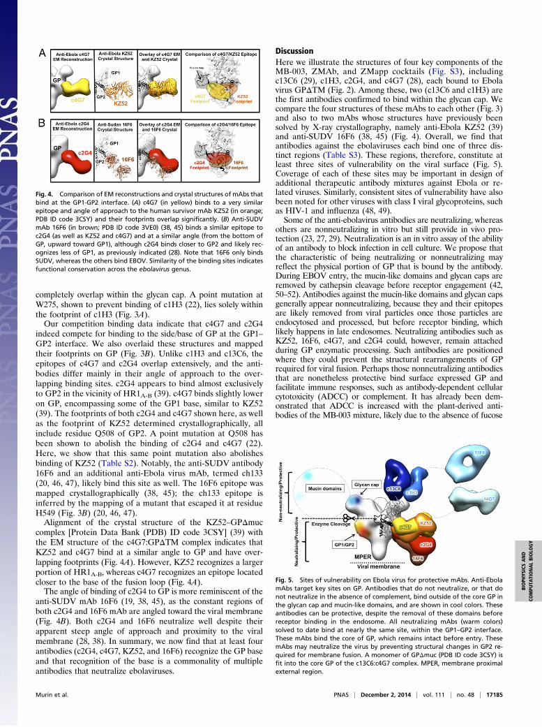
completely overlap within the glycan cap. A point mutation atW275, shown to prevent binding of c1H3 (22), lies solely withinthe footprint of c1H3 (Fig. 3A).Our competition binding data indicate that c4G7 and c2G4
indeed compete for binding to the side/base of GP at the GP1–GP2 interface. We also overlaid these structures and mappedtheir footprints on GP (Fig. 3B). Unlike c1H3 and c13C6, theepitopes of c4G7 and c2G4 overlap extensively, and the anti-bodies differ mainly in their angle of approach to the over-lapping binding sites. c2G4 appears to bind almost exclusivelyto GP2 in the vicinity of HR1A-B (39). c4G7 binds slightly loweron GP, encompassing some of the GP1 base, similar to KZ52(39). The footprints of both c2G4 and c4G7 shown here, as wellas the footprint of KZ52 determined crystallographically, allinclude residue Q508 of GP2. A point mutation at Q508 hasbeen shown to abolish the binding of c2G4 and c4G7 (22).Here, we show that this same point mutation also abolishesbinding of KZ52 (Table S2). Notably, the anti-SUDV antibody16F6 and an additional anti-Ebola virus mAb, termed ch133(20, 46, 47), likely bind this site as well. The 16F6 epitope wasmapped crystallographically (38, 45); the ch133 epitope isinferred by the mapping of a mutant that escaped it at residueH549 (Fig. 3B) (20, 46, 47).Alignment of the crystal structure of the KZ52–GPΔmuc
complex [Protein Data Bank (PDB) ID code 3CSY] (39) withthe EM structure of the c4G7:GPΔTM complex indicates thatKZ52 and c4G7 bind at a similar angle to GP and have over-lapping footprints (Fig. 4A). However, KZ52 recognizes a largerportion of HR1A-B, whereas c4G7 recognizes an epitope locatedcloser to the base of the fusion loop (Fig. 4A).The angle of binding of c2G4 to GP is more reminiscent of the
anti-SUDV mAb 16F6 (19, 38, 45), as the constant regions ofboth c2G4 and 16F6 mAb are angled toward the viral membrane(Fig. 4B). Both c2G4 and 16F6 neutralize well despite theirapparent steep angle of approach and proximity to the viralmembrane (28, 38). In summary, we now find that at least fourantibodies (c2G4, c4G7, KZ52, and 16F6) recognize the GP baseand that recognition of the base is a commonality of multipleantibodies that neutralize ebolaviruses.
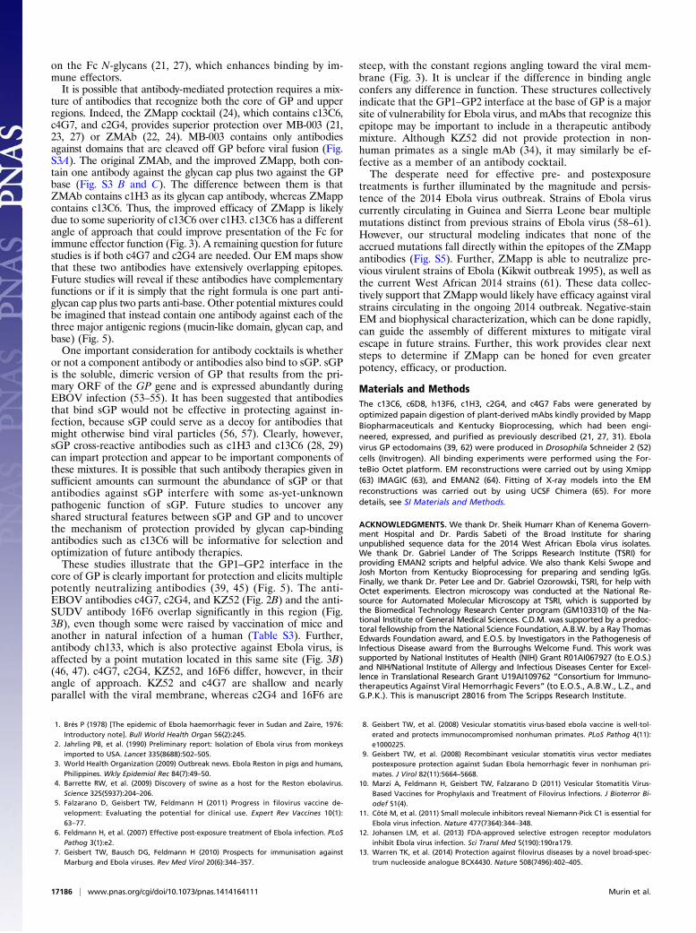
DiscussionHere we illustrate the structures of four key components of theMB-003, ZMAb, and ZMapp cocktails (Fig. S3), includingc13C6 (29), c1H3, c2G4, and c4G7 (28), each bound to Ebolavirus GPΔTM (Fig. 2). Among these, two (c13C6 and c1H3) arethe first antibodies confirmed to bind within the glycan cap. Wecompare the four structures of these mAbs to each other (Fig. 3)and also to two mAbs whose structures have previously beensolved by X-ray crystallography, namely anti-Ebola KZ52 (39)and anti-SUDV 16F6 (38, 45) (Fig. 4). Overall, we find thatantibodies against the ebolaviruses each bind one of three dis-tinct regions (Table S3). These regions, therefore, constitute atleast three sites of vulnerability on the viral surface (Fig. 5).Coverage of each of these sites may be important in design ofadditional therapeutic antibody mixtures against Ebola or re-lated viruses. Similarly, consistent sites of vulnerability have alsobeen noted for other viruses with class I viral glycoproteins, suchas HIV-1 and influenza (48, 49).Some of the anti-ebolavirus antibodies are neutralizing, whereas
others are nonneutralizing in vitro but still provide in vivo pro-tection (23, 27, 29). Neutralization is an in vitro assay of the abilityof an antibody to block infection in cell culture. We propose thatthe characteristic of being neutralizing or nonneutralizing mayreflect the physical portion of GP that is bound by the antibody.During EBOV entry, the mucin-like domains and glycan caps areremoved by cathepsin cleavage before receptor engagement (42,50–52). Antibodies against the mucin-like domains and glycan capsgenerally appear nonneutralizing, because they and their epitopesare likely removed from viral particles once those particles areendocytosed and processed, but before receptor binding, whichlikely happens in late endosomes. Neutralizing antibodies such asKZ52, 16F6, c4G7, and c2G4 could, however, remain attachedduring GP enzymatic processing. Such antibodies are positionedwhere they could prevent the structural rearrangements of GPrequired for viral fusion. Perhaps those nonneutralizing antibodiesthat are nonetheless protective bind surface expressed GP andfacilitate immune responses, such as antibody-dependent cellularcytotoxicity (ADCC) or complement. It has already been dem-onstrated that ADCC is increased with the plant-derived anti-bodies of the MB-003 mixture, likely due to the absence of fucose
Fig. 4. Comparison of EM reconstructions and crystal structures of mAbs thatbind at the GP1-GP2 interface. (A) c4G7 (in yellow) binds to a very similarepitope and angle of approach to the human survivor mAb KZ52 (in orange;PDB ID code 3CSY) and their footprints overlap significantly. (B) Anti-SUDVmAb 16F6 (in brown; PDB ID code 3VE0) (38, 45) binds a similar epitope toc2G4 (as well as KZ52 and c4G7) and at a similar angle (from the bottom ofGP, upward toward GP1), although c2G4 binds closer to GP2 and likely rec-ognizes less of GP1, as previously indicated (28). Note that 16F6 only bindsSUDV, whereas the others bind EBOV. Similarity of the binding sites indicatesfunctional conservation across the ebolavirus genus.
Fig. 5. Sites of vulnerability on Ebola virus for protective mAbs. Anti-EbolamAbs target key sites on GP. Antibodies that do not neutralize, or that donot neutralize in the absence of complement, bind outside of the core GP inthe glycan cap and mucin-like domains, and are shown in cool colors. Theseantibodies can be protective, despite the removal of these domains beforereceptor binding in the endosome. All neutralizing mAbs (warm colors)solved to date bind at nearly the same site, within the GP1–GP2 interface.These mAbs bind the core of GP, which remains intact before entry. ThesemAbs may neutralize the virus by preventing structural changes in GP2 re-quired for membrane fusion. A monomer of GPΔmuc (PDB ID code 3CSY) isfit into the core GP of the c13C6:c4G7 complex. MPER, membrane proximalexternal region.
Murin et al. PNAS | December 2, 2014 | vol. 111 | no. 48 | 17185
BIOPH
YSICSAND
COMPU
TATIONALBIOLO
GY
on the Fc N-glycans (21, 27), which enhances binding by im-mune effectors.It is possible that antibody-mediated protection requires a mix-
ture of antibodies that recognize both the core of GP and upperregions. Indeed, the ZMapp cocktail (24), which contains c13C6,c4G7, and c2G4, provides superior protection over MB-003 (21,23, 27) or ZMAb (22, 24). MB-003 contains only antibodiesagainst domains that are cleaved off GP before viral fusion (Fig.S3A). The original ZMAb, and the improved ZMapp, both con-tain one antibody against the glycan cap plus two against the GPbase (Fig. S3 B and C). The difference between them is thatZMAb contains c1H3 as its glycan cap antibody, whereas ZMappcontains c13C6. Thus, the improved efficacy of ZMapp is likelydue to some superiority of c13C6 over c1H3. c13C6 has a differentangle of approach that could improve presentation of the Fc forimmune effector function (Fig. 3). A remaining question for futurestudies is if both c4G7 and c2G4 are needed. Our EM maps showthat these two antibodies have extensively overlapping epitopes.Future studies will reveal if these antibodies have complementaryfunctions or if it is simply that the right formula is one part anti-glycan cap plus two parts anti-base. Other potential mixtures couldbe imagined that instead contain one antibody against each of thethree major antigenic regions (mucin-like domain, glycan cap, andbase) (Fig. 5).One important consideration for antibody cocktails is whether
or not a component antibody or antibodies also bind to sGP. sGPis the soluble, dimeric version of GP that results from the pri-mary ORF of the GP gene and is expressed abundantly duringEBOV infection (53–55). It has been suggested that antibodiesthat bind sGP would not be effective in protecting against in-fection, because sGP could serve as a decoy for antibodies thatmight otherwise bind viral particles (56, 57). Clearly, however,sGP cross-reactive antibodies such as c1H3 and c13C6 (28, 29)can impart protection and appear to be important components ofthese mixtures. It is possible that such antibody therapies given insufficient amounts can surmount the abundance of sGP or thatantibodies against sGP interfere with some as-yet-unknownpathogenic function of sGP. Future studies to uncover anyshared structural features between sGP and GP and to uncoverthe mechanism of protection provided by glycan cap-bindingantibodies such as c13C6 will be informative for selection andoptimization of future antibody therapies.These studies illustrate that the GP1–GP2 interface in the
core of GP is clearly important for protection and elicits multiplepotently neutralizing antibodies (39, 45) (Fig. 5). The anti-EBOV antibodies c4G7, c2G4, and KZ52 (Fig. 2B) and the anti-SUDV antibody 16F6 overlap significantly in this region (Fig.3B), even though some were raised by vaccination of mice andanother in natural infection of a human (Table S3). Further,antibody ch133, which is also protective against Ebola virus, isaffected by a point mutation located in this same site (Fig. 3B)(46, 47). c4G7, c2G4, KZ52, and 16F6 differ, however, in theirangle of approach. KZ52 and c4G7 are shallow and nearlyparallel with the viral membrane, whereas c2G4 and 16F6 are
steep, with the constant regions angling toward the viral mem-brane (Fig. 3). It is unclear if the difference in binding angleconfers any difference in function. These structures collectivelyindicate that the GP1–GP2 interface at the base of GP is a majorsite of vulnerability for Ebola virus, and mAbs that recognize thisepitope may be important to include in a therapeutic antibodymixture. Although KZ52 did not provide protection in non-human primates as a single mAb (34), it may similarly be ef-fective as a member of an antibody cocktail.The desperate need for effective pre- and postexposure
treatments is further illuminated by the magnitude and persis-tence of the 2014 Ebola virus outbreak. Strains of Ebola viruscurrently circulating in Guinea and Sierra Leone bear multiplemutations distinct from previous strains of Ebola virus (58–61).However, our structural modeling indicates that none of theaccrued mutations fall directly within the epitopes of the ZMappantibodies (Fig. S5). Further, ZMapp is able to neutralize pre-vious virulent strains of Ebola (Kikwit outbreak 1995), as well asthe current West African 2014 strains (61). These data collec-tively support that ZMapp would likely have efficacy against viralstrains circulating in the ongoing 2014 outbreak. Negative-stainEM and biophysical characterization, which can be done rapidly,can guide the assembly of different mixtures to mitigate viralescape in future strains. Further, this work provides clear nextsteps to determine if ZMapp can be honed for even greaterpotency, efficacy, or production.
Materials and MethodsThe c13C6, c6D8, h13F6, c1H3, c2G4, and c4G7 Fabs were generated byoptimized papain digestion of plant-derived mAbs kindly provided by MappBiopharmaceuticals and Kentucky Bioprocessing, which had been engi-neered, expressed, and purified as previously described (21, 27, 31). Ebolavirus GP ectodomains (39, 62) were produced in Drosophila Schneider 2 (S2)cells (Invitrogen). All binding experiments were performed using the For-teBio Octet platform. EM reconstructions were carried out by using Xmipp(63) IMAGIC (63), and EMAN2 (64). Fitting of X-ray models into the EMreconstructions was carried out by using UCSF Chimera (65). For moredetails, see SI Materials and Methods.
ACKNOWLEDGMENTS. We thank Dr. Sheik Humarr Khan of Kenema Govern-ment Hospital and Dr. Pardis Sabeti of the Broad Institute for sharingunpublished sequence data for the 2014 West African Ebola virus isolates.We thank Dr. Gabriel Lander of The Scripps Research Institute (TSRI) forproviding EMAN2 scripts and helpful advice. We also thank Kelsi Swope andJosh Morton from Kentucky Bioprocessing for preparing and sending IgGs.Finally, we thank Dr. Peter Lee and Dr. Gabriel Ozorowski, TSRI, for help withOctet experiments. Electron microscopy was conducted at the National Re-source for Automated Molecular Microscopy at TSRI, which is supported bythe Biomedical Technology Research Center program (GM103310) of the Na-tional Institute of General Medical Sciences. C.D.M. was supported by a predoc-toral fellowship from the National Science Foundation, A.B.W. by a Ray ThomasEdwards Foundation award, and E.O.S. by Investigators in the Pathogenesis ofInfectious Disease award from the Burroughs Welcome Fund. This work wassupported by National Institutes of Health (NIH) Grant R01AI067927 (to E.O.S.)and NIH/National Institute of Allergy and Infectious Diseases Center for Excel-lence in Translational Research Grant U19AI109762 “Consortium for Immuno-therapeutics Against Viral Hemorrhagic Fevers” (to E.O.S., A.B.W., L.Z., andG.P.K.). This is manuscript 28016 from The Scripps Research Institute.
1. Brès P (1978) [The epidemic of Ebola haemorrhagic fever in Sudan and Zaire, 1976:Introductory note]. Bull World Health Organ 56(2):245.
2. Jahrling PB, et al. (1990) Preliminary report: Isolation of Ebola virus from monkeysimported to USA. Lancet 335(8688):502–505.
3. World Health Organization (2009) Outbreak news. Ebola Reston in pigs and humans,Philippines. Wkly Epidemiol Rec 84(7):49–50.
4. Barrette RW, et al. (2009) Discovery of swine as a host for the Reston ebolavirus.Science 325(5937):204–206.
5. Falzarano D, Geisbert TW, Feldmann H (2011) Progress in filovirus vaccine de-velopment: Evaluating the potential for clinical use. Expert Rev Vaccines 10(1):63–77.
6. Feldmann H, et al. (2007) Effective post-exposure treatment of Ebola infection. PLoSPathog 3(1):e2.
7. Geisbert TW, Bausch DG, Feldmann H (2010) Prospects for immunisation againstMarburg and Ebola viruses. Rev Med Virol 20(6):344–357.
8. Geisbert TW, et al. (2008) Vesicular stomatitis virus-based ebola vaccine is well-tol-erated and protects immunocompromised nonhuman primates. PLoS Pathog 4(11):e1000225.
9. Geisbert TW, et al. (2008) Recombinant vesicular stomatitis virus vector mediatespostexposure protection against Sudan Ebola hemorrhagic fever in nonhuman pri-mates. J Virol 82(11):5664–5668.
10. Marzi A, Feldmann H, Geisbert TW, Falzarano D (2011) Vesicular Stomatitis Virus-Based Vaccines for Prophylaxis and Treatment of Filovirus Infections. J Bioterror Bi-odef S1(4).
11. Côté M, et al. (2011) Small molecule inhibitors reveal Niemann-Pick C1 is essential forEbola virus infection. Nature 477(7364):344–348.
12. Johansen LM, et al. (2013) FDA-approved selective estrogen receptor modulatorsinhibit Ebola virus infection. Sci Transl Med 5(190):190ra179.
13. Warren TK, et al. (2014) Protection against filovirus diseases by a novel broad-spec-trum nucleoside analogue BCX4430. Nature 508(7496):402–405.
17186 | www.pnas.org/cgi/doi/10.1073/pnas.1414164111 Murin et al.
14. Geisbert TW, et al. (2006) Postexposure protection of guinea pigs against a lethalebola virus challenge is conferred by RNA interference. J Infect Dis 193(12):1650–1657.
15. Geisbert TW, et al. (2010) Postexposure protection of non-human primates againsta lethal Ebola virus challenge with RNA interference: A proof-of-concept study.Lancet 375(9729):1896–1905.
16. Saphire EO (2013) An update on the use of antibodies against the filoviruses. Im-munotherapy 5(11):1221–1233.
17. Wong G, Qiu X, Olinger GG, Kobinger GP (2014) Post-exposure therapy of filovirusinfections. Trends Microbiol 22(8):456–463.
18. Qiu X, Kobinger GP (2014) Antibody therapy for Ebola: Is the tide turning around?Hum Vaccin Immunother 10(4):964–967.
19. Dye JM, et al. (2012) Postexposure antibody prophylaxis protects nonhuman primatesfrom filovirus disease. Proc Natl Acad Sci USA 109(13):5034–5039.
20. Marzi A, et al. (2012) Protective efficacy of neutralizing monoclonal antibodies ina nonhuman primate model of Ebola hemorrhagic fever. PLoS ONE 7(4):e36192.
21. Zeitlin L, et al. (2011) Enhanced potency of a fucose-free monoclonal antibody beingdeveloped as an Ebola virus immunoprotectant. Proc Natl Acad Sci USA 108(51):20690–20694.
22. Qiu X, et al. (2012) Successful treatment of ebola virus-infected cynomolgus macaqueswith monoclonal antibodies. Sci Transl Med 4(138):138ra181.
23. Pettitt J, et al. (2013) Therapeutic intervention of Ebola virus infection in rhesusmacaques with the MB-003 monoclonal antibody cocktail. Sci Transl Med 5(199):ra113.
24. Qiu X, et al. (2014) Reversion of advanced Ebola virus disease in nonhuman primateswith ZMapp. Nature 514(7520):47–53.
25. Hart MK, Wilson JA, Schmaljohn AL (2003) US Patent 6,630,144 B1.26. Qiu X, et al. (2012) Ebola GP-specific monoclonal antibodies protect mice and guinea
pigs from lethal Ebola virus infection. PLoS Negl Trop Dis 6(3):e1575.27. Olinger GG, Jr, et al. (2012) Delayed treatment of Ebola virus infection with plant-
derived monoclonal antibodies provides protection in rhesus macaques. Proc NatlAcad Sci USA 109(44):18030–18035.
28. Qiu X, et al. (2011) Characterization of Zaire ebolavirus glycoprotein-specific mono-clonal antibodies. Clin Immunol 141(2):218–227.
29. Wilson JA, et al. (2000) Epitopes involved in antibody-mediated protection fromEbola virus. Science 287(5458):1664–1666.
30. Giritch A, et al. (2006) Rapid high-yield expression of full-size IgG antibodies in plantscoinfected with noncompeting viral vectors. Proc Natl Acad Sci USA 103(40):14701–14706.
31. Strasser R, et al. (2008) Generation of glyco-engineered Nicotiana benthamiana forthe production of monoclonal antibodies with a homogeneous human-like N-glycanstructure. Plant Biotechnol J 6(4):392–402.
32. Maruyama T, et al. (1999) Ebola virus can be effectively neutralized by antibodyproduced in natural human infection. J Virol 73(7):6024–6030.
33. Parren PW, Geisbert TW, Maruyama T, Jahrling PB, Burton DR (2002) Pre- and post-exposure prophylaxis of Ebola virus infection in an animal model by passive transferof a neutralizing human antibody. J Virol 76(12):6408–6412.
34. Oswald WB, et al. (2007) Neutralizing antibody fails to impact the course of Ebolavirus infection in monkeys. PLoS Pathog 3(1):e9.
35. Marzi A, et al. (2013) Antibodies are necessary for rVSV/ZEBOV-GP-mediated pro-tection against lethal Ebola virus challenge in nonhuman primates. Proc Natl Acad SciUSA 110(5):1893–1898.
36. Audet J, et al. (2014) Molecular characterization of the monoclonal antibodies com-posing ZMAb: A protective cocktail against Ebola virus. Sci Rep 4:6881.
37. Kajihara M, et al. (2012) Inhibition of Marburg virus budding by nonneutralizingantibodies to the envelope glycoprotein. J Virol 86(24):13467–13474.
38. Dias JM, et al. (2011) A shared structural solution for neutralizing ebolaviruses. NatStruct Mol Biol 18(12):1424–1427.
39. Lee JE, et al. (2008) Structure of the Ebola virus glycoprotein bound to an antibodyfrom a human survivor. Nature 454(7201):177–182.
40. Lee JE, et al. (2008) Complex of a protective antibody with its Ebola virus GP peptideepitope: Unusual features of a V lambda x light chain. J Mol Biol 375(1):202–216.
41. Tran EE, et al. (2014) Spatial localization of the Ebola virus glycoprotein mucin-likedomain determined by cryo-electron tomography. J Virol 88(18):10958–10962.
42. Lee JE, Saphire EO (2009) Ebolavirus glycoprotein structure and mechanism of entry.Future Virol 4(6):621–635.
43. Martinez O, Tantral L, Mulherkar N, Chandran K, Basler CF (2011) Impact of Ebolamucin-like domain on antiglycoprotein antibody responses induced by Ebola virus-like particles. J Infect Dis 204(Suppl 3):S825–S832.
44. Olal D, et al. (2012) Structure of an antibody in complex with its mucin domain linearepitope that is protective against Ebola virus. J Virol 86(5):2809–2816.
45. Bale S, et al. (2012) Structural basis for differential neutralization of ebolaviruses.Viruses 4(4):447–470.
46. Takada A, Ebihara H, Jones S, Feldmann H, Kawaoka Y (2007) Protective efficacy ofneutralizing antibodies against Ebola virus infection. Vaccine 25(6):993–999.
47. Takada A, et al. (2003) Identification of protective epitopes on ebola virus glyco-protein at the single amino acid level by using recombinant vesicular stomatitis vi-ruses. J Virol 77(2):1069–1074.
48. Burton DR, Poignard P, Stanfield RL, Wilson IA (2012) Broadly neutralizing antibodiespresent new prospects to counter highly antigenically diverse viruses. Science337(6091):183–186.
49. Julien JP, Lee PS, Wilson IA (2012) Structural insights into key sites of vulnerability onHIV-1 Env and influenza HA. Immunol Rev 250(1):180–198.
50. Bale S, et al. (2011) Ebola virus glycoprotein needs an additional trigger, beyondproteolytic priming for membrane fusion. PLoS Negl Trop Dis 5(11):e1395.
51. Brecher M, et al. (2012) Cathepsin cleavage potentiates the Ebola virus glycoproteinto undergo a subsequent fusion-relevant conformational change. J Virol 86(1):364–372.
52. Chandran K, Sullivan NJ, Felbor U, Whelan SP, Cunningham JM (2005) Endosomalproteolysis of the Ebola virus glycoprotein is necessary for infection. Science308(5728):1643–1645.
53. Barrientos LG, Martin AM, Rollin PE, Sanchez A (2004) Disulfide bond assignment ofthe Ebola virus secreted glycoprotein SGP. Biochem Biophys Res Commun 323(2):696–702.
54. Sanchez A, et al. (1998) Biochemical analysis of the secreted and virion glycoproteinsof Ebola virus. J Virol 72(8):6442–6447.
55. Volchkova VA, Feldmann H, Klenk HD, Volchkov VE (1998) The nonstructural smallglycoprotein sGP of Ebola virus is secreted as an antiparallel-orientated homodimer.Virology 250(2):408–414.
56. Basler CF (2013) A novel mechanism of immune evasion mediated by Ebola virussoluble glycoprotein. Expert Rev Anti Infect Ther 11(5):475–478.
57. Mohan GS, Li W, Ye L, Compans RW, Yang C (2012) Antigenic subversion: A novelmechanism of host immune evasion by Ebola virus. PLoS Pathog 8(12):e1003065.
58. Baize S, et al. (2014) Emergence of Zaire Ebola virus disease in Guinea. N Engl J Med371(15):1418–1425.
59. Dudas G, Rambaut A (2014) Phylogenetic analysis of Guinea 2014 EBOV Ebolavirusoutbreak. PLoS Curr 6:6.
60. Calvignac-Spencer S, Schulze JM, Zickmann F, Renard BY (2014) Clock rooting furtherdemonstrates that Guinea 2014 EBOV is a member of the Zaïre lineage. PLoS Curr 6:6.
61. Gire SK, et al. (2014) Genomic surveillance elucidates Ebola virus origin and trans-mission during the 2014 outbreak. Science 345(6202):1369–1372.
62. Lee JE, et al. (2009) Techniques and tactics used in determining the structure of thetrimeric ebolavirus glycoprotein. Acta Crystallogr D Biol Crystallogr 65(Pt 11):1162–1180.
63. van Heel M, Harauz G, Orlova EV, Schmidt R, Schatz M (1996) A new generation ofthe IMAGIC image processing system. J Struct Biol 116(1):17–24.
64. Tang G, et al. (2007) EMAN2: An extensible image processing suite for electron mi-croscopy. J Struct Biol 157(1):38–46.
65. Pettersen EF, et al. (2004) UCSF Chimera—A visualization system for exploratory re-search and analysis. J Comput Chem 25(13):1605–1612.
Murin et al. PNAS | December 2, 2014 | vol. 111 | no. 48 | 17187
BIOPH
YSICSAND
COMPU
TATIONALBIOLO
GY