Embed Size (px)
Citation preview
research papers
784 doi:10.1107/S0907444912011912 Acta Cryst. (2012). D68, 784–793
Acta Crystallographica Section D
BiologicalCrystallography
ISSN 0907-4449
Structures of Staphylococcus aureus peptidedeformylase in complex with two classes of newinhibitors
Sang Jae Lee,a,b Seung-Jae Lee,c
Seung Kyu Lee,d Hye-Jin Yoon,b
Hyung Ho Lee,e Kyeong Kyu
Kim,f Bong Jin Lee,d,g* Byung Il
Leea* and Se Won Suhb,h*
aBiomolecular Function Research Branch,
Division of Convergence Technology, Research
Institute, National Cancer Center, Goyang,
Gyeonggi 410-749, Republic of Korea,bDepartment of Chemistry, College of Natural
Sciences, Seoul National University,
Seoul 151-742, Republic of Korea, cHoward
Hughes Medical Institute and W. M. Keck
Structural Biology Laboratory, Cold Spring
Harbor Laboratory, 1 Bungtown Road, Cold
Spring Harbor, NY 11724, USA, dResearch
Institute of Pharmaceutical Sciences, College of
Pharmacy, Seoul National University,
Seoul 151-742, Republic of Korea,eDepartment of Chemistry, Kookmin University,
861-1 Jeongneung, Seongbuk, Seoul 136-702,
Republic of Korea, fDepartment of Molecular
Cell Biology, Sungkyunkwan University of
Medicine, Suwon, Gyeonggi 440-746,
Republic of Korea, gProMediTech Ltd,
College of Pharmacy, Seoul National University,
Seoul 151-742, Republic of Korea, andhDepartment of Biophysics and Chemical
Biology, College of Natural Sciences,
Seoul National University, Seoul 151-742,
Republic of Korea
Correspondence e-mail: [email protected],
[email protected], [email protected]
# 2012 International Union of Crystallography
Printed in Singapore – all rights reserved
Peptide deformylase (PDF) catalyzes the removal of the
formyl group from the N-terminal methionine residue in
newly synthesized polypeptides, which is an essential process
in bacteria. Four new inhibitors of PDF that belong to two
different classes, hydroxamate/pseudopeptide compounds
[PMT387 (7a) and PMT497] and reverse-hydroxamate/non-
peptide compounds [PMT1039 (15e) and PMT1067], have
been developed. These compounds inhibited the growth of
several pathogens involved in respiratory-tract infections,
such as Streptococcus pneumoniae, Moraxella catarrhalis and
Haemophilus influenzae, and leading nosocomial pathogens
such as Staphylococcus aureus and Klebsiella pneumoniae
with a minimum inhibitory concentration (MIC) in the range
0.1–0.8 mg ml�1. Interestingly, the reverse-hydroxamate/non-
peptide compounds showed a 250-fold higher antimicrobial
activity towards S. aureus, although the four compounds
showed similar Ki values against S. aureus PDF enzymes, with
Ki values in the 11–85 nM range. To provide a structural basis
for the discovery of additional PDF inhibitors, the crystal
structures of S. aureus PDF in complex with the four inhibitors
were determined at resolutions of 1.90–2.30 A. The inhibitor-
bound structures displayed distinct deviations depending on
the inhibitor class. The distance between the Zn2+ ion and
the carbonyl O atom of the hydroxamate inhibitors (or the
hydroxyl O atom of the reverse-hydroxamate inhibitors)
appears to be correlated to S. aureus inhibition activity. The
structural information reported in this study should aid in the
discovery of new PDF inhibitors that can be used as novel
antibacterial drugs.
Received 25 November 2011
Accepted 19 March 2012
PDB References: peptide
deformylase, complex with
PMT387, 3u7k; complex with
PMT497, 3u7l; complex with
PMT1039, 3u7m; complex
with PMT1067, 3u7n.
1. Introduction
Bacterial infections are one of the leading causes of human
death in the world (Boucher et al., 2009; World Health
Organization, 2004). Furthermore, the development of new
antibacterial drugs is lagging behind the increasing rate of
antibiotic resistance, which is becoming a major threat to
human health (Boucher et al., 2009; Spellberg et al., 2004).
Many currently used antibiotics will eventually become in-
effective because of the acquisition of resistance mechanisms
by pathogenic bacteria (Aubart & Zalacain, 2006; Boucher et
al., 2009). Therefore, there is an urgent need for the discovery
of new antibacterial targets and the development of novel
antibacterial agents. Peptide deformylase (PDF), a metallo-
enzyme that is highly conserved in bacteria, has been
proposed as one attractive such target (Meinnel et al., 1993;
Solbiati et al., 1999). It catalyzes the removal of a formyl group
from the N-terminal methionine residue of newly synthesized
polypeptides. Removal of the N-formyl group from nascent
polypeptide chains is an essential process in bacteria (Guillon
et al., 1992; Mazel et al., 1994).
Bacterial PDFs are classified into two types. Type I PDFs
are found in Gram-negative and Gram-positive bacteria, while
type II PDFs are only found in Gram-positive bacteria
(Guilloteau et al., 2002). The sequence identity between type I
and type II PDFs is relatively low (27–40%). Although the
sequence identity ranges from 52 to 61% among PDFs of the
same type, the C-terminal regions are markedly different
despite similarity in the structures of type I and type II PDFs.
In type I PDFs the C-terminal region consists of �-helices,
whereas in type II PDFs the C-terminal region is comprised of
�-strands that fold back onto themselves to form �-sheets. In
addition, type I and type II PDFs share structurally conserved
active sites that contain a metal ion. Crystal structures of
inhibitor-bound PDFs are available for type I and type II
enzymes. Type I PDFs from Escherichia coli (Clements et al.,
2001; Guilloteau et al., 2002; Smith et al., 2003), Pseudomonas
aeruginosa (Guilloteau et al., 2002; Molteni et al., 2004; Yoon
et al., 2004), Bacillus cereus (Moon et al., 2005), Helicobacter
pylori (Cai et al., 2006) and Mycobacterium tuberculosis
(Pichota et al., 2008) have been structurally characterized. The
crystal structures of type II PDFs from B. stearothermophilus
(Guilloteau et al., 2002), Streptococcus pneumoniae (Smith
et al., 2003) and S. aureus (Yoon et al., 2004) have also been
reported. The structure of the Leptospira interrogans PDF,
which is not classified as either type I or type II, has also been
determined (Zhou et al., 2004). Plasmodium falciparum is a
unicellular eukaryotic protozoan that is a major human
pathogen and the causative agent of the most deadly form of
malaria; a crystal structure of its type I PDF has been reported
(Robien et al., 2004).
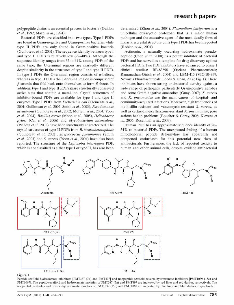
Actinonin, a naturally occurring hydroxamate pseudo-
peptide (Chen et al., 2000), is a potent inhibitor of bacterial
PDFs and has served as a template for drug discovery against
bacterial PDFs. Two PDF inhibitors have advanced to phase I
clinical studies: BB-83698 (Oscient Pharmaceuticals;
Ramanathan-Girish et al., 2004) and LBM-415 (VIC-104959;
Novartis Pharmaceuticals; Leeds & Dean, 2006; Fig. 1). These
inhibitors have shown strong antibacterial activity against a
wide range of pathogens, particularly Gram-positive aerobes
and some Gram-negative anaerobes (Guay, 2007). S. aureus
and K. pneumoniae are the main causes of hospital- and
community-acquired infections. Moreover, high frequencies of
methicillin-resistant and vancomycin-resistant S. aureus, as
well as ceftazidime/ceftriaxone-resistant K. pneumoniae, pose
serious health problems (Boucher & Corey, 2008; Klevens et
al., 2006; Rosenthal et al., 2009).
Human PDF has an approximate sequence identity of 28–
34% to bacterial PDFs. The unexpected finding of a human
mitochondrial peptide deformylase has apparently not
dampened enthusiasm for this potential new class of
antibacterials. Furthermore, the lack of reported toxicity to
human and other animal cells, despite evident antibacterial
research papers
Acta Cryst. (2012). D68, 784–793 Lee et al. � Peptide deformylase 785
Figure 1Peptide-scaffold hydroxamate inhibitors [PMT387 (7a) and PMT497] and nonpeptide-scaffold reverse-hydroxamate inhibitors [PMT1039 (15e) andPMT1067]. The peptide-scaffold and hydroxamate moieties of PMT387 (7a) and PMT497 are indicated by red lines and red dashes, respectively. Thenonpeptide scaffolds and reverse-hydroxamate moieties of PMT1039 (15e) and PMT1067 are indicated by blue lines and blue dashes, respectively.
action, has indicated that the human mitochondrial PDF
protein may not be functional or that the tested inhibitors may
not reach the mitochondrion (Nguyen et al., 2003; Robien et
al., 2004). The inhibitors of human PDF have been reported to
be potent anticancer drugs that promote cell death or prolif-
eration arrest in a wide variety of cancer cell lines (Grujic et
al., 2005; Lee et al., 2004; Xu et al., 1998). Interestingly, it has
been reported that actinonin prevents bacterial survival and
has a considerable influence on innate immune reactions in
humans (Fu et al., 2006; Mader et al., 2010). Such pro-
inflammatory consequences can be beneficial for the host and
serve to clear localized infections (Mader et al., 2010). More-
over, all of the resistant mutants have a slow-growth pheno-
type similar to that of the the fmt/def double-knockout
mutants, which are less virulent than the PDF-inhibitor-
sensitive parental strains (Apfel et al., 2000; Margolis et al.,
2000; Watters et al., 2006). These studies show the considerable
promise of bacterial PDF as a novel antibacterial drug
candidate.
Presently, only one crystal structure of inhibitor-bound
S. aureus PDF complexed with actinonin is available (Yoon et
al., 2004). In this study, we have performed structural analysis
of S. aureus PDF complexed with the hydroxamate/pseudo-
peptide inhibitors PMT387 (7a) and PMT497 and the reverse-
hydroxamate/nonpeptide scaffold compounds PMT1039 (15e)
and PMT1067 at resolutions of 1.90–2.30 A, which provided a
detailed atomic level view of the binding modes of the PDF
inhibitors (Fig. 1). These PDF inhibitors were highly effective
against infectious respiratory-tract pathogens, including
S. pneumoniae, M. catarrhalis and H. influenza, as well as
leading nosocomial pathogens such as S. aureus and
K. pneumoniae. Of these inhibitors, PMT1039 (15e) and
PMT1067, with nonpeptide scaffolds, were highly effective in
inhibiting the S. aureus and K. pneumoniae PDFs.
When we compared and analyzed the four new inhibitor-
bound PDF structures, distinct structural changes that were
dependent on the inhibitor class were observed. The structural
changes appear to be driven by the binding of the inhibitor
and are likely to be involved in inhibition, particularly of
S. aureus.
2. Materials and methods
2.1. Measurement of PDF activity
The PDF activity of S. aureus PDF was determined.
Recombinant S. aureus PDF with a C-terminal hexahistidine-
containing tag was expressed and purified. The purification
procedures were identical to those used for the production of
crystals except for the cell-disruption procedure. The wet cells
were homogenized with a Dounce glass homogenizer (100 ml
size) on ice instead of sonication. For the assay, 50 ml of a
reaction mixture consisting of 50 mM HEPES pH 7.0, 10 mM
NaCl, 0.1% Triton X-100 was used. Assays were performed for
10 min at 303 K with an inhibitor concentration in the range
0.001–100 mM and 500 nM S. aureus PDF. The reaction was
initiated by the addition of substrate (50 ml 5 mM formyl-Met-
Ala-Ser; Bachem). The formation of the product was detected
using fluorescamine. For the assay, 50 ml 150 mg ml�1 fluor-
escamine and 50 ml 50 mM borate–sodium hydroxide buffer
pH 9.5 were added. The fluorescence was quantified with a
Gemini EM Spectrofluorometer Microplate Reader (Mole-
cular Devices) using an excitation wavelength of 390 nm and
an emission wavelength of 465 nm. The Ki values of the
inhibitors were calculated using GraphPad Prism (GraphPad
Software). Protein concentrations were determined using the
Bradford method with bovine serum albumin as a standard
(Bradford, 1976). Enzymatic assay results are presented in
Table 1.
2.2. Bacteria, reagents and miminal inhibitory concentration(MIC) tests
S. aureus (ATCC 6538p), K. pneumoniae (ATCC 10031),
E. coli (ATCC 25922), S. pneumoniae (ATCC 6305 and ATCC
49619), M. catarrhalis (ATCC 43617), H. influenzae (ATCC
51907) and P. aeruginosa (ATCC 27853) were tested using in
vitro susceptibility tests (MIC). S. aureus, K. pneumoniae,
E. coli and P. aeruginosa were cultured in Mueller Hinton
broth (Oxoid) at 310 K for 24 h. S. pneumoniae was cultured
in tryptic soy broth (Difco) with 5% sheep blood at 310 K for
48 h in a 5% CO2 incubator. M. catarrhalis and H. influenzae
were grown in brain–heart infusion broth (Difco) supple-
research papers
786 Lee et al. � Peptide deformylase Acta Cryst. (2012). D68, 784–793
Table 1Results of enzymatic assays and MICs.
MIC (mg ml�1)
S. aureus(ATCC 6538p)
K. pneumoniae(ATCC 10031)
E. coli(ATCC 25922)
S. pneumoniae(ATCC 6305)
M. catarrhalis(ATCC 43617)
H. influenzae(ATCC 49247)
P. aeruginosa(ATCC 27853)
Inhibitor Ki (nM) Gram-positive Gram-negative Gram-negative Gram-positive Gram-negative Gram-negative Gram-negative
PMT387 (7a)† 84.9 � 12.8 50 0.4–0.8 25 0.2‡ 0.1‡ 0.2‡ 100PMT497 52.4 � 6.5 50 6.3 25 0.8 0.1 0.1 200PMT1039 (15e)† 35.1 � 3.1 0.2‡ 0.1 12.5 0.1–0.2‡§ 0.1‡ 0.1‡ 200PMT1067 11.2 � 1.6 0.2 0.4 12.5 0.1‡ 0.4 0.1 200Actinonin 319.2 � 39.3 6.3–12.5 0.8–3.2 25–50 3.2–6.3 0.1–0.2 1.6–3.2 100–200Ampicillin No data 0.2 25–100 1.6 0.1 0.1–0.4 3.2 200LBM-415} No data 2 32 32 1 0.5 4–8 N.A.
† The nomenclature in parentheses was used in previous reports (Lee et al., 2010, 2011). ‡ The results of antibacterial inhibition assays have been described in previous reports (Lee etal., 2010, 2011). § Antibacterial inhibition assays were performed on both strains of S. pneumoniae (ATCC 6305 and ATCC 49619). } The inhibition-assay data were taken fromCredito et al. (2004), Fritsche et al. (2005) and Watters et al. (2006).
mented with 2.5% horse serum, 10 mg NAD+ and 5 mg
haemin per litre for 24 and 48 h, respectively, in a 5% CO2
incubator. All bacterial strains were obtained from the
American Type Culture Collection. Ampicillin, an anti-
bacterial agent used as a control, was purchased from Sigma–
Aldrich (USA). The inhibitor compounds [PMT387 (7a),
PMT497, PMT1039 (15e), PMT1067 and actinonin] were
synthesized as described previously (Lee et al., 2010, 2011).
Other compounds were obtained from their respective
manufacturers. MIC tests were performed using the Clinical
and Laboratory Standards Institute (CLSI, formerly NCCLS)
broth microplate method (National Committee for Clinical
Laboratory Standards, 2003), with a starting inoculum of
approximately 106 CFU ml�1 for all isolates. The MIC was
defined as the lowest concentration of antimicrobial agent that
inhibited visible growth. The results of the MIC tests are
summarized in Table 1.
2.3. Protein purification and crystallization
The def (SACOL1100) gene from S. aureus COL was
amplified by polymerase chain reaction using the genomic
DNA as the template. The forward and reverse oligonucleo-
tide primers were designed using the published genome
sequence (Gill et al., 2005) and were 50-G GAA TTC CAT
ATG TTA ACA ATG AAA GAC ATC ATT AGC G-30 and
50-CCG CCG CTC GAG AAC TTC TAC TGC ATC TGT
ATG TGG-30, respectively. The sequences in bold are NdeI
and XhoI restriction-enzyme sites, respectively. The recom-
binant protein sequence contained an eight-residue tag
(LEHHHHHH) that was added to the carboxy-terminus of
the recombinant protein. The gene was cloned into the pET-
21a(+) expression vector (Novagen). Recombinant protein
with a C-terminal hexahistidine-containing tag was over-
expressed in E. coli C41 (DE3) cells in Terrific Broth. Protein
expression was induced by the addition of 0.5 mM isopropyl
�-d-1-thiogalactopyranoside and the cells were incubated for
an additional 20 h at 303 K following growth to mid-log phase
at 310 K. The cells were lysed by sonication in buffer A
(20 mM Tris–HCl pH 7.9, 50 mM imidazole, 500 mM NaCl)
containing 10%(v/v) glycerol. The crude lysate was centri-
fuged at approximately 36 000g for 60 min. The supernatant
was applied onto a HiTrap Chelating HP affinity chromato-
graphy column (GE Healthcare) which was equilibrated with
buffer A. After elution with a gradient of imidazole in buffer
A, the protein was further purified by gel filtration on a
Superdex 200 prep-grade column (GE Healthcare) which was
equilibrated with 20 mM Tris–HCl buffer pH 7.5 containing
research papers
Acta Cryst. (2012). D68, 784–793 Lee et al. � Peptide deformylase 787
Table 2Data-collection and model-refinement statistics.
Values in parentheses are for the highest resolution shell.
Data set PMT387 (7a)† PMT497 PMT1039 (15e)† PMT1067
Data collectionX-ray wavelength (A) 0.9722 0.9722 1.0000 1.0000Space group C2221 C2221 C2221 C2221
Unit-cell parameters (A)a 93.77 94.78 94.58 93.75b 120.27 120.21 119.70 120.65c 47.75 47.79 47.27 47.56
Resolution range (A) 20–1.9 (1.97–1.90) 50–2.0 (2.07–2.0) 20–2.15 (2.23–2.15) 20–2.3 (2.38–2.30)Total/unique reflections 127541/21933 96279/18445 110518/14864 67943/12218Completeness (%) 99.5 (99.9) 99.7 (100) 99.4 (94.3) 98.6 (96.0)hI/�(I)i 22.2 (4.0) 20.9 (4.3) 53.4 (13.9) 30.6 (7.1)Rmerge‡ 0.167 (0.496) 0.116 (0.469) 0.055 (0.164) 0.090 (0.190)
Model refinementRwork/Rfree§ 0.216/0.266 0.208/0.235 0.196/0.223 0.195/0.228No. of non-H atoms/average B factor (A2)
Protein atoms 1462/16.6 1462 22.0 1462/27.1 1462/32.4Water O atoms 202/28.5 121/29.3 113/35.0 94/37.0Inhibitor 31/23.2 31/32.9 27/57.9 27/58.8
Wilson B factor (A2) 26.2 31.2 39.4 46.3R.m.s.d.s from ideal geometry
Bond lengths (A) 0.011 0.010 0.009 0.010Bond angles (�) 1.33 1.36 1.37 1.23
R.m.s. Z scores}Bond lengths 0.452 0.421 0.379 0.427Bond angles 0.562 0.554 0.563 0.524
Ramachandran plot†† (%)Favoured 99.5 100.0 98.9 99.5Outliers 0.0 0.0 0.0 0.0
Rotamer outliers†† (%) 0.63 0.63 0.63 0.0
† The nomenclature in parentheses was used in previous reports (Lee et al., 2010, 2011). ‡ Rmerge =P
hkl
Pi jIiðhklÞ � hIðhklÞij=
Phkl
Pi IiðhklÞ, where I(hkl) is the intensity of
reflection hkl,P
hkl is the sum over all reflections andP
i is the sum over i measurements of reflection hkl. § R =P
hkl
��jFobsj � jFcalcj
��=P
hkl jFobsj; Rfree is calculated for a randomlyselected set of 5% of the reflections which were not used for structure refinement and Rwork is calculated for the remaining reflections. } Values obtained using REFMAC (Murshudovet al., 2011). †† Values obtained using MolProbity (Chen et al., 2010).
120 mM NaCl. For crystallization, the fractions containing
S. aureus PDF were concentrated to 30 mg ml�1 using an
Amicon Ultra-15 centrifugal filter unit (Millipore).
To grow crystals of the protein–inhibitor complexes, the
protein solution was incubated at 297 K for 1 h with the
inhibitors, which were dissolved in a 6.6-fold molar excess of
dimethyl sulfoxide. The crystals were grown by the hanging-
drop vapour-diffusion method at 297 K by mixing equal
volumes (2 ml each) of protein solution (30 mg ml�1 concen-
tration in 20 mM Tris–HCl buffer pH 7.5 containing 120 mM
NaCl) and reservoir solution consisting of 23%(w/v) PEG
4000, 50 mM Tris–HCl pH 8.5, 15%(v/v) glycerol, 100 mM
MgCl2, 20 mM CaCl2. The crystals grew to approximate
dimensions of 0.04 � 0.02 � 0.4 mm within one week.
2.4. X-ray data collection, structure determination andrefinement
X-ray diffraction data were collected at 100 K using an
ADSC Quantum 4R CCD detector on beamline BL-38B1 at
SPring-8 (� = 0.9722 A), Japan or an ADSC Quantum 315
CCD detector system on beamline BL-5A at Photon Factory
(� = 1.0000 A), Japan. The crystal was rotated by 1� for each
image and the raw data were processed and scaled using the
HKL-2000 program suite (Otwinowski & Minor, 1997). The
crystals belonged to the orthorhombic space group C2221.
One monomer was present in each asymmetric unit of the
crystal.
The structures of S. aureus PDF in complex with four
inhibitors were determined by molecular replacement using
the program MOLREP (Vagin & Teplyakov, 2010). A model
of S. aureus PDF (Yoon et al., 2004) was used as a search
model. In addition, 5% of the data were randomly set aside as
a test set for calculating Rfree (Brunger, 1992). The models
were built manually using the program Coot (Emsley et al.,
2010) and refined with the program REFMAC (Murshudov et
al., 2011), including bulk-solvent correction. The inhibitors
[PMT387 (7a), PMT497, PMT1039 (15e) and PMT1067] and
water molecules were assigned according to mFo � DFc maps
calculated with the model phases. Inhibitor and water mole-
cules were added using Coot and were manually inspected. All
of the models presented excellent stereochemistry, which was
evaluated using the program MolProbity (Chen et al., 2010).
Structural deviation was calculated using SUPERPOSE
(Krissinel & Henrick, 2004). The refinement statistics are
summarized in Table 2. The atomic coordinates and structure
factors of S. aureus PDF in complex with the four new inhi-
bitors [PMT387 (7a), PMT497, PMT1039 (15e) and PMT1067]
have been deposited in the Protein Data Bank and their PDB
codes are listed in Table 2.
research papers
788 Lee et al. � Peptide deformylase Acta Cryst. (2012). D68, 784–793
Figure 2The binding modes of PMT387 (7a), PMT497, PMT1039 (15e), PMT1067 and actinonin (PDB entry 1q1y) to the active site of S. aureus PDF. Theintermolecular interactions and inhibitor–Zn2+ ion interactions are depicted. The residues binding to PMT387 (7a), PMT497, PMT1039 (15e), PMT1067and actinonin are coloured blue, green, orange and grey according to the inhibitor-class binding characteristics. The binding residues that only interactwith peptide-scaffold hydroxamate inhibitors are coloured blue. The binding residues that only interact with nonpeptide scaffold reverse hydroxamateinhibitors are coloured orange. Green residues indicate interactions with both classes; however, more interactions were observed with peptide-scaffoldhydroxamate inhibitors. The residues with similar interactions or no characteristics are coloured grey. Intermolecular interactions are designated by reddashes.
3. Results and discussion
3.1. Antibacterial activities of the four inhibitors
In our exploration of inhibitors of PDFs, our structure–
activity relationship study of retro-amide PDF inhibitors led
us to discover PMT387 (7a) and PMT497, which enabled us to
develop the nonpeptide and reverse-hydroxamate inhibitors
PMT1039 (15e) and PMT1067 (Lee et al., 2010, 2011). The
inhibitors used in this study are categorized into two types:
peptide scaffolds [PMT387 (7a) and PMT497] and nonpeptide
scaffolds [PMT1039 (15e) and PMT1067] (Fig. 1). The Ki
values for inhibition of S. aureus PDF by the four compounds
PMT387 (7a), PMT497, PMT1039 (15e) and PMT1067 were
measured as 84.9 � 12.8, 52.4 � 6.5, 35.1 � 3.1 and 11.2 �
1.6 nM, respectively (Table 1). The reverse-hydroxamate/
nonpeptide compounds [PMT1039 (15e) and PMT1067] were
found to have a higher binding affinity for S. aureus PDF than
the hydroxamate/pseudopeptide compounds [PMT387 (7a)
and PMT497].
When we tested the four inhibitors PMT387 (7a),
PMT497, PMT1039 (15e) and PMT1067 against S. aureus,
K. pneumoniae, E. coli, S. pneumoniae, M. catarrhalis and
H. influenzae, all four inhibitors had MICs of between 0.1 and
0.8 mg ml�1 for the pathogens that are related to respiratory-
tract infections (S. pneumoniae, M. catarrhalis and
H. influenzae; Table 1). Interestingly, although the four inhi-
bitors exhibited very similar MICs for pathogens related to
respiratory-tract infections, PMT1039 (15e) and PMT1067
presented superior inhibition activities against S. aureus
compared with PMT387 (7a) and PMT497, with MICs of
0.2 mg ml�1. The antimicrobial activities of PMT1039 (15e)
and PMT1067 against S. aureus were 250-fold higher than
those of PMT387 (7a) and PMT497. Therefore, the critical
issue of the optimization of these compounds can be empha-
sized by understanding the limited correlation between
enzymatic inhibition and MIC data, as well as by under-
standing the molecular interactions with PDF. The detailed
assay data are summarized in Table 1.
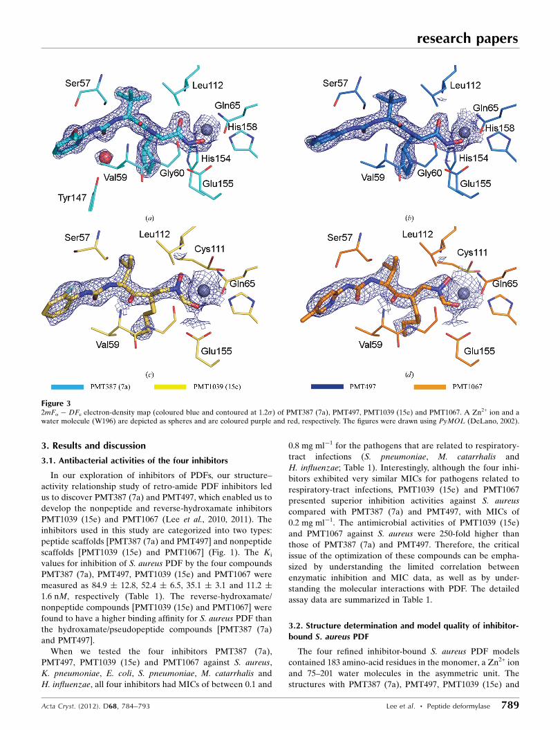
3.2. Structure determination and model quality of inhibitor-bound S. aureus PDF
The four refined inhibitor-bound S. aureus PDF models
contained 183 amino-acid residues in the monomer, a Zn2+ ion
and 75–201 water molecules in the asymmetric unit. The
structures with PMT387 (7a), PMT497, PMT1039 (15e) and
research papers
Acta Cryst. (2012). D68, 784–793 Lee et al. � Peptide deformylase 789
Figure 32mFo � DFc electron-density map (coloured blue and contoured at 1.2�) of PMT387 (7a), PMT497, PMT1039 (15e) and PMT1067. A Zn2+ ion and awater molecule (W196) are depicted as spheres and are coloured purple and red, respectively. The figures were drawn using PyMOL (DeLano, 2002).
PMT1067 had Rwork/Rfree values of 0.216/0.266, 0.208/0.235,
0.196/0.223 and 0.195/0.228 for the resolution ranges 20–1.9,
50–2.0, 20–2.15 and 20–2.3 A, respectively. In these four
models, the six histidine residues in the C-terminal fusion tag
were disordered. The four inhibitor-bound structures of
S. aureus PDF were highly similar to each other and had r.m.s.
deviations (r.m.s.d.s) of 0.08–0.20 A for 183 C�-atom pairs. The
OMIT map around Cys111 clearly shows that the functional
sulfhydryl group of Cys111 in the active site is oxidized to
sulfinic acid (Cys-SO2H; Supplementary Fig. S11) and this
observation is in good agreement with previous studies of
PDFs from S. aureus, S. pneumoniae and Thermotoga mari-
tima, in which the corresponding Cys residue is found in the
form of sulfinic acid/sulfonic acid (Kreusch et al., 2003; Yoon
et al., 2004). Although it remains unclear how the oxidation of
Cys111 can be achieved under the reducing conditions of the
physiological environment in the bacterial cell, it is possible
that the oxidation is caused by superoxide radical or another
unidentified reactive species (Rajagopalan & Pei, 1998). More
extensive studies are required to elucidate the biological
relevance of cysteine oxidation.
3.3. Structures of S. aureus PDF bound to four differentinhibitors
The structures of S. aureus PDF bound to four different
inhibitors [PMT387 (7a), PMT497, PMT1039 (15e) and
PMT1067] and an actinonin-bound PDF structure (PDB entry
1q1y; Yoon et al., 2004) were compared with an inhibitor-free
structure (PDB code 1lmh; Baldwin et al., 2002).
In PMT387 (7a)-bound S. aureus PDF the hydroxamate and
peptide-scaffold moieties of PMT387 (7a) are recognized
by residues Gly60/Gln65/Leu112/Glu155/His154/His158/Zn2+
and Ser57/Val59/Tyr147, respectively (Figs. 2 and 3). PMT497
is bound to the active site in a highly similar manner to
PMT387 (7a). However, PMT497 does not interact with either
Tyr147 or the side chain of Ser57 in S. aureus PDF, unlike
PMT387 (7a) (Figs. 2 and 3). The binding modes of PMT387
(7a) and PMT497 are similar to that of actinonin. Actinonin
makes an additional interaction with Cys111, whereas it does
not interact with Ser57. A unique feature of the PMT387 (7a)-
bound and PMT497-bound structures is the carbonyl oxygen–
Zn2+ ion and hydroxyl oxygen–Zn2+ ion distances in the
hydroxamate moieties (Fig. 3). Unlike actinonin, which has a
symmetric geometry between two O atoms and the Zn2+ ion
(2.4 and 2.4 A), PMT387 (7a) and PMT497 present distorted
geometries, with carbonyl oxygen–Zn2+ ion distances of 3.0
and 3.1 A and hydroxyl oxygen–Zn2+ ion distances of 2.1 and
2.2 A, respectively. The reverse-hydroxamate nonpeptide
inhibitors PMT1039 (15e) and PMT1067 share similar binding
modes. However, fewer interactions were observed compared
with PMT387 (7a) and PMT497. In PMT1039 (15e)-bound
S. aureus PDF, the reverse-hydroxamate and nonpeptide
scaffold moieties of PMT1039 (15e) were recognized by
residues Gln65/Cys111/Leu112/Glu155/Zn2+ and Ser57/Val59,
respectively (Figs. 2 and 3). PMT1067 is bound to the active
site in a manner highly similar to PMT1039 (15e), apart from
an interaction with Ser57 (Figs. 2 and 3). Unlike PMT1039
(15e), two N atoms in the peptide-scaffold moiety of PMT1067
were shown to interact with Ser57. These two reverse-
hydroxamate nonpeptide inhibitors [PMT1039 (15e) and
PMT1067] have an isosceles-geometry bonding mode between
the hydroxyl O atom and the Zn2+ ion and between the
carbonyl O atom and the Zn2+ ion in the reverse-hydroxamate
moieties. The distances between the two O atoms and Zn2+ are
2.8 and 2.7 A in the PMT1039 (15e) complex and 2.4 and 2.5 A
in the PMT1067 complex.
Like the other PDFs, S. aureus PDF has three inhibitor-
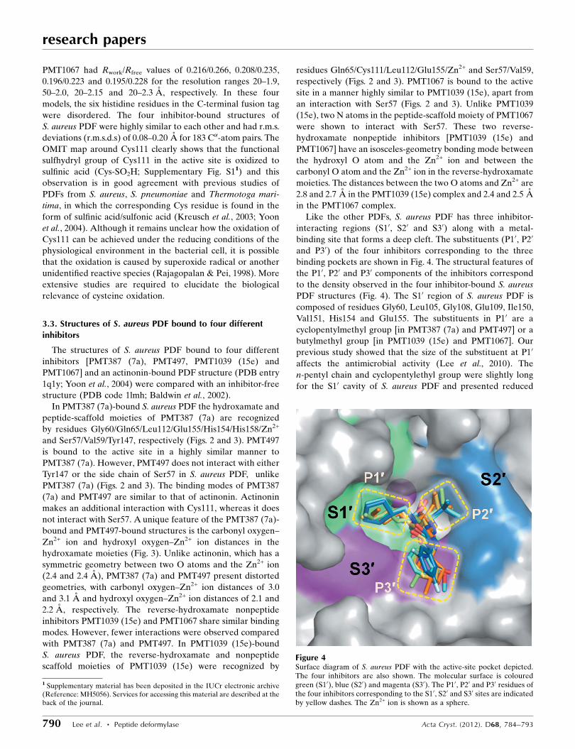
interacting regions (S10, S20 and S30) along with a metal-
binding site that forms a deep cleft. The substituents (P10, P20
and P30) of the four inhibitors corresponding to the three
binding pockets are shown in Fig. 4. The structural features of
the P10, P20 and P30 components of the inhibitors correspond
to the density observed in the four inhibitor-bound S. aureus
PDF structures (Fig. 4). The S10 region of S. aureus PDF is
composed of residues Gly60, Leu105, Gly108, Glu109, Ile150,
Val151, His154 and Glu155. The substituents in P10 are a
cyclopentylmethyl group [in PMT387 (7a) and PMT497] or a
butylmethyl group [in PMT1039 (15e) and PMT1067]. Our
previous study showed that the size of the substituent at P10
affects the antimicrobial activity (Lee et al., 2010). The
n-pentyl chain and cyclopentylethyl group were slightly long
for the S10 cavity of S. aureus PDF and presented reduced
research papers
790 Lee et al. � Peptide deformylase Acta Cryst. (2012). D68, 784–793
Figure 4Surface diagram of S. aureus PDF with the active-site pocket depicted.The four inhibitors are also shown. The molecular surface is colouredgreen (S10), blue (S20) and magenta (S30). The P10, P20 and P30 residues ofthe four inhibitors corresponding to the S10, S20 and S30 sites are indicatedby yellow dashes. The Zn2+ ion is shown as a sphere.
1 Supplementary material has been deposited in the IUCr electronic archive(Reference: MH5056). Services for accessing this material are described at theback of the journal.
activity. Cyclopropylmethyl and cyclobutylmethyl groups had
a reduced inhibitory effect on bacterial growth.
These compounds do not fit into the S10 pocket very well.
Accordingly, we screened and selected cyclopentylmethyl
for PMT387 (7a) and PMT497 and butylmethyl for PMT1039
(15e) and PMT1067. This is in agreement with our previous
reports (Lee et al., 2010, 2011) concerning inhibitor-bound
structures.
The S20 region of S. aureus PDF is composed of residues
Arg56, Cys111 and Leu112. The P20 region, which corresponds
to S20, was optimized as t-leucine [in PMT387 (7a) and
PMT497] or valine [in PMT1039 (15e) and PMT1067]. The
size and length of the substituents in the P20 region have
significant effects on the antimicrobial activity, especially
against S. aureus (Lee et al., 2011). These effects arise from the
size of the S20 region, which is not wider than the S10 region. In
the case of PMT387 (7a) and PMT497, a shorter or longer side
chain than valine was shown to reduce the antimicrobial
activity, which led to the discovery of t-leucine as the optimal
residue for inhibiting the growth of bacteria. Therefore, when
the side chain was t-leucine the antimicrobial activity of the
reverse-hydroxamate nonpeptide inhibitors against S. aureus
PDF was significantly reduced. This result allowed us to select
valine as an effective P20 substituent in the development of the
reverse-hydroxamate nonpeptide inhibitors (Lee et al., 2011).
The S30 region of S. aureus PDF is composed of residues
Ser57, Gly58, Val59 and Tyr147. Our intensive search for
P30 substituents led us to discover a 2-methoxyphenyl ring
[PMT387 (7a)] and a 3,5-difluorophenyl ring, both of which
significantly increased antimicrobial activity against the tested
research papers
Acta Cryst. (2012). D68, 784–793 Lee et al. � Peptide deformylase 791
Figure 5Structural changes arising from the effects of inhibitors binding to S. aureus PDF. Residues are coloured using a linear ramp from blue for unperturbedstructures (r.m.s.d. < 0.4 A) to red for perturbations greater than an r.m.s.d. of 1.0 A. Residues shown in blue exhibited no measurable change in r.m.s.d.,while red residues were more flexible in the complex structures. Residues 41–51, 54–58, 80–81, 107–110, 115–116, 117–120 and 171–175 are indicated by a,b, c, d, e, f and g, respectively. The Zn2+ ion is shown as a magenta sphere and the five inhibitors (including actinonin) are shown as sticks with atomscoloured by type.
strains (Lee et al., 2010). For the reverse-hydroxamate non-
peptide inhibitors, a 2-fluorophenyl ring [PMT1039 (15e)]
substitution was shown to have the most potent activity and
3-methylpiperidine (PMT1067) was also effective (Lee et al.,
2010, 2011; Table 1).
3.4. Effects of inhibitors on the structure of S. aureus PDF
There were measurable structural differences between the
four inhibitor-bound S. aureus PDF structures and an inhibitor-
free structure. Therefore, we hypothesized that induced-fit
recognition may play a role in inhibitor binding. To obtain
structural insights into the role of protein flexibility in mole-
cular recognition, we plotted the r.m.s.d.s of five separate
structures of S. aureus PDF bound to five inhibitors (including
actinonin) and compared the structures with the inhibitor-free
structure (Fig. 5). Interestingly, the structural comparisons
indicated that the structure of S. aureus PDF undergoes a
small conformational change that depends on the type of
inhibitor: hydroxamate pseudopeptide inhibitors [PMT387
(7a) and PMT497] or reverse-hydroxamate nonpeptide inhi-
bitors [PMT1039 (15e) and PMT1067]. The key findings are
summarized in Fig. 5 and Supplementary Fig. S2. Residues 41–
51 (corresponding to S30), 54–58 (corresponding to S20), 107–
110 (corresponding to S10), 117–120 and 171–175 present
greater deviations in the PMT387 (7a)- and PMT497-bound
PDF structures compared with structures in which PDF was
bound to the two reverse-hydroxamate nonpeptide inhibitors
PMT1039 (15e) and PMT1067 or actinonin (PDB entry 1q1y).
The two reverse-hydroxamate inhibitor-bound PDF structures
present greater deviations of residues 115–116. For residues
80–81 (corresponding to S20), the PMT387 (7a)- and PMT1039
(15e)-bound PDF structures present greater deviations.
Residues 41–51, 54–58 and 107–110 are located in the active
site. In the case of the hydroxamate peptide inhibitors,
deviations of residues 41–51, 54–58 and 107–110 were found.
In addition to the affected residues 107–110 mentioned above,
their flanking residues 115–120 as well as the neighbouring
residues 171–175 also showed concurrent structural changes.
The structural rearrangements induced by the hydroxamate
peptide inhibitors involved local changes in the active-site
pocket and global structural changes transmitted via inter-
action networks to other parts of the protein, while the
reverse-hydroxamate nonpeptide inhibitors were not shown to
have such effects on conformational change. These ligand–
enzyme complexes cause a specific structural perturbation that
affects regions remote from the active site. These results are in
agreement with previous findings that indicated that local
entropy changes in an active site can be redistributed from one
site to another in a protein, which has important thermo-
dynamic implications (Amero et al., 2009).
The structure of PDF is rigid in the absence of ligands, in
contrast to other members of the metallohydrolase super-
family (Amero et al., 2009; Moy et al., 2002). This lack of
flexibility may be related to the narrow specificity of PDF for
substrates that are peptides bearing N-formylated methionine
residues (Amero et al., 2009; Hu et al., 1999; Ragusa et al.,
1999). Despite the high rigidity of the S. aureus structure, two
different classes of PDF inhibitors induced local structural
changes. We observed that these structures displayed distinct
structural features depending on the inhibitor class. The
distances between the hydroxamate carbonyl O atom and the
Zn2+ ion and between the reverse-hydroxamate hydroxyl O
atom and the Zn2+ ion in the hydroxamate or reverse-
hydroxamate moiety appeared to be correlated with selective
and strong inhibition activity against S. aureus (Table 1).
When we compared the two classes of inhibitor-bound struc-
tures, a distinctive change in the S. aureus PDF structure was
also observed which was driven by the binding of specific
inhibitors. As shown in Fig. 2, the binding modes of the inhi-
bitors demonstrate comparable characteristics that were
dependent on the inhibitor class. These structural changes
appear to be involved in the inhibition activity against
S. aureus.
Most known PDF inhibitors consist of a hydroxamate
moiety and a pseudopeptide backbone. Although a
hydroxamate compound such as actinonin is regarded as an
extremely effective inhibitor, such a compound typically
displays poor selectivity by chelating the metals of other
metalloproteins that are cytotoxic to mammalian cells and also
shows poor oral bioavailability related to characteristic rapid
metabolization (Aubart & Zalacain, 2006; Antczak et al., 2011;
Fisher & Mobashery, 2006). In terms of resolving the problems
of poor selectivity as well as oral bioavailability, the use of
either a reverse-hydroxamate moiety or a nonpeptide back-
bone may be considered to be one alternative method of
improving current PDF inhibitors. The study that we present
here provides new structural information on four inhibitor
complexes of S. aureus PDF which will aid in the design of new
potential inhibitors and will provide important data for
selective antimicrobial strategies. Further studies combining
structural (X-ray crystallographic or nuclear magnetic reso-
nance) evidence from PDFs from other strains or different
types of enzymes will aid in improving rational drug design.
The binding of an inhibitor to an enzyme is more complex
than is generally assumed; therefore, we suggest that accurate
information on local and long-range contributions to mole-
cular behaviour will be required for the rational design of an
inhibitor, especially for antibacterial drugs.
We thank the beamline staff at the Photon Factory (BL-5A)
and SPring-8 (BL38-B1), Japan for assistance in the X-ray
diffraction experiments. This work was partially supported by
the Innovative Drug Research Center for Metabolic and
Inflammatory Disease, Basic Science Outstanding Scholars
Program, World-Class University Program and Korea
Healthcare Technology R&D Project, Ministry for Health,
Welfare and Family Affairs, Republic of Korea (grant No.
A092006) (SWS) and the Mid-career Researcher Program
(2011-0029294) and the Bio and Medical Technology Devel-
opment Program (2011-0030032) through the National
Research Foundation of Korea (NRF) and funded by the
Ministry of Education, Science and Technology and National
research papers
792 Lee et al. � Peptide deformylase Acta Cryst. (2012). D68, 784–793
Cancer Center Research Grant (grant No. 1110012) (BIL).
S-JL is a Howard Hughes Medical Institute postdoctoral
fellow. SJL is supported by funding from the Fostering Next-
Generation Researchers program, which was granted by NRF,
Republic of Korea (NRF-2011-355-C00118).
References
Amero, C. D., Byerly, D. W., McElroy, C. A., Simmons, A. & Foster,M. P. (2009). Biochemistry, 48, 7595–7607.
Antczak, C., Shum, D., Bassit, B., Frattini, M. G., Li, Y., de Stanchina,E., Scheinberg, D. A. & Djaballah, H. (2011). Bioorg. Med. Chem.Lett. 21, 4528–4532.
Apfel, C., Banner, D. W., Bur, D., Dietz, M., Hirata, T., Hubschwerlen,C., Locher, H., Page, M. G., Pirson, W., Rosse, G. & Specklin, J. L.(2000). J. Med. Chem. 43, 2324–2331.
Aubart, K. & Zalacain, M. (2006). Prog. Med. Chem. 44, 109–143.Baldwin, E. T., Harris, M. S., Yem, A. W., Wolfe, C. L., Vosters, A. F.,
Curry, K. A., Murray, R. W., Bock, J. H., Marshall, V. P., Cialdella,J. I., Merchant, M. H., Choi, G. & Deibel, M. R. (2002). J. Biol.Chem. 277, 31163–31171.
Boucher, H. W. & Corey, G. R. (2008). Clin. Infect. Dis. 46, Suppl. 5,S344–S349.
Boucher, H. W., Talbot, G. H., Bradley, J. S., Edwards, J. E., Gilbert,D., Rice, L. B., Scheld, M., Spellberg, B. & Bartlett, J. (2009). Clin.Infect. Dis. 48, 1–12.
Bradford, M. M. (1976). Anal. Biochem. 72, 248–254.Brunger, A. T. (1992). Nature (London), 355, 472–475.Cai, J., Han, C., Hu, T., Zhang, J., Wu, D., Wang, F., Liu, Y., Ding, J.,
Chen, K., Yue, J., Shen, X. & Jiang, H. (2006). Protein Sci. 15, 2071–2081.
Chen, D. Z., Patel, D. V., Hackbarth, C. J., Wang, W., Dreyer, G.,Young, D. C., Margolis, P. S., Wu, C., Ni, Z.-J., Trias, J., White, R. J.& Yuan, Z. (2000). Biochemistry, 39, 1256–1262.
Chen, V. B., Arendall, W. B., Headd, J. J., Keedy, D. A., Immormino,R. M., Kapral, G. J., Murray, L. W., Richardson, J. S. & Richardson,D. C. (2010). Acta Cryst. D66, 12–21.
Clements, J. M., Beckett, R. P., Brown, A., Catlin, G., Lobell, M.,Palan, S., Thomas, W., Whittaker, M., Wood, S., Salama, S., Baker,P. J., Rodgers, H. F., Barynin, V., Rice, D. W. & Hunter, M. G.(2001). Antimicrob. Agents Chemother. 45, 563–570.
Credito, K., Lin, G., Ednie, L. M. & Appelbaum, P. C. (2004).Antimicrob. Agents Chemother. 48, 4033–4036.
DeLano, W. L. (2002). PyMOL. http://www.pymol.org.Emsley, P., Lohkamp, B., Scott, W. G. & Cowtan, K. (2010). Acta
Cryst. D66, 486–501.Fisher, J. F. & Mobashery, S. (2006). Cancer Metastasis Rev. 25,
115–136.Fritsche, T. R., Sader, H. S., Cleeland, R. & Jones, R. N. (2005).
Antimicrob. Agents Chemother. 49, 1468–1476.Fu, H., Karlsson, J., Bylund, J., Movitz, C., Karlsson, A. & Dahlgren,
C. (2006). J. Leukoc. Biol. 79, 247–256.Gill, S. R. et al. (2005). J. Bacteriol. 187, 2426–2438.Grujic, M., Zavasnik-Bergant, T., Pejler, G. & Renko, M. (2005).
Cancer Lett. 223, 211–218.Guay, D. R. (2007). Ther. Clin. Risk Manag, 3, 513–525.Guillon, J.-M., Mechulam, Y., Schmitter, J. M., Blanquet, S. & Fayat,
G. (1992). J. Bacteriol. 174, 4294–4301.Guilloteau, J.-P., Mathieu, M., Giglione, C., Blanc, V., Dupuy, A.,
Chevrier, M., Gil, P., Famechon, A., Meinnel, T. & Mikol, V. (2002).J. Mol. Biol. 320, 951–962.
Hu, Y.-J., Wei, Y., Zhou, Y., Rajagopalan, P. T. R. & Pei, D. (1999).Biochemistry, 38, 643–650.
Klevens, R. M., Edwards, J. R., Tenover, F. C., McDonald, L. C.,Horan, T. & Gaynes, R. (2006). Clin. Infect. Dis. 42, 389–391.
Kreusch, A., Spraggon, G., Lee, C. C., Klock, H., McMullan, D., Ng,K., Shin, T., Vincent, J., Warner, I., Ericson, C. & Lesley, S. A.(2003). J. Mol. Biol. 330, 309–321.
Krissinel, E. & Henrick, K. (2007). J. Mol. Biol. 372, 774–797.Lee, S. K., Choi, K. H., Lee, S. J., Lee, J. S., Park, J. Y., Kim, B. M. &
Lee, B. J. (2011). Bioorg. Med. Chem. Lett. 21, 133–136.Lee, S. K., Choi, K. H., Lee, S. J., Suh, S. W., Kim, B. M. & Lee, B. J.
(2010). Bioorg. Med. Chem. Lett. 20, 4317–4319.Lee, M. D., She, Y., Soskis, M. J., Borella, C. P., Gardner, J. R., Hayes,
P. A., Dy, B. M., Heaney, M. L., Philips, M. R., Bornmann, W. G.,Sirotnak, F. M. & Scheinberg, D. A. (2004). J. Clin. Invest. 114,1107–1116.
Leeds, J. A. & Dean, C. R. (2006). Curr. Opin. Pharmacol. 6, 445–452.Mader, D., Rabiet, M.-J., Boulay, F. & Peschel, A. (2010). Microbes
Infect. 12, 415–419.Margolis, P. S., Hackbarth, C. J., Young, D. C., Wang, W., Chen, D.,
Yuan, Z., White, R. & Trias, J. (2000). Antimicrob. AgentsChemother. 44, 1825–1831.
Mazel, D., Pochet, S. & Marliere, P. (1994). EMBO J. 13, 914–923.Meinnel, T., Mechulam, Y. & Blanquet, S. (1993). Biochimie, 75,
1061–1075.Molteni, V., He, X., Nabakka, J., Yang, K., Kreusch, A., Gordon, P.,
Bursulaya, B., Warner, I., Shin, T., Biorac, T., Ryder, N. S.,Goldberg, R., Doughty, J. & He, Y. (2004). Bioorg. Med. Chem.Lett. 14, 1477–1481.
Moon, J. H., Park, J. K. & Kim, E. E. (2005). Proteins, 61, 217–220.Moy, F. J., Chanda, P. K., Chen, J., Cosmi, S., Edris, W., Levin, J. I.,
Rush, T. S., Wilhelm, J. & Powers, R. (2002). J. Am. Chem. Soc. 124,12658–12659.
Murshudov, G. N., Skubak, P., Lebedev, A. A., Pannu, N. S., Steiner,R. A., Nicholls, R. A., Winn, M. D., Long, F. & Vagin, A. A. (2011).Acta Cryst. D67, 355–367.
National Committee for Clinical Laboratory Standards (2003).Susceptibility Testing of Mycobacteria, Nocardia, and OtherAerobic Actinomycetes. Approved Standard M24-A. Wayne, PA:National Committee for Clinical Laboratory Standards.
Nguyen, K. T., Hu, X., Colton, C., Chakrabarti, R., Zhu, M. X. & Pei,D. (2003). Biochemistry, 42, 9952–9958.
Otwinowski, Z. & Minor, W. (1997). Methods Enzymol. 276, 307–326.Pichota, A. et al. (2008). Bioorg. Med. Chem. Lett. 18, 6568–6572.Ragusa, S., Mouchet, P., Lazennec, C., Dive, V. & Meinnel, T. (1999).
J. Mol. Biol. 289, 1445–1457.Rajagopalan, P. T. & Pei, D. (1998). J. Biol. Chem. 273, 22305–22310.Ramanathan-Girish, S., McColm, J., Clements, J. M., Taupin, P.,
Barrowcliffe, S., Hevizi, J., Safrin, S., Moore, C., Patou, G., Moser,H., Gadd, A., Hoch, U., Jiang, V., Lofland, D. & Johnson, K. W.(2004). Antimicrob. Agents Chemother. 48, 4835–4842.
Robien, M. A., Nguyen, K. T., Kumar, A., Hirsh, I., Turley, S., Pei, D.& Hol, W. G. J. (2004). Protein Sci. 13, 1155–1163.
Rosenthal, V. D. et al. (2010). Am. J. Infect. Control, 38, 95–104.Smith, K. J., Petit, C. M., Aubart, K., Smyth, M., McManus, E., Jones,
J., Fosberry, A., Lewis, C., Lonetto, M. & Christensen, S. B. (2003).Protein Sci. 12, 349–360.
Solbiati, J., Chapman-Smith, A., Miller, J. L., Miller, C. G. & Cronan,J. E. (1999). J. Mol. Biol. 290, 607–614.
Spellberg, B., Powers, J. H., Brass, E. P., Miller, L. G. & Edwards, J. E.(2004). Clin. Infect. Dis. 38, 1279–1286.
Vagin, A. & Teplyakov, A. (2010). Acta Cryst. D66, 22–25.Watters, A. A., Jones, R. N., Leeds, J. A., Denys, G., Sader, H. S. &
Fritsche, T. R. (2006). J. Antimicrob. Chemother. 57, 914–923.World Health Organization (2004). World Health Report 2004, Annex
Table 2. http://www.who.int/whr/2004/annex/topic/en/annex_2_en.pdf.Xu, Y., Lai, L. T., Gabrilove, J. L. & Scheinberg, D. A. (1998). Clin.
Cancer Res. 4, 171–176.Yoon, H.-J., Kim, H. L., Lee, S.-K., Kim, H.-W., Kim, H.-W., Lee, J. Y.,
Mikami, B. & Suh, S. W. (2004). Proteins, 57, 639–642.Zhou, Z., Song, X., Li, Y. & Gong, W. (2004). J. Mol. Biol. 339,
207–215.
research papers
Acta Cryst. (2012). D68, 784–793 Lee et al. � Peptide deformylase 793