Embed Size (px)
Citation preview
Summer 2009 Workshop in Biology and Multimedia for High School TeachersHarvard University Life Sciences – HHMI Outreach Program
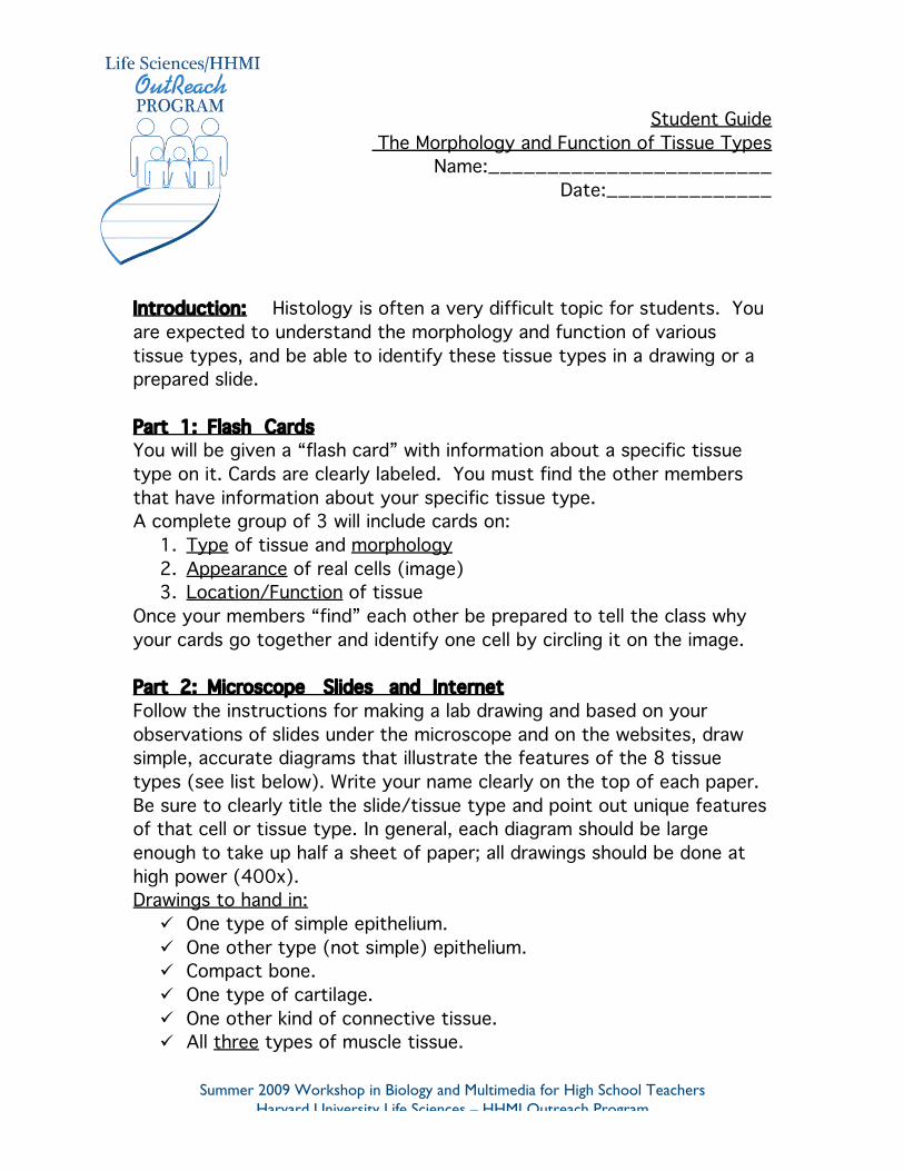
Student Guide The Morphology and Function of Tissue Types
Name:________________________Date:______________
Introduction: Histology is often a very difficult topic for students. Youare expected to understand the morphology and function of varioustissue types, and be able to identify these tissue types in a drawing or aprepared slide.
Part 1: Flash CardsYou will be given a “flash card” with information about a specific tissuetype on it. Cards are clearly labeled. You must find the other membersthat have information about your specific tissue type.A complete group of 3 will include cards on:
1. Type of tissue and morphology2. Appearance of real cells (image)3. Location/Function of tissue
Once your members “find” each other be prepared to tell the class whyyour cards go together and identify one cell by circling it on the image.
Part 2: Microscope Slides and InternetFollow the instructions for making a lab drawing and based on yourobservations of slides under the microscope and on the websites, drawsimple, accurate diagrams that illustrate the features of the 8 tissuetypes (see list below). Write your name clearly on the top of each paper.Be sure to clearly title the slide/tissue type and point out unique featuresof that cell or tissue type. In general, each diagram should be largeenough to take up half a sheet of paper; all drawings should be done athigh power (400x).Drawings to hand in:
One type of simple epithelium. One other type (not simple) epithelium. Compact bone. One type of cartilage. One other kind of connective tissue. All three types of muscle tissue.
Summer 2009 Workshop in Biology and Multimedia for High School TeachersHarvard University Life Sciences – HHMI Outreach Program
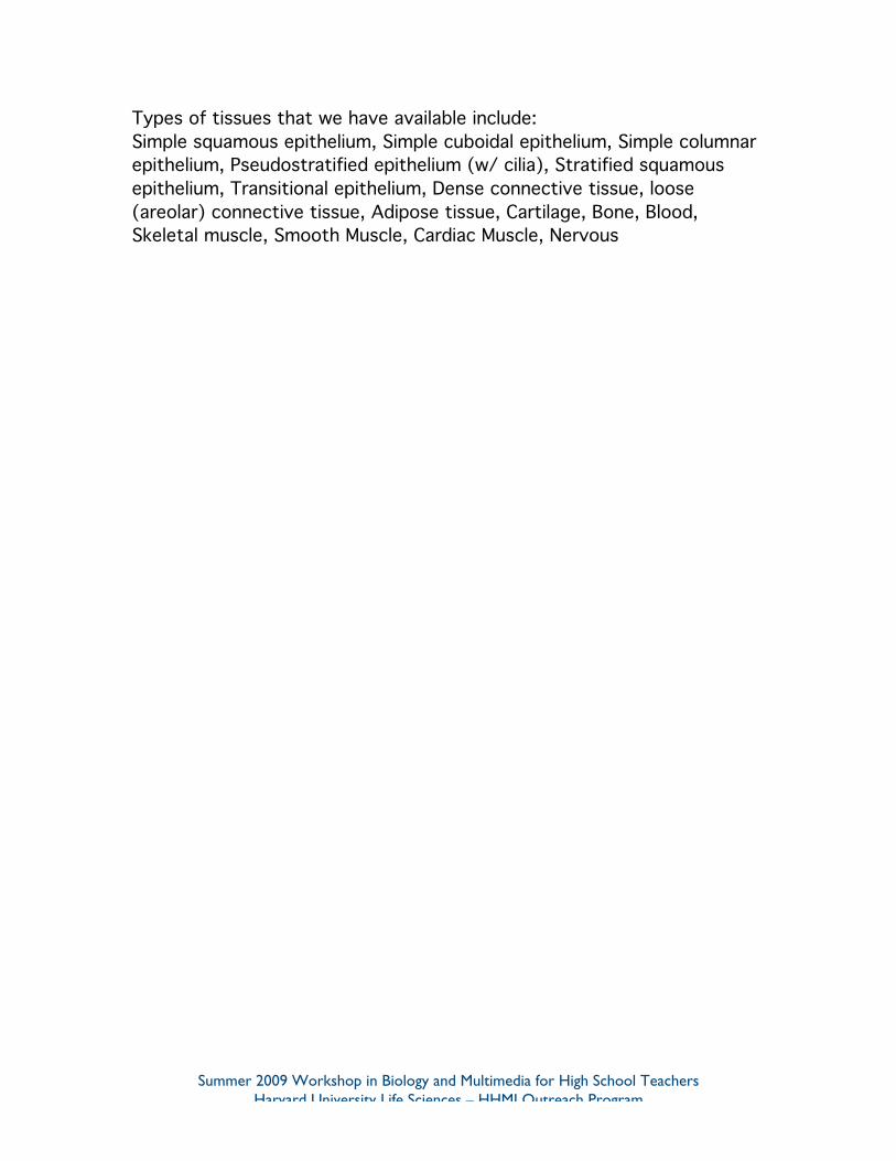
Types of tissues that we have available include:Simple squamous epithelium, Simple cuboidal epithelium, Simple columnarepithelium, Pseudostratified epithelium (w/ cilia), Stratified squamousepithelium, Transitional epithelium, Dense connective tissue, loose(areolar) connective tissue, Adipose tissue, Cartilage, Bone, Blood,Skeletal muscle, Smooth Muscle, Cardiac Muscle, Nervous
Summer 2009 Workshop in Biology and Multimedia for High School TeachersHarvard University Life Sciences – HHMI Outreach Program
Part 1: Flash Cards
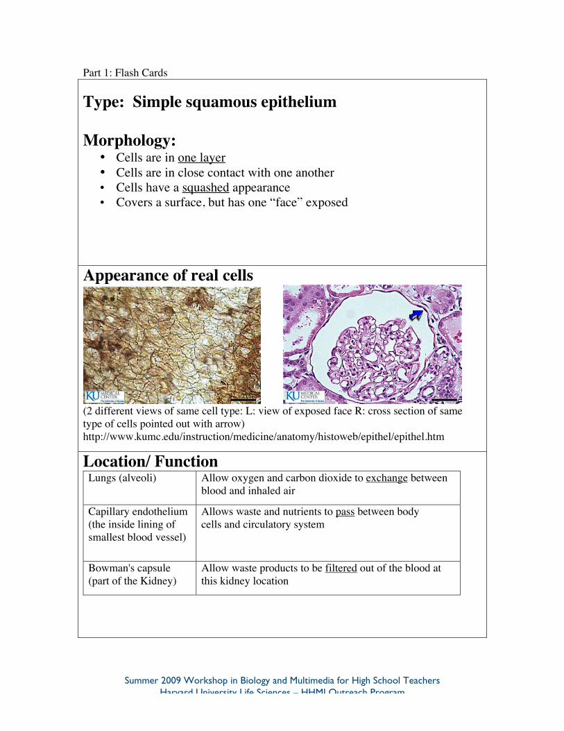
Type: Simple squamous epithelium
Morphology:• Cells are in one layer• Cells are in close contact with one another• Cells have a squashed appearance• Covers a surface, but has one “face” exposed
Appearance of real cells
(2 different views of same cell type: L: view of exposed face R: cross section of sametype of cells pointed out with arrow)http://www.kumc.edu/instruction/medicine/anatomy/histoweb/epithel/epithel.htm
Location/ FunctionLungs (alveoli) Allow oxygen and carbon dioxide to exchange between
blood and inhaled air
Capillary endothelium(the inside lining ofsmallest blood vessel)
Allows waste and nutrients to pass between bodycells and circulatory system
Bowman's capsule(part of the Kidney)
Allow waste products to be filtered out of the blood atthis kidney location
Summer 2009 Workshop in Biology and Multimedia for High School TeachersHarvard University Life Sciences – HHMI Outreach Program
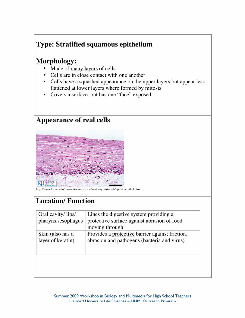
Type: Stratified squamous epithelium
Morphology:• Made of many layers of cells• Cells are in close contact with one another• Cells have a squashed appearance on the upper layers but appear less
flattened at lower layers where formed by mitosis• Covers a surface, but has one “face” exposed
Appearance of real cells
http://www.kumc.edu/instruction/medicine/anatomy/histoweb/epithel/epithel.htm
Location/ Function
Oral cavity/ lips/pharynx /esophagus
Lines the digestive system providing aprotective surface against abrasion of foodmoving through
Skin (also has alayer of keratin)
Provides a protective barrier against friction,abrasion and pathogens (bacteria and virus)
Summer 2009 Workshop in Biology and Multimedia for High School TeachersHarvard University Life Sciences – HHMI Outreach Program
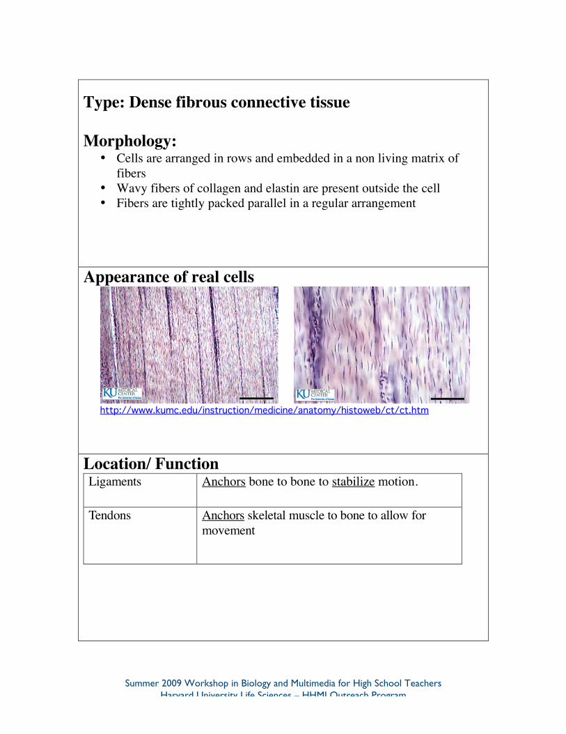
Type: Dense fibrous connective tissue
Morphology:• Cells are arranged in rows and embedded in a non living matrix of
fibers• Wavy fibers of collagen and elastin are present outside the cell• Fibers are tightly packed parallel in a regular arrangement
Appearance of real cells
http://www.kumc.edu/instruction/medicine/anatomy/histoweb/ct/ct.htm
Location/ FunctionLigaments Anchors bone to bone to stabilize motion.
Tendons Anchors skeletal muscle to bone to allow formovement
Summer 2009 Workshop in Biology and Multimedia for High School TeachersHarvard University Life Sciences – HHMI Outreach Program
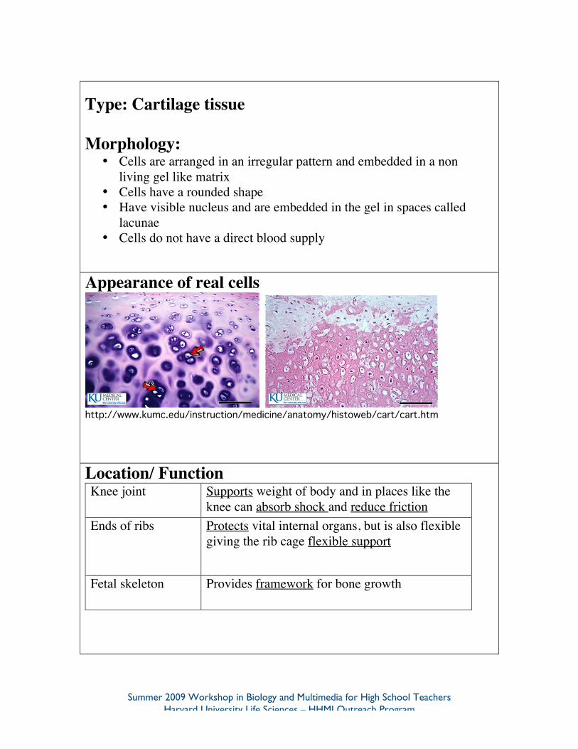
Type: Cartilage tissue
Morphology:• Cells are arranged in an irregular pattern and embedded in a non
living gel like matrix• Cells have a rounded shape• Have visible nucleus and are embedded in the gel in spaces called
lacunae• Cells do not have a direct blood supply
Appearance of real cells
http://www.kumc.edu/instruction/medicine/anatomy/histoweb/cart/cart.htm
Location/ FunctionKnee joint Supports weight of body and in places like the
knee can absorb shock and reduce frictionEnds of ribs Protects vital internal organs, but is also flexible
giving the rib cage flexible support
Fetal skeleton Provides framework for bone growth
Summer 2009 Workshop in Biology and Multimedia for High School TeachersHarvard University Life Sciences – HHMI Outreach Program
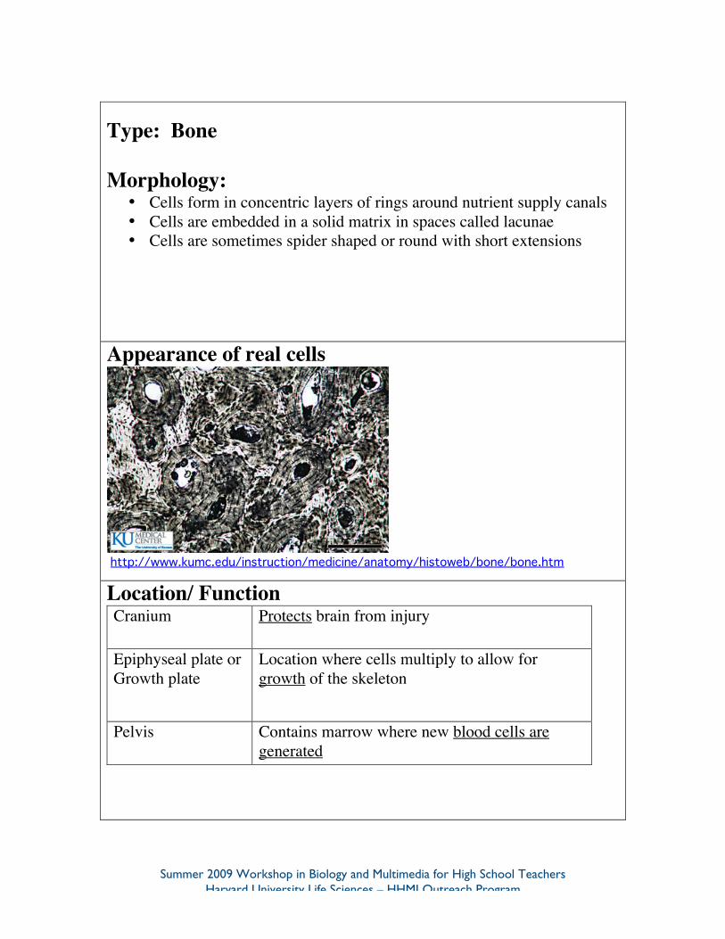
Type: Bone
Morphology:• Cells form in concentric layers of rings around nutrient supply canals• Cells are embedded in a solid matrix in spaces called lacunae• Cells are sometimes spider shaped or round with short extensions
Appearance of real cells
http://www.kumc.edu/instruction/medicine/anatomy/histoweb/bone/bone.htm
Location/ FunctionCranium Protects brain from injury
Epiphyseal plate orGrowth plate
Location where cells multiply to allow forgrowth of the skeleton
Pelvis Contains marrow where new blood cells aregenerated
Summer 2009 Workshop in Biology and Multimedia for High School TeachersHarvard University Life Sciences – HHMI Outreach Program
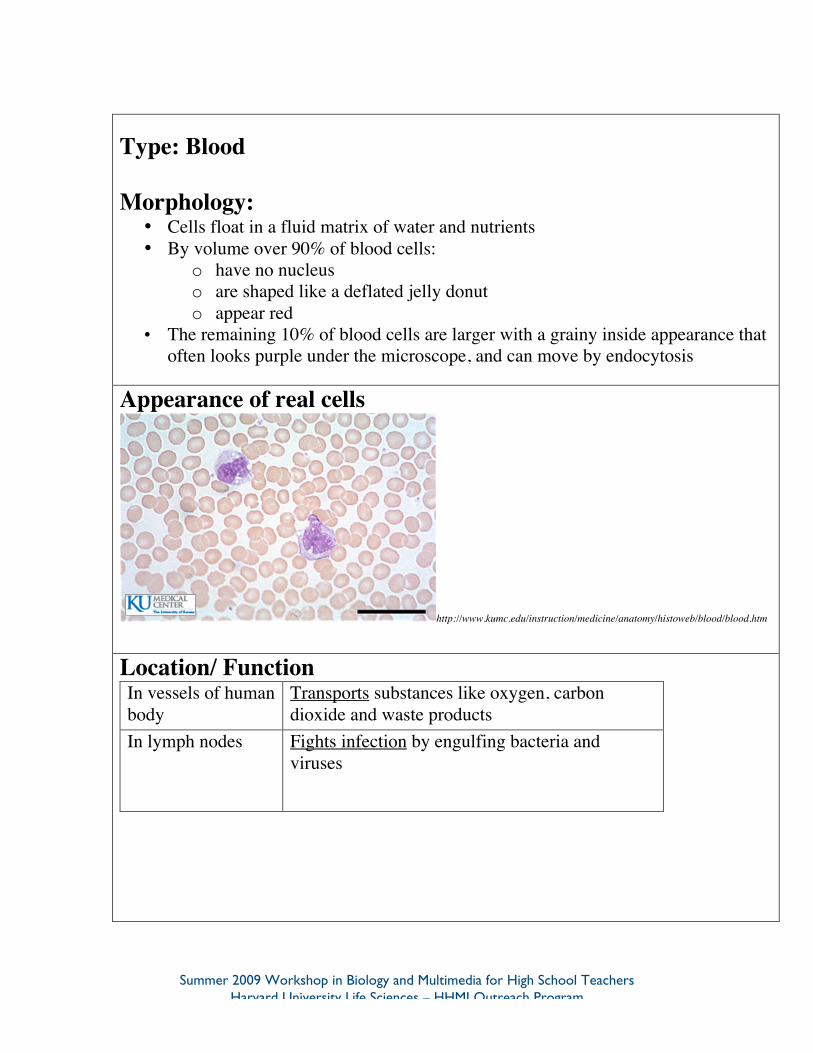
Type: Blood
Morphology:• Cells float in a fluid matrix of water and nutrients• By volume over 90% of blood cells:
o have no nucleuso are shaped like a deflated jelly donuto appear red
• The remaining 10% of blood cells are larger with a grainy inside appearance thatoften looks purple under the microscope, and can move by endocytosis
Appearance of real cells
http://www.kumc.edu/instruction/medicine/anatomy/histoweb/blood/blood.htm
Location/ FunctionIn vessels of humanbody
Transports substances like oxygen, carbondioxide and waste products
In lymph nodes Fights infection by engulfing bacteria andviruses
Summer 2009 Workshop in Biology and Multimedia for High School TeachersHarvard University Life Sciences – HHMI Outreach Program
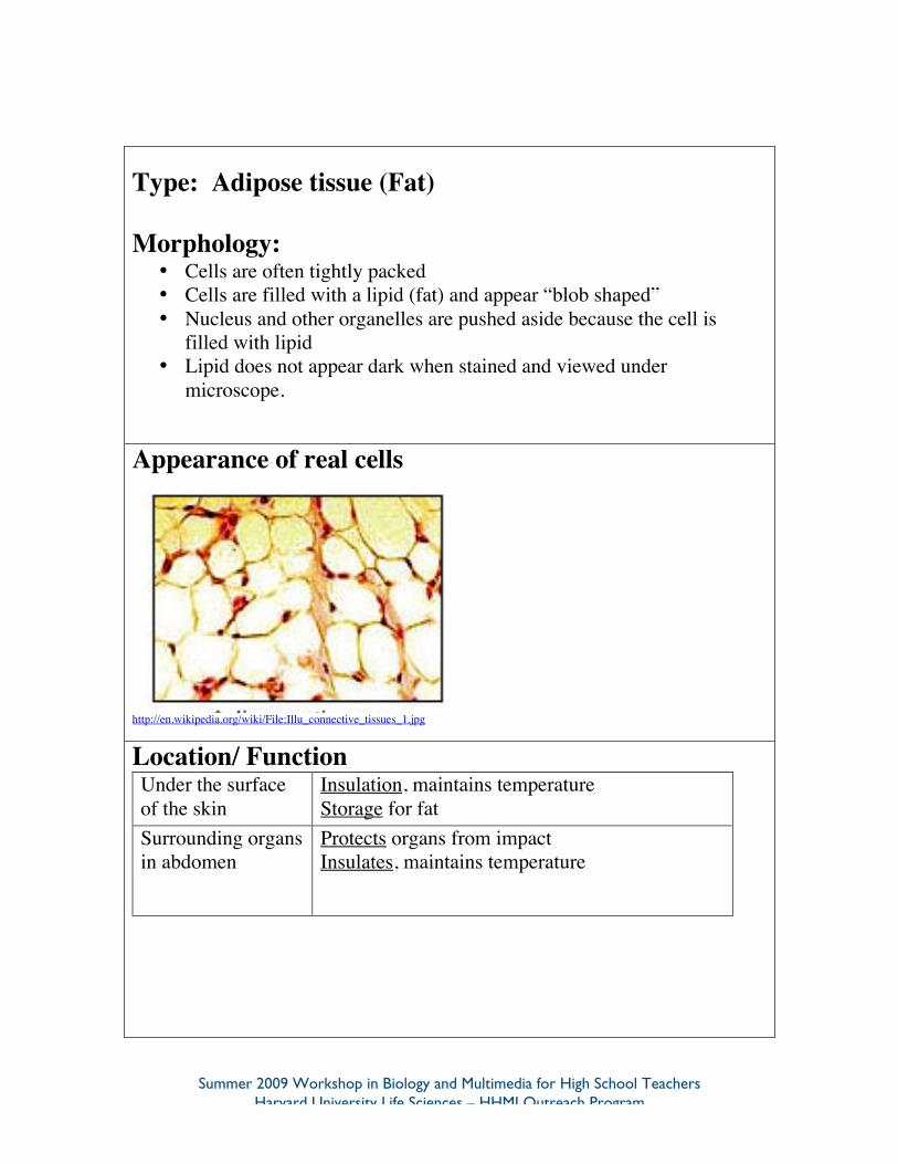
Type: Adipose tissue (Fat)
Morphology:• Cells are often tightly packed• Cells are filled with a lipid (fat) and appear “blob shaped”• Nucleus and other organelles are pushed aside because the cell is
filled with lipid• Lipid does not appear dark when stained and viewed under
microscope.
Appearance of real cells
http://en.wikipedia.org/wiki/File:Illu_connective_tissues_1.jpg
Location/ FunctionUnder the surfaceof the skin
Insulation, maintains temperatureStorage for fat
Surrounding organsin abdomen
Protects organs from impactInsulates, maintains temperature
Summer 2009 Workshop in Biology and Multimedia for High School TeachersHarvard University Life Sciences – HHMI Outreach Program
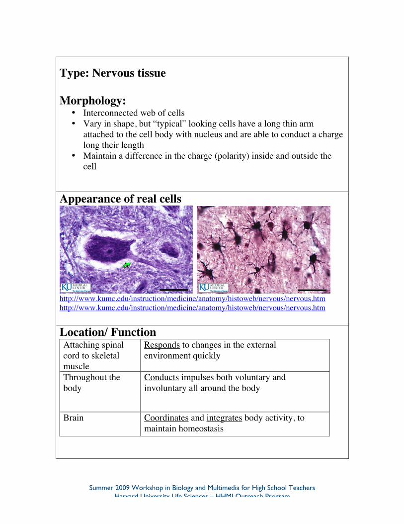
Type: Nervous tissue
Morphology:• Interconnected web of cells• Vary in shape, but “typical” looking cells have a long thin arm
attached to the cell body with nucleus and are able to conduct a chargelong their length
• Maintain a difference in the charge (polarity) inside and outside thecell
Appearance of real cells
http://www.kumc.edu/instruction/medicine/anatomy/histoweb/nervous/nervous.htmhttp://www.kumc.edu/instruction/medicine/anatomy/histoweb/nervous/nervous.htm
Location/ FunctionAttaching spinalcord to skeletalmuscle
Responds to changes in the externalenvironment quickly
Throughout thebody
Conducts impulses both voluntary andinvoluntary all around the body
Brain Coordinates and integrates body activity, tomaintain homeostasis
Summer 2009 Workshop in Biology and Multimedia for High School TeachersHarvard University Life Sciences – HHMI Outreach Program
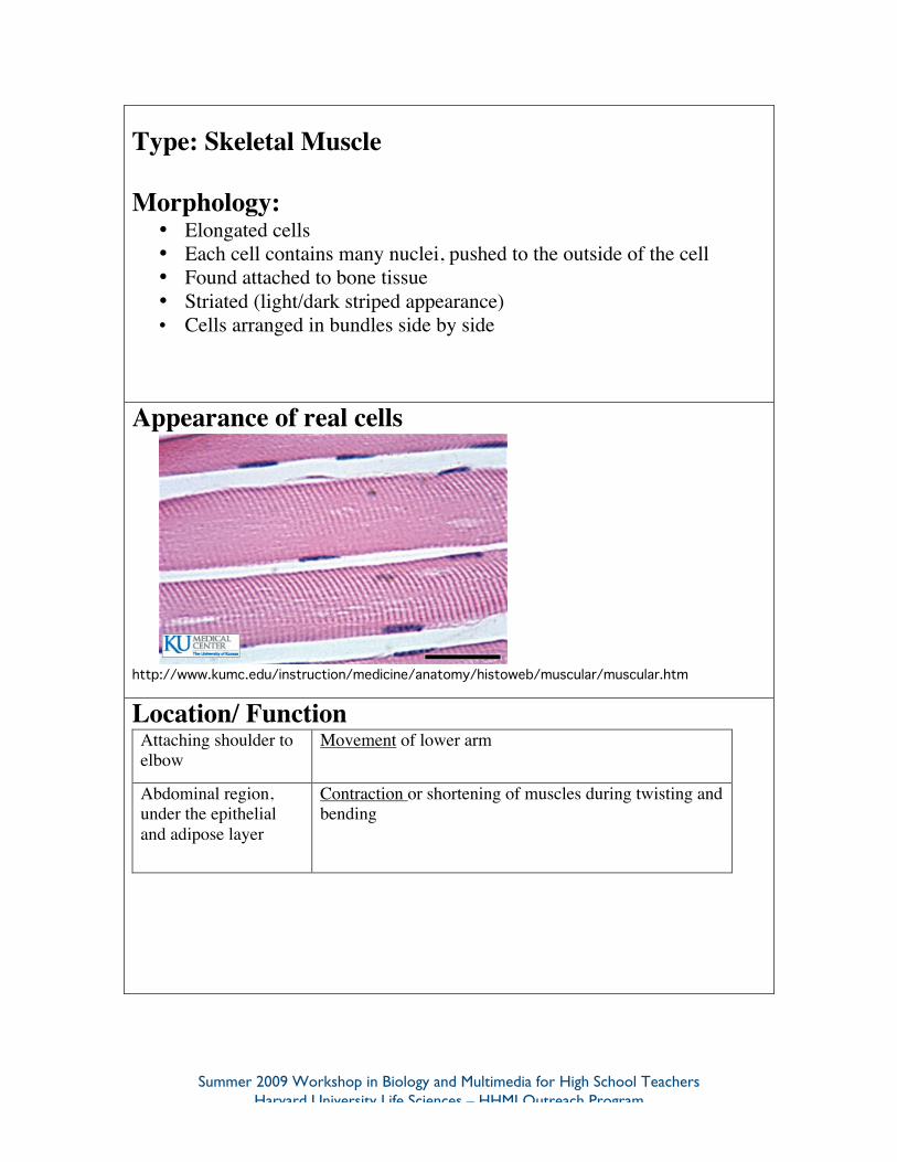
Type: Skeletal Muscle
Morphology:• Elongated cells• Each cell contains many nuclei, pushed to the outside of the cell• Found attached to bone tissue• Striated (light/dark striped appearance)• Cells arranged in bundles side by side
Appearance of real cells
http://www.kumc.edu/instruction/medicine/anatomy/histoweb/muscular/muscular.htm
Location/ FunctionAttaching shoulder toelbow
Movement of lower arm
Abdominal region,under the epithelialand adipose layer
Contraction or shortening of muscles during twisting andbending
Summer 2009 Workshop in Biology and Multimedia for High School TeachersHarvard University Life Sciences – HHMI Outreach Program
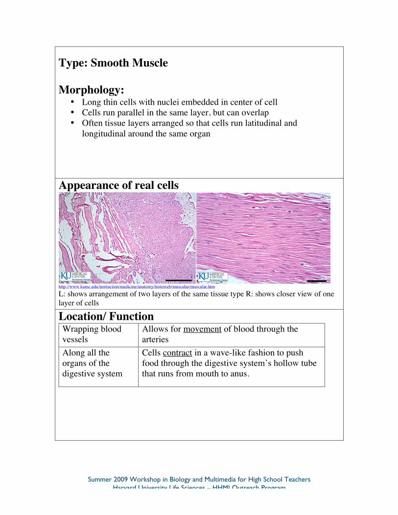
Type: Smooth Muscle
Morphology:• Long thin cells with nuclei embedded in center of cell• Cells run parallel in the same layer, but can overlap• Often tissue layers arranged so that cells run latitudinal and
longitudinal around the same organ
Appearance of real cells
http://www.kumc.edu/instruction/medicine/anatomy/histoweb/muscular/muscular.htm
L: shows arrangement of two layers of the same tissue type R: shows closer view of onelayer of cells
Location/ FunctionWrapping bloodvessels
Allows for movement of blood through thearteries
Along all theorgans of thedigestive system
Cells contract in a wave-like fashion to pushfood through the digestive system’s hollow tubethat runs from mouth to anus.
Summer 2009 Workshop in Biology and Multimedia for High School TeachersHarvard University Life Sciences – HHMI Outreach Program
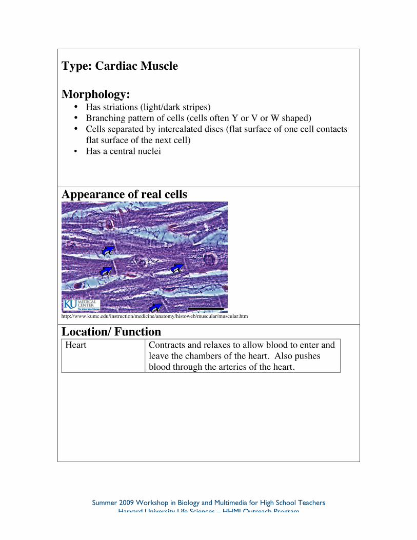
Type: Cardiac Muscle
Morphology:• Has striations (light/dark stripes)• Branching pattern of cells (cells often Y or V or W shaped)• Cells separated by intercalated discs (flat surface of one cell contacts
flat surface of the next cell)• Has a central nuclei
Appearance of real cells
http://www.kumc.edu/instruction/medicine/anatomy/histoweb/muscular/muscular.htm
Location/ FunctionHeart Contracts and relaxes to allow blood to enter and
leave the chambers of the heart. Also pushesblood through the arteries of the heart.